Pulmonary Hypertension - PHA Online University
Pulmonary Hypertension - PHA Online University
Pulmonary Hypertension - PHA Online University

Create successful ePaper yourself
Turn your PDF publications into a flip-book with our unique Google optimized e-Paper software.
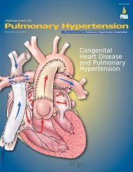
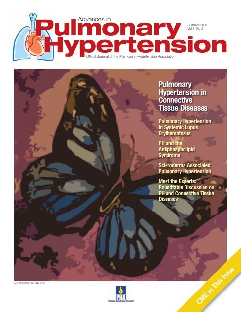
Advances in<strong>Pulmonary</strong><strong>Hypertension</strong>Official Journal of the <strong>Pulmonary</strong> <strong>Hypertension</strong> AssociationSummer 2008Vol 7, No 2<strong>Pulmonary</strong><strong>Hypertension</strong> inConnectiveTissue Diseases<strong>Pulmonary</strong> <strong>Hypertension</strong>in Systemic LupusErythematosusPH and theAntiphospholipidSyndromeScleroderma Associated<strong>Pulmonary</strong> <strong>Hypertension</strong>Meet the Experts:Roundtable Discussion onPH and Connective TissueDiseasesSee description on page 262ge 2.CME in This Issue
Table of ContentsGuest Editor for this issue:Kristin Highland, MD, MSCRAssistant ProfessorDivision of <strong>Pulmonary</strong> and CriticalCare MedicineDirector, <strong>Pulmonary</strong> <strong>Hypertension</strong> ClinicMedical <strong>University</strong> of South CarolinaCharleston, South Carolina264 Profiles in <strong>Pulmonary</strong><strong>Hypertension</strong>:Virginia D. Steen, MD277 International Corner:Combination Therapy in PAH278 CME Section280 <strong>Pulmonary</strong> <strong>Hypertension</strong>in Systemic LupusErythematosus286 <strong>Pulmonary</strong> <strong>Hypertension</strong> andthe AntiphospholipidSyndrome292 Scleroderma Associated<strong>Pulmonary</strong> <strong>Hypertension</strong>299 CME Self-AssessmentExamination301 <strong>Pulmonary</strong> <strong>Hypertension</strong>Roundtable: PAH andSclerodermaPublisher<strong>Pulmonary</strong> <strong>Hypertension</strong> AssociationMichael D. McGoon, MD, Chair of the BoardRino Aldrighetti, PresidentDonica Merhazion, Associate Director of Medical Services<strong>PHA</strong> Office<strong>Pulmonary</strong> <strong>Hypertension</strong> Association801 Roeder Rd. Suite 400Silver Spring, MD 20910-4496301-565-3004, 301-565-3994 (fax)www.phassociation.org© 2008 by <strong>Pulmonary</strong> <strong>Hypertension</strong> Association. All rightsreserved. None of the contents may be reproduced in anyform whatsoever without the written permission of <strong>PHA</strong>.ISSN: 1933-088X (print); 1933-0898 (online)Editorial OfficesAdvances in <strong>Pulmonary</strong> <strong>Hypertension</strong>, DataMedica,P.O. Box 1688, Westhampton Beach, NY 11978Tel (631) 288-7733 Fax (631) 288-7744E-mail: sbelsonchapman@aol.comPublishing StaffStu Chapman, Executive EditorSusan Chapman, Managing EditorHeidi Green, Associate EditorGloria Catalano, Production DirectorMichael McClain, Design DirectorAdvances in <strong>Pulmonary</strong> <strong>Hypertension</strong> is circulated to cardiologists,pulmonologists, rheumatologists, and other selectedphysicians by the <strong>Pulmonary</strong> <strong>Hypertension</strong> Association. Thecontents are independently determined by the Editor and theEditorial Advisory Board. All past issues of the journal areavailable at: www.<strong>PHA</strong>ssociation.org/Medical/Advances_in_PH/Cover ImageThis illustration symbolizes the malar ("butterfly") rash that ischaracteristic of patients with systemic lupus erythematosus.The artwork was provided by Martin J. Somsky.Guest Editor’s MemoTargeting the Connection BetweenPAH and Connecive Tissue Disease<strong>Pulmonary</strong> hypertension (PH) is associated with all of the connectivetissue diseases (CTDs), but it is most commonly seen insystemic sclerosis (scleroderma), followed by systemic lupus erythematosus,and the antiphospholipid antibody syndrome. Although theprognosis for CTD-associated PH has improved with the availabilityof numerous pulmonary arterial hypertension therapies, it stillcarries a substantially poorer prognosis than idiopathic pulmonaryarterial hypertension, and necessitates additional research into itstreatment and prevention.Further complicating treatment, CTD-associated PH is often multifactorial andcan be caused by any of the World Health Organization categories of PH. In thisissue of Advances in <strong>Pulmonary</strong> <strong>Hypertension</strong>, the contributors review what is knownregarding the epidemiology, pathobiology, and treatment of PH, with a focus on pulmonaryarterial hypertension in systemic sclerosis, systemic lupus erythematosus,and antiphospholipid syndrome.—Kristin B. Highland, MD, MSCREditor’s MemoHighlighting Current and FuturePerspectives in the JournalThe recent increase in awareness and recognition of pulmonaryhypertension is in part related to the efforts of Rheumatologistswho are now screening many of their patients for this deadly disease.This issue, guest edited by Dr Kristin Highland, focuses onthe spectrum of connective tissue diseases and their relation topulmonary hypertension. While there is still much to learn abouthow and why patients with connective tissue diseases develop pulmonaryhypertension, the benefits of treatment for many of thesepatients are apparent. This issue also spotlights rheumatologist,Dr Virginia Steen, whose efforts in the world of pulmonary hypertension, particularlyon screening and early diagnosis, are close to my heart. For our new InternationalCorner, Dr Nick Morrell provides commentary on combination therapy issues coveredin the previous issue of Advances, with an international perspective.I am pleased to preview important content to be published in Advances in <strong>Pulmonary</strong><strong>Hypertension</strong> in the next 2 issues. The journal will be focusing on 2 meetingsheld earlier this year that provided relevant and timely updates on pulmonaryhypertension. First, physicians who were unable to attend the World Health Organization4 th World Symposium on <strong>Pulmonary</strong> <strong>Hypertension</strong> will be pleased to seethe next issue and its comprehensive discussion of key findings from this meeting.Among the topics to be covered are: current perspectives on pathophysiology,diagnosis and assessment of pulmonary arterial hypertension (PAH), interventionaland surgical modalities, endpoints and clinical trial design in PAH, as well asinflammation, growth factors, and pulmonary vascular remodeling.In the following issue, the journal will feature content from another importantmeeting, this year’s 8 th International Conference and Scientific Sessions of the<strong>Pulmonary</strong> <strong>Hypertension</strong> Association. The information from this meeting will begleaned from physicians who led the sessions and who are among the leadinginvestigators in the field. We look forward to providing articles on these importantissues and welcome your feedback. Please send your comments and suggestionsto our executive editor at Stulink@aol.com.Ronald J. Oudiz, MDEditor-in-Chief
Editorial Advisory BoardEditor-in-ChiefRonald J. Oudiz, MDAssociate Professor of MedicineUCLA School of MedicineDirector, Liu Center for <strong>Pulmonary</strong><strong>Hypertension</strong>Division of CardiologyLos Angeles Biomedical ResearchInstitute at Harbor-UCLAMedical CenterTorrance, CaliforniaImmediate Past EditorVallerie V. McLaughlin, MDAssociate Professor of MedicineDirector, <strong>Pulmonary</strong> <strong>Hypertension</strong>Program<strong>University</strong> of Michigan Health SystemAnn Arbor, MichiganEditor-in-Chief ElectRichard Channick, MDProfessor of Clinical Medicine<strong>Pulmonary</strong> and Critical Care Division<strong>University</strong> of California, San DiegoMedical CenterSan Diego, CaliforniaAssociate EditorsErika Berman Rosenzweig, MDAssistant Professor of PediatricsDepartment of PediatricsColumbia College of Physiciansand SurgeonsNew York, New YorkTodd Bull, MDAssociate Professor of MedicineMedical Director, ICU AnshutzInpatient PavilionDivision of <strong>Pulmonary</strong> Sciences andCritical Care Medicine<strong>University</strong> of Colorado Health SciencesCenterDenver, ColoradoRobert Schilz, DO, PhDMedical Director of Lung Transplantationand <strong>Pulmonary</strong> Vascular Disease<strong>University</strong> Hospital of ClevelandCase Western Reserve <strong>University</strong>Cleveland, OhioEditorial BoardTeresa De Marco, MDDirector, Heart Failure and<strong>Pulmonary</strong> <strong>Hypertension</strong> Program<strong>University</strong> of California, San FranciscoSan Francisco, CaliforniaEli Gabbay, MDAssociate Professor<strong>University</strong> of Western AustraliaSchool of Medicine and PharmacologyMedical Director, Advanced Lung Diseaseand <strong>Pulmonary</strong> Vascular UnitRoyal Perth HospitalPerth, AustraliaKristin Highland, MDAssistant ProfessorDivision of <strong>Pulmonary</strong> and CriticalCare MedicineDirector, <strong>Pulmonary</strong> <strong>Hypertension</strong> ClinicMedical <strong>University</strong> of South CarolinaCharleston, South CarolinaOmar Minai, MDStaff PhysicianCleveland ClinicCleveland, OhioMyung H. Park, MDDirector, <strong>Pulmonary</strong> VascularDiseases Program<strong>University</strong> of Maryland School ofMedicineBaltimore, MarylandIoana Preston, MDAssistant Professor of MedicineTufts-New England Medical CenterBoston, MassachusettsZeenat Safdar, MDAssistant Professor of MedicineDepartment of Medicine,<strong>Pulmonary</strong> & Critical Care Section<strong>Pulmonary</strong> <strong>Hypertension</strong> CenterBaylor College of MedicineHouston, TexasRajan Saggar, MDAssistant Professor of MedicineDivision of <strong>Pulmonary</strong> and Critical CareMedicine and HospitalistsDavid Geffen School ofMedicine at UCLALos Angeles, CaliforniaFrancisco Soto, MDAssistant ProfessorDirector, <strong>Pulmonary</strong> <strong>Hypertension</strong>ProgramMedical College of WisconsinMilwaukee, WisconsinFernando Torres, MDDirector, <strong>Pulmonary</strong> <strong>Hypertension</strong>ProgramUT Southwestern Medical CenterDallas, TexasProgram DescriptionThe mission of Advances in <strong>Pulmonary</strong><strong>Hypertension</strong> is to serve as the premiereforum for state of the art information regardingdiagnosis, pathophysiology, and treatment ofpulmonary hypertension. The 2003 Venicerevision of the World Health OrganizationClassification serves as a guide to categoriesof pulmonary hypertension addressed by theJournal. While focusing on WHO Group I PAH,the other categories (Group II, Left heartdisease; Group III, Associated with lung diseaseand/or hypoxemia; Group IV, Thromboticand/or Embolic Disease; Group V, Miscellaneous)are also addressed. This mission isachieved by a combination of invited reviewarticles, Roundtable discussions with panelsconsisting of international experts in PH, andoriginal contributions. In addition, a specialsection entitled “Profiles in <strong>Pulmonary</strong><strong>Hypertension</strong>”recognizes major contributors tothe field and serves as an inspiring reminderof the rich and collegial history of dedicationto advancing the field.Objectives• Provide up-to-date information regardingdiagnosis, pathophysiology, and treatmentof pulmonary hypertension.• Serve as a forum for presentation and discussionof important issues in the field,including new paradigms of disease understandingand investigational trial design.• Recognize and preserve the rich history ofindividuals who have made major contributionsto the field via dedication to patientcare, innovative research, and furtheringthe mission of the PH community to curepulmonary hypertension.The Scientific LeadershipCouncil of the <strong>Pulmonary</strong><strong>Hypertension</strong> AssociationThe scientific program of the <strong>Pulmonary</strong><strong>Hypertension</strong> Association is guided bythe association’s Scientific LeadershipCouncil. The Council includes thefollowing health care professionals:Vallerie V. McLaughlin, MDSLC Chair<strong>University</strong> of Michigan Health SystemAnn Arbor, MichiganDavid B. Badesch, MDSLC Immediate Past Chair<strong>University</strong> of Colorado HealthSciences CenterDenver, ColoradoJohn H. Newman, MDSLC Chair ElectVanderbilt Medical SchoolNashville, TennesseeRobyn J. Barst, MDNew York, New YorkRaymond L. Benza, MD<strong>University</strong> of Alabama Health SystemBirmingham, AlabamaTodd Bull, MD<strong>University</strong> of Colorado HealthSciences CenterDenver, ColoradoRichard N. Channick, MDUCSD Medical CenterSan Diego, CaliforniaC. Gregory Elliott, MDLDS Hospital<strong>University</strong> of Utah School of MedicineSalt Lake City, UtahKaren A. Fagan, MD<strong>University</strong> of South AlabamaCollege of MedicineMobile, AlabamaAdaani Frost, MDBaylor College of MedicineHouston, TexasJohn Granton, MDToronto General HospitalToronto, CanadaNazzareno Galiè, MDInstitute of Cardiology<strong>University</strong> of BolognaBologna, ItalyNicholas S. Hill, MDDivision of <strong>Pulmonary</strong>, Critical Careand Sleep MedicineTufts-New England Medical CenterBoston, MassachusettsMarius Hoeper, MDHannover Medical schoolHannover, GermanyDunbar Ivy, MD<strong>University</strong> of Colorado HealthSciences CenterDenver, ColoradoZhi-Cheng Jing, MDFu wai Heart HospitalBeijing, ChinaAnne M. Keogh, MDSt. Vincent’s Public HospitalSydney, AustraliaMichael J. Krowka, MDMayo ClinicRochester, MinnesotaJames E. Loyd, MDVanderbilt <strong>University</strong> Medical CenterNashville, TennesseeMichael D. McGoon, MDChair, <strong>PHA</strong> Board of Trustees<strong>Pulmonary</strong> <strong>Hypertension</strong> ClinicMayo ClinicRochester, MinnesotaSrinivas Murali, MDAllegheny General HospitalPittsburgh, PennsylvaniaRonald J. Oudiz, MDLiu Center for <strong>Pulmonary</strong> <strong>Hypertension</strong>Los Angeles Biomedical ResearchInstituteHarbor-UCLA Medical CenterTorrance, CaliforniaMarlene Rabinovitch, MDStanford <strong>University</strong> School of MedicineStanford, CaliforniaErica Berman-Rosenzweig, MDColumbia-Presbyterian Medical CenterNew York, New YorkIvan M. Robbins, MDSLC Scientific Sessions CommitteeVanderbilt <strong>University</strong>Nashville, TennesseeJulio Sandoval, MDCardiopulmonary DepartmentNational Institute of Cardiologyof MexicoTlalpan, MexicoRichard Silver, MDMedical <strong>University</strong> of South CarolinaCharleston, South CarolinaVictor F. Tapson, MDDivision of <strong>Pulmonary</strong> and CriticalCare MedicineDuke <strong>University</strong> Medical CenterDurham, North CarolinaLiaisonsArlene Schiro, RN, MA, ACNP-BCChair, PH Resource NetworkMassachusetts General HospitalBoston, MassachusettsJoanne Sperando SchmidtPatient LiaisonEmeritus MembersBruce H. Brundage, MDSt. Charles Medical Center-BendBend, OregonAlfred P. Fishman, MD<strong>University</strong> of Pennsylvania HealthSystemPhiladelphia, PennsylvaniaThe Mission of the Scientific LeadershipCouncil is to provide medical and scientificguidance and support to the <strong>PHA</strong> by:• Developing and disseminating knowledgefor diagnosing and treating pulmonaryhypertension• Advocating for patients with pulmonaryhypertension• Increasing involvement of basic and clinicalresearchers and practitionersMore information on <strong>PHA</strong>’s ScientificLeadership Council and associatedcommittees can be found at:www.<strong>PHA</strong>ssociation.org/SLC/Advances in <strong>Pulmonary</strong> <strong>Hypertension</strong> 263
Virginia D. Steen, MD:Pushing the Envelope of Research,Redefining the Link BetweenScleroderma and PAHVirginia D.Steen, MDFor Virginia D. Steen, MD, the journeyessentially began at the <strong>University</strong> ofPittsburgh in the 1980s where she wasa Fellowship in Rheumatology andbegan to see her first patients with sclerodermaand an “isolated” pulmonaryhypertension independent of pulmonaryfibrosis. In this era before any treatmentswere approved, before the landmarkstudies that would revolutionizethe treatment of the disease, rheumatologists remainedon the fringes of PH research. Although rheumatologistsgenerally do not participate in the clinical trials in PH,largely because of the relatively smaller number ofpatients in their care who can be evaluated, efforts byDr Steen and her colleagues over more than 20 years havehelped research the connection between scleroderma andpulmonary arterial hypertension. This has led to a tremendouslyimproved understanding of the natural history ofPH in scleroderma, the risk factors for predicting PH inpatients with this coexisting disease, and the response tothe newer therapies approved within the last 5 to 6 years.The initial work, however, and the impetus for extendingher study of PH in scleroderma, began with Dr Steen’sfirst paper on the natural history of the association. “Afterthe initial observation of PH occurring in scleroderma, welooked at 30 patients who had PH and scleroderma andcompared them to similar scleroderma patients who didnot have PH,” recalled Dr. Steen. This study showedpatients with PH in scleroderma did not have significantfibrosis but had a very low diffusing capacity. Some hadthis finding prior to the diagnosis of PH. “In our nextstudy, using the Pittsburgh Scleroderma Database, ourprospective collected database, we performed a retrospectivecase control analyses, looking at pulmonary functionand echo[cardiogram]s prior to the diagnosis. The pulmonaryfunction tests showed that even 5 years beforetheir diagnosis their DLCO [diffusing capacity of the lungfor carbon monoxide] was decreased.”Dr Steen and colleagues have published more than 50articles and editorials that form the core of the mostextensive database of articles in this clinical setting.Studies that addressed topics such as predictors of endstage lung disease in systemic sclerosis and other findingsthat contributed to the evidence-based clinical practiceguidelines of the American College of Chest Physicians.In these and ensuing years, she became Professor ofMedicine at the <strong>University</strong> of Pittsburgh and then, in1995, moved to her current position as Professor ofMedicine at Georgetown <strong>University</strong>, Washington DC, whereshe also serves as Program Director of the RheumatologyFellowship Program.As their research progressed, Dr Steen and her teambegan to identify features of scleroderma patients thathelped differentiate subsets within the overall category.“There are those typical PAH [pulmonary arterial hypertension]patients who look—other than their age and havingscleroderma—very similar to idiopathic PAH patients. Butthen there is another group of scleroderma patients whohave more interstitial disease with their PH. And there isa group who have more heart disease but still have vascularpulmonary hypertension. The recognition of thesedifferent groups has evolved as more treatments havebecome available.”The focus of much of her current research centersaround the <strong>PHA</strong>ROS: <strong>Pulmonary</strong> <strong>Hypertension</strong> Assessmentand Recognition of Outcomes in Scleroderma. This is amulticenter, 18-site study of systemic scleroderma (systemicsclerosis) patients in the United States who are athigh risk for developing pulmonary hypertension as wellas those with newly diagnosed pulmonary hypertension.This is an extremely important study since pulmonaryhypertension is the most common cause of sclerodermadeaths. It is looking specifically at patients who are athigh risk for PAH, those who have specific abnormalitieson pulmonary function tests (PFTs) or echocardiogramsas well as those who are newly diagnosed with PAH.“We have close to 300 patients now and almost athird of them have definite PH,” said Dr Steen. “We arefollowing these patients’ response to therapy to determinetheir outcome. We are also following patients who are athigh risk for PH over 5 years to determine who developsPH based on various risk factors. For example, I am doingan exercise study and we’ve shown that if a sclerodermapatients is at high risk he or she has a 40% chance ofhaving exercise-induced PH even if one has restingnormal pressures.”The findings coming from the rheumatology communityare an essential component in the expanding translationalresearch that Dr Steen and her colleagues hope willenable clinicians to identify risk factors much earlier inscleroderma and initiate preventive therapy at a stage thatcan make a real difference in altering the natural historyof the disease. ■264 Advances in <strong>Pulmonary</strong> <strong>Hypertension</strong>
Advances in <strong>Pulmonary</strong> <strong>Hypertension</strong>Author Guidelines 2008Scope of ManuscriptsAdvances in <strong>Pulmonary</strong> <strong>Hypertension</strong> considersthe following types of manuscripts for publication:• Reviews that summarize and synthesize peerreviewedliterature to date on relevant topics in ascholarly fashion and format.• Letters to the Editor• Clinical Case StudiesManuscript SubmissionAuthors are required to submit their manuscriptsin an electronic format, preferably by email tothe Editor-in-Chief, Ronald J. Oudiz, MD, atoudiz@humc.edu. Please provide manuscripts ina word processing program. Images should besubmitted electronically as well.All material reproduced from previously published,copyrighted material should contain a full credit lineacknowledging the original source. Authors are responsiblefor obtaining permission to reproducesuch material.Contact information: List all authors, including mailingaddress, titles and affiliations, phone, fax, and email.Please note corresponding author.Peer Review and Editing: Manuscripts will be peerreviewed. Accepted manuscripts will be edited for clarity,spelling, punctuation, grammar, and consistencywith American Medical Association (AMA) style.Manuscript PreparationLength: Full-length manuscripts should not exceed4,000 words, including references. Please limit thereference list to 50 citations. Manuscripts should beaccompanied by figures and/or tables. Generally, 4 to 5figures and 2 to 3 tables are preferred for each manuscript.Please include a brief description to accompanythese items, as well as a key for all abbreviatedwords.Spacing: One space after commas and periods.Manuscripts should be double spaced. Manuscriptsshould not contain an abstract but an introduction isrecommended.References: All submissions should include numberedreferences that are referred to in the text by superscriptsand that conform to AMA style. Example:Lewczuk J, Piszko P, Jagas J, et al. Prognostic factorsin medically treated patients with chronic pulmonaryembolism. Chest. 2001;119:818-823.Copyright: Manuscripts and accompanying materialare accepted for exclusive publication in Advances in<strong>Pulmonary</strong> <strong>Hypertension</strong>. None of the contents maybe reproduced without permission of the <strong>Pulmonary</strong><strong>Hypertension</strong> Association. To request permission,please contact Stu Chapman, Executive Editor, (516)356-5006; email: stulink@aol.com.Advances in <strong>Pulmonary</strong> <strong>Hypertension</strong> 265
Request YourCopy NowABreakthroughinMedical EducationComplimentaryCD-ROM Available<strong>Pulmonary</strong> <strong>Hypertension</strong>: An Interactive Guide to DiagnosisThis companion piece to the Fall issue of Advancesin <strong>Pulmonary</strong> <strong>Hypertension</strong> assists with diagnosis ofpulmonary hypertension and is an invaluable resourcefor medical professionals in pulmonology, cardiology,rheumatology and primary care. Please go towww.<strong>PHA</strong>ssociation.org/medical/Advances_in_PHto view the Fall issue of the journal.Featuring comprehensive diagnosticinformation on:Physical examinationIntroduction on jugular venous pulsePlease go towww.<strong>PHA</strong>ssociation.org/medical/cd.aspto request your complimentary copy orcheck the box on the reply card found atthe front of the journal.The production of this CD-ROM was supported by GrantNumber Purchase Request (PR)# HCL33-2005-23060 andContract Award # 254-2005-M-13200 and Purchase Request(PR)# HCL33-2004-09925 and Contract Award#200-2004-M-10076fromtheCentersforDiseaseControland Prevention. Its contents are solely the responsibility ofthe <strong>Pulmonary</strong> <strong>Hypertension</strong> Association and do not necessarilyrepresent the official views of the Centersfor Disease Control and Prevention.The distribution of this CD-ROM is being made possible byan unrestricted educational grant from Myogen, Inc.7casesprovidingcomprehensivediagnosticinformation on:•Valvularpulmonicstenosis•Patentductusarteriosuswithpulmonaryhypertension(Eisenmenger syndrome)•Restrictiveventricularseptaldefect(VSD)•Non-restrictiveVSDwithpulmonaryhypertension(Eisenmenger)•Hypertensiveheartdisease,atrialfibrillation,PH,and tricuspid regurgitation•<strong>Pulmonary</strong>arterialhypertensionwithtricuspidregurgitation•<strong>Pulmonary</strong>arterialhypertensionwithtricuspidandpulmonic regurgitationInitial Diagnostic TestingIncludes comprehensive and interactive information on:•ECG •Echocardiography•Chestx-ray •Computedtomography•V/Qscan •Rightheartcatheterization•MRI
START WITH CONFIDENCEREVATIO: for patients with PAH as early as class II• REVATIO is indicated for the treatment ofpulmonary arterial hypertension (WHOGroup I) to improve exercise ability– WHO Group I• A first-line treatment for class II andclass III 1– Updated American College of Chest Physiciansevidence-based clinical practice guidelines• The lowest-priced oral PAH therapy*– REVATIO 20 mg tid*Based on wholesale acquisition cost: First DataBank Inc., 2008. Actualpharmacy or out-of-pocket costs may vary. Price comparisons donot imply comparable efficacy and safety. The pivotal trial for REVATIOincluded patients who were predominantly functional classes II and III,and the pivotal trial for Tracleer ® included patients who werepredominantly functional class III.REVATIO is indicated for the treatment of pulmonary arterial hypertension (WHO Group I) to improve exercise ability. The efficacy of REVATIO has not beenevaluated in patients currently on bosentan therapy.The use of REVATIO and organic nitrates in any form, at any time, is contraindicated.Co-administration of REVATIO with potent CYP3A4 inhibitors (eg, ketoconazole, itraconazole, and ritonavir) is not recommended as serumconcentrations of sildenafil substantially increase. Co-administration of REVATIO with CYP3A4 inducers, including bosentan; and more potent inducerssuch as barbiturates, carbamazepine, phenytoin, efavirenz, nevirapine, rifampin, and rifabutin, may alter plasma levels of either or bothmedications. Dosage adjustment may be necessary.Before starting REVATIO, physicians should carefully consider whether their patients with underlying conditions could be adversely affected by the mildand transient vasodilatory effects of REVATIO on blood pressure. <strong>Pulmonary</strong> vasodilators may significantly worsen the cardiovascular status of patientswith pulmonary veno-occlusive disease (PVOD) and administration of REVATIO to these patients is not recommended. Should signs ofpulmonary edema occur when sildenafil is administered, the possibility of associated PVOD should be considered.The most common side effects of REVATIO (placebo-subtracted) were epistaxis (8%), headache (7%), dyspepsia (6%), flushing (6%), and insomnia (6%).Adverse events were generally transient and mild to moderate.Caution is advised when PDE5 inhibitors, such as REVATIO, are administered with -blockers as both are vasodilators with blood pressure lowering effects.REVATIO should be used with caution in patients with anatomical deformation of the penis or patients who have conditions which may predispose them to priapism.In PAH patients, the concomitant use of vitamin K antagonists and REVATIO resulted in a greater incidence of reports of bleeding (primarily epistaxis)versus placebo. The incidence of epistaxis was higher in patients with PAH secondary to CTD (sildenafil 13%, placebo 0%) than in PPH patients(sildenafil 3%, placebo 2%).Non-arteritic anterior ischemic optic neuropathy (NAION) has been reported rarely post-marketingin temporal association with the use of PDE5 inhibitors for the treatment of erectile dysfunction,including sildenafil. It is not possible to determine if these events are related to PDE5inhibitors or to other factors. Physicians should advise patients to seek immediate medicalattention in the event of sudden loss of vision while taking PDE5 inhibitors, including REVATIO.Sudden decrease or loss of hearing has been reported in temporal association with the intakeof PDE5 inhibitors, including REVATIO. It is not possible to determine whether these events arerelated directly to the use of PDE5 inhibitors or to other factors. Physicians should advisepatients to seek prompt medical attention in the event of sudden decrease or loss ofhearing while taking PDE5 inhibitors, including REVATIO.Tracleer (bosentan) is a registered trademark of Actelion Pharmaceuticals.Please see brief summary of prescribing information on adjacent page.REVATIO contains sildenafil citrate, thesame active ingredient found in Viagra ®www.pfizerpro.com
Physicians from 56 countries helped supportthe <strong>Pulmonary</strong> <strong>Hypertension</strong> Association.Thank you!During the Gilead Sciences event at the 2008American Thoracic Society (ATS) International Conference,you helped raise $75,000 for the <strong>Pulmonary</strong> <strong>Hypertension</strong> Association.Physicians from around the globe participated:Argentina, Australia, Austria, Belgium, Brazil, Bulgaria, Canada, Chile, China, Colombia, Croatia, Ecuador, Egypt,El Salvador, Estonia, France, Germany, Greece, Honduras, India, Iran, Ireland, Israel, Italy, Japan, Jordan, Kuwait, Lebanon,Mexico, Netherlands, Norway, Pakistan, Panama, Peru, Philippines, Poland, Romania, Russia, Saudi Arabia, Serbia,Singapore, Slovakia, South Africa, South Korea, Spain, Sweden, Switzerland, Taiwan, Thailand, Trinidad and Tobago,Turkey, United Arab Emirates, United Kingdom, United States, Uruguay, VenezuelaProudly sponsored byFor the benefit of<strong>PHA</strong>—A member of the ATS Public Advisory Roundtable© 2008 Gilead Sciences, Inc. All rights reserved. Gilead and the Gilead logo are trademarks of Gilead Sciences, Inc. ABS0129 July 2008ATS, ATS International Conference and ATS Public Advisory Roundtable trademarks are used with permission from the American Thoracic Society.
IN THE TREATMENT OF PAH (WHO GROUP 1, CLASS II OR III SYMPTOMS)1 pill 1Once a day 1Please see below for important safety information, including boxed WARNINGS information on the possible risk of liverinjury and the risk of serious birth defects, and brief summary of full prescribing information on next page.95% of patients alive at 1 year (Kaplan-Meier estimates; N=383) 1,2• Ongoing, multicenter, open-label study of the long-term safety and efficacy of LETAIRIS; there is no control comparator,and long-term safety is the primary endpoint 1,2– These uncontrolled observations do not allow comparison with a group not given LETAIRIS and cannot be used todetermine the long-term effect of LETAIRIS on survival 1• 94% of patients were receiving LETAIRIS monotherapy at 1 year (278 of 295 patients with 48 weeks of follow-up) 1,2INDICATION: LETAIRIS is an endothelin receptor antagonistindicated for the treatment of pulmonary arterial hypertension(PAH) (WHO Group 1) in patients with WHO Class II orIII symptoms to improve exercise capacity and delayclinical worsening.Clinical worsening is defined as the first occurrence of death,lung transplantation, hospitalization for PAH, atrial septostomy,study withdrawal due to the addition of other PAH therapeuticagents, or study withdrawal due to early escape. 1,2Early escape criteria were two or more of the following after aminimum treatment period of 4 weeks: ≥20% decrease in6-minute walk distance; worsening WHO functional class;worsening right ventricular failure; rapidly progressing cardiac,hepatic, or renal failure; and refractory systolic hypotension5× ULN or if elevations are accompaniedby bilirubin >2× ULN or by signs or symptoms ofliver dysfunction• May cause fetal harm if taken during pregnancy• Must exclude pregnancy before the start of treatment• Prevent pregnancy thereafter by the use of two reliablemethods of contraceptionImportant safety information regarding hepatotoxicityLETAIRIS is not recommended in patients with elevatedaminotransferases (>3× ULN) at baseline because monitoringliver injury may be more difficult. If aminotransferase elevationsare accompanied by clinical symptoms of liver injury (such asanorexia, nausea, vomiting, fever, malaise, fatigue, right upperquadrant abdominal discomfort, itching, or jaundice) orincreases in bilirubin >2× ULN, LETAIRIS treatment should bestopped. There is no experience with the reintroduction ofLETAIRIS in these circumstances.Contraindication• Do not administer LETAIRIS to a pregnant woman becauseit can cause fetal harmWarnings and precautions• Decreases in hemoglobin have been observed within the firstfew weeks of treatment with LETAIRIS; measure hemoglobinprior to initiation, at 1 month, and periodically thereafter• Mild to moderate peripheral edema. Peripheral edemaoccurred more frequently in elderly patients (age ≥65 years)receiving LETAIRIS (29%; 16/56) compared to placebo(4%; 1/28)
1 important stepLiver function abnormalities similar to placebo 1• For all patients treated with LETAIRIS (N=483), the 12-week incidence of aminotransferase elevations >3× ULN was 0.8%compared with 2.3% for patients treated with placebo (N=132)• The 1-year rate of aminotransferase elevations >3× ULN was 2.8% and >8× ULN was 0.5%• In the post-marketing period, at least one patient receiving another endothelin receptor antagonist (ERA) exhibited latepresentation (after 20 months of treatment) of pronounced elevations in aminotransferases and bilirubin levels. Thisreinforces the importance of monthly liver monitoring for the duration of treatment• Peripheral edema is a known class effect of endothelinreceptor antagonists. In addition, there have been postmarketingreports of fluid retention occurring within weeksafter starting LETAIRIS which required intervention with adiuretic, fluid management, or, in some cases, hospitalizationfor decompensating heart failureDrug interactions• Use caution when LETAIRIS is coadministered withcyclosporine A• Use caution when LETAIRIS is coadministered with strongCYP3A inhibitors (e.g., ketoconazole) or CYP2C19 inhibitors(e.g., omeprazole)• Use caution when LETAIRIS is coadministered with inducers ofP-gp, CYPs, and UGTs• No significant interactions of LETAIRIS with warfarin orsildenafil have been observedReferences: 1. LETAIRIS [Prescribing Information]. Foster City, Calif: Gilead Sciences, Inc;February 2008. 2. Data on file. Gilead Sciences, Inc.Adverse eventsAdverse Events in >3% of PAH Patients ReceivingLETAIRIS and More Frequent Than PlaceboPlacebo(n=132)LETAIRIS(n=261)Adverse event n (%) n (%)Placeboadjusted,%Peripheral edema 14 (11) 45 (17) 6Nasal congestion 2 (2) 15 (6) 4Sinusitis 0 (0) 8 (3) 3Flushing 1 (1) 10 (4) 3Palpitations 3 (2) 12 (5) 3Nasopharyngitis 1 (1) 9 (3) 2Abdominal pain 1 (1) 8 (3) 2Constipation 2 (2) 10 (4) 2Dyspnea 4 (3) 11 (4) 1Headache 18 (14) 38 (15) 1Note: This table includes all adverse events >3% incidence in the combined LETAIRIStreatment group and more frequent than in the placebo group, with a difference of ≥1%between the LETAIRIS and placebo groups.Because of the risks of liver injury and birth defects, LETAIRISis available only through a special restricted distributionprogram called the LETAIRIS Education and Access Program(LEAP), by calling 1-866-664-LEAP (5327). Only prescribers andpharmacies registered with LEAP may prescribe and distributeLETAIRIS. In addition, LETAIRIS may be dispensed only topatients who are enrolled in and meet all conditions of LEAP.The efficacy and safety of LETAIRIS were evaluated in two 12-week,randomized, double-blind, placebo-controlled, multicenter studies.Please see next page for brief summary of full prescribing information, including boxed WARNINGS.© 2008 Gilead Sciences, Inc. All rights reserved. ABS0084 April 2008 LETAIRIS is a registered trademark and Gilead and the Gilead logo are trademarks of Gilead Sciences, Inc.
Combination Therapy in <strong>Pulmonary</strong> Artery <strong>Hypertension</strong>Nicholas W. Morrell, MA, MD, FRCPProfessor of Cardiopulmonary Medicine/Honorary Consultant,<strong>University</strong> of Cambridge School of Clinical MedicineAddress for correspondence: <strong>University</strong> of Cambridge School of ClinicalMedicine Box 157, Addenbrooke's Hospital, Hills Road, Cambridge CB22QQ, United Kingdom; e-mail: nwm23@cam.ac.uk.In her excellent article Iona Preston 1 systematically evaluatesthe current status of combination therapy in the treatment ofpatients with pulmonary artery hypertension (PAH) in theUnited States. The United Kingdom recently published itsown consensus statement on the management of PAH in clinicalpractice, which included the current use of combinationtherapy. 2Despite the current lack of definitive clinical trial data,combination therapy remains an attractive option for the clinicianfaced with a patient who is getting worse or a patient whois failing to improve on monotherapy. In the UK, the mostcommon way to combine therapies is to add a second drugwhen the patient is not improving or getting worse on optimaldoses of single therapy. Despite the concerns over pharmacologicalinteractions, the most common combination in the UKis the addition of an endothelin receptor antagonist (ETRA) tosildenafil or vice versa, which accounts for 65% of combinationtherapy prescriptions.In rapidly deteriorating patients, a parenteral prostanoidmay be added to the original oral therapy. The combination ofa prostanoid with sildenafil accounts for 23% of combinationprescriptions in the UK. Combined use of a prostanoid andETRA accounts for the remaining 12% of prescriptions. It is ofsome concern that the funding arrangements in the UK aremaking it harder to prescribe combination therapy in theabsence of definitive trial data and a favourable cost-benefitanalysis. Thus it is recommended that where combination therapyis being considered that all patients should be entered intoa clinical trial where possible. When it is not possible toinclude patients in clinical trials, we recommend that at thevery least the patient’s response to treatment is carefully monitoredand that data are submitted to national databases foraudit purposes.References1. Preston IR. Combination therapies in pulmonary arterial hypertension.Adv <strong>Pulmonary</strong> Hypertens. 2008;7:235-242.2. National <strong>Pulmonary</strong> <strong>Hypertension</strong> Centres of the UK and Ireland.Consensus statement on the management of pulmonary hypertension inclinical practice in the UK and Ireland. Thorax. 2008;63:1-41.<strong>PHA</strong> has just completed fundingfor the first year’s budget for our newMedical Education FundThis new two million dollar Fund will supportfour new <strong>PHA</strong> programs:Thirty-City Medical Education TourTo present information on the diagnosis and management of PAH to physicians and otherhealth professionals located in areas that lack recognized experts or centers for PAH.Preceptorship ProgramTo facilitate direct education and training of medical professionals, particularlycardiologists, pulmonologists and rheumatologists, by experienced pulmonary hypertensionspecialists in clinical settings.<strong>PHA</strong> <strong>Online</strong> <strong>University</strong>To create a clear, focused website for medical education in pulmonary hypertension,segmented by levels of expertise and medical interestMedical Education for Patients Regional SessionsTo present information on the mechanisms, diagnosis and treatment of PAH to patientsand family members in regional face-to-face settingsMade possible by unrestricted educational grants from our sponsors:Platinum: Actelion Gold: Gilead and Pfizer Silver: United TheraputicsWatch for more details and schedules in coming issues.
Advances in <strong>Pulmonary</strong> <strong>Hypertension</strong> CME SectionProgram Overview<strong>Pulmonary</strong> arterial hypertension (PAH), an incurabledisease, is characterized by medial hypertrophy, intimalfibrosis, and in situ thrombi in small muscular pulmonaryarteries PAH was considered a rapidly fatal illness witha median survival of 2.8 years in the 1980s when noevidence-based therapies were available. Since thenthe treatment of this disease has made tremendousadvances, and the last 10 years have seen the discoveryof new medications that have positively influenced theprognosis and survival of patients with PAH.This self-study activity is based on 3 articles that reviewthe latest information on new treatments, combinations oftherapies, and data from phase 1 and 2 clinical trials.This activity is jointly sponsored by the <strong>University</strong> ofMichigan Medical School and the <strong>Pulmonary</strong> <strong>Hypertension</strong>Association and supported by an unrestrictededucation grant from Actelion Pharmaceuticals US, Inc,Encysive Pharmaceuticals, Inc, Gilead Sciences, Inc,Pfizer, Inc, and United Therapeutics Corporation.Target AudienceThis self-study activity is appropriate for cardiologists,pulmonologists, rheumatologists, and other physicianswho treat patients with pulmonary hypertension.Learning ObjectivesUpon completion of this activity participants will beable to:1. To review the prevalence, demographics, and riskfactors of pulmonary hypertension associated withsystemic lupus erythematosus.2. To review the pathology/pathogenesis of pulmonaryhypertension associated with systemic lupuserythematosus.3. To review the treatment and prognosis of pulmonaryhypertension associated with systemic lupuserythematosus.4. To review the association of antiphospholipidantibodies and pulmonary arterial hypertension.5. To review the treatment for chronic thromboembolicpulmonary hypertension.6. To review the epidemiology, risk factors, andbiomarkers for pulmonary hypertension associatedwith scleroderma.7. To review diagnosis and screening of pulmonaryhypertension associated with scleroderma.8. To review the treatment of pulmonary hypertensionassociated with scleroderma.Self-Assessment ExaminationSee pages 299 and 300 for self-assessmentquestions, answer key, and evaluation form.FacultyChairKristin B. Highland, MD, MSCRDivision of <strong>Pulmonary</strong>, Critical Care,Allergy, and Sleep Medicine, andDivision of Rheumatology and ImmunologyContributing AuthorsKristin B. Highland, MD, MSCRDivision of <strong>Pulmonary</strong>, Critical Care,Allergy, and Sleep Medicine, andDivision of Rheumatology and ImmunologyGary Gilkeson, MDDivision of Rheumatology and ImmunologyDepartment of MedicineMedical <strong>University</strong> of South CarolinaRaj S. Kasthuri, MDRobert A. S. Roubey, MDDepartment of Medicine and ThurstonArthritis Research CenterThe <strong>University</strong> of North Carolinaat Chapel HillVictoria K. Shanmugam, MDVirginia D. Steen, MDDivision of Rheumatology, Immunology,and Allergy,Georgetown <strong>University</strong> Hospital278 Advances in <strong>Pulmonary</strong> <strong>Hypertension</strong>
Accreditation StatementThis activity has been planned and implemented inaccordance with the Essential Areas and Policies of theAccreditation Council for Continuing Medical Education(ACCME) through the joint sponsorship of the <strong>University</strong>of Michigan Medical School and the <strong>Pulmonary</strong> <strong>Hypertension</strong>Association. The <strong>University</strong> of Michigan isaccredited by the ACCME to provide continuing medicaleducation to physicians.Credit DesignationThe <strong>University</strong> of Michigan Medical School designatesthis activity for a maximum of 2.0 AMA PRA Category 1Credits. Physicians should claim credit commensuratewith the extent of their participation in the activity.Instructions for Earning CreditThis activity is a self-study program; a self-assessmentexamination is included on page 299 to help physiciansreview important points. A form is also included on page300 for physicians to evaluate the CME activity. Completionof this activity involves reading the journal andcompleting the self-assessment examination and evaluationform, which may take up to 2 hours. Credits for thisself-study program are available from September 15,2008 through September 15, 2009. There is no feefor this program.Please note that this self-study program may also beviewed online at: http://www.cme.med.umich.edu<strong>University</strong> of Michigan Privacy Statementhttp://www.cme.med.umich.edu/privacy.aspSponsorshipThis CME self-study program is jointly sponsored bythe <strong>University</strong> of Michigan Medical School and the<strong>Pulmonary</strong> <strong>Hypertension</strong> Association.SupportThis CME self-study program is supported by an educationalgrant from Actelion Pharmaceuticals US, Inc.,Encysive Pharmaceuticals, Inc., Gilead Sciences, Inc.,Pfizer, Inc., and United Therapeutics Corporation.Oversite and AccreditationArlene Bradford, BAAssistant DirectorOffice of CME<strong>University</strong> of Michigan Medical SchoolDisclosuresThe Accreditation Council for Continuing Medical Educationand the Association of American Colleges havestandards and guidelines to ensure that individualsparticipating in CME activities are aware of relationshipsbetween authors and commercial companies that couldpotentially affect the information presented. The <strong>University</strong>of Michigan Medical School follows these nationalpolicies to ensure balance, independence, objectivity,and scientific rigor in all its CME activities. Each authorwas asked to complete a disclosure information form forthis activity. Disclosures are reported below.Kristin Highland, MD, is on the Speaker’s Bureau ofActelion Pharmaceuticals, United Therapueitcs, andGilead Sciences. She receives grant/research supportfrom Actelion Pharmaceuticals, United Therapeutics,and Gilead Sciences.Gary Gilkeson, MD, serves as a consultant for and ison the Speaker’s Bureau of Genentech, He is a stockshareholder in Taligen Therapeutics.Raj Kasthuri, MD, has no relevant personal financialrelationship to disclose.Robert Roubey, MD, has no relevant personal financialrelationship to disclose.Victoria Shanmugam, MD, has no relevant personalrelationship to disclose.Virginia Steen, MD, serves as a consultant for ActelionPharmaceuticals and Gilead Sciences. She is on theSpeaker’s Bureau of Actelion Pharmaceuticals, GileadSciences and Abbott Laboratories. She receives grant/research support from Actelion Pharmaceuticals andGilead Sciences.Arlene Bradford, BA, has no relevant personal financialrelationships to disclose.CME ReviewerKevin M. Chan, MDAssistant Professor of MedicineDivision of <strong>Pulmonary</strong> and Critical Care Medicine<strong>University</strong> of Michigan Health SystemsAnn Arbor, MichiganDr Chan has no relevant personal financial relationshipsto disclose.Advances in <strong>Pulmonary</strong> <strong>Hypertension</strong> 279
Continuing Medical Education Section<strong>Pulmonary</strong> <strong>Hypertension</strong>in Systemic Lupus ErythematosusKristin B.Highland, MD, MSCRGary Gilkeson, MDKristin B. Highland, MD, MSCR 1,2 andGary Gilkeson, MD 21Division of <strong>Pulmonary</strong>, Critical Care,Allergy and Sleep MedicineDepartment of MedicineMedical <strong>University</strong> of South CarolinaCharleston, SC2Division of Rheumatology andImmunology, Department of MedicineMedical <strong>University</strong> of South CarolinaCharleston, SC<strong>Pulmonary</strong> and cardiac manifestations (Table 1) are commonin systemic lupus erythematosus (SLE). They occur inthe vast majority of patients, and as a result, patients withSLE have a marked decrease in exercise compared with controls.1,2 Although pulmonary hypertension (PH) is less frequentlyreported, exercise hemodynamics are abnormal inpatients with SLE, with higher pulmonary artery pressures atrest and for each stage of exercise when compared with controls.2 This occurs in the setting of similar cardiac indexes,which suggests that the mechanism for exercise intoleranceis an increase in pulmonary vascular resistance. In additionto cardiopulmonary complications, exercise intolerance inSLE may be caused by overwhelming fatigue, physicaldeconditioning, peripheral neuropathy, arthralgias/arthritis,and muscle weakness, which further complicates the evaluationof dyspnea in patients with SLE.All 5 World Health Organization (WHO) categories(Figure 1) of PH can be found in patients with SLE. Thewhole spectrum of pulmonary arterial hypertension (PAH) inSLE has also been reported, including pulmonary venoocclusivedisease and pulmonary capillary hemangiomatosis.3,4 <strong>Pulmonary</strong> arterial hypertension as a consequence ofnoncirrhotic portal hypertension has also been reported. 5,6<strong>Pulmonary</strong> venous hypertension is often seen as a result ofleft ventricular dysfunction from diastolic dysfunction,myocarditis, ischemic heart disease, or left ventricular valvulardysfunction secondary to Libman Sachs endocarditis.<strong>Pulmonary</strong> hypertension in SLE may be a consequence ofinterstitial lung disease, diaphragmatic dysfunction, andchronic thromboembolic disease. <strong>Pulmonary</strong> hypertensionhas also been associated with pulmonary vasculitis with orwithout alveolar hemorrhage. 7-9Key Words—<strong>Pulmonary</strong> hypertension; systemic lupus erythematosus;lupus.Address for reprints and other correspondence: Kristin B. Highland, MD,MSCR, Associate Professor of Medicine, Director, <strong>Pulmonary</strong> <strong>Hypertension</strong>Program, Division of <strong>Pulmonary</strong>, Critical Care, Allergy, and SleepMedicine, Division of Rheumatology and Immunology, 96 Jonathan LucasStreet, 812 CSB, Charleston, SC 29425; email: highlakb@musc.edu.Table 1. Cardiopulmonary Manifestations ofSystemic Lupus ErythematosusAcute lupus pneumonitisAcute reversible hypoxemiaAlveolar hemorrhageAtelectasisCoronary artery diseaseDiaphragmatic dysfunctionDiastolic dysfunctionInterstitial lung diseaseMyocarditisObstructive lung diseasePericardial diseasePleural effusionPleurisyPneumothorax<strong>Pulmonary</strong> embolism<strong>Pulmonary</strong> hypertensionUremic pulmonary edemaValvular lesionsPrevalence, Demographics, and Risk FactorsStudies of patients with SLE have found a prevalence of PHranging from 0.5% to 43%, although the degree of PH istypically modest. 10-16 The prevalence varied based on themethod used for detection, and many patients in thesereports either had significant restrictive lung disease or datareported were inadequate to determine the etiology of thePH. The prevalence of comorbid SLE and PH (SLE-PH) hasbeen shown to increase over time. In a serial study of 28patients with SLE, the prevalence of PH measured byechocardiogram increased from 14% to 43% with 5 years offollow-up. 16 In an autopsy series of 20 patients with SLE, 8(40%) patients had evidence of pulmonary vascular involvement;although clinically overt PH was present in only 1patient. 17Clinical manifestations (Table 2) of SLE-PH are variable,but the predominant features include the insidious onset ofshortness of breath, fatigue, and chest pain. Unfortunately,the disease process is usually far advanced with irreversiblechanges of the pulmonary vasculature by the time symptomsor signs develop. An isolated diffusion defect may be predictiveof PH in patients with SLE. 18The characteristics of patients with SLE-PH are similarto those of patients with idiopathic pulmonary arterial hyper-280 Advances in <strong>Pulmonary</strong> <strong>Hypertension</strong>
<strong>Pulmonary</strong>Arterial<strong>Hypertension</strong>Table 2. Possible Risk Factors for theDevelopment of <strong>Pulmonary</strong> <strong>Hypertension</strong> inSystemic Lupus Erythematosus<strong>Pulmonary</strong>Venous<strong>Hypertension</strong>ChronicThromboembolicDiseaseInterstitialLung disease/DiaphragmaticDysfunctionVasculitisFemale sexIsolated reduction in diffusionRaynaud phenomenonRenal diseaseDigital gangreneCutaneous vasculitis/livedo reticularisRheumatoid factorAntiribonuclear proteinAntiphospholipid antibodiesAntiendothelial antibodiesFigure 1. Etiology of pulmonary hypertension in systemic lupuserythematosus.tension (IPAH), which raises the possibility of shared etiologies.Patients are predominantly women of child-bearingpotential: aged from 18 to 40 years with a 10 to 1 ratio offemale over male. 19,20 In a study that compared 20 patientswith SLE-PH with 34 patients with IPAH, those with SLE-PHhad a significantly shorter time from symptom onset to diagnosisand were more likely to have Raynaud phenomenonand the presence of autoantibodies. 21 Patients with SLE-PHwere also more likely to have a pericardial effusion and wereless likely to be vasoresponsive to nitric oxide during rightheart catheterization. In addition, SLE-PH patients had lesshypoxemia and better hemodynamics, but a significantlyincreased mortality risk.Extrapulmonic manifestations can be found with IPAHincluding Raynaud phenomenon (30%), arthralgias, andarthritis. 22-24 Serologic abnormalities such as hypergammaglobulinemia,positive antinuclear antibody (ANA), rheumatoidfactor, and biological false-tests for syphilis have alsobeen reported, which suggests that some patients with IPAHmay have an autoimmune disease confined to the pulmonaryvasculature. 22-25 Alternatively, these patients may be at riskfor developing an underlying connective tissue disease(CTD), such as SLE, later on in the disease course.Study findings indicate that the duration of SLE does notcorrelate with the development of PH, although manypatients with SLE develop PH within the first 5 years.<strong>Pulmonary</strong> hypertension may be a presenting manifestationof SLE that necessitates close follow-up of all patients newlydiagnosed with IPAH. 26 The occurrence of PH also appearsunrelated to the severity or activity of SLE such as high antidoublestranded (ds)DNA and/or grossly elevated erythrocytesedimentation rate (ESR) and can occur when nonpulmonarydisease activity is quiescent. 19,20,26,27 This is in contrastto a study by Simonson and colleagues 13 that showedthat the duration of SLE and the duration of steroid therapytended to be shorter in SLE patients with PH, although theuse of anti-inflammatory agents was more common whencompared to a population of SLE patients without PH. Anadditional study showed that PH, as recognized by right ventricularechocardiography, occurred during 288 acute flaresof SLE, which suggests that a reversible increase in pulmonaryvasoconstrictor tone may be the first hemodynamicdisturbance, with fixed PH developing later. 28As many as 75% of patients with SLE-PH have Raynaudphenomenon, which is higher than the expected rate of 25%among all patients with SLE. 13,19,20,25,29,30 Asherson and colleagues19,20 have reported that 63% of patients in theirstudy had renal disease and approximately one-third hadevidence of peripheral cutaneous vasculitis, livedo reticularis,and digital gangrene.AutoantibodiesPatients with SLE-PH are universally positive for ANA.Antibodies to ribonuclear protein (RNP) and rheumatoid factor(RF) are often present in SLE-PH, although no pathogenicrole has been postulated. Frequently, patients haveantiphospholipid antibodies (aPL) and antiendothelial antibodies(aECA). 10,19,20,30,31 The prevalence of RNP in SLE-PHis reported in a majority of patients, which is greater thanthe reported prevalence of 25%, which occurs in all patientswith SLE. 30 The prevalence of RF has been reported to be ashigh as 50% to 80% in SLE-PH. 10,19 The frequency of PHin patients with SLE and a positive aPL is considerably higherthan in patients with SLE and negative aPL (83% versus25%). 30PathologyAutopsy findings suggest that SLE-PH may be multifactorialin origin. 25,30-32 Findings include acute fibrinoid necrosisand vasculitis, as well as chronic intimal fibrosis, medialhypertrophy, alteration of elastic laminae, periadventitialfibrosis, aneurysmal dilation, and plexiform lesions, whichare virtually identical to the alterations seen in patients withIPAH. 9,17,29 These changes occurred in arteries, arterioles,and veins. Occasional cases with thrombotic arteriopathyhave also been reported and were found to correlate with ahypercoagulable status, including positive lupus anticoagulantand anticardiolipin antibodies. 6,33Acute inflammation of small pulmonary arteries and arterioleshas also been found on autopsy in patients withSLE. 30 Deposition of circulating immune complexes (IgGand C1q) with antinuclear and anti-dsDNA activity has beenAdvances in <strong>Pulmonary</strong> <strong>Hypertension</strong> 281
described. The presence of diffuse interstitial fibrosis inaffected vessels further supports the likelihood of chronicinflammation that occurs as a result of the deposition ofsuch complexes and/or direct injury to the vessel wall. 10,34PathogenesisThe causal relationship between SLE and PH has never beenestablished. However, multiple small vessel inflammationand/or vasculitis as well as sustained vasoconstriction, insitu thrombosis, and/or thromboembolism and interstitialpulmonary fibrosis, all features of SLE, may damage andreduce the pulmonary vascular bed and lead to PH. 20,35There is an imbalance between vasoconstrictors andvasodilators in SLE-PH. Higher serum endothelin levels werefound in patients with SLE-PH compared with non-PHpatients with SLE and healthy controls. 36 There is also animbalance of thromboxane and prostacyclin that results inendothelial dysfunction, vascular damage, and remodelingthat is felt to be pathophysiologically important. 37 The inhibitionof prostacyclin production by endothelial cells is possiblyrelated to the action of aPL on the endothelial surface.38,39 In addition, when antiphospholipid antibodiesbind to the phospholipids on the endothelial surface, thereis resultant in-situ thrombosis and the release of solublemediators and subsequent vascular injury. 9,37Antiendothelial cell antibodies may also play a key pathogenicrole in the development of SLE-PH. Systemic lupuserythematosus is an autoimmune disease characterized bypolyclonal B cell activation. One factor that stimulates Bcells to produce immunoglobulin is interleukin 6 (IL-6), andendothelial cells are an important source of IL-6. Serumtiters of aECA are elevated in patients with active SLE, particularlyin patients with PH, digital vasculitis, Raynaud phenomenon,or serositis. Binding of antiendothelial cell antibodiesor immune complexes to endothelial cells may augmentthe release of IL-6 and result in vascular injury andensuing intimal and medial proliferation and in situ thrombosis.40The striking correlation between the occurrence ofRaynaud phenomenon and SLE-PH suggests that pulmonaryarterial vasospasm may also be involved in the pathogenesisof SLE-PH. Raynaud phenomenon is part of a systemic vascularresponse that includes a decrease in size of the pulmonarycapillary bed, which may in turn result in muscularnecrosis and secondary inflammation. 41,42 However, sincethe vast majority of patients with Raynaud phenomenon donot develop PH, this would suggest that other factors areoperative in those who are prone to develop PH.Alternatively, in conjunction with the high prevalence ofRNP found in SLE-PH, this subset of SLE patients mayactually belong to the scleroderma spectrum of diseasewhere PH is more common.TreatmentThere are no independent consensus guidelines for thetreatment of SLE-PH; instead, treatment recommendationsare generalized for PAH from all causes. There are no reportsof the efficacy of calcium channel blockers in patients withSLE-PH and vasoreactivity is rare.Endothelin receptor antagonists. Endothelin is a keypathogenic mediator of PAH secondary to CTD. 43 A post-hocanalysis of the CTD subgroup from the pivotal studies ofbosentan and their open-label extensions included 8patients (12%) with SLE. 43 Patients with PAH secondary toCTD who were treated with bosentan were stable during the6-minute walking distance test (+19.5 m, 95% confidenceinterval [CI] –3.2 to 42.2), whereas patients treated withplacebo deteriorated (–2.6 m, 95% CI –54.0 to 48.7). In asecond small uncontrolled study composed of patients withscleroderma and SLE, long-term treatment with bosentanwas effective in improving exercise capacity and pulmonaryhemodynamics in patients with CTD-associated PAH. 44Patients with SLE were also included in the pivotal trials forambrisentan and sitaxsentan; the CTD subgroup analyses inthese trials have yet to be published.Phosphodiesterase type-5 inhibitors. Case reports haveshown that sildenafil improved quality of life in patientswith SLE-PH and a minority of patients with SLE was includedin the pivotal trial leading to the regulatory approval ofsildenafil. 45,46Prostanoid therapy. Treatment of PAH with intravenousepoprostenol has been shown to improve hemodynamics,exercise tolerance, functional status, and quality of life inpatients with IPAH and PAH related to the scleroderma spectrumof disease and is felt to be of significant benefit in otherforms of CTD. There are multiple case reports describing abenefit from epoprostenol in patients with SLE-PAH. Thelargest case series (n = 6) of patients with SLE showed thatepoprostenol improved functional class in all patients with adose ranging from 4 to 46 ng/kg/min. 26 Four of the 6 patientsunderwent repeat hemodynamic evaluation (9 to 16 monthsafter starting epoprostenol) and had a 38 ± 21% improvementin their mean pulmonary artery pressure and a 58 ±12% improvement in their pulmonary vascular resistance.The adverse effects from epoprostenol did not differ fromthose seen in patients with IPAH, and except for one patient,there was no exacerbation of SLE. All patients were treatedwith anticoagulation; nevertheless, one patient with aPLdeveloped a right subclavian and jugular vein thrombosisthat required removal of a Hickman catheter and subsequentlysevere thrombocytopenia developed. Severe refractorythrombocytopenia has been reported in a second caseseries of patients with SLE-PH. 47A subgroup analysis of 2 multicenter, randomized double-blindplacebo-controlled prospective trials of treprostinilversus placebo in 470 patients with PAH included 90patients with CTD, 25 (28%) of whom had SLE. 48 Therewere no statistically significant differences in pretreatmentand posttreatment hemodynamic variables between patientswith different CTDs. Modest statistically significant improvementswere seen in cardiac index and pulmonary vascularresistance. After 12 weeks, the placebo-corrected medianimprovement from baseline in the 6-minute walking distancetest was 25 m in treprostinil-treated patients (P =.055); this improvement appeared to be dose related.Dyspnea-fatigue scores also improved in the treprostinilgroup compared with the placebo group (P = .014). Adverseeffects included infusion site pain and typical side effects282 Advances in <strong>Pulmonary</strong> <strong>Hypertension</strong>
IIImmunosuppressivetherapy aloneSLE- or MCTD-associated PAHIII (CI>3.1L/min/m 2 )Clinical and hemodynamicevaluation 4-6 months afterNYHANo response Response Response No responseStop immunosuppressivetherapy unless indicatedstart pulmonary vasodilatorsConventional therapy(anticoagulant with an INR from 1.5to 2.5, diuretics and oxygen if needed)III (CI
of SLE-PH. 58,59 The maternal mortality secondary to SLE-PHhas been reported to be 66%, which is higher than the 56%mortality that has been reported in a systematic review ofpulmonary vascular disease in pregnancy. 60,61Transplantation. Patients with multisystem involvementfrom CTD are generally excluded from consideration fromheart-lung and lung transplantation because of profounddonor organ shortages and complications of comorbidities asa result of systemic disease. However, heart-lung and lungtransplantation for PAH has resulted in long-term survival inpatients with SLE. 62,63 This is consistent with the favorableoutcomes that can be expected after renal transplantation,an organ that is frequently transplanted in patients withSLE. 62SurvivalDeath due to PH is rare in several western series of SLEpatients, accounting for less than 1% to 15.7% of the total,and PH is often unrecognized for a long period of time inthose patients who eventually die from it. This is in contrastto SLE patients in Korea, where PH is the third leadingcause of death in SLE patients. 64 The overall mortality rateof SLE-PH is 25% to 50% at 2 years after PH is diagnosed,although these studies are largely from the pretreatmentera. 11,13,14,35,36,56,65 Even with improved mortality in today’streatment era, SLE-PH has a worse prognosis than IPAH.Conclusion<strong>Pulmonary</strong> and cardiac manifestations are common in SLE,and all 5 WHO categories of PH can be found in patientswith SLE. Since there is no relationship between the severityor duration of SLE and the development of pulmonaryhypertension, the association of these 2 conditions shouldbe kept in mind by all clinicians who treat these patients.PAH can be a presenting manifestation of SLE, so patientswith newly diagnosed IPAH need to be carefully evaluatedfor the development SLE. Consideration should be given toscreening SLE patients with Raynaud phenomenon, positiveaPL, RNP, RF, aECA, or those considering pregnancy. Inpatients with SLE-PH, experts suggest that the underlyingSLE should be aggressively treated with immunosuppressivetherapy in addition to PAH-specific therapies. ■References1. Gladman DD, Sternberg L. <strong>Pulmonary</strong> hypertension in systemiclupus erythematosus. J Rheumatol. 1985;12:365-367.2. Winslow TM, Ossipov M, Redbert RF, et al. Exercise capacity andhemodynamics in systemic lupus erythematosus: a Doppler echocardiographicexercise study. Am Heart J. 1993;126:410-414.3. Corrin B, Spencer H, Turner-Warwick M, et al. <strong>Pulmonary</strong> venoocclusion—animmune-complex disease? Virchows Arch Pathol Anat.1974;364:81-914. Fernández-Alonso J, Zulueta T, Reyes-Ramirez JR, et al. <strong>Pulmonary</strong>capillary hemangiomatosis as cause of pulmonary hypertension in ayoung woman with systemic lupus erythematosus. J Rheumatol. 1999;26:231-233.5. Woolf D, Voigt MD, Jaskiewicz K, Kalla AA. <strong>Pulmonary</strong> hypertensionassociated with non-cirrhotic portal hypertension in systemic lupus erythematosus.Postgrad Med J. 1994;70:41-43.6. DeClerck LS, Michielsen PP, Ramael MR, et al. Portal and pulmonaryvessel thrombosis associated with systemic lupus erythematosusand anticardiolipin antibodies. J Rheumatol. 1991;18:1919-1921.7. Hübscher O, Ermon A, Elmer B, Arana RM. Fatal postpartum pulmonaryvasculitis in systemic lupus erythematosus. Clin Rheumatol.1984;3:547-508. Haupt H, Moore GW, Hutchins GH. The lung in SLE. Am J Med.1981;71:791-989. Rubin LA, Geran A, Rose TH, Cohen H. A fatal pulmonary complicationof lupus in pregnancy. Arthritis Rheum. 1995;38:710-714.10. Quismorio FP, Sharma O, Koss M, et al. Immuno-pathologic andclinical studies in pulmonary hypertension associated with systemiclupus erythematosus. Semin Arthritis Rheum. 1984;13:349-359.11. Badui E, Garcia-Rubi D, Robles E, et al. Cardiovascular manifestationsin systemic lupus erythematosus: prospective review of 100patients. Angiology. 1985;36:431-441.12. Perez HD, Kramer N, <strong>Pulmonary</strong> hypertension in systemic lupuserythematosus: report of four cases and review of the literature. SeminArthritis Rheum. 1981;11:177-181.13. Simonson JS, Schiller NB, Petri M, Hellmann DB. <strong>Pulmonary</strong>hypertension in systemic lupus erythematosus. J Rheumatol. 1989;16:918-925.14. Shen JY, Chen SL, Wu YX et al. <strong>Pulmonary</strong> hypertension in systemiclupus erythematosus. Rheumatol Int. 1999;18:147-151.15. Pan TL, Thumboo J, Boey ML. Primary and secondary pulmonaryhypertension in systemic lupus erythematosus. Lupus. 2000;9:338-342.16. Winslow TM, Ossipov MA, Fazio GP, et al. Five-year follow-up studyof the prevalence and progression of pulmonary hypertension in systemiclupus erythematosus. Am Heart J. 1995;129:510-515.17. Fayemi AO. <strong>Pulmonary</strong> vascular disease in systemic lupus erythematosus.Am J Clin Pathol. 1976;65:284-290.18. Hodson P, Klemp P, Meyers OL. <strong>Pulmonary</strong> hypertension in systemiclupus erythematosus: a report of four cases. Clin Exper Rheumatol.1983;1:241-245.19. Asherson RA, Higenbottam TW, Xuan ATD, et al. <strong>Pulmonary</strong> hypertensionin a lupus clinic. Experience with twenty-four patients. JRheumatol. 1990;17:1792-1798.20. Asherson RA, Mackworth-young CG, Boey ML et al. <strong>Pulmonary</strong>hypertension in systemic lupus erythematosus. Brit Med J. 1983;287:1024-1025.21. Chung S, Lee C, Lee E, et al. Clinical aspects of pulmonary hypertensionin patients with systemic lupus erythematosus and in patientswith idiopathic pulmonary arterial hypertension. Clin Rheumatol.2006;25:866-872.22. Walcott G, Burchell HB, Brown AL. Primary pulmonary hypertension.Am J Med. 1970;49:70-79.23. Winters WL, Joseph RR, Learner N. Primary pulmonary hypertensionand Raynaud’s phenomenon. Arch Intern Med. 1964;114:821-829.24. Rawson AJ, Woske HM. A study of etiologic factors in so-called primarypulmonary hypertension. Arch Intern Med. 1960;105:233-243.25. Kanemoto N, Gonda N, Katsu M, et al. Case report: Two cases ofpulmonary hypertension with Raynaud’s phenomenon: primary pulmonaryhypertension and systemic lupus erythematosus. Jpn Heart J.1975;16:354-360.26. Robbins IM, Gaine SP, Schilz R, et al. Epoprostenol for treatmentof pulmonary hypertension in patients with systemic lupus erythematosus.Chest. 2000;117:14-18.27. Boumpas DT, Austin HA III, Fessler BJ, et al. Systemic lupus erythematosus:emerging concepts. Part 1: renal, neuropsychiatric, cardiovascular,pulmonary, and hematologic disease. Ann Intern Med.1995;122:940-950.28. Ito M, Kagiyama Y, Omura I, et al. Cardiovascular manifestationsin systemic lupus erythematosus. Jpn Cir J. 1979;43:985-994.29. Sivaramkrishnan S, Nair MD, Askari MD et al. <strong>Pulmonary</strong> hypertensionand systemic lupus erythematosus. Arch Intern Med. 1980;140:109-111.30. Asherson RA, Oakley CM. <strong>Pulmonary</strong> hypertension and systemiclupus erythematosus. J Rheumatol. 1986;13:1-6.31. Wilson L, Tomita T, Braniecki M. Fatal pulmonary hypertension inidentical twins with systemic lupus erythematosus. Hum Pathol.1991;22:295-297.284 Advances in <strong>Pulmonary</strong> <strong>Hypertension</strong>
32. Yokoi T, Tomita Y, Fukaya M, et al. <strong>Pulmonary</strong> hypertension associatedwith systemic lupus erythematosus. Arch Pathol Lab Med.1998;122:467-470.33. Yutani C, Imakita M, Ishibashi-Ueda H, et al. <strong>Pulmonary</strong> thromboembolichypertension in systemic lupus erythematosus with lupusanticoagulant: histopatholical analysis of localization and distributionof thromboemboli in pulmonary vasculature. Intern Med. 1995;34:1030-1034.34. Brentjens J, Ossi E, Albini B, et al. Disseminated immune depositsin lupus erythematosus. Arthritis Rheum. 1977;20:962-968.35. Orens JB, Martinez FJ, Lynch JP III. Pleuropulmonary manifestationsof systemic lupus erythematosus. Rheum Dis Clin North Am.1994;20:159-193.36. Fagan KA, Badesch DB. <strong>Pulmonary</strong> hypertension associated withconnective tissue disease. Prog Cardiovasc Dis. 2002;45:225-234.37. Yoshio T, Masuyama J, Kohda N. Association of interleukin 6release from endothelial cells and pulmonary hypertension in SLE. JRheumatol. 1997;24:489-495.38. Roncoroni AJ, Alvarez C, Molinas F. Plexogenic arteriopathy associatedwith pulmonary vasculitis in systemic lupus erythematosus.Respiration. 1992;59:52-56.39. Rustin MHA, Bull HA, Machin SJ, et al. Effects of the lupus anticoagulantin patients with systemic lupus erythematosus on endometrialcell prostacyclin release and procoagulant activity. J InvestDermatol. 1988;90:744-748.40. Yoshida T, Kameda H, Akizuki M, et al. Abnormal prostanoid metabolismin systemic lupus erythematosus with pulmonary hypertension.J Rheumatol. 1994;2393-2394.41. Naslund MJ, Pearson TA, Ruitter JM. A documented episode ofpulmonary vasoconstriction in systemic sclerosis. Johns Hopkins MedJ. 1981;148:78-80.42. Fahey PJ, Utell MJ, Condemi JJ, et al. Raynaud’s phenomenon ofthe lung. Am J Med. 1984;76:263-269.43. Denton CP, Humbert M, Rubin L et al. Bosentan treatment for pulmonaryarterial hypertension related to connective tissue disease: asubgroup analysis of the pivotal clinical trials and their open-labelextensions. Ann Rheum Dis. 2006;65:1336-1340.44. Cozzi F, Montisci R, Marotta H, et al. Bosentan therapy of pulmonaryarterial hypertension in connective tissue diseases. Eur J ClinInvest. 2006;36 (Suppl 3): 49-53.45. Watanebe H, Ohashi K, Takeuchi. Sildenafil for primary and secondarypulmonary hypertension. Clin Pharmacol Ther. 2002;71:398-402.46. Molina J, Lucero E, Luluaga S. Systemic lupus erythematosusassociatedpulmonary hypertension: good outcome following sildenafiltherapy. Lupus. 2003;12:321-323.47. Horn EM, Barst RJ, Poon M,. Epoprostenol for treatment of pulmonaryhypertension in patients with systemic lupus erythematosus.Chest. 2000;118:1229-1230.48. Oudiz RJ, Schilz RJ, Barst RJ, et al. Treprostinil, a prostacyclinanalogue, in pulmonary arterial hypertension associated with connectivetissue disease. Chest. 2004;126:420-427.49. Olschewski H, Simonneau G, Galie N, et al. Inhaled iloprost forsevere pulmonary hypertension. N Engl J Med. 2002;347:322-329.50. Yeo PP, Sinniah R. Lupus cor pulmonale with electron microscopeand immunofluorescent antibody studies. Ann Rheum Dis.1975;34:457-460.51. Cummings P. Primary pulmonary hypertension and SLE. N Engl JMed. 1973;88:1078-1079.52. Schwartzberg M, Lieberman DH, Getzoff B, et al. Systemic lupuserythematosus and pulmonary vascular hypertension. Arch Intern Med.1984;144:605-607.53. Pines A, Kaplinsky N, Olchovsky D, et al. Pleuro-pulmonary manifestationsof systemic lupus erythematosus; clinical features of its subgroups,prognostic and therapeutic implications. Chest. 1985;88:129-135.54. Sanchez O, Sitbon O, Jais X, et al. Immunosuppressive therapy inconnective tissue diseases-associated pulmonary arterial hypertension.Chest. 2006;130:182-189.55. Jais X, Launy D, Yaici A, et al. Immunosuppressive therapy inlupus- and mixed connective tissue disease- associated pulmonaryarterial hypertension. Arthritis Rheum. 2008;58:521-531.56. Tanaka E, Harigai M, Tanaka M, et al. <strong>Pulmonary</strong> hypertension insystemic lupus erythematosus. Evaluation of clinical characteristicsand response to immunosuppressive treatment. J Rheumatol. 2002;29:282-287.57. Schwarz EB, Manzi S. Risk of unintended pregnancy amongwomen with systemic lupus erythematosus. Arthritis Care Res.2008;59:863-866.58. Greenstone MA. Delayed diagnosis of systemic lupus erythematosusassociated pulmonary hypertension. Brit J Rheumatol. 1991;1991:390-396.59. Ray J, Sermer M. Systemic lupus erythematosus and pulmonaryhypertension during pregnancy: report of a case fatality. Can J Cardiol.1996;12:753-756.60. Martin WL, Gordon C, Kilby MD. Systemic lupus erythematosus.Lancet. 2001;358:586.61. Weiss BM, Zemp L, Seifert B, Hess OM. Outcome of pulmonaryvascular disease in pregnancy: a systematic overview from 1978through 1996. J Am Coll Cardiol. 1998;31:1650-1657.62. Levy RD, Guerraty AJ, Yacoub MH, Loertscher. Prolonged survivalafter heart-lung transplantation in systemic lupus erythematosus.Chest. 1993;104:1903-1905.63. Hutter JA, Despins P, Higenbottam T, et al. Heartlung transplantation:better use of resources. Am J Med. 1988;85:4-11.64. Kim WU, Min JK, Lee SH, et al. Causes of death in Korean patientswith systemic lupus erythematosus: a single center retrospective study.Clin Exper Rheumatol. 1999;17:539-545.65. Li EK, Tam LS. <strong>Pulmonary</strong> hypertension in systemic lupus erythematosus.Clinical association and survival in 18 patients. J Rheumatol.1999;26:1923-1929.Advances in <strong>Pulmonary</strong> <strong>Hypertension</strong> 285
Continuing Medical Education Section<strong>Pulmonary</strong> <strong>Hypertension</strong> and theAntiphospholipid SyndromeRaj S. Kasthuri, MD Robert A. S.Roubey, MDRaj S. Kasthuri, MD andRobert A. S. Roubey, MDDepartment of Medicine andThurston Arthritis Research CenterThe <strong>University</strong> of North Carolinaat Chapel HillAntiphospholipid antibodies (aPL) have been implicated in thedevelopment of both idiopathic pulmonary arterial hypertension(PAH) and PAH associated with connective tissue disease (CTD)and chronic thromboembolic pulmonary hypertension (CTEPH).<strong>Pulmonary</strong> venous hypertension (PVH) can also develop as asequela of aPL-associated valvular heart disease (Libman-Sacksendocarditis).Antiphospholipid antibodies are a group of autoantibodieswith an apparent specificity for anionic phospholipids. Inthe clinical laboratory, aPL are typically detected in anticardiolipinassays and by their ability to prolong coagulationtests in lupus anticoagulant assays. Most aPL are directedagainst certain phospholipid-binding plasma proteins ratherthan phospholipids. The best characterized antigenic targetsare b 2-glycoprotein I (b2GPI) and prothrombin. Anti-b2GPIantibodies are detected in anticardiolipin assays. Lupusanticoagulant assays detect certain anti-b2GPI antibodies aswell as antiprothrombin antibodies. Immunoassays usingpurified b2GPI as the antigen are also available, and antiprothrombinimmunoassays have recently been developed.The Antiphospholipid SyndromeThe antiphospholipid syndrome (APS) is the association ofpersistent aPL with arterial or venous thrombosis and/orrecurrent pregnancy losses. Other clinical features associated,or possibly associated with aPL, include a form of valvularheart disease (Libman-Sacks endocarditis), livedo reticularis,and certain nonstroke neurological problems. Internationalconsensus criteria for the classification of definiteAPS were proposed in 1999 and updated in 2005. 1,2 Antiphospholipidsyndrome can occur in association with systemiclupus erythematosus (SLE), or related conditions (secondaryAPS), or in the absence of other autoimmune disease(primary APS).Key Words—<strong>Pulmonary</strong> hypertension; antiphospholipid syndrome; chronicthromboembolic pulmonary hypertension; systemic lupus erythematosus.Address for reprints and other correspondence to: Robert A. S. Roubey,MD, Division of Rheumatology, UNC-CH, CB #7280, Rm. 3330 ThurstonBldg, Chapel Hill, NC, 27599-7280; email: roubey@med.unc.edu.Antiphospholipid Antibodies and<strong>Pulmonary</strong> <strong>Hypertension</strong>Antiphospholipid antibodies have been detected in the plasmaof patients with PH in 4 general settings (Figure): (1)patients with APS may have one or more pulmonary embolileading to CTEPH; (2) aPL have been detected in patientswith PAH associated with SLE, scleroderma, and otherCTDs, in the absence of a history of venous thromboembolism;(3) aPL have been detected in some patients withidiopathic PAH (IPAH); and (4) aPL are associated withLibman-Sacks endocarditis and left-sided valvular diseasecan lead to pulmonary venous hypertension.Chronic Thromboembolic <strong>Pulmonary</strong>Arterial <strong>Hypertension</strong><strong>Pulmonary</strong> hypertension is an infrequent albeit feared complicationof pulmonary embolism (PE). <strong>Pulmonary</strong> hypertensionassociated with thrombosis of the pulmonary vasculature,referred to as CTEPH, is classified as a unique entityin the Venice classification of PH (class IV). 3 Until recently,this was believed to be a rare complication that occurred inless than 1% of patients with PE. 4,5 Newer data suggest thatCTEPH may be present in up to 4% to 5% of patients followingPE. 6,7 Given the number of patients with undiagnosedand/or asymptomatic PE, the true incidence ofCTEPH may be higher than these estimates. CTEPH canoccur from months to many years following the initial thromboembolicevent, and the natural history of the disease ispoorly understood until the time the patient develops symptoms.Similar to PAH, untreated CTEPH has a uniformly poorprognosis that correlates with the extent of PH.Antiphospholipid syndrome. Antiphospholipid syndromeis the most common acquired cause of venous thromboembolism(VTE) and accounts for 15% to 20% of VTE in theUnited States. While there are case reports on the developmentof CTEPH in patients with APS, good systematic studieswith significant numbers of patients are lacking. The lackof data is probably due to the low incidence of CTEPH inpatients with PE, as discussed above, and the long lag periodbetween occurrence of PE and the subsequent developmentof CTEPH.286 Advances in <strong>Pulmonary</strong> <strong>Hypertension</strong>
CTEPHSLE and otherconnectivetissue diseasesPH withantiphospholipidantibodiesIPAHA European study of 114 patients with APS found theprevalence of PH to be 3.5% in primary APS and 1.8% insecondary APS. 8 Interestingly, Wolf and colleagues 9observed aPL in 20% of patients with CTEPH. These dataare consistent with the estimated frequency of APS as acommon acquired cause of VTE and do not necessarily suggestthat patients with PE due to APS are more likely todevelop CTEPH than patients with PE related to other hypercoagulableconditions.Pathophysiology. The pathophysiology of CTEPH isunlikely to be related solely to pulmonary artery occlusionfrom VTE, as the vast majority of the thrombi resolve withina few weeks of the acute event. 10 It has been postulated thatVTE is the inciting event following which pulmonary vascularremodeling occurs over time, which results in the developmentof PH. 11 In addition, in situ thrombosis in the pulmonaryvasculature is also well described. These thrombi arehistologically indistinguishable from thromboemboli.Whether genetic and environmental factors influence thedevelopment of in situ pulmonary arterial thrombi or affectthe resolution of emboli and thereby confer a susceptibilityto development of CTEPH is not known. A causative role foraPL in the development of CTEPH has not be established,although a number of other risk factors for CTEPH have beenidentified, such as splenectomy and chronic inflammatory/infectious conditions. 12Clinical manifestations. The clinical manifestations ofCTEPH are similar to those of PH of any etiology and includeprogressive dyspnea with exertion, decreased exercise tolerance,and fatigue. As the disease progresses, patients eventuallydevelop evidence of right heart failure such as edemaand ascites. A unique but rare finding in patients withCTEPH is the presence of bruits over the peripheral lungfields in the lower lobes. 13 These have been reported in upto 10% of patients with CTEPH. 11 It is important to realizethat a history of VTE is a poor screening tool for CTEPHbecause the majority of patients with CTEPH do not report aPVH secondaryto valvular heartdiseaseFigure. Antiphospholipid antibody associated PH. The different scenarios in which aPL havebeen reported in PH.history of symptomatic VTE. 10 Therefore,CTEPH should be considered in the evaluationof all patients in whom PH is suspected.Diagnostic imaging. Diagnostic imagingfor these patients includes echocardiography,ventilation and perfusionscans (V/Q scan) or computed tomographyangiogram, and pulmonary angiography,which remains the gold standard forthe diagnosis of CTEPH. The observedrelatively high prevalence of aPL inCTEPH, as discussed above, supportstesting for these antibodies as part of theevaluation. However, it is unclear whetherthe diagnosis of APS would significantlyalter management of CTEPH perse (as discussed below). The presence ofaPL may be helpful in calling the physician’sattention to other manifestationsof APS and in the evaluation of comorbidconditions, overall risk assessment,and patient education. There is no evidence for an increasedprevalence of inherited thrombophilias in patients withCTEPH. 9,14Treatment. Treatment of CTEPH differs significantly fromthat of other forms of PH in that there is a well-defined rolefor anticoagulation and pulmonary endarterectomy (PEA),which is the treatment of choice. Lifelong anticoagulation isrecommended for all patients with CTEPH in order to preventrecurrent VTE and progressive PH. Vitamin K antagonists(eg, warfarin) are used for anticoagulation with the goalof maintaining a therapeutic International Normalized Ratio(INR) between 2 and 3. While baseline prolongation of theINR has been reported in patients with aPL, this is rare.When there is concern over the reliability of INR for monitoringanticoagulation, alternative approaches such as measuringfactor II activity or chromogenic factor X activity maybe used concurrently to assess the accuracy of INR measurements.15The use of unfractionated or low molecular weightheparins for long-term anticoagulation is not routinely recommendedexcept in patients with recurrent thromboembolismwhile taking therapeutic warfarin. <strong>Pulmonary</strong> endarterectomysignificantly improves symptoms and cardiopulmonaryhemodynamics in these patients. 16,17 The exactstage of the disease at which PEA should be performed isunclear.Surgery early in the course of the disease is recommendedas there appears to be a correlation between preoperativepulmonary vascular resistance and perioperative mortality. 18In addition, the degree of residual PH after surgery is astrong predictor of mortality and it has been proposed thatPEA should be considered only if a significant improvementin pulmonary vascular resistance (> 50%) is expected followingsurgery. 16The benefit of placing inferior vena cava filters prior toPEA has not been established. While this is routinely performedat certain centers, this practice has not been univer-Advances in <strong>Pulmonary</strong> <strong>Hypertension</strong> 287
From its beginnings, <strong>PHA</strong> has led the fig1990: The United Patient’sAssociation for <strong>Pulmonary</strong><strong>Hypertension</strong> (UPAPH) is formed,which distributes fifty copies of itspatient newsletter Pathlight.1994: The UPAPH holds its first conferencewith 200 attendees in Stone Mountain, GA. Theconference is used as an opportunity to takeresearch blood draws, paving the way forunderstanding the genetics of PH.1998: <strong>PHA</strong>’s <strong>Pulmonary</strong><strong>Hypertension</strong>:APatientʼsSurvivalGuide is first published.2000: The first PHresearch fellowshis awarded.19901992: The Scientific AdvisoryBoard (now known as theScientific Leadership Council,or SLC) is formed.1997: UPAPH becomes the<strong>Pulmonary</strong> <strong>Hypertension</strong>Association (<strong>PHA</strong>).1999: PH ResourceNetwork is formedand <strong>PHA</strong> hires firststaff member.pWith your involvemenJoin us as we move toward a fJoin PH Clinicians andResearchers (PHCR)Aprofessionalsectionwithin<strong>PHA</strong>forphysicians and researchers interested inpulmonary hypertension.Apply for PHCR membership onlineat www.<strong>PHA</strong>ssociation.org/PHCR or contact thePHCR Medical Services Associate, RebekahMacfie, by calling 301-565-3004 x130 ore-mail phcr@<strong>PHA</strong>ssociation.org.Learn more about two essential medBenefits of membership i•Subscriptionto<strong>PHA</strong>’smedicaljournal•AfreecopyofthethirdeditioAPatientʼsS•Educationalma•Freemembe•SubscriptiontothePHCRorPHCR membe•Listingonthe“FindADocto•$100discountfor<strong>PHA</strong>’sbienAdvance:We are often confronted with treatment issues with no clearanswers. Those of us who practice in this field find ourselvesnavigating through uncharted territories, with the decisions wemake on behalf of our patients impacting their outcome. It isso imperative to have a community of colleagues who you canrely on for sound and trusted advice and opinions – that’swhere PHCR comes in.Myung H. Park, MD, FACC<strong>University</strong> of Maryland School of MedicineBaltimore, MDConnect:PHCR has been invaluable in strengthening the networkbetween physicians and patients. It allows physicians to interactwith other physicians as well as with patients. This is avery unique opportunity not seen in any other organization.Fernando Torres, MDUT Southwestern Medical Center-DallasDallas, TX
ht against PH and given hope to many.Aip2002: <strong>PHA</strong>’s medicaljournal Advances in <strong>Pulmonary</strong><strong>Hypertension</strong> is launched as theonly journal dedicated to PH.2005: <strong>PHA</strong> holds its first series ofregional medical education sessions.The PH Research Act is introducedin Congress by Rep. Lantos andRep. Brady.2007: Over 350 nurses and alliedhealth professionals gather to attendthe 3rd PH Resource NetworkSymposium in Arlington, VA for twodays of educational sessions and aLobby Day.20082001: <strong>PHA</strong> establishes aclinical research fundingartnership with the NHLBI.2004: PH Doctor, now known asPH Clinicians and Researchers(PHCR), is formed.2006: <strong>PHA</strong> holds its seventhbiennal international conferencein Minneapolis, MN with over1100 attendees.t, more can be done.2008: PHCR remains an active sectionof <strong>PHA</strong>’s membership, with over 350members from 23 countries across theglobe. PH Resource Network continuesto grow, with nearly 600 members fromdifferent allied healthprofessions.uture that brings us to a cure.ical sections for PH professionals.n both sections include:, Advances in <strong>Pulmonary</strong> <strong>Hypertension</strong>n of <strong>Pulmonary</strong> <strong>Hypertension</strong>:urvival Guideterial discountsrship in <strong>PHA</strong>PH Resource Network listservrs also enjoy:r” section of the <strong>PHA</strong> websitenial international conferenceLearn:It is so reassuring to know that if I have a question that Ican’t answer, I have many, many PH specialists (we nurses,of course!) right at my fingertips!Stephanie Harris, RN, BSN<strong>University</strong> of Washington Medical CenterSeattle, WAJoin PH Resource NetworkAprofessionalsectionwithin<strong>PHA</strong>fornurses,nurse practitioners, physician assistants,pharmacists, respiratory therapists, techniciansand other allied PH-healing health professionals.Join online at www.<strong>PHA</strong>ssociation.org/PHRNor contact the PH Resource Network Medical ServicesAssociate, Robyn Duarte, by calling301-565-3004 x121 or e-mailphrn@<strong>PHA</strong>ssociation.org.Empower:Being relatively new to the pulmonary hypertension field,Ihavetrulyappreciatedtheopportunitytolearnfromtheexpertise of much more experienced nurses. The resourcesand educational programs available have been invaluable,and I am always amazed at how much knowledge is out thereand how much I learn from other involved professionals. Ithelps to keep me energized in my job and to continue tostrive to provide the best care possible to our patients.Cathy Sunday, RN<strong>University</strong> of Colorado HospitalDenver, CO
sally adopted. Patients that are ineligible for surgery havebeen treated with prostanoids, phosphodiesterase 5inhibitors, and endothelin receptor antagonists with variablesuccess. Finally, there are case reports of significant benefitwith use of prednisone in patients with APS and CTEPH,which may reflect an immune component of the pathophysiologyin at least the subset of patients with CTEPH and aPLor APS. 19,20<strong>Pulmonary</strong> <strong>Hypertension</strong>Associated With Connective Tissue DiseaseAntiphospholipid antibodies are commonly present inpatients with SLE and other CTDs. Development of PAH hasbeen reported in these patients. 21-24 The prevalence of aPLin patients with SLE and scleroderma is 30% to 50% andabout 7%, respectively. 25 In the majority of cases thesepatients do not have APS based on current criteria.<strong>Pulmonary</strong> arterial hypertension is part of the spectrum ofthese diseases, and a role for aPL in the pathogenesis ofPAH in these conditions has not been established.The clinical manifestations of PAH in these patients aresimilar to and indistinguishable from those seen with IPAHand PAH secondary to other causes. In general, the diagnosticevaluation and management of PAH in these patientsis also similar to other forms of PAH. Because the primarydisorders in this subgroup are immunological, the additionof immunosuppressive agents to the therapeutic regimenmay be of benefit. It is important to consider CTEPH in allpatients with CTDs and PH as the management of CTEPH issignificantly different from other forms of PH, as discussedabove.Idiopathic <strong>Pulmonary</strong> Arterial <strong>Hypertension</strong>IPAH (previously termed primary pulmonary hypertension) isdefined as the development of PAH in the absence of anyother associated disease or cause. Antiphospholipid antibodieshave been reported in these patients as well. 26 Onceagain, no data are available to demonstrate a causative rolefor these antibodies, although such a role has been speculated.Proposed mechanisms by which aPL lead to the developmentof IPAH include platelet and endothelial activation,which leads to pulmonary vascular remodeling and PAH.Increased levels of endothelin-1, a potent vasoconstrictor,have been demonstrated in patients with PAH and it is widelybelieved that endothelin-1 plays a role in the pathogenesisof PAH. 27 Increased levels of circulating endothelin-1have been reported in patients with aPL and this has beenproposed as a mechanism for the development of PAH inpatients with aPL with or without thrombosis. 28<strong>Pulmonary</strong> Venous <strong>Hypertension</strong>Associated With Valvular Heart DiseaseValvular heart disease is a well-recognized cardiac manifestationin APS and SLE. The classic abnormality is the presenceof verrucous vegetations on the valve leaflets firstdescribed by Libman and Sacks. 29 This is variably referredto as Libman-Sacks endocarditis, verrucous endocarditis, ornonbacterial endocarditis. While reports on the incidence ofLibman-Sacks endocarditis vary widely, it is conservativelyestimated to be present in about 20% to 30% of patientswith SLE and about a third of patients with primary APS. 30-33There are reports of an increased incidence of valvular heartdisease in patients with aCL, with or without coexistingCTD. 32,34 Libman-Sacks endocarditis commonly involves themitral and aortic valves and can result in significant regurgitationand eventually result in PVH. The exact incidence ofPVH in patients with Libman-Sacks endocarditis is notknown. Treatment with anticoagulation or antiplatelet agentsdoes not improve the valvular disease in these patients. 35-37Whether there is a role for immunosuppression in thesepatients is unclear as there are conflicting reports in the literature.38,39Key Points• There is significant overlap between the different subsetsof PH. Indeed, patients with IPAH or PAH in the settingof SLE can subsequently develop in situ pulmonary arterialthrombosis, which further complicates this issue.• Although aPL are associated with venous thromboembolism,it is not clear that they play any role in the pathogenesisof PH per se.• It is essential to identify patients with CTEPH as the therapeuticapproach includes pulmonary endarterectomy,which significantly improves the prognosis and quality oflife in these patients.• The presence of aPL in patients with PAH secondary toCTD or apparent IPAH should prompt evaluation forCTEPH.• The role of immune suppression in patients with aPL-associatedPH should be evaluated prospectively. ■References1. Wilson WA, Gharavi AE, Koike T, et al. International consensus statementon preliminary classification criteria for definite antiphospholipidsyndrome: report of an international workshop. Arthritis Rheum.1999;42:1309-1311.2. Miyakis S, Lockshin MD, Atsumi T, et al. International consensusstatement on an update of the classification criteria for definiteantiphospholipid syndrome (APS). J Thromb Haemost. 2006;4:295-306.3. Simonneau G, Galie N, Rubin LJ, et al. Clinical classification of pulmonaryhypertension. J Am Coll Cardiol. 16 2004;43(12 Suppl S):5S-12S.4. Fedullo PF, Auger WR, Kerr KM, Rubin LJ. Chronic thromboembolicpulmonary hypertension. N Engl J Med. 15 2001;345:1465-1472.5. Moser KM, Auger WR, Fedullo PF. Chronic major-vessel thromboembolicpulmonary hypertension. Circulation. 1990;81:1735-1743.6. Pengo V, Lensing AW, Prins MH, et al. Incidence of chronic thromboembolicpulmonary hypertension after pulmonary embolism. N EnglJ Med. 27 2004;350:2257-2264.7. Ribeiro A, Lindmarker P, Johnsson H, Juhlin-Dannfelt A, Jorfeldt L.<strong>Pulmonary</strong> embolism: one-year follow-up with echocardiography dopplerand five-year survival analysis. Circulation. 16 1999;99:1325-1330.8. Vianna JL, Khamashta MA, Ordi-Ros J, et al. Comparison of the primaryand secondary antiphospholipid syndrome: a EuropeanMulticenter Study of 114 patients. Am J Med. 1994;96:3-9.9. Wolf M, Boyer-Neumann C, Parent F, et al. Thrombotic risk factorsin pulmonary hypertension. Eur Respir J. 2000;15:395-399.10. Lang IM. Chronic thromboembolic pulmonary hypertension—not sorare after all. N Engl J Med. 27 2004;350:2236-2238.290 Advances in <strong>Pulmonary</strong> <strong>Hypertension</strong>
11. Hoeper MM, Mayer E, Simonneau G, Rubin LJ. Chronic thromboembolicpulmonary hypertension. Circulation. 25 2006;113:2011-2020.12. Bonderman D, Jakowitsch J, Adlbrecht C, et al. Medical conditionsincreasing the risk of chronic thromboembolic pulmonary hypertension.Thromb Haemost. 2005;93:512-516.13. ZuWallack RL, Liss JP, Lahiri B. Acquired continuous murmurassociated with acute pulmonary thromboembolism. Chest. 1976;70:557-559.14. Lang IM, Klepetko W, Pabinger I. No increased prevalence of thefactor V Leiden mutation in chronic major vessel thromboembolic pulmonaryhypertension (CTEPH). Thromb Haemost. 1996;76:476-477.15. Kasthuri RS, Roubey RA. Warfarin and the antiphospholipid syndrome:does one size fit all? Arthritis Rheum. 2007;57:1346-1347.16. Dartevelle P, Fadel E, Mussot S, et al. Chronic thromboembolicpulmonary hypertension. Eur Respir J. 2004;23:637-648.17. Klepetko W, Mayer E, Sandoval J, et al. Interventional and surgicalmodalities of treatment for pulmonary arterial hypertension. J Am CollCardiol. 2004;43(12 Suppl S):73S-80S.18. Jamieson SW, Kapelanski DP, Sakakibara N, et al. <strong>Pulmonary</strong>endarterectomy: experience and lessons learned in 1,500 cases. AnnThorac Surg. 2003;76:1457-1464.19. Maggiorini M, Knoblauch A, Schneider J, Russi EW. Diffusemicrovascular pulmonary thrombosis associated with primary antiphospholipidantibody syndrome. Eur Respir J. 1997;10:727-730.20. Nguyen VA, Gotwald T, Prior C, Oberrnoser G, Sepp N. Acute pulmonaryedema, capillaritis and alveolar hemorrhage: pulmonary manifestationscoexistent in antiphospholipid syndrome and systemic lupuserythematosus? Lupus. 2005;14:557-560.21. Asherson RA, Hackett D, Gharavi AE, Harris EN, Kennedy HG,Hughes GR. <strong>Pulmonary</strong> hypertension in systemic lupus erythematosus:a report of three cases. J Rheumatol. 1986;13:416-420.22. Asherson RA, Mackworth-Young CG, Boey ML, et al. <strong>Pulmonary</strong>hypertension in systemic lupus erythematosus. Br Med J. 1983;287:1024-1025.23. Mackworth-Young CG, Gharavi AE, Boey ML, Hughes GR. Portaland pulmonary hypertension in a case of systematic lupus erythematosus:possible relationship with a clotting abnormality. Eur J RheumatolInflamm. 1984;7:71-74.24. Miyata M, Suzuki K, Sakuma F, et al. Anticardiolipin antibodies areassociated with pulmonary hypertension in patients with mixed connectivetissue disease or systemic lupus erythematosus. Int ArchAllergy Immunol. 1993;100:351-354.25. Merkel PA, Chang Y, Pierangeli SS, Convery K, Harris EN, PolissonRP. The prevalence and clinical associations of anticardiolipin antibodiesin a large inception cohort of patients with connective tissuediseases. Am J Med. 1996;101:576-583.26. Asherson RA, Higenbottam TW, Dinh Xuan AT, Khamashta MA,Hughes GR. <strong>Pulmonary</strong> hypertension in a lupus clinic: experience withtwenty-four patients. J Rheumatol. 1990;17:1292-1298.27. Cacoub P, Dorent R, Nataf P, et al. Endothelin-1 in the lungs ofpatients with pulmonary hypertension. Cardiovasc Res. 1997;33:196-200.28. Atsumi T, Khamashta MA, Haworth RS, et al. Arterial disease andthrombosis in the antiphospholipid syndrome: a pathogenic role forendothelin 1. Arthritis Rheum. 1998;41:800-807.29. Libman E, Sacks B. A hithero undescribed form of valvular andmural endocarditis. Arch Intern Med. 1924;33:701-737.30. Brenner B, Blumenfeld Z, Markiewicz W, Reisner SA. Cardiacinvolvement in patients with primary antiphospholipid syndrome. J AmColl Cardiol. Oct 1991;18(4):931-936.31. Galve E, Ordi J, Barquinero J, Evangelista A, Vilardell M, Soler-Soler J. Valvular heart disease in the primary antiphospholipid syndrome.Ann Intern Med. 1992;116:293-298.32. Khamashta MA, Cervera R, Asherson RA, et al. Association of antibodiesagainst phospholipids with heart valve disease in systemic lupuserythematosus. Lancet. 1990;335:1541-1544.33. Leung WH, Wong KL, Lau CP, Wong CK, Liu HW. Associationbetween antiphospholipid antibodies and cardiac abnormalities inpatients with systemic lupus erythematosus. Am J Med. 990;89:411-419.34. Cervera R, Font J, Pare C, et al. Cardiac disease in systemic lupuserythematosus: prospective study of 70 patients. Ann Rheum Dis. 1992;51:156-159.35. Espinola-Zavaleta N, Vargas-Barron J, Colmenares-Galvis T, et al.Echocardiographic evaluation of patients with primary antiphospholipidsyndrome. Am Heart J. 1999;137:973-978.36. Turiel M, Sarzi-Puttini P, Peretti R, et al. Five-year follow-up bytransesophageal echocardiographic studies in primary antiphospholipidsyndrome. Am J Cardiol. 2005;96:574-579.37. Zavaleta NE, Montes RM, Soto ME, Vanzzini NA, Amigo MC.Primary antiphospholipid syndrome: a 5-year transesophageal echocardiographicfollowup study. J Rheumatol. 2004;31:2402-2407.38. Nesher G, Ilany J, Rosenmann D, Abraham AS. Valvular dysfunctionin antiphospholipid syndrome: prevalence, clinical features, andtreatment. Semin Arthritis Rheum. 1997;27:27-35.39. Shahian DM, Labib SB, Schneebaum AB. Etiology and managementof chronic valve disease in antiphospholipid antibody syndromeand systemic lupus erythematosus. J Card Surg. 1995;10:133-139.Advances in <strong>Pulmonary</strong> <strong>Hypertension</strong> 291
Continuing Medical Education SectionScleroderma Associated <strong>Pulmonary</strong> <strong>Hypertension</strong>Victoria K. Shanmugam, MD andVirginia D. Steen, MDDivision of Rheumatology,Immunology, and AllergyGeorgetown <strong>University</strong> HospitalVictoria K.Shanmugam, MDVirginia D. Steen, MDWith the introduction of angiotensin-converting enzymeinhibitors as an effective therapy for scleroderma renal crisis,pulmonary hypertension is now one of the leading causes ofscleroderma-related deaths. 1 In this review, we will summarizethe current evidence to support screening for scleroderma-associatedpulmonary hypertension (SScPH), and we will review theavailable therapies for SScPH.EpidemiologyEstimates of the prevalence of SScPH vary, depending onthe population studied and whether echocardiogram orcatheterization criteria are used for the diagnosis. A recentlongitudinal 4-year follow-up study of 794 patients withscleroderma who had been referred to a tertiary referral centerin the United Kingdom identified a prevalence of 12%using right heart catheterization for diagnosis. 2 It has beenpostulated that such studies may underestimate the trueprevalence of SScPH, since only clinically severe and symptomaticpatients are referred to university centers. TheUNCOVER study was a multicenter study of 50 communityrheumatology practices in the United States, which evaluatedpatients with scleroderma and mixed connective tissuedisease. Using doppler echocardiography the study found aprevalence of 26.7% for pulmonary hypertension (PH). 3Furthermore, in patients with limited scleroderma who diefrom scleroderma-related complications, PH is the cause ofdeath in up to 50% of patients.Before the introduction of current therapies, PH had theworst prognosis of all scleroderma organ involvements, witha 2-year cumulative survival rate of 50% and limited survivalbeyond 5 years. 4 Earlier studies found that despite similarhemodynamics SScPH carries a higher risk of death thanidiopathic pulmonary arterial hypertension (IPAH), and, inmost of the available clinical trials, SScPH patients had aless robust clinical response (as measured by the 6-minuteKey Words—Scleroderma; pulmonary hypertension; idiopathic pulmonaryarterial hypertension; antibodies.Address for reprints and other correspondence: Victoria K. Shanmugam,MD, Division of Rheumatology, Immunology, and Allergy, Georgetown<strong>University</strong> Hospital, 3800 Reservoir Road NW, Washington, DC 20007;email: vks4@gunet.georgetown.edu.Table 1. Risk Factors for DevelopingIsolated SScPHLimited sclerodermaLong history of Raynaud phenomenonLow DLCO (typically with normal FVC and minimal fibrosis)FVC/DLCO ratio >1.6Nucleolar ANAU3-RNP antibodyDLCO, carbon monoxide diffusing capacity; FVC, forced vitalcapacity; ANA, antinuclear antibody.walk test) than patients with IPAH. 5,6 With the introductionof newer therapies for PH, the prognosis for SScPH hasimproved compared with historical controls. 7 Mukerjee andcolleagues 2 found that the 148 patients with SScPH in theircohort who received treatment had a 56% three-year survivalrate, which was comparable to cohorts with IPAH.Risk Factors for SScPHThe mechanisms by which scleroderma patients develop PHvary depending on the underlying scleroderma phenotype(Table 1). In limited scleroderma, a primary vasculopathydevelops late in the course of the disease, without any interstitiallung disease or fibrosis. In patients with diffuse scleroderma,longstanding fibrosis and chronic hypoxia can leadto secondary PH. A subgroup of patients with sclerodermahad moderate pulmonary fibrosis but developed PH out ofproportion to the degree of fibrosis. 8 Finally, findings indicatethat some patients with scleroderma develop diastolicdysfunction, which may be confounded with PH.A retrospective case-control study evaluating the patientsin the Pittsburgh Scleroderma Databank who developed isolatedPH found that a decreasing carbon monoxide diffusingcapacity (DLCO) was a predictor of the subsequent developmentof PH. 4 Patients with PH had a mean DLCO of 52%predicted an average of 4.5 years before diagnosis ofSScPH, whereas subjects who did not develop pulmonary292 Advances in <strong>Pulmonary</strong> <strong>Hypertension</strong>
Table 2. Organ Involvement in Patients WithScleroderma Specific Autoantibodies 9Antibody PositiveAutoantibody Clinical Association Patients (%)Anticentromere Limited scleroderma 95(antinuclear antibody) Joint involvement 60Digital ulcers 61Calcinosis 46Acroosteolysis 27Gangrene 18Isolated pulmonary hypertension* 19Antitopoisomerase Diffuse scleroderma 71(Scl-70) Joint involvement 86Tendon rubs 50Digital ulcers 63Acroosteolysis 28Severe pulmonary fibrosis* 23Cardiac disease 16Gangrene 13Renal crisis 10U1RNP Joint involvement 94Muscle inflammation 27Severe pulmonary fibrosis* 22Severe gastrointestinal involvement 14RNA polymerase-3 Joint involvement 88Carpal tunnel 43Tendon rubs 61Diffuse scleroderma 85Renal crisis 28Antinucleolar Joint involvement 89antibodies U3RNP Diffuse scleroderma 64Digital ulcers 58Tendon rubs 42Severe gastrointestinal involvement 25Severe pulmonary fibrosis* 24Isolated pulmonary hypertension* 24Calcinosis 22Severe cardiac disease 18Muscle inflammation 18Th/To Joint involvement 60Isolated pulmonary hypertension* 32Calcinosis 22Severe pulmonary fibrosis* 16Severe gastrointestinal involvement 13Anti-Pm/Scl Joint involvement 75Calcinosis 39Muscle inflammation 58Severe pulmonary fibrosis* 27Acroosteolysis 32*<strong>Pulmonary</strong> association.hypertension had a mean DLCO of 81% predicted(P < .0001).AutoantibodiesAlthough autoantibodies are not thought to play a role in thepathogenesis of scleroderma, they have been shown to beuseful in predicting disease manifestations. 9 At least 7autoantibodies are known to be associatedwith scleroderma phenotypes.Table 2 lists their major clinical associations.The highest frequency of isolatedpulmonary hypertension is seen inpatients with anticentromere antibodies.The presence of the nucleolar pattern onthe antinuclear antibody immunofluorescenceis strongly associated with thedevelopment of SScPH and usually suggeststhe presence of anti-Th/To, anti-U3RNP, or anti-Pm/Scl antibodies. Inthis group of patients, interstitial lungdisease develops early in the diseasecourse; patients are stable for severalyears before developing severe and oftenfatal pulmonary hypertension. The PH inthis group is probably secondary to vasculopathysince the mean forced vitalcapacity (FVC) is preserved, and patientsdo not have sufficient hypoxia orfibrosis to account for the elevation inpulmonary artery pressure.The 2 antibodies most commonlyassociated with diffuse scleroderma arethe antitopoisomerase antibody (Scl-70)and the anti-RNA polymerase III antibody(Pol-3). Interstitial lung disease isseen in 25% of patients with Scl-70antibody, but patients with this antibodyhave a low risk of developing PH. 4 Incontrast, patients with Pol-3 have astriking absence of severe interstitiallung disease, similar to that seen inpatients with anticentromere positivelimited scleroderma.Clinical PresentationIn patients with IPAH one of the earliestclinical symptoms of developing PH isdyspnea on exertion. However, in scleroderma,exercise limitation due to thedevelopment of contractures and lowerextremity ulcers leads to progressiveadaptation to reduced exercise tolerance.As a result, dyspnea is often unrecognized,and it is not uncommon forpatients with SScPH to present withacute right heart failure, pronunciationof the pulmonary component of the secondheart sound, parasternal heave, elevatedjugular venous pressure, and peripheral edema.Diagnosis and ScreeningAs with IPAH, the gold-standard diagnostic test for SScPHis right heart catheterization. Now that newer therapies arebeing developed for SScPH there is a need for a noninvasivescreening test to identify patients at high risk for SScPH, soAdvances in <strong>Pulmonary</strong> <strong>Hypertension</strong> 293
that the disease may be identified before the vasculopathybecomes irreversible.As discussed earlier, one of the features that has beenshown to identify patients with higher risk for developing PHis the isolated reduction in DLCO and an FVC/DLCO ratioless than 1.6. 4,10 Regular pulmonary function testing withDLCO is recommended every 12 months for patients withscleroderma. 11Several studies have shown that asymptomatic pulmonaryhypertension by echocardiographic criteria inpatients with scleroderma is underrecognized. This findinghas led to the recommendation for regular echocardiographicscreening for at-risk scleroderma patients. 3,6 The implicationof identifying echocardiographic evidence of PH inpatients with scleroderma is not known at this time, but 2studies have shown that not all patients meeting echocardiographiccriteria for SScPH develop progressive disease.Chang and colleagues 10 evaluated a cohort of 457 patientswith scleroderma who were being followed at John HopkinsHospital. Study participants underwent serial echocardiogramsover a mean follow-up period of 3.2 years. Findingsof the study indicate that, of the 361 patients without initialevidence of PH, 25.5% went on to develop mild to moderatePH, and 13.6% progressed to severe PH. Of the patientswith mild to moderate PH at baseline, 17.7% progressed tosevere PH as measured by serial echocardiograms, and15.6% regressed to having no evidence of PH. Finally, in thegroup with severe PH at baseline, 25% regressed to mild tomoderate PH, while 3% regressed to having no evidence ofPH. MacGregor and colleagues 6 followed sclerodermapatients with elevation of pulmonary artery systolic pressureas measured by echocardiogram (> 35 mmHg) and foundthat, although 20% of patients died during the 3-year follow-up,65% did not have any deterioration at 3 years. 6 Todate, it has not been possible to identify features that predictthose patients at risk of death from SScPH.The presence of exercise-induced elevation of the pulmonaryartery pressure is included in the catheterization criteriafor PH. Based on studies of familial pulmonary arterialhypertension, exercise-induced PH measured by echocardiogramhas been proposed as a preclinical predictor of PH. 12Using an exercise echocardiogram protocol in a populationof patients with scleroderma at risk for PH but with normalresting pulmonary artery systolic pressure, we identified47% with exercise induced elevation of pulmonary arterysystolic pressure (> 20 mmHg above resting pulmonaryartery systolic pressure) as measured by exercise echocardiogram.13 Generally, this finding correlates well with thepresence of exercise-induced PH at catheterization.However, a small number of patients had false-positive exerciseechocardiogram studies, and these patients had evidenceof diastolic dysfunction at catheterization, substantiatingthe importance of right heart catheterization to confirma diagnosis of PH.Other researchers have shown that patients with SScPHhave abnormal cardiopulmonary exercise tests compared withscleroderma patients without PH, which provides further supportfor exercise testing and cardiopulmonary evaluation as noninvasivescreening tests for PH in patients with scleroderma. 14The implications of preclinical detection of PH remainunclear. It is not known whether intervention at a clinicallyasymptomatic stage can prevent development or delay progressionof SScPH. A longitudinal follow-up study is ongoing,and, as with other diseases, it is hoped that earlier diagnosisand treatment may result in better outcomes.BiomarkersThe most promising potential biomarker for SScPH is the N-terminal pro-BNP (NT-proBNP), which has been shown to bea marker of disease severity in IPAH and is independentlyassociated with mortality. 15 High NT-proBNP levels havebeen shown to identify SScPH with a sensitivity and specificityof 90%, positive predictive value of 69%, and negativepredictive value of 96%. 16 In a prospective cohort of101 patients with scleroderma without evidence of PAH atbaseline, an NT-proBNP greater than 97% of normal was apredictor of developing SScPH during the 36-month followup(P = .005). Use of the NT-proBNP in conjunction with adiffusion capacity to alveolar volume (DLCO/Va) ratio lessthan 70% was highly predictive of the development of PHduring follow-up (hazard ratio 47.20, 95% confidence interval4.9-450.33). 17 Finally, NT-proBNP has been shown tocorrelate with severity of SScPH (P = .02), and serialchanges in NT-proBNP during therapy are highly predictiveof survival. 18PathogenesisThe pathogenesis of SScPH is unknown. The vasculopathyin SScPH is very similar to that of IPAH, with autopsy specimensthat show microvascular luminal obliteration withmedial and adventitial fibrosis, proliferation, and intimalhyperplasia. Altered expression of the transforming growthfactor signaling pathway has been implicated, and endothelialcell activation is reported, in early SScPH. 19-21 Certainly,vasospasm is not thought to be the major factor in establishedSScPH. The response to vasodilator agents generallyoccurs over days or weeks, which suggests that structuralremodeling rather than vasodilatation is the mechanism forthe response. Profibrotic pathways probably play a role inSScPH. Endothelin receptor blockade is antifibrotic, and iloprost,the synthetic analogue of prostacyclin (PGI 2), downregulates connective tissue growth factor, a downstreamprofibrotic mediator, which lends support to the role offibrotic mechanisms in the development of SScPH. 22,23TreatmentThere are currently no consensus guidelines for treatment ofSScPH. Only small numbers of patients with SScPH havebeen included in PH clinical trials because associatedcomorbidities such as diastolic dysfunction, intersitital lungdisease, and renal disease frequently preclude their inclusion.The American College of Chest Physicians recentlyrevised their clinical practice guidelines for PH. 24 In theabsence of dedicated guidelines for managing scleroderma,most experts extrapolate the treatment recommendations foridiopathic PH to the scleroderma population. We review theavailable data that support the use of each modality specificallywith regard to that population (Table 3).294 Advances in <strong>Pulmonary</strong> <strong>Hypertension</strong>
Table 3. Available Therapies for SScPHTherapySupplemental oxygenCalcium channel blockersAnticoagulationEndothelin antagonistsProstacyclin analoguesPhosphodiesterase 5 inhibitorsDrugBosentan (nonselectiveET inhibitor)Sitaxsentan (selectiveETA inhibitor)Ambrisentan (selectiveETA inhibitor)EpoprostenolTreprostinilIloprostSildenafilSupplemental oxygen. Supplemental oxygen is well-recognizedas a treatment for the hypoxic vasoconstriction seenin chronic hypoxic lung disease from a variety of causes.Morgan and colleagues 25 studied the acute vasodilatorresponse to oxygen in 8 patients with SScPH and 7 patientswith primary PH. They found that in patients with sclerodermahigh-flow–oxygen therapy significantly lowered the elevatedpulmonary vascular resistance from 797.6+/-179.2 to610+/-151.6 dynes/s/cm 2 (P < .01). This decrease correlatedwith baseline PAP (r = 0.86, P < .025) and PaO 2(r =0.77, P < .05) before oxygen therapy, which suggests thatlong-term domiciliary oxygen therapy may be beneficial inthe treatment of hypoxic patients with SScPH.Calcium channel blockers. In some patients with vasoresponsivePAH, calcium channel blockers have been shown tocause a sustained reduction in pulmonary vascular resistanceand increased cardiac output. However, increasingly itis felt that their role in IPAH is limited to those patients withevidence of vasoreactivity. 24 It is standard care to perform anacute vasoreactivity test during the catheterization, exceptin those patients with low cardiac output or elevated wedgepressures in whom vasoreactivity testing can precipitatecongestive heart failure. 26 Most patients with sclerodermaare already taking calcium channel blockers or are intolerantof the adverse effects. Since a positive vasoreactivityresponse is rarely seen in SScPH, calcium channel blockersare generally not considered helpful.Oral anticoagulation. Oral anticoagulation in the form ofwarfarin has been shown to have a survival benefit inIPAH. 27 It is postulated that patients with IPAH haveincreased risk of thrombosis due to right ventricular failure.SScPH is associated with positive anticardiolipin antibodiesand these antibodies may contribute to endothelial injury,which suggests that there may be a role for anticoagulationin SScPH. 28 However, patients with scleroderma may haveother comorbidities, including gastric antral vascular ectasiathat can lead to an increased risk of gastrointestinal bleeding;therefore, treatment with an anticoagulation agent isgenerally considered on a case-by-case basis.Endothelin antagonists. Endothelin-1 concentrations areelevated in patients with SScPH. 29 Endothelin is a potentvasoconstrictor that also stimulates proliferation of smoothmuscle. The actions of endothelin on smooth muscle cellsare mediated through 2 receptors. Endothelin-A receptorscause smooth muscle proliferation and vasoconstriction,while endothelin-B receptors are involved with clearance ofendothelin-1 and vasodilation.The nonselective endothelin receptor antagonist bosentanhas been approved for the treatment of IPAH and connectivetissue disease associated pulmonary arterial hypertension(CTD-PAH). In a double-blind, placebo-controlledstudy that evaluated 213 patients with PH and included22% of patients with SScPH, bosentan improved exercisecapacity in both patients with IPAH and those with SScPH.However, in a retrospective study that compared IPAH andSScPH patients treated with bosentan, the subset ofpatients with SScPH had no improvement in functionalclass and a worse 2-year survival, although this did not reachstatistical significance. 30With a view to targeting the vasoconstrictive actions ofendothelin, 2 selective endothelin-A receptor antagonistshave been developed. Ambrisentan has been approved foruse in PH in the United States, while sitaxsentan is availableonly in Europe. There are no studies specifically addressingthe use of these selective endothelin-A receptor antagonistsin SScPH. However, a recent post hoc analysis of 42patients with CTD-PAH who had been treated with sitaxsentandemonstrated improved exercise capacity, quality of life,and hemodynamics, although elevated levels in liver functiontests were reported in 2 patients. 31Phosphodiesterase-5 inhibitors. Phosphodiesterase-5inihibitors hinder the metabolism of cyclic guanosinemonophosphate, which is required to mediate the effects ofnitric oxide. Inhibition of this enzyme slows the proliferationof vascular smooth muscle cells, and, in a 12-week doubleblindstudy in which 45% of patients had SScPH, sildenafilwas shown to have beneficial effects on hemodynamics,exercise capacity, and functional class. 32Prostacyclin analogues. Prostacyclin stimulates the productionof cyclic adenosine monophosphate, which leads tosmooth muscle relaxation, inhibition of smooth muscle cellgrowth, and inhibition of platelet aggregation. Severalprostanoid formulations are available for treatment of PAH.A 3-month randomized controlled trial that evaluated theuse of intravenous epoprostenol in 111 patients with SScPHdemonstrated improved exercise capacity, functional class,and cardiopulmonary hemodynamics. 33 Similarly, a studydesigned to evaluate the short-term and long-term effects ofepoprostenol showed sustained response at 2 years in thesmall number of patients studied, which suggests that thisdrug may have a role in vascular remodeling. 34 Continuoussubcutaneous infusion of treprostinil has been studied inpatients with CTD-PH, and demonstrated improvement inexercise capacity, hemodynamics, and symptoms at 3months. 35 There have been no studies evaluating iloprost, aninhaled prostacyclin analogue with a longer duration ofAdvances in <strong>Pulmonary</strong> <strong>Hypertension</strong> 295
action, in SScPH. However, iloprost has been shown to significantlyimprove hemodynamics, the 6-minute walk test,functional class, and quality of life in 203 patients, including17% of patients with underlying connective tissue disease.36Combination therapy. The addition of sildenafil to bosentanmonotherapy in a group of 13 patients with IPAH and 12with SScPH found that although there was improvement infunctional class and the 6-minute walk distance in thepatients with IPAH, similar improvement was not seen inpatients with SScPH. 37 The lack of improvement in thisstudy may reflect the advanced disease in this patientgroup, and larger studies evaluating patients earlier in thecourse of their disease are needed.Lung transplantation. Lung transplantation has beenused in small numbers of patients with severe pulmonarydysfunction from scleroderma, but there remain only limiteddata on the outcomes of transplantation in SScPH. Themedical records of all patients undergoing lung transplantationat 2 major centers in the United States were evaluatedin a retrospective study. The findings indicate that patientswith scleroderma (38% of whom had SScPH) had a smallincrease in early mortality at 6 months compared withpatients who were undergoing transplant for IPAH and idiopathicpulmonary fibrosis. However, the results of a followupat 2 years showed that there was convergence in the survivalrates: the 2-year cumulative survival for all patients wascomparable. 38 Therefore, this study supports the use of lungtransplantation as a viable therapeutic option for patientswith advanced lung disease from scleroderma.ConclusionThe prevalence of SScPH is between 13% and 30% inpatients with scleroderma with high associated mortality.With the advent of new therapies for SScPH, includingendothelin antagonists, phosphodiesterase-5 antagonists,and prostacyclin analogues, it is hoped that the prognosis forthis condition will continue to improve. Longitudinal studiesare ongoing to identify an effective screening test forSScPH, so that therapy can be started before the diseasebecomes irreversible. ■References1. Steen VD, Medsger TA. Changes in causes of death in systemic sclerosis,1972-2002. Ann Rheum Dis. 2007;66:940-944.2. Mukerjee D, St George D, Coleiro B, et al. Prevalence and outcomein systemic sclerosis associated pulmonary arterial hypertension: applicationof a registry approach. Ann Rheum Dis. 2003;62:1088-1093.3. Wigley F, Lima J, Mayes M, McLain D, Chapin J, Ward-Able C. Theprevalence of undiagnosed pulmonary arterial hypertension in subjectswith connective tissue disease at the secondary health care level ofcommunity-based rheumatologists (the UNCOVER study). ArthritisRheum. 2005;52:2125-2132.4. Virginia Steen, Thomas A. Medsger J. Predictors of isolated pulmonaryhypertension in patients with systemic sclerosis and limitedcutaneous involvement. Arthritis Rheum. 2003;48:516-522.5. Kawut SM, Taichman DB, Archer-Chicko CL, Palevsky HI, KimmelSE. Hemodynamics and Survival in Patients With <strong>Pulmonary</strong> Arterial<strong>Hypertension</strong> Related to Systemic Sclerosis. Chest. 2003;123:344-350.6. MacGregor AJ, Canavan R, Knight C, et al. <strong>Pulmonary</strong> hypertensionin systemic sclerosis: risk factors for progression and consequences forsurvival. Rheumatol. 2001;40:453-459.7. Williams MH, Das C, Handler CE, et al. Systemic sclerosis associatedpulmonary hypertension: improved survival in the current era. Heart.2006;92:926-932.8. Sacks D, Okano Y, Steen V, Curtiss E, Shapiro L, Medsger TA.Isolated pulmonary hypertension in systemic sclerosis with diffusecutaneous involvement: association with serum anti-U3RNP antibody.J Rheumatol. 1996;23:639-642.9. Steen VD. Autoantibodies in systemic sclerosis. Sem ArthritisRheum. 2005;35:35-42.10. Chang B, Schachna L, White B, Wigley F, Wise R. Natural historyof mild-moderate pulmonary hypertension and the risk factors forsevere pulmonary hypertension in scleroderma. J Rheumatol. 2006;33:269-274.11. McGoon M, Gutterman D, Steen V, et al. Screening, early detection,and diagnosis of pulmonary arterial hypertension: ACCP evidencebasedclinical practice guidelines. Chest. 2004;126:14S-34.12. Grunig E, Janssen B, Mereles D, et al. Abnormal pulmonary arterypressure response in asymptomatic carriers of primary pulmonaryhypertension gene. Circulation. 2000;102:1145-1150.13. Steen V, Chou M, Shanmugam V, Mathias M, Kuru T, Morrissey R.Exercise induced pulmonary arterial hypertension in patients with systemicsclerosis. Chest. 2008;134:146-151.14. Morelli S, Ferrante L, Sgreccia A, et al. <strong>Pulmonary</strong> hypertension isassociated with impaired exercise performance in patients with systemicsclerosis. Scand J Rheumatol. 2000;29:236-242.15. Andreassen AK, Wergeland R, Simonsen S, Geiran O, Guevara C,Ueland T. N-terminal Pro-B-type natriuretic peptide as an indicator ofdisease severity in a heterogeneous group of patients with chronic precapillarypulmonary hypertension. Am J Cardiol. 2006;98:525-529.16. Allanore Y, Borderie D, Meune C, et al. N-terminal pro-brain natriureticpeptide as a diagnostic marker of early pulmonary artery hypertensionin patients with systemic sclerosis and effects of calcium-channelblockers. Arthritis Rheum. 2003;48:3503-3508.17. Allanore Y, Borderie D, Avouac J, et al. High N-terminal pro-brainnatriuretic peptide levels and low diffusing capacity for carbon monoxideas independent predictors of the occurrence of precapillary pulmonaryarterial hypertension in patients with systemic sclerosis.Arthritis Rheum. 2008;58:284-291.18. Williams MH, Handler CE, Akram R, et al. Role of N-terminal brainnatriuretic peptide (N-TproBNP) in scleroderma-associated pulmonaryarterial hypertension. Eur Heart J. 2006;27:1485-1489.19. Crilly A, Hamilton J, Clark CJ, Jardine A, Madhok R. Analysis oftransforming growth factor ß1 gene polymorphisms in patients withsystemic sclerosis. Ann Rheum Dis. 2002;61:678-681.20. Susol E, Rands AL, Herrick A, et al. Association of markers forTGFß3, TGFß2 and TIMP1 with systemic sclerosis. Rheumatol. 2000;39:1332-1336.21. Stratton RJ, Coghlan JG, Pearson JD, et al. Different patterns ofendothelial cell activation in renal and pulmonary vascular disease inscleroderma. QJM. 1998;91:561-566.22. Shi-wen X, Kennedy L, Renzoni EA, et al. Endothelin is a downstreammediator of profibrotic responses to transforming growth factorbeta in human lung fibroblasts. Arthritis Rheum. 2007;56:4189-4194.23. Stratton R, Shiwen X, Martini G., et al. Iloprost suppresses connectivetissue growth factor production in fibroblasts and in the skin ofscleroderma patients. J Clin Invest. 2001;108:241-250.24. Badesch DB, Abman SH, Simonneau G, Rubin LJ, McLaughlin VV.Medical therapy for pulmonary arterial hypertension: updated ACCPevidence-based clinical practice guidelines. Chest. 2007;131:1917-1928.25. Morgan JM, Griffiths M, du Bois RM, Evans TW. Hypoxic pulmonaryvasoconstriction in systemic sclerosis and primary pulmonaryhypertension. Chest. 1991;99:551-556.26. Alam S, Palevsky HI. Standard therapies for pulmonary arterialhypertension. Clin Chest Med. 2007;28:91-115.27. Johnson SR, Mehta S, Granton JT. Anticoagulation in pulmonaryarterial hypertension: a qualitative systematic review. Eur Respir J.2006;28:999-1004.296 Advances in <strong>Pulmonary</strong> <strong>Hypertension</strong>
28. Assous N, Allanore Y, Batteaux F, et al. Prevalence of antiphospholipidantibodies in systemic sclerosis and association with primitivepulmonary hypertension and endothelial injury. Clin Exp Rheumatol.2005;23:199-204.29. Channick RN, Sitbon O, Barst RJ, Manes A, Rubin LJ. Endothelinreceptor antagonists in pulmonary arterial hypertension. J Am CollCardiol. 2004;43:S62-S67.30. Girgis RE, Mathai SC, Krishnan JA, Wigley FM, Hassoun PM. Longtermoutcome of bosentan treatment in idiopathic pulmonary arterialhypertension and pulmonary arterial hypertension associated with thescleroderma spectrum of diseases. J Heart Lung Transplant. 2005;24:1626-1631.31. Girgis RE, Frost AE, Hill NS, et al. Selective endothelinA receptorantagonism with sitaxsentan for pulmonary arterial hypertension associatedwith connective tissue disease. Ann Rheum Dis. 2007;66:1467-1472.32. Badesch DB, Hill NS, Burgess G, et al. Sildenafil for pulmonaryarterial hypertension associated with connective tissue disease. JRheumatol. 2007;34:2417-2422.33. Badesch DB, Tapson VF, McGoon MD, et al. Continuous intravenousepoprostenol for pulmonary hypertension due to the sclerodermaspectrum of disease: a randomized, controlled trial. Ann InternMed. 2000;132:425-434.34. Klings ES, Hill NS, Ieong MH, Simms RW, Korn JH, Farber HW.Systemic sclerosis-associated pulmonary hypertension: Short- and longtermeffects of epoprostenol (prostacyclin). Arthritis Rheum. 1999;42:2638-2645.35. Oudiz RJ, Schilz RJ, Barst RJ, et al. Treprostinil, a prostacyclinanalogue, in pulmonary arterial hypertension associated with connectivetissue disease. Chest. 2004;126:420-427.36. Olschewski H, Simonneau G, Galie N, et al. Inhaled iloprost forsevere pulmonary hypertension. N Engl J Med. 2002;347:322-329.37. Mathai SC, Girgis RE, Fisher MR, et al. Addition of sildenafil tobosentan monotherapy in pulmonary arterial hypertension. Eur RespirJ. 2007;29:469-475.38. Schachna L, Medsger TA, Dauber JH, et al. Lung transplantationin scleroderma compared with idiopathic pulmonary fibrosis and idiopathicpulmonary arterial hypertension. Arthritis Rheum. 2006;54:3954-3961.Advances in <strong>Pulmonary</strong> <strong>Hypertension</strong> 297
Congratulations to Recent<strong>PHA</strong> Research Award RecipientsThe <strong>Pulmonary</strong> <strong>Hypertension</strong> Association funds three distinctresearch programs.Since the first awards were presented in 2000, over $7,000,000has been committed to advance research in the field.<strong>PHA</strong> salutes our recent award recipients. We look forward to supportingfuture research leading to a better life for patients and, ultimately, a cure.1. <strong>PHA</strong> Research Fellowship AwardEach year, <strong>PHA</strong> selects two outstandingindividuals who have recently obtained aPhD, MD, DO, or equivalent degree forthis two-year grant.This award is intended for individualswho seek additional supervised researchtraining. Applicants with more than fiveyears of postgraduate research experienceor faculty rank are not eligible.Recipients of <strong>PHA</strong>’s Research Fellowshipawards for 2008 are:Yinzhong Zhang, MDThe Feinstein Institute forMedical ResearchTitle of Project:Role of macrophage migration inhibitoryfactor (MIF) in hypoxia inducedpulmonary vascular remodelingMetin Aytekin, PhDCleveland Clinic FoundationTitle of Project:Hyaluronan in idiopathic pulmonaryarterial hypertension2. Mentored Clinical Scientist Award(K08)/Mentored Patient-OrientedResearch Career Development Award(K23) A <strong>PHA</strong>/NHLBI Partnership<strong>PHA</strong>'s partnership with the NationalHeart, Lung, and Blood Institute (NHLBI)provides funding for research in pulmonaryhypertension through...The Mentored Clinical Scientist DevelopmentAward (K08), which supports thedevelopment of outstanding clinicianresearch scientists in the area of pulmonaryhypertension.The Mentored Patient-Oriented ResearchCareer Development Award (K23), whichsupports the career development ofinvestigators who have made a commitmentto focus their research endeavorson patient-oriented research.Each year <strong>PHA</strong> awards one grant, eitheraKO8oraK23,for3-5yearsofsupervisedresearch.Recipient of the 2008 <strong>Pulmonary</strong><strong>Hypertension</strong> Association K08 MentoredClinical Scientist Development award is:Daniel Greif, MDStanford <strong>University</strong> School of MedicineTitle of Project:Morphogenesis of the pulmonary arterysmooth muscle layer3. The <strong>Pulmonary</strong> <strong>Hypertension</strong>Association and American ThoracicSociety Partnership Grant for<strong>Pulmonary</strong> <strong>Hypertension</strong>Co-funded by the American ThoracicSociety (ATS) and <strong>PHA</strong>, this partnershipprogram annually supports two researchersconducting novel studies ofpulmonary hypertension.This award is intended for new facultylevelinvestigators who are within one tofive years of completing their training.One of the investigators must be an ATSmember at the time of the application,and the principal investigator must beanATSmemberatthetimethatthegrant is awarded.Recipients of the current round ofATS/<strong>PHA</strong> partnership grants are:Asosingh Kewal, MDCleveland Clinic FoundationTitle of Project:Role of pulmonary vascular wall residentendothelial progenitors and circulatingbone marrow-derived angiogenic precursorsin idiopathic pulmonary arterialhypertensionQing Lu, MD, PhDBrown <strong>University</strong>Title of Project:TGF-beta1 and pulmonary artery hypertensionFor more information on how to apply, visit:www.<strong>PHA</strong>ssociation.org/support/ResearchFunding.aspor send questions to: medical@<strong>PHA</strong>ssociation.org
Self-Assessment ExaminationSee answer key on next page1. Which autoantibody may have a pathogenic role inpulmonary hypertension in systemic lupus erythematosus?a. Ribonuclear proteinb. Antiphospholipid antibodyc. Antiendothelial antibodyd. All of the abovee. C and D2. What may be predictive of a response to immunosuppressivetherapy in a patient with lupus associatedpulmonary hypertension?a. Cardiac index > 3.1 L/min/m2b. World Health Organization functional class III/IVc. <strong>Pulmonary</strong> vascular resistance greater than 6.6 Woodunitsd. Active systemic lupus erythematosus3. What is NOT part of the definition of antiphospholipidsyndrome?a. Presence on one occasion of a positive antiphospholipidantibodyb. Recurrent pregnancy lossc. Venous thrombosisd. Arterial thrombosis4. Which statement is FALSE?a. Antiphospholipid syndrome is the most commonacquired cause of venous thromboembolismb. Chronic thromboembolic hypertension may occur inas many as 4% of patients following a pulmonaryembolismc. 50% of patients with chronic thromboembolicpulmonary hypertension have an antiphospholipidantibodyd. None of the above5. Which treatment(s) are considered standard of care forpatients with chronic thromboembolic pulmonary hypertensiona. Prednisoneb. Vena cava filterc. Lifelong anticoagulationd. Cyclophosphamidee. A, B, and C6. What is/are the risk factor(s) for scleroderma-associatedpulmonary hypertension?a. Anticentromere antibodyb. Low or decreasing diffusion capacityc. Diffuse skin diseased. A and Be. All of the above7. What is the autoimmune disease that is most commonlyassociated with pulmonary arterial hypertension?a. Antiphospholipid syndromeb. Sclerodermac. Systemic lupus erythematosusd. Sjogren syndromee. Rheumatoid arthritis8. What categories of pulmonary hypertension can be seenin connective tissue disease?a. <strong>Pulmonary</strong> arterial hypertensionb. <strong>Pulmonary</strong> venous hypertensionc. <strong>Pulmonary</strong> hypertension secondary to hypoxemiad. <strong>Pulmonary</strong> hypertension due to chronic thromboembolicdiseasee. All of the above9. Which autoantibody is NOT associated with isolatedpulmonary hypertension in systemic sclerosis?a. Anticentromereb. Antitopoisomerasec. Anti-U3RNPd. Anti-Th/To10. Which statement is TRUE?a. Patients with systemic lupus erythematosusassociatedpulmonary arterial hypertension have abetter mortality than patients with idiopathicpulmonary arterial hypertension.b. Patients with systemic lupus erythematosus have adecreased exercise tolerance compared with controlsbecause of a lower cardiac index.c. Older, male patients with lupus erythematosus are atincreased risk for the development of pulmonaryhypertensiond. Patients with idiopathic pulmonary arterial hypertensionmay have a positive antinuclear antibody andRaynaud phenomenonAdvances in <strong>Pulmonary</strong> <strong>Hypertension</strong> 299
ACCP Update and New Treatments for PAHProject # 406579Individuals wishing CME credit for this self-study activityshould read the text, answer the self-assessment examinationquestions, complete the form below,* and sendby US mail or fax to the following address by September15, 2009. You should receive a score of 70% or higherfor CME credit. Your test will be scored and your participationwill be entered into the CME records at the<strong>University</strong> of Michigan Medical School.Office of Continuing Medical EducationAttn: Pamela LittleTowsley Center—1500 East Medical Center Drive<strong>University</strong> of Michigan Medical SchoolAnn Arbor, MI 48109Fax: (734) 936-1641Your certificate will be mailed within 3 weeks of receiptof request.Evaluation of CME Activity (see page 278)Poor Satisfactory Excellent1. Extent to which objectives were met1 2 3 4 52. Potential impact on your practice1 2 3 4 53. Avoidance of commercial bias or influence1 2 3 4 54. Your overall evaluation of this self-study activity1 2 3 4 5Self-Assessment Answer KeyCircle one correct answerAdditional comments about this self-study activity:1. a b c d e 6. a b c d e2. a b c d 7. a b c d e3. a b c d 8. a b c d e4. a b c d 9. a b c d5. a b c d e 10. a b c dNameDegree(s)Specialty(Please print)Suggestions for future topics:StreetCityStateZipE-mail AddressDate Test CompletedCheck number of CME credits requested■ 1.0 ■ 1.5 ■ 2.0* Self-assessment examination may also becompleted online at: http://cme.med.umich.eduSignature300 Advances in <strong>Pulmonary</strong> <strong>Hypertension</strong>
<strong>Pulmonary</strong> <strong>Hypertension</strong> Roundtable<strong>Pulmonary</strong> Arterial <strong>Hypertension</strong>Associated With SclerodermaHarrison Farber, MDRichard M. Silver, MDVirginia D. Steen, MDCharles Strange, MDThis discussion was moderated by Harrison (Hap)Farber, MD, Professor, Department of Medicine,Boston <strong>University</strong> School of Medicine, and Director,<strong>Pulmonary</strong> <strong>Hypertension</strong> Center, Boston MedicalCenter, Boston, Massachusetts. Panel membersincluded Richard M. Silver, MD, Professor ofMedicine and Pediatrics and Director of theDivision of Rheumatology and Immunology, Medical<strong>University</strong> of South Carolina, Charleston,South Carolina; Virginia D. Steen, MD, Proffessorof Medicine, Georgetown <strong>University</strong>, Washington,DC; and Charles Strange, MD, Professor of <strong>Pulmonary</strong>Medicine, Division of <strong>Pulmonary</strong> and CriticalCare Medicine, Medical <strong>University</strong> of SouthCarolina, Charleston, South Carolina.Dr Farber: What do you think is the real incidenceof PAH [pulmonary arterial hypertension] in peoplewith scleroderma?Dr Steen: The incidence is very hard to determinebecause there are so many different kinds of pulmonaryhypertension in scleroderma. We havepatients that have fairly typical pulmonary arterialhypertension, similar to idiopathic pulmonaryhypertension. These are the classic limited sclerodermapatients that are typically anticentromerepositive. Probably 10% of patients with limitedscleroderma will have pulmonary arterial hypertension.There is also a group of patients that havesecondary pulmonary hypertension as a consequenceof severe interstitial lung disease, whichare mostly the topoisomerase-positive patients.Another group of scleroderma patients have acombination of both fibrosis and pulmonary arterialproblems, and another group has diastolicdysfunction resulting in pulmonary venous hypertension.Adding these groups together, probably30% of patients with scleroderma will eventuallyget some form of pulmonary hypertension.Dr Strange: The only thing to add to that is thatpulmonary hypertension may occur in many of theother connective tissue diseases, not just scleroderma.Dr Silver: I think the data are even softer whenyou look at lupus and myositis patients. Inpatients with lupus or mixed connective tissuedisease, you have another variable and that is thepatients who have antiphospholipid antibodiesresulting in a hypercoagulable state and subsequentchronic thromboembolic disease, addingyet another form of pulmonary hypertension to thelist. In terms of the actual incidence or prevalenceof pulmonary hypertension in lupus orMCTD, I think it is anybody’s guess. It is probablyfar less than what we see in scleroderma.Wouldn’t you agree Ginny?Dr Steen: Yes, and there is even another form ofpulmonary hypertension seen in the connectivetissue diseases and that is an inflammatory pulmonaryvasculitis.Dr Silver: Right, definitely a true vasculitis of thepulmonary circulation. It think that is very rareand would actually be in the lupus/MCTD [mixedconnective tissue disease] group and thosepatients often will improve with immunosuppressivetherapy. That’s probably the one group wherewe can say that immunosuppressive therapy isgenerally indicated.Dr Steen: Agreed.Dr Strange: One of the other difficulties in determiningincidence and prevalence, stems fromreferral bias, ie, whether these patients are seenin a pulmonary hypertension clinic, interstitiallung disease clinic, [by a] general pulmonologist,or general rheumatology practice, which wouldresult in a different denominator.Dr Farber: Do you think the effect of these differentforms of pulmonary hypertension is equalamong the groups? Is the effect on life expectancysimilar?Dr Steen: I don’t think that the people with diastolicdysfunction have quite the horrible outcomeas the PAH patients. My experience with patientsthat have pulmonary hypertension secondary topulmonary fibrosis is also not as bad as the PAHgroup. This is in contrast to what has been reportedby the Hopkins group, who report a worse mortalitythan the pulmonary arterial hypertensionpatients.Dr Strange: My experience has not been differentfrom what is reported by the Hopkins group. TheAdvances in <strong>Pulmonary</strong> <strong>Hypertension</strong> 301
interstitial lung disease-related pulmonary hypertensionpatients have a much worse outcome now that we haveeffective therapies for isolated PAH.Dr Silver: That has not been my experience.different scleroderma centers seem to be reporting differentkinds of patients. Are we seeing the same patients and lookingat them differently, or are we truly, in fact, seeing differentpatients depending on maybe where we are, what ourreferral patterns are, whatever?Dr Steen: Not mine either.Dr Silver: We see the worst outcome in those patients whohave isolated pulmonary arterial hypertension. Most of themhave limited disease, many of which are centromere positive.I think if you add in diastolic dysfunction, those patientsdo even worse.Dr Steen: That’s been my experience too, which is supportedby our data. Patients who have a mild secondary hypoxic-driventype of pulmonary hypertensionare not the ones with the worst outcome.I wonder if the Hopkins group is describinga group of patients with vasculopathyon top of hypoxic-driven pulmonaryhypertension. This is a group of patientsthat aren’t likely to be antitopoisomerasesor anticentromeres. They have a combinationof both fibrosis and primary vasculopathyand have bad disease.Dr Silver: Do you have a particular autoantibody profile?Dr Silver: There are some disparities in terms of race, so insome areas we will see more African-Americans. I wouldimagine that Hopkins sees more African-Americans thanPittsburgh, but I don’t know about Georgetown. Ginny?Dr Steen: We [Georgetown] have 23%, Hopkins has 23%,Pittsburgh has 8%, you have nearly 30%. I don’t know whatBoston has. There are clearly different patient populations,but also I think that people are looking at it differently. Iknow at Hopkins they haven’t paid as much attention to theautoantibody profile when characterizingNucleolars are foundin only maybe 15% ofscleroderma patients,yet there is a highprevalence of bad lungdisease in this cohort.–Dr Steentheir patients with pulmonary hypertension.So, I think there may be 2 differentgroups of patients with pulmonary fibrosisand pulmonary hypertension.Dr Strange: Ginny, I guess I will just challengeyou. How much fibrosis does it taketo give you a combination patient asopposed to an isolated, knowing that a lotof people have a little bit of reticular change on their baselineCT scan?Dr Steen: Those are the nucleolar antibodies.Dr Strange: In my experience as the one pulmonologistshere, the people that do the worst seemingly are the peoplewho have bad interstitial lung disease and have pulmonaryartery pressures out of proportion to what would be expectedby the interstitial lung disease alone, suggesting concomitantPAH. No matter what you do for these patients, itdoesn’t seem to work.Dr Steen: But that’s different than secondary hypoxic-drivenpulmonary hypertension.Dr Strange: I agree.Dr Silver: Ginny, how big or small is that group?Dr Steen: You know, I’m seeing a lot of nucleolar antibodiesin African-Americans, but also in some Caucasian patients.Up to 30% of the patients with nucleolar antibodies canhave severe pulmonary hypertension; it’s a very high percentage.Even though the nucleolar antibodies are a smallgroup of the patients compared to the autoantibodies suchas centromere, the topos, and the RNA polymerase III,which are each found in 20% to 25% of sclerodermapatients. The nucleolars are found in only maybe 15% ofscleroderma patients, yet there is a high prevalence of badlung disease in this cohort.Dr Strange: I have actually been impressed by the fact thatDr Steen: I think you can have a reasonable amount. I liketo think that the patient has to have been hypoxic at sometime before they develop secondary pulmonary hypertension.Dr Silver: So the question to the pulmonologist is how muchrestrictive disease by PFTs [pulmonary function tests] or diseaseby CT [computed tomography] does one have to havebefore you have exercise-induced hypoxemia?Dr Strange: The answer is, nobody knows. It seems to bevariable from patient to patient.Dr Steen: But there are patients that have very mild FVCs[forced vital capacity] of 65%, never have been hypoxemic,and yet develop PA [pulmonary artery] pressures that areclearly vasculopathic with high PA pressures without hypoxemia.Dr Silver: Is this secondary to scleroderma, or would you seethe same in patients with IPF [idiopathic pulmonary fibrosis],Charlie?Dr Strange: You don’t. Usually in IPF, FVCs are clearly below60% or 50% before you start seeing pulmonary hypertension.By the time you get to lung transplant referral, 50% ofall IPF patients have pulmonary hypertension.Dr Steen: But what degree of pulmonary hypertension is it,Charlie? Is it the really severe kind or is it sort of a moderatedegree?302 Advances in <strong>Pulmonary</strong> <strong>Hypertension</strong>
Dr Strange: There are varying kinds. We will occasionally seepeople with IPF with pulmonary pressures of 70 to 80 or100 systolic. Although this is not the norm.Dr Farber: So the norm is usually less than 60, don’t youagree?Dr Strange: But every once in a while you get these PAH-likelooking ones. One of the last ones I actually saw had scleroderma,but nobody had diagnosed it.where spirometry and diffusion are not available, the othertest that I always thought was helpful was the 6-minute walktest. The problem in a connective tissue disease patientthough are Raynaud [disease] and its impact on detectingdesaturation. Most of our interstitial lung disease and pulmonaryhypertension patients will desaturate, even at milddegrees of disease. It is really the presence of desaturationrather than the walk distance, which is often limited byarthralgias and connective tissue disease symptoms, that Iuse as a screen.Dr Farber: When we observe these out-of-proportion PA pressuresin an IPF patient, it makes us look for connective tissuedisease.Dr Steen: Right.Dr Strange: Definitely.Dr Farber: So given all of these issues, do you think weshould be adhering to the ACR [American College ofRheumatology] recommendations aboutPFTs and echocardiograms every year inscleroderma patients? Is this sufficient?Do we need some other type of screeningtest?Dr Steen: I really think that the screeningtest to look for the potential of pulmonaryhypertension is the pulmonary functiontests, specifically the DLCO [carbonmonoxide diffusing capacity] and theFVC/DLCO ratio. Patients with pulmonary fibrosis with concomitantpulmonary hypertension have a lower DLCO thansomeone who has pulmonary fibrosis alone. Although pulmonaryfunction tests are not diagnostic, they are a goodscreening test in conjunction with the echocardiogram,although a right heart catheterization is still required forconfirmation.Dr Silver: I agree, and I think more people need to be awareof Ginny’s paper where she and her colleagues went backand looked at a group of limited patients, with or withoutpulmonary arterial hypertension. There was evidence of afalling DLCO 5, 10, and 15 years before the clinical diagnosisof pulmonary arterial hypertension. By itself, the diffusioncapacity is not diagnostic and can be highly variable,but it is a very sensitive test. If there is a consistent fall inthe DLCO, particularly if the FVC is normal, that should bea red flag to the rheumatologist or the pulmonologist thatthese patients have pulmonary vascular disease.Dr Steen: That is even true in the patients that have somefibrosis.Dr Strange: One of the lessons we have learned fromattempts at COPD [chronic obstructive pulmonary disease]screening is how difficult it is to push spirometry into areaswhere it isn’t part of the culture. I guess in those placesOne of the lessonswe have learned fromattempts at COPD[chronic obstructivepulmonary disease]screening is howdifficult it is to push spirometry intoareas where it isn’t part of theculture.–Dr StrangeDr Silver: As Charlie indicated, there is a technical issue inthe scleroderma or connective tissue disease patient thatusually is not considered in your average pulmonary patient,and that is just the fact that the oximeter on the earlobe orthe fingertip doesn’t work well and is not reliable in thesepatients who have Raynaud phenomenon.Dr Strange: That’s why we use the forehead probe whichdoes take a level of sophistication that many office-basedpractices don’t have.Dr Farber: This actually brings up a veryimportant issue. How good do we believethe 6-minute walk test is in people withconnective tissue disease? Or, is thereany kind of screening test that might bebetter?Dr Steen: The biggest problem with the6-minute walk [test] in scleroderma,besides the technical issues, is that itdoesn’t differentiate between pulmonary fibrosis, pulmonaryhypertension, or functional difficulties. There are numerousstudies, including many abstracts that will be presented thisyear at ACR, that highlight the limitations of the 6-minutewalk test. Our initial studies were primarily looking at pulmonaryhypertension or high-risk pulmonary hypertension,and we were able to show, in that group of patients, that the6-minute walk correlated with symptoms based on thehealth assessment questionnaire, scleroderma visual analogscale, as well as the dyspnea index of the <strong>University</strong> ofCalifornia at San Diego. In this patient population, the 6-minute walk test correlated quite well, including serially.However, it is less clear whether this test is as effective inscleroderma patients with pulmonary fibrosis versus pulmonaryhypertension. I think the 6-minute walk test will bedifficult to use as a diagnostic tool, but it is not clearwhether it might be useful to determine the effectiveness oftreatment within an individual.Dr Silver: One legitimate value of this test, especially if it isdone correctly with the forehead probe, is that if the patientdoes desaturate during exercise, that patient needs supplementaloxygen, at least during exercise.Dr Steen: That is particularly important in the interstitiallung disease patients that I think rheumatologists haven’tbeen very perceptive or concerned about. Again, hypoxemiaAdvances in <strong>Pulmonary</strong> <strong>Hypertension</strong> 303
is a driving factor for pulmonary hypertension in pulmonaryfibrosis. That has never really been studied.Dr Strange: We do have a little bit of information on how the6-minute walk does not perform very well in the sclerodermainterstitial lung disease population. The Build-2 studywas a study in which the 6-minute walk test was used as aprimary end point in a treatment study for scleroderma interstitiallung disease. It didn’t work very well in this cohort.Dr Steen: But you don’t know whether the 6-minute walkdidn’t work well because the drug didn’t work, or because itdoesn’t work. It certainly hasn’t been validated, but withthat kind of study it is hard to tell.Dr Silver: The problem with using the 6-minute walk seriallyin a given patient is that unlike idiopathic pulmonary arterialhypertension or idiopathic pulmonary fibrosis, patientswith scleroderma and other connectivetissue disease have a multisystem disease,and so their decline in distancewalked in a given period of time may ormay not relate to pulmonary vascular orlung function. For example, it may relateto worsening skin disease, joint disease,deconditioning, overlapping myositis, anda host of other nonpulmonary factors.Dr Steen: But it is still not clear, Rick, whether that on a serialbasis these other factors will make that much difference.Dr Silver: Yes, I think you just don’t know until studies aredone, but we have all had patients whose skin disease waxesand wanes, or joint disease or muscle disease waxes andwanes. You could certainly predict that the 6-minute walkwould change, as a result, and this may be reflective of nonpulmonaryinvolvement.Dr Strange: The other thing we have noticed is a fair numberof patients that complain of fatigue during their 6-minute walk. I wonder if this is a correlate of decreased cardiacoutput related either to diastolic dysfunction or a consequenceof small vessel disease in the muscles, which areother variables influencing the utility of the 6-minute walkin the scleroderma population as opposed to patients withpure pulmonary disorders.Dr Farber: As a final point, it has been shown in most of theclinical trials that for the same functional class, sclerodermapatients walk a lesser distance than individuals withother forms of PAH. And, once again, it is an issue ofwhether it is actually because of the PAH itself, the interstitialfibrosis, or some other form of the systemic disease,such as diastolic dysfunction. I think it is a very difficultgroup in which to use a 6-minute walk; I echo Rick’s pointthat if they do deteriorate, I often do not know whether it isbecause of the pulmonary disease, be it fibrosis or vasculopathy,or because the patient had a bad day because of aflare of arthritis.304 Advances in <strong>Pulmonary</strong> <strong>Hypertension</strong>It has been shown inmost of the clinicaltrials that for the samefunctional class, sclerodermapatients walka lesser distance thanindividuals with other forms of PAH.–Dr FarberDr Strange: What we end up doing there is defaulting intothe CT scan, full spirometry with diffusions and lung volumes,echocardiogram, and even right heart catheterizationto try and sort it all out.Dr Silver: Plus enzymes, sedimentation rate, and things ofthat sort as well.Dr Farber: So, should we approach scleroderma patientswith PAH differently than other patients in the PAH group?Dr Silver: I think we have to look at them differently becauseif you look at the results of virtually any study, these patientsdo not fare as well as those with idiopathic pulmonary arterialhypertension for whatever reason, whether it’s lung fibrosis,or cardiac disease, or other factors.Dr Steen: There really are different populations of patients.Also, the question is how long their diseaseduration has been and whether theyhave really had it longer than the IPAHpatients, whether they are older and havecardiac issues. Unfortunately, I think ifyou really look back at the clinical trialsand the comparison between an IPAHgroup which tends to be a pretty puregroup, and the scleroderma group whichis a very unpure group, it is hard to tellwhat the differences are due to. If you took a pure isolatedpulmonary arterial hypertensive patient at the onset of his orher disease, early on, when you could first diagnose it, itmay be the same. But, the problem is that we don’t makethis diagnosis right away. These people have adapted to theirshortness of breath. They have compensated and said, “Oh,it is just related to my disease.” We really have to fight withthem to get them to even acknowledge that they are short ofbreath, so that by the time they are diagnosed with pulmonaryhypertension they may have had it for a much longerperiod of time.Dr Farber: Okay. Charlie, what do you think?Dr Strange: I struggle half the time to figure out who mightrespond to immunosuppressive therapy. I think we are allaware of the single case here and there that would suggestthe presence of an inflammatory vasculitis with pulmonaryhypertension that would respond to immunosuppressivetherapy. I guess I am interested in what Rick and Ginny haveto say about how to find that person.Dr Silver: I guess I don’t really know how to find them and Ihave always felt that they are most likely to be present inthose patients who have an overlap, particularly with lupus,which we know is an inflammatory vasculitis. So, these arepatients with RNP [ribonucleoprotein] antibodies and lowcomplements as well as features of lupus on top of their featuresof scleroderma.Dr Steen: I have never really been impressed in patients with
pure scleroderma, even in somebody that has significantpulmonary fibrosis, that the treatment of their fibrosis hasany effect on the pulmonary hypertension.of the medicines might not affect that in some way, but youhave so much vascular drop-out, making this a different disease.Dr Silver: I agree with that.Dr Strange: I would agree with that too. I haven’t seen anybodyyet respond to anti-inflammatories. I know they are outthere, but I sure haven’t found them.Dr Silver: They don’t look like scleroderma, at least at thebeginning when they are having pulmonary symptoms. Theylook more like lupus with puffy hands and arthritis, but theyhave Raynaud phenomenon, and they tend to have RNPantibodies. They also have dermatitis. If they survive theinflammatory vasculitic portion of the disease, later they willstart to look more like a typical scleroderma patient.Dr Farber: Are there any particular reasonswhy individuals with connective tissuedisease respond to vasodilator therapyless well than individuals with otherforms of PAH?Dr Steen: Again I think it’s such a multitudeof different problems. There are fewpure pulmonary vasculopathies. I don’tknow how many of the patients that havebeen studied in these comparison groupsare pure vasculopathy. They are likely to have some fibrosis,likely to have some diastolic dysfunction. They are older.They have had a longer duration of their disease. It’s veryhard to compare a typical type of patient that is an IPAH versusa scleroderma.Dr Silver: I think the difference lies in the fact that the sclerodermapatient has a widespread systemic vascular disease.It involves, as Charlie said, the peripheral muscles, cardiactissue, as well as the lungs and other organs. This is far differentthan that young female patient with idiopathic pulmonaryarterial hypertension whose disease is restricted tothe pulmonary circulation. Even if you match sclerodermapatients on the basis of cardiac parameters, they do less wellthan the idiopathic PAH patients. I think that is telling usthat there is cardiac disease that we are not able to detectroutinely, or that it’s just a generalized widespread vasculopathythat makes them less responsive to treatment.Dr Strange: I have always been impressed looking at the cardiopulmonaryexercise test, how early an anaerobic thresholdis met. Basically all of the papers on CPET [cardiopulmonaryexercise testing] and scleroderma have shown thatthere is this cardiovascular limitation. The problem in sortingthis out though, is how much is intrinsic heart diseaseand how much is actually coming from the peripheral musclesand small vessel disease. It is my opinion that this reallyis a small vessel disease at the muscular level that is reallydriving the lack of response. That’s not to say that someWe need our professionalsociety toestablish practiceguidelines that wouldinclude PFTs andechocardiogramsand right heart catheterizations ina certain patient who has PFT orecho [cardiogram] findings.–Dr SilverDr Farber: Actually, that is an interesting point, which leadsto the next question. It is interesting that although scleroderma-associatedPAH is included in the PAH group, thepathology doesn’t look exactly identical. Furthermore, theincidence of genetic abnormalities, at least the ones that weknow of at this point, is markedly less, if even present, in theconnective tissue population. So, do you think this mayrequire a sub or reclassification?Dr Silver: Well, I think it does and actually it may be furthercomplicated if you consider the other connective tissue diseases.So, lupus may be more vasculitic or inflammatory.Myositis may be more inflammatory. Scleroderma is lessinflammatory, but as Charlie indicated, iswidespread and characterized byendothelial cell dysfunction and drop-outof capillaries that is widespread.Dr Strange: I think we underestimate thebenefit that has come from lumpinginstead of splitting in this whole diseasecategory of PAH and when we start doingsubgroups of diseases that are subtly differentwe lose a lot of the benefits, atleast at the FDA level of the drugs that wecan use, and boy I sure do like to keep these all in the samesubgroup while recognizing the differences at the sametime.Dr Steen: I definitely agree with that. I think that if we startseparating them and saying that they are not pulmonaryhypertensive, we are going to really get into trouble and nothave anything to treat them with. I think we have to go evenfurther and look at the subsets of patients that haven’t beenlooked at in clinical trials for PAH such as the fibrosispatients that have pulmonary hypertension. We also have tolook at what these drugs do in patients with concomitantdiastolic dysfunction. I don’t know where the sitaxsentantrial that looked at diastolic dysfunction is, but the resultsmay be available soon. It may be that our PAH drugs arehelpful in some of these other groups, but they haven’t beenwell studied, so we don’t know.Dr Farber: Well, it is interesting that you bring that up, sincesitaxsentan, seemingly was at least equally or maybe even alittle more beneficial in the subset of connective tissue PAHpatients.Dr Steen: Which is not available?Dr Farber: Well, unless you are in Europe. So, would we allbe interested in seeing trials that address connective tissuepatients alone?Advances in <strong>Pulmonary</strong> <strong>Hypertension</strong> 305
Dr Steen: I think that this is really important because includingthe connective tissue disease patients alone and recognizingthe different subsets, even though they are all includedin the study, would be really important to see whichgroups of patients are responsive or not. It would also benice to study very early patients and even consider doing apreventive study. These are studies that take a long time todo and it has been tough to think about that, but at least ifwe can do [a study on] pure connective tissue diseasepatients that have different types of disease, I think thatwould be very helpful in understanding their responses.Dr Strange: I think we probably ought to at least touch onthe whole controversy around heart catheterization and howimportant it is in the scleroderma subgroup, not only to pullout the left ventricular dysfunction patients. If people aren’tdoing well over time, it may be due to the increased cardiacoutput caused by some of the PAH drugs. This may changediastolic dynamics of the left ventricle. Repeat heartcatheterization has never been studied in the connective tissuedisease population and probably should as an isolatedstudy group rather than grouping everybody together as PAH.Dr Farber: I’m with that. In our experience, we have hadpatients with connective tissue disease who clearly hadPAH, were started on vasodilator therapy, deteriorated duringtherapy, and at recatheterization had evidence of diastolicheart disease. Treatment with vasodilators had obviouslyunmasked this.Dr Silver: That’s because they have a vasculopathy thatdecreases capillary capacity in the myocardium, and theycan’t respond appropriately. Again, it points to the systemicvascular disease that these patients have, which is not presentin the idiopathic PAH group.Dr Steen: How do you tell the difference between somebodywho has pulmonary hypertension out of proportion to thedegree of diastolic dysfunction? Is it somebody that hasmean PA pressures of 60 and a wedge of 17? I mean, that’sa group of patients that perhaps still has primarily vasculopathy,even though they have diastolic dysfunction. Andthen we have the group, the patients that I have been lookingat recently, that are the exercise-induced group in whichthey have resting normal pressures, resting wedge, and youexercise them and they get increased wedges and increasedmean pulmonary pressures. So, that is going to be an issueas well.Dr Strange: I think what all this brings out is the fact thatwe have evolved to the point where we thought we had verysimplistic groups of patients, or at least some of us did whowere naive, and these patients are incredibly complicated.Every one of them is probably different. It impresses me thatevery one of them is hemodynamically different.Dr Steen: The antibody profile may help. Hunter Championand I have an abstract for our ACR meeting looking at diastolicdysfunction; 50% of them had a nucleolar antibody.Dr Farber: There may be ways to categorized these patientsbased on antibodies or other factors that we have not yetfound.Dr Steen: Antibodies, genetics, pulmonary function tests, Ithink all of these play a role. I think we used to think verynaively 15 years ago when Rick and I first started in thisarea that pulmonary hypertension was all isolated, all centromere,and it wasn’t really until we had these drugs thatwe started screening for these patients. Prior to availabletherapy, we didn’t want to know that they had pulmonaryhypertension.Dr Farber: I think one very important point to which we haveall alluded, and which Charlie emphasized, is the importanceof right heart catheterization given the complexity ofthese patients and the hemodynamic changes over time. Notonly is right heart catheterization imperative for diagnosis,but also to characterize patients if they deteriorate.Dr Silver: I think that most rheumatologists have gotten themessage on the importance of right heart catheterization forinitial diagnosis, although we still see patients who are puton drugs without right heart catheterization. I think we doneed to emphasize the point that right heart catheterizationshould be considered to either evaluate the effect of thetreatment or to reassess the patient who is not responding inthe way that we anticipate they should.Dr Steen: I think that rheumatologists need to understand alittle bit more about the presence of diastolic dysfunctionand concomitant cardiac involvement in the connective tissuedisease patient.Dr Strange: Conversely, the pulmonologist and cardiologistneeds to understand the nuances of the connective tissuedisease patient.Dr Farber: What lies in the future?Dr Steen: The first thing we have to do is to get the connectivetissue disease patient evaluated and diagnosed soonerrather than later. These patients are not good at telling youthat they are short of breath, so you can’t use that as aparameter. They will inevitably say, “Oh, I’m fine, I’m notshort of breath, don’t worry.” You have to rely on theirecho[cardiogram] and particularly their pulmonary functiontests. Diagnose them early and get them treated early. Idon’t know whether that is going to make any difference. Itcertainly makes sense that it should make a difference, butuntil we consistently do this in all our patients with scleroderma,we are not going to know.Dr Strange: Ginny, I do think we need that trial that randomizesWHO [World Health Organization] functional class Ipatients to early treatment in order to answer your questions.Dr Steen: I don’t think we are ever going to get a companyto do that.306 Advances in <strong>Pulmonary</strong> <strong>Hypertension</strong>
Dr Silver: Ginny, I go back to your article, which I quotemany times, where you looked at the differences betweenthe group of patients that developed pulmonary hypertensionand the group that did not, and found that the groupthat developed pulmonary hypertension was less likely tohave received a calcium channel blocker. Am I correct?Dr Steen: That is definitely correct.Dr Silver: That would suggest that early treatment, althoughin this case it was treatment of Raynaud phenomenon,might make a difference. I would think that this would beammunition one might use to get such a study.Dr Steen: It’s ammunition, but that’s all it is. All we can sayis that at least one company has chosen to do a study lookingat a catheterization at one point and 3 years later anothercatheterization as a “time to event.”Dr Farber: Do you think the majority of patients with connectivetissue disease are getting screened on a yearly basisor getting screened when they become symptomatic?Dr Steen: No. As much as my area knows about my interestsand experience, I still get patients who are referred maybefor something else and have not had PFTs in 6 years. I don’tunderstand this. Providers are still relying on symptoms.Dr Farber: How about South Carolina?Dr Silver: Well we know from the UNCOVER study that if yougo into a community’s rheumatology practices, 20% to 25%of the patients who have an echo[cardiogram] because ofevidence of pulmonary hypertension have never even had anecho[cardiogram] offered before that study. So, it’s not onthe radar screen of many rheumatologists in the community.Dr Farber: It would seem relatively easy to educate physiciansto do; however, it obviously has been difficult.Dr Steen: I think that there has been an emphasis on theecho[cardiogram] and not enough emphasis on the pulmonaryfunction tests. The echoe[cardiogram]s are so variablein that middle range. I used to think that everybodywith scleroderma had a PA pressure of 30 or 35, and didn’trealize that normal people really were lower than that. So,that range of 30 to 45 is sort of a very hazy group. You don’tknow what their PA pressure is until you do the catheterization.Dr Silver: I think we need our professional society to establishpractice guidelines that would include PFTs andechocardiograms and right heart catheterizations in a certainpatient who has PFT or echo[cardiogram] findings.That’s probably the only thing that is going to drive thisbecause, otherwise, we know that what we have done so farhas not really gotten the attention of the community ofrheumatologists.Dr Strange: Should we redefine the diseases while we aredoing it Rick?Dr Silver: Well, I would say let’s take one thing at a time. ■308 Advances in <strong>Pulmonary</strong> <strong>Hypertension</strong>
FOR PATIENTS WITH PULMONARY ARTERIAL HYPERTENSION (PAH) WITH NYHA CLASS II-IV SYMPTOMSJoanneREMODULIN patientInfused with POSSIBILITIESWhen initial PAH therapy loses its momentum, think REMODULINThe first and only prostacyclin available for both SC and IV infusion Improves symptoms associated with exercise 1,2 Improves hemodynamics 1 May be titrated to effect Multiple pump options No ice packs Up to 72 hours (SC) or 48 hours (IV) between reservoir changesIndications: REMODULIN ® (treprostinil sodium) Injection is indicated for the treatment of pulmonary arterial hypertension in patients with NYHAClass II-IV symptoms to diminish symptoms associated with exercise. It may be administered as a continuous subcutaneous infusion or continuousintravenous infusion; however, because of the risks associated with chronic indwelling central venous catheters, including serious blood streaminfections, continuous intravenous infusion should be reserved for patients who are intolerant of the subcutaneous route, or in whom these risksare considered warranted.REMODULIN is indicated to diminish the rate of clinical deterioration in patients requiring transition from Flolan ® (epoprostenol sodium) forInjection; the risks and benefits of each drug should be carefully considered prior to transition.Important Safety Information: Chronic intravenous infusions of REMODULIN are delivered using an indwelling central venous catheter.This route is associated with the risk of blood stream infections (BSI) and sepsis, which may be fatal.REMODULIN is contraindicated in patients with hypersensitivity to REMODULIN, its ingredients, or similar drugs. REMODULIN is a potent vasodilator.It lowers blood pressure, which may be further lowered by other drugs that also reduce blood pressure. REMODULIN inhibits platelet aggregationand therefore, may increase the risk of bleeding, particularly in patients on anticoagulants. Abrupt withdrawal or sudden large reductions in dosageof REMODULIN may result in worsening of PAH symptoms and should be avoided. Caution should be used in patients with hepatic or renal problems.The most common side effects of REMODULIN included those related to the method of infusion. For subcutaneous infusion, infusion site pain andinfusion site reaction (redness and swelling) occurred in the majority of patients. These symptoms were often severe and could lead to treatment withnarcotics or discontinuation of REMODULIN. For intravenous infusion, line infections, sepsis, arm swelling, paresthesias, hematoma and pain were mostcommon. General side effects (>5% more than placebo) were diarrhea, jaw pain, vasodilation, and edema.References: 1. Simonneau G, Barst RJ, Galie N, et al. Continuous subcutaneous infusion of treprostinil,a prostacyclin analogue, in patients with pulmonary arterial hypertension: a double-blind, randomized,placebo-controlled trial. Am J Respir Crit Care Med. 2002;165(6):800-804. 2. REMODULIN [package insert].United Therapeutics Corporation; 2008.For important safety and other information, please see brief summary offull prescribing information on the back of this page.REMODULIN is a registered trademark of United Therapeutics Corporation.Flolan is a registered trademark of GlaxoSmithKline.REM_JAd_JUN08v.4Empowering Prostacyclin
REMODULIN ® (treprostinil sodium) InjectionBRIEF SUMMARYThe following is a brief summary of the full prescribing information on Remodulin(treprostinil sodium) Injection. Please review the full prescribing information priorto prescribing Remodulin.INDICATIONS AND USAGERemodulin is indicated for the treatment of pulmonary arterial hypertension inpatients with NYHA Class II-IV symptoms to diminish symptoms associated withexercise. It may be administered as a continuous subcutaneous (SC) infusion orcontinuous intravenous (IV) infusion; however, because of the risks associatedwith chronic indwelling central venous catheters, including serious blood streaminfections, continuous IV infusion should be reserved for patients who areintolerant of the subcutaneous route, or in whom these risks are consideredwarranted.Remodulin is indicated to diminish the rate of clinical deterioration in patientsrequiring transition from Flolan ® ; the risks and benefits of each drug should becarefully considered prior to transition.DESCRIPTIONRemodulin ® (treprostinil sodium) Injection is a sterile sodium salt supplied in 20 mLvials in four strengths, containing 1 mg/mL, 2.5 mg/mL, 5 mg/mL or 10 mg/mL oftreprostinil. Each mL also contains 5.3 mg sodium chloride (except for the 10mg/mL strength which contains 4.0 mg sodium chloride), 3.0 mg metacresol, 6.3mg sodium citrate, and water for injection.CONTRAINDICATIONSRemodulin is contraindicated in patients with known hypersensitivity to the drug orto structurally related compounds.WARNINGSAdverse Events Attributable to the Intravenous Drug Delivery SystemChronic IV infusions of Remodulin are delivered using an indwelling centralvenous catheter. This route is associated with the risk of blood stream infections(BSIs) and sepsis, which may be fatal.In an open-label study of IV treprostinil (n=47), there were seven catheter-relatedline infections during approximately 35 patient years, or about 1 BSI event per 5years of use. A CDC survey of seven sites that used IV treprostinil for thetreatment of PAH found approximately 1 BSI (defined as any positive bloodculture) event per 3 years of use.PRECAUTIONSGeneralRemodulin should be used only by clinicians experienced in the diagnosis andtreatment of PAH. Remodulin is a potent pulmonary and systemic vasodilator.Initiation of Remodulin must be performed in a setting with adequate personneland equipment for physiological monitoring and emergency care. Therapy withRemodulin may be used for prolonged periods, and the patient’s ability toadminister Remodulin and care for an infusion system should be carefullyconsidered. Dose should be increased for lack of improvement in, or worsening of,symptoms and it should be decreased for excessive pharmacologic effects or forunacceptable infusion site symptoms. Abrupt withdrawal or sudden largereductions in dosage of Remodulin may result in worsening of PAH symptoms andshould be avoided.Information for PatientsPatients receiving Remodulin should be given the following information:Remodulin is infused continuously through a SC or surgically placed indwellingcentral venous catheter, via an infusion pump. Therapy with Remodulin will beneeded for prolonged periods, possibly years, and the patient's ability to acceptand care for a catheter and to use an infusion pump should be carefullyconsidered. In order to reduce the risk of infection, aseptic technique must beused in the preparation and administration of Remodulin. Additionally, patientsshould be aware that subsequent disease management may require the initiationof an alternative IV prostacyclin therapy, Flolan ® (epoprostenol sodium).Drug InteractionsReduction in blood pressure caused by Remodulin may be exacerbated by drugsthat by themselves alter blood pressure, such as diuretics, antihypertensiveagents, or vasodilators. Since Remodulin inhibits platelet aggregation, there isalso a potential for increased risk of bleeding, particularly among patientsmaintained on anticoagulants. During clinical trials, Remodulin was usedconcurrently with anticoagulants, diuretics, cardiac glycosides, calcium channelblockers, analgesics, antipyretics, nonsteroidal anti-inflammatories, opioids,corticosteroids, and other medications. Remodulin has not been studied inconjunction with Flolan or Tracleer ® (bosentan).Effect of Other Drugs on RemodulinIn vivo studies: Acetaminophen - Analgesic doses of acetaminophen, 1000 mgevery 6 hours for seven doses, did not affect the pharmacokinetics of Remodulin,at a SC infusion rate of 15 ng/kg/min.Effect of Remodulin on Other DrugsIn vitro studies: Remodulin did not significantly affect the plasma protein binding ofnormally observed concentrations of digoxin or warfarin.In vivo studies: Warfarin - Remodulin does not affect the pharmacokinetics orpharmacodynamics of warfarin. The pharmacokinetics of R- and S- warfarin andthe INR in healthy subjects given a single 25 mg dose of warfarin were unaffectedby continuous SC Remodulin at an infusion rate of 10 ng/kg/min.Hepatic and Renal ImpairmentCaution should be used in patients with hepatic or renal impairment.Carcinogenesis, Mutagenesis, Impairment of FertilityLong-term studies have not been performed to evaluate the carcinogenic potentialof treprostinil. In vitro and in vivo genetic toxicology studies did not demonstrateany mutagenic or clastogenic effects of treprostinil. Treprostinil sodium did notaffect fertility or mating performance of male or female rats given continuous SCinfusions at rates of up to 450 ng treprostinil/kg/min [about 59 times therecommended starting human rate of infusion (1.25 ng/kg/min) and about 8 timesthe average rate (9.3 ng/kg/min) achieved in clinical trials, on a ng/m2 basis]. Inthis study, males were dosed from 10 weeks prior to mating and through the 2-week mating period. Females were dosed from 2 weeks prior to mating untilgestational day 6.PregnancyPregnancy Category B - In pregnant rats, continuous SC infusions of treprostinilsodium during organogenesis and late gestational development, at rates as highas 900 ng treprostinil/kg/min (about 117 times the starting human rate of infusion,on a ng/m 2 basis and about 16 times the average rate achieved in clinical trials),resulted in no evidence of harm to the fetus. In pregnant rabbits, effects ofcontinuous SC infusions of treprostinil during organogenesis were limited to anincreased incidence of fetal skeletal variations (bilateral full rib or right rudimentaryrib on lumbar 1) associated with maternal toxicity (reduction in body weight andfood consumption) at an infusion rate of 150 ng treprostinil/kg/min (about 41 timesthe starting human rate of infusion, on a ng/m 2 basis, and 5 times the average rateused in clinical trials). In rats, continuous SC infusion of treprostinil fromimplantation to the end of lactation, at rates of up to 450 ng treprostinil/kg/min, didnot affect the growth and development of offspring. Because animal reproductionstudies are not always predictive of human response, Remodulin should be usedduring pregnancy only if clearly needed.Labor and deliveryNo treprostinil sodium treatment-related effects on labor and delivery were seen inanimal studies. The effect of treprostinil sodium on labor and delivery in humans isunknown.Nursing mothersIt is not known whether treprostinil is excreted in human milk or absorbedsystemically after ingestion. Because many drugs are excreted in human milk,caution should be exercised when Remodulin is administered to nursing women.Pediatric useSafety and effectiveness in pediatric patients have not been established. Clinicalstudies of Remodulin did not include sufficient numbers of patients aged 40 ng/kg/min. Abrupt cessation of infusion should beavoided (see PRECAUTIONS). Restarting a Remodulin infusion within a fewhours after an interruption can be done using the same dose rate. Interruptions forlonger periods may require the dose of Remodulin to be re-titrated.AdministrationSC InfusionRemodulin is administered subcutaneously by continuous infusion, via a selfinsertedSC catheter, using an infusion pump designed for SC drug delivery. Toavoid potential interruptions in drug delivery, the patient must have immediateaccess to a backup infusion pump and SC infusion sets. The ambulatory infusionpump used to administer Remodulin should: (1) be small and lightweight, (2) beadjustable to approximately 0.002 mL/hr, (3) have occlusion/no delivery, lowbattery, programming error and motor malfunction alarms, (4) have deliveryaccuracy of ±6% or better and (5) be positive pressure driven. The reservoirshould be made of polyvinyl chloride, polypropylene or glass.For SC infusion, Remodulin is delivered without further dilution at a calculatedSC Infusion Rate (mL/hr) based on a patient’s Dose (ng/kg/min), Weight (kg), andthe Vial Strength (mg/mL) of Remodulin being used. During use, a singlereservoir (syringe) of undiluted Remodulin can be administered up to 72 hours at37C. The SC Infusion rate is calculated using the following formula:SC Infusion Rate(mL/hr)*Conversion factor of 0.00006 = 60 min/hour x 0.000001 mg/ngIV InfusionRemodulin must be diluted with either Sterile Water for Injection or 0.9%Sodium Chloride Injection and is administered intravenously by continuousinfusion, via a surgically placed indwelling central venous catheter, using aninfusion pump designed for intravenous drug delivery. If clinically necessary, atemporary peripheral intravenous cannula, preferably placed in a large vein, maybe used for short term administration of Remodulin. Use of a peripheralintravenous infusion for more than a few hours may be associated with anincreased risk of thrombophlebitis. To avoid potential interruptions in drugdelivery, the patient must have immediate access to a backup infusion pump andinfusion sets. The ambulatory infusion pump used to administer Remodulinshould: (1) be small and lightweight, (2) have occlusion/no delivery, low battery,programming error and motor malfunction alarms, (3) have delivery accuracy of±6% or better of the hourly dose, and (4) be positive pressure driven. Thereservoir should be made of polyvinyl chloride, polypropylene or glass. DilutedRemodulin has been shown to be stable at ambient temperature for up to 48 hoursat concentrations as low as 0.004 mg/mL (4,000 ng/mL). When using anappropriate infusion pump and reservoir, a predetermined intravenous infusionrate should first be selected to allow for a desired infusion period length of up to 48hours between system changeovers. Typical intravenous infusion systemreservoirs have volumes of 50 or 100 mL. With this selected Intravenous InfusionRate (mL/hr) and the patient’s Dose (ng/kg/min) and Weight (kg), the DilutedIntravenous Remodulin Concentration (mg/mL) can be calculated using thefollowing formula:Step 1Diluted IVRemodulinConc. (mg/mL)The Amount of Remodulin Injection needed to make the required DilutedIntravenous Remodulin Concentration for the given reservoir size can then becalculated using the following formula:Step 2Amount ofRemodulinInjection(mL)=Diluted IVRemodulin Conc.(mg/mL)Remodulin VialStrength (mg/mL)xTotal Volume ofDiluted RemodulinSolution inReservoir(mL)The calculated amount of Remodulin Injection is then added to the reservoir alongwith the sufficient volume of diluent (Sterile Water for Injection or 0.9% SodiumChloride Injection) to achieve the desired total volume in the reservoir.In patients requiring transition from Flolan:Transition from Flolan to Remodulin is accomplished by initiating the infusion ofRemodulin and increasing it, while simultaneously reducing the dose ofintravenous Flolan. The transition to Remodulin should take place in a hospitalwith constant observation of response (e.g., walk distance and signs andsymptoms of disease progression). During the transition, Remodulin is initiated ata recommended dose of 10% of the current Flolan dose, and then escalated asthe Flolan dose is decreased (see table below for recommended dose titrations).Patients are individually titrated to a dose that allows transition from Flolan therapyto Remodulin while balancing prostacyclin-limiting adverse events. Increases inthe patient’s symptoms of PAH should be first treated with increases in the dose ofRemodulin. Side effects normally associated with prostacyclin and prostacyclinanalogs are to be first treated by decreasing the dose of Flolan.Recommended Transition Dose ChangesStep Flolan Dose Remodulin Dose1 Unchanged 10% Starting Flolan Dose2 80% Starting Flolan Dose 30% Starting Flolan Dose3 60% Starting Flolan Dose 50% Starting Flolan Dose4 40% Starting Flolan Dose 70% Starting Flolan Dose5 20% Starting Flolan Dose 90% Starting Flolan Dose6 5% Starting Flolan Dose 110% Starting Flolan Dose7 0Dose(ng/kg/min)110% Starting Flolan Dose +additional 5-10% increments asneededHOW SUPPLIEDRemodulin ® is supplied in 20 mL multi-use vials at concentrations of 1 mg/mL, 2.5mg/mL, 5 mg/mL, and 10 mg/mL treprostinil, as sterile solutions in water forinjection, individually packaged in a carton. Unopened vials of Remodulin arestable until the date indicated when stored at 15 to 25 o C (59 to 77 o F). Store at25 o C (77 o F), with excursions permitted to 15-30 o C (59-86 o F) [see USP ControlledRoom Temperature].During use, a single reservoir (syringe) of undiluted Remodulin can beadministered up to 72 hours at 37 o C. Diluted Remodulin Solution can beadministered up to 48 hours at 37 o C when diluted to concentrations as low as0.004 mg/mL in Sterile Water for Injection or 0.9% Sodium Chloride Injection. Asingle vial of Remodulin should be used for no more than 30 days after the initialintroduction into the vial.Parenteral drug products should be inspected visually for particulate matter anddiscoloration prior to administration whenever solution and container permit. Ifeither particulate matter or discoloration is noted, Remodulin should not beadministered.United Therapeutics Corp., Research Triangle Park, NC 27709©Copyright 2008 United Therapeutics Corp. All rights reserved.Rx onlyFebruary 2008Refer to Full Package Insertfor Complete InformationREM_PIBrief_FEB08v.2==Dose(ng/kg/min)xxWeight(kg)Remodulin Vial Strength(mg/mL)Weight(kg)IV Infusion Rate (mL/hr)x 0.00006*x 0.00006
SECONDWINDIN PAH
FLOLAN:Over a Decade ofExperience in PAHINDICATION: FLOLAN is indicated for the long-term intravenous treatment of primary pulmonary hypertension and pulmonary hypertension associatedwith the scleroderma spectrum of disease in NYHA Class III and Class IV patients who do not respond adequately to conventional therapy.IMPORTANT SAFETY INFORMATION: Chronic use of FLOLAN is contraindicated in patients with congestive heart failure due to severe left ventricularsystolic dysfunction.FLOLAN should not be used chronically in patients who develop pulmonary edema during dose initiation.FLOLAN must be reconstituted only as directed using STERILE DILUENT for FLOLAN. FLOLAN must not be reconstituted or mixed with any otherparenteral medications or solutions prior to or during administration.Abrupt withdrawal or reductions in delivery of FLOLAN, as well as overdoses, may result in hemodynamic instability, including rebound pulmonaryhypertension or fatal hypotension.FLOLAN should be used only by clinicians experienced in the diagnosis and treatment of pulmonary hypertension.FLOLAN is a potent inhibitor of platelet aggregation. Therefore, an increased risk for hemorrhagic complications should be considered, particularly forpatients with other risk factors for bleeding.During chronic use, FLOLAN is delivered continuously on an ambulatory basis through a permanent indwelling central venous catheter. Unlesscontraindicated, anticoagulant therapy should be administered to PPH and PH/SSD patients receiving FLOLAN to reduce the risk of pulmonarythromboembolism or systemic embolism through a patent foramen ovale. In order to reduce the risk of infection, aseptic technique must be used in thereconstitution and administration of FLOLAN as well as in routine catheter care. Dosage of FLOLAN during chronic use should be adjusted at the firstsign of recurrence or worsening of symptoms.Chronic adverse events reported during clinical trials include headache, jaw pain, flushing, diarrhea, nausea and vomiting, flu-like symptoms,and anxiety/nervousness.Serious adverse events have been reported during post-approval use of FLOLAN. These include sepsis, anemia, hypersplenism, thrombocytopenia,pancytopenia, splenomegaly, and hyperthyroidism.Excessive doses of FLOLAN may acutely result in systemic hypotension, tachycardia, headache, flushing, nausea and vomiting, or diarrhea; excessivedoses administered chronically can lead to the development of a hyperdynamic state and high-output cardiac failure.** Badesch DB, Abman SH, Ahearn GS, et al. Medical therapy for pulmonary arterial hypertension:ACCP evidence-based clinical practice guidelines. Chest. 2004;126(1, suppl):35S-62S.Please see adjacent page for brief summary of full prescribing information.© 2008 Gilead Sciences, Inc. All rights reserved. FLO07203PAD May 2008Gilead and the Gilead logo are trademarks of Gilead Sciences, Inc.FLOLAN is a registered trademark of GlaxoSmithKline Group of Companies.
FLOLAN ® (epoprostenol sodium) for InjectionBrief Summary of full prescribing information. See full prescribing information. Rx only.INDICATIONS AND USAGE:FLOLAN is indicated for the long-term intravenous treatment of primary pulmonary hypertension and pulmonaryhyperten sion associated with the scleroderma spectrum of disease in NYHA Class III and Class IV patients who donot respond ade quately to conventional therapy.CONTRAINDICATIONS:The chron ic use of FLOLAN in patients with congestive heart failure due to severe left ventricular systolic dysfunctionis therefore con traindicated. Some patients with pulmonary hypertension have developed pulmonary edema duringdose initiation, which may be asso ciated with pulmonary veno-occlusive disease. FLOLAN should not be usedchronically in patients who develop pulmonary edema during dose initiation. FLOLAN is also contraindicated in patientswith known hypersensitivity to the drug or to structurally related compounds.WARNINGS:FLOLAN must be reconstituted only as directed using Sterile Diluent for FLOLAN. FLOLAN must not be reconstitutedor mixed with any other parenteral medications or solutions prior to or during administration. AbruptWithdrawal: Abrupt withdrawal (including interruptions in drug delivery) or sudden large reductions in dosage of FLOLANmay result in symptoms associated with rebound pulmonary hypertension, including dyspnea, dizziness, and asthenia. Inclinical trials, one Class III PPH patient’s death was judged attributable to the interruption of FLOLAN. Abrupt withdrawalshould be avoided. Sepsis: See ADVERSE REACTIONS: Adverse Events Attributable to the Drug Delivery System.PRECAUTIONS:General: FLOLAN should be used only by clinicians experienced in the diagnosis and treatment of pulmonary hypertension.FLOLAN is a potent pulmonary and systemic vasodilator. Dose initiation with FLOLAN must be performed in asetting with adequate personnel and equipment for physiologic monitoring and emergency care. Dose initiation incontrolled PPH clini cal trials was performed during right heart catheterization. In uncontrolled PPH and controlledPH/SSD clinical trials, dose initiation was performed without cardiac catheterization. The risk of cardiac catheterizationin patients with pulmonary hyper tension should be carefully weighed against the potential benefi ts. During dose initiation,asymptomatic increases in pul monary artery pressure coincident with increases in cardiac output occurred rarely.In such cases, dose reduction should be considered, but such an increase does not imply that chronic treatment iscontraindicated. FLOLAN is a potent inhibitor of platelet aggregation. Therefore, an increased risk for hemorrhagiccomplications should be considered, particularly for patients with other risk factors for bleeding (see PRECAUTIONS:Drug Interactions). During chronic use, FLOLAN is delivered continuously on an ambulatory basis through a permanentindwelling central venous catheter. Unless contraindicated, anticoagulant therapy should be administered to PPH andPH/SSD patients receiv ing FLOLAN to reduce the risk of pulmonary thromboembolism or systemic embolism through apatent foramen ovale. In order to reduce the risk of infection, aseptic technique must be used in the reconstitution andadministration of FLOLAN as well as in routine catheter care. Because FLOLAN is metabolized rapidly, even brief interruptionsin the delivery of FLOLAN may result in symptoms associated with rebound pulmonary hypertension includingdyspnea, dizziness, and asthenia. The decision to initiate therapy with FLOLAN should be based upon the understandingthat there is a high likelihood that intra venous therapy with FLOLAN will be needed for prolonged periods, possiblyyears, and the patient’s ability to accept and care for a permanent intravenous catheter and infusion pump should becarefully considered. Dosage of FLOLAN during chronic use should be adjusted at the fi rst sign of recurrence or worseningof symptoms attributable to pulmonary hypertension or the occurrence of adverse events associated with FLOLAN(see DOSAGE AND ADMINISTRATION). Following dosage adjustments, standing and supine blood pressure and heartrate should be monitored closely for several hours. Information for Patients: Patients receiving FLOLAN should receivethe following information. FLOLAN must be recon stituted only with Sterile Diluent for FLOLAN. FLOLAN is infusedcontinuously through a permanent indwelling central venous catheter via a small, portable infusion pump. Thus, therapywith FLOLAN requires commitment by the patient to drug reconstitution, drug administration, and care of the permanentcentral venous catheter. Sterile technique must be adhered to in preparing the drug and in the care of thecatheter, and even brief interruptions in the delivery of FLOLAN may result in rapid symptomatic deterioration. A patient’sdecision to receive FLOLAN should be based upon the understanding that there is a high likelihood that therapywith FLOLAN will be needed for prolonged periods, possibly years. The patient’s ability to accept and care for a permanentintravenous catheter and infusion pump should also be carefully considered. Drug Interactions: Additional reductionsin blood pressure may occur when FLOLAN is administered with diuretics, antihypertensive agents, or other vasodilators.When other antiplatelet agents or anticoagulants are used concomitantly, there is the potential for FLOLAN toincrease the risk of bleeding. However, patients receiving infusions of FLOLAN in clinical trials were maintained onanticoagulants without evidence of increased bleeding. In clinical trials, FLOLAN was used with digoxin, diuretics, anticoagulants,oral vasodilators, and supplemental oxygen. In a pharmacokinetic substudy in patients with congestiveheart failure receiving furosemide or digoxin in whom therapy with FLOLAN was initiated, apparent oral clearance valuesfor furosemide (n = 23) and digoxin (n = 30) were decreased by 13% and 15%, respectively, on the second day oftherapy and had returned to baseline values by day 87. The change in furosemide clearance value is not likely to beclinically signifi cant. However, patients on digoxin may show elevations of digoxin concentrations after initiation oftherapy with FLOLAN, which may be clinically signifi cant in patients prone to digoxin toxicity. Carcinogenesis, Mutagenesis,Impairment of Fertility: Long-term studies in animals have not been performed to evaluate carcinogenicpotential. A micronucleus test in rats revealed no evidence of mutagenicity. The Ames test and DNA elution tests werealso negative, although the instability of epoprostenol makes the signifi cance of these tests uncertain. Fertility was notimpaired in rats given FLOLAN by subcutaneous injection at doses up to 100 mcg/kg/day (600 mcg/m 2 /day, 2.5 timesthe recommended human dose [4.6 ng/kg/min or 245.1 mcg/m 2 /day, IV] based on body surface area). Pregnancy:Pregnancy Category B. Reproductive studies have been performed in pregnant rats and rabbits at doses up to100 mcg/kg/day (600 mcg/m 2 /day in rats, 2.5 times the recommended human dose, and 1,180 mcg/m 2 /day in rabbits,4.8 times the recommended human dose based on body surface area) and have revealed no evidence of impaired fertilityor harm to the fetus due to FLOLAN. There are, however, no adequate and well-controlled studies in pregnant women.Because animal reproduction studies are not always predictive of human response, this drug should be used duringpregnancy only if clearly needed. Labor and Delivery: The use of FLOLAN during labor, vaginal delivery, or cesareansection has not been adequately studied in humans. Nursing Mothers: It is not known whether this drug is excreted inhuman milk. Because many drugs are excreted in human milk, caution should be exercised when FLOLAN is administeredto a nursing woman. Pediatric Use: Safety and effectiveness in pediatric patients have not been established.Geriatric Use: Clinical studies of FLOLAN in pulmonary hypertension did not include suffi cient numbers of subjectsaged 65 and over to determine whether they respond differently from younger patients. Other reported clinical experiencehas not identifi ed differences in responses between the elderly and younger patients. In general, dose selectionfor an elderly patient should be cautious, usually starting at the low end of the dosing range, refl ecting the greaterfrequency of decreased hepatic, renal, or cardiac function and of concomitant disease or other drug therapy.ADVERSE REACTIONS:During clinical trials, adverse events were classifi ed as follows: (1) adverse events during dose initiation and escalation,(2) adverse events during chronic dosing, and (3) adverse events associated with the drug delivery system.Adverse Events During Dose Initiation and Escalation: During early clinical trials, FLOLAN was increased in 2-ng/kg/min increments until the patients developed symptomatic intolerance. The most common adverse events and theadverse events that limited further increases in dose were generally related to vasodilation, the major pharmacologiceffect of FLOLAN. The most common dose-limiting adverse events (occurring in ≥1% of patients) were nausea, vomiting,headache, hypoten sion, and fl ushing, but also include chest pain, anxiety, dizziness, bradycardia, dyspnea, abdominalpain, musculoskeletal pain, and tachycardia. Adverse events reported in ≥1% of patients receiving FLOLAN(n = 391) during dose initiation and escalation are: fl ushing 58%; headache 49%; nausea/vomiting 32%; hypotension16%; anxiety, nervousness, agitation 11%; chest pain 11%; dizziness 8%; bradycardia 5%; abdominal pain 5%; musculoskeletalpain 3%; dyspnea 2%; back pain 2%; sweating 1%; dyspepsia 1%; hypesthesia/paresthesia 1%; andt ach y c ar dia 1%. Adverse Events During Chronic Administration: Interpretation of adverse events is complicated bythe clinical features of PPH and PH/SSD, which are similar to some of the pharmacologic effects of FLOLAN (e.g., dizziness,syncope). Adverse events probably related to the underlying disease include dyspnea, fatigue, chest pain, edema,hypoxia, right ventricular fail ure, and pallor. Several adverse events, on the other hand, can clearly be attributed toFLOLAN. These include headache, jaw pain, fl ushing, diarrhea, nausea and vomiting, fl u-like symptoms, and anxiety/nervousness. Adverse Events During Chronic Administration for PPH: In an effort to separate the adverse effects ofthe drug from the adverse effects of the underlying disease, the following is a listing of adverse events that occurred ata rate at least 10% dif ferent in the 2 groups [FLOLAN (n = 52), conventional therapy (n = 54)] in controlled trials forPPH (events are listed by incidence for FLOLAN followed by conventional therapy): Occurrence More Common withFLOLAN: General: chills/fever/sepsis/fl u-like symptoms (25%, 11%); Cardiovascular: tachycardia (35%, 24%), fl ushing(42%, 2%); Gastrointestinal: diarrhea (37%, 6%), nausea/vomiting (67%, 48%); Musculoskeletal: jaw pain(54%, 0%), myalgia (44%, 31%), nonspecifi c musculoskeletal pain (35%, 15%); Neurological: anxiety/nervousness/tremor (21%, 9%), dizziness (83%, 70%), headache (83%, 33%), hypesthesia, hyper esthesia, paresthesia (12%, 2%).Occurrence More Common With Standard Therapy: Cardiovascular: heart failure (31%, 52%), syncope (13%,24%), shock (0%, 13%); Respiratory: hypoxia (25%, 37%). Thrombocytopenia has been reported during uncontrolledclinical trials in patients receiving FLOLAN.Additional adverse events that occurred at a rate with less than 10% difference reported in PPH patients receivingFLOLAN ® (epoprostenol sodium) for injection plus conventional therapy (n = 52) compared to conventional therapy alone(n = 54) during controlled clinical trials (events are listed by incidence for FLOLAN followed by conventional therapy):General: asthenia (87%, 81%); Cardiovascular: angina pectoris (19%, 20%), arrhythmia (27%, 20%), bradycardia(15%, 9%), supraventricu lar tachycardia (8%, 0%), pallor (21%, 30%), cyanosis (31%, 39%), palpitation (63%, 61%),cerebrovascular accident (4%, 0%), hemorrhage (19%, 11%), hypotension (27%, 31%), myocardial ischemia (2%,6%); Gastrointestinal: abdominal pain (27%, 31%), anorexia (25%, 30%), ascites (12%, 17%), constipation (6%,2%); Metabolic: edema (60%, 63%), hypokalemia (6%, 4%), weight reduction (27%, 24%), weight gain (6%, 4%);Musculoskeletal: arthralgia (6%, 0%), bone pain (0%, 4%), chest pain (67%, 65%); Neurological: confusion (6%,11%), convulsion (4%, 0%), depression (37%, 44%), insomnia (4%, 4%); Respiratory: cough increase (38%, 46%),dyspnea (90%, 85%), epistaxis (4%, 2%), pleural effusion (4%, 2%); Skin and Appendages: pruritus (4%, 0%), rash(10%, 13%), sweating (15%, 20%); Special Senses: amblyopia (8%, 4%), vision abnormality (4%, 0%). AdverseEvents During Chronic Administration for PH/SSD: In an effort to separate the adverse effects of the drug from theadverse effects of the underlying disease, the following is a listing of adverse events that occurred at a rate at least10% different in the 2 groups [FLOLAN (n = 56) and conventional therapy (n = 55)] in the controlled trial for patientswith PH/SSD (events are listed by incidence for FLOLAN followed by conventional therapy): Occurrence More CommonWith FLOLAN: Cardiovascular: fl ushing (23%, 0%), hypotension (13%, 0%); Gastrointestinal: anorexia (66%, 47%),nausea/vomiting (41%, 16%), diarrhea (50%, 5%); Musculoskeletal: jaw pain (75%, 0%), pain/neck pain/arthralgia(84%, 65%); Neurological: headache (46%, 5%); Skin and Appendages: skin ulcer (39%, 24%), eczema/rash/urticaria(25%, 4%). Occurrence More Common With Conventional Therapy: Cardiovascular: cyanosis (54%, 80%),pallor (32%, 53%), syncope (7%, 20%); Gastrointestinal: ascites (23%, 33%), esophageal refl ux/gastritis (61%,73%); Metabolic: weight decrease (45%, 56%); Neurological: dizziness (59%, 76%); Respiratory: hypoxia (55%,65%). Additional adverse events that occurred at a rate with less than 10% difference reported in PH/SSD patientsreceiving FLOLAN plus conventional therapy (n = 56) or conventional therapy alone (n = 55) during controlled clinicaltrials (adverse events occurred in at least 2 patients in either treatment group and are listed by inci dence for FLOLANfollowed by conventional therapy): General: asthenia (100%, 98%), hemorrhage/hemorrhage injection site/hemorrhagerectal (11%, 2%), infection/rhinitis (21%, 20%), chills/fever/sepsis/fl u-like symptoms (13%, 11%); Blood andLymphatic: thrombocytopenia (4%, 0%); Cardiovascular: heart failure/heart failure right (11%, 13%), myocardialinfarction (4%, 0%), palpitation (63%, 71%), shock (5%, 5%), tachycardia (43%, 42%), vascular disorder peripheral(96%, 100%), vascular disorder (95%, 89%); Gastrointestinal: abdominal enlargement (4%, 0%), abdominal pain(14%, 7%), con stipation (4%, 2%), fl atulence (5%, 4%); Metabolic: edema/edema peripheral/edema genital (79%,87%), hypercalcemia (48%, 51%), hyperkalemia (4%, 0%), thirst (0%, 4%); Musculoskeletal: arthritis (52%, 45%),back pain (13%, 5%), chest pain (52%, 45%), cramps leg (5%, 7%); Respiratory: cough increase (82%, 82%), dyspnea(100%, 100%), epistaxis (9%, 7%), pharyngitis (5%, 2%), pleural effusion (7%, 0%), pneumonia (5%, 0%),pneumothorax (4%, 0%), pulmonary edema (4%, 2%), respiratory disorder (7%, 4%), sinusitis (4%, 4%); Neurological:anxiety/hyperkinesia/nervousness/tremor (7%, 5%), depression/depression psychotic (13%, 4%), hyperesthesia/hypesthesia/paresthesia (5%, 0%), insomnia (9%, 0%), somnolence (4%, 2%); Skin and Appendages: collagen disease(82%, 84%), pruritus (4%, 2%), sweat (41%, 36%); Urogenital: hematuria (5%, 0%), urinary tract infection (7%,0%). Although the relationship to FLOLAN administration has not been established, pulmonary embolism has been reportedin several patients taking FLOLAN and there have been reports of hepatic failure. Adverse Events Attributableto the Drug Delivery System: Chronic infusions of FLOLAN are delivered using a small, portable infusion pump throughan indwelling central venous catheter. During controlled PPH trials of up to 12 weeks’ dura tion, up to 21% of patientsreported a local infection and up to 13% of patients reported pain at the injection site. During a controlled PH/SSDtrial of 12 weeks’ duration, 14% of patients reported a local infection and 9% of patients reported pain at the injectionsite. During long-term follow-up in the clinical trial of PPH, sepsis was reported at least once in 14% of patients andoccurred at a rate of 0.32 infections/patient per year in patients treated with FLOLAN. This rate was higher than reportedin patients using chronic indwelling central venous catheters to administer parenteral nutrition, but lower than reportedin oncology patients using these catheters. Malfunctions in the delivery system resulting in an inadvertent bolusof or a reduc tion in FLOLAN were associated with symptoms related to excess or insuffi cient FLOLAN, respectively (seeADVERSE REACTIONS: Adverse Events During Chronic Administration). Observed During Clinical Practice: In additionto adverse reactions reported from clinical trials, the following events have been identifi ed during post-approval use ofFLOLAN. Because they are reported voluntarily from a population of unknown size, estimates of frequency cannot bemade. These events have been chosen for inclusion due to a combination of their seriousness, frequency of reporting,or potential causal connection to FLOLAN. Blood and Lymphatic: Anemia, hypersplenism, pancytopenia, splenomegaly.Endocrine and Metabolic: Hyperthyroidism.OVERDOSAGE:Signs and symptoms of excessive doses of FLOLAN during clinical trials are the expected dose-limiting pharmacologic effectsof FLOLAN, including flushing, headache, hypotension, tachycardia, nausea, vomiting, and diarrhea. Treatment willordinarily require dose reduction of FLOLAN. One patient with secondary pulmonary hypertension accidentally received 50 mLof an unspecifi ed concentration of FLOLAN. The patient vomited and became unconscious with an initially unrecordableblood pressure. FLOLAN was dis continued and the patient regained consciousness within seconds. In clinical practice,fatal occurrences of hypoxemia, hypotension, and respiratory arrest have been reported following overdosage of FLOLAN.DOSAGE AND ADMINISTRATION:Important Note: FLOLAN must be reconstituted only with STERILE DILUENT for FLOLAN. Reconstituted solutions ofFLOLAN must not be diluted or administered with other parenteral solutions or medications (see WARNINGS). Dosage:Continuous chronic infusion of FLOLAN should be administered through a central venous catheter. Temporary peripheralintravenous infusion may be used until central access is established. Chronic infusion of FLOLAN should be ini tiated at2 ng/kg/min and increased in increments of 2 ng/kg/min every 15 minutes or longer until dose-limiting pharmaco logiceffects are elicited or until a tolerance limit to the drug is established and further increases in the infusion rate are notclinically warranted (see Dosage Adjustments). If dose-limiting pharmacologic effects occur, then the infusion rate shouldbe decreased to an appropriate chronic infusion rate whereby the pharmacologic effects of FLOLAN are tolerated. In clinicaltri als, the most common dose-limiting adverse events were nausea, vomiting, hypotension, sepsis, headache, abdominalpain, or respiratory disorder (most treatment-limiting adverse events were not serious). If the initial infusion rate of 2 ng/kg/min is not tolerated, a lower dose that is tolerated by the patient should be identifi ed. In the controlled 12-week trial inPH/SSD, for example, the dose increased from a mean starting dose of 2.2 ng/kg/min. During the first 7 days of treatment,the dose was increased daily to a mean dose of 4.1 ng/kg/min on day 7 of treatment. At the end of week 12, the mean dosewas 11.2 ng/kg/min. The mean incremental increase was 2 to 3 ng/kg/min every 3 weeks. Dosage Adjustments: Changesin the chronic infusion rate should be based on persistence, recurrence, or worsening of the patient’s symptoms of pulmonaryhypertension and the occurrence of adverse events due to excessive doses of FLOLAN. In general, increases in dosefrom the initial chronic dose should be expected. Increments in dose should be considered if symptoms of pulmonary hypertensionpersist or recur after improving. The infu sion should be increased by 1- to 2-ng/kg/min increments at intervalssuffi cient to allow assessment of clinical response; these intervals should be at least 15 minutes. Following establishmentof a new chronic infusion rate, the patient should be observed, and standing and supine blood pressure and heart ratemonitored for several hours to ensure that the new dose is tolerated. During chronic infusion, the occurrence of dose-limitingpharmacological events may necessitate a decrease in infusion rate, but the adverse event may occasionally resolvewithout dosage adjustment. Dosage decreases should be made gradually in 2-ng/kg/min decrements every 15 minutes orlonger until the dose-limiting effects resolve. Abrupt withdrawal of FLOLAN or sudden large reductions in infusion ratesshould be avoided. Except in life-threatening situations (e.g., unconsciousness, collapse, etc.), infusion rates of FLOLANshould be adjusted only under the direction of a physician. Administration: FLOLAN is administered by continuous intravenousinfusion via a central venous catheter using an ambu latory infusion pump. During initiation of treatment, FLOLANmay be administered peripherally.To avoid potential interruptions in drug delivery, the patient should have access to a backup infusion pump and intravenousinfusion sets. A multi-lumen catheter should be considered if other intravenous therapies are routinely administered. Tofacilitate extended use at ambient temperatures exceeding 25°C (77°F), a cold pouch with frozen gel packs was used inclinical trials. Any cold pouch used must be capable of maintaining the temperature of reconstituted FLOLAN between 2°and 8°C for 12 hours. Reconstitution: FLOLAN is stable only when reconstituted with STERILE DILUENT for FLOLAN.FLOLAN must not be reconstituted or mixed with any other parenteral medications or solutions prior to or duringadministration. Storage and Stability: Unopened vials of FLOLAN are stable until the date indicated on the packagewhen stored at 15° to 25°C (59° to 77°F) and protected from light in the carton. Unopened vials of STERILE DILUENT forFLOLAN are stable until the date indicated on the package when stored at 15° to 25°C (59° to 77°F). Prior to use, reconstitutedsolutions of FLOLAN must be protected from light and must be refrigerated at 2° to 8°C (36° to 46°F) if not usedimmediately. Do not freeze reconstituted solutions of FLOLAN. Discard any reconstituted solution that has beenfrozen. Discard any reconstituted solution if it has been refrigerated for more than 48 hours.©2008, GlaxoSmithKline. All rights reserved. January 2008 FLL:1PI
Building MedicalEducationinPH:APartnershipInitiativetoAdvance Medical Understanding of<strong>Pulmonary</strong> <strong>Hypertension</strong>Upcoming Events forMedical Professionals Include:Building Medical Education in PH events are designedto foster partnerships between <strong>PHA</strong> and PH Centers topromote continuing education in the field of <strong>Pulmonary</strong><strong>Hypertension</strong> through CME educational events.Saturday October 4, 2008Manhasset, NY2nd Annual Symposium onPAH and Advanced Lung DiseaseNorth Shore <strong>University</strong> HospitalParticipants of this symposium will be able to understand evidence basedtreatment options, incorporate the latest guidelines on PAH managementand review recent advances in the management of interstitial lung diseaseand COPD. Visit www.northshorelij.edu/cme or call 516-465-2500.October 11, 2008Covington, KYWhatisNewin<strong>Pulmonary</strong><strong>Hypertension</strong> 2008The Ohio Heart & Vascular Centerand the Lindner CenterThis symposium will provide a broad perspective on the problem of PAH.Discussions include the symptoms and complexities of PAH and the benefitsof an early diagnosis. Contact Gail Welsch at welsch.lctc@fuse.net or513-585-1924.Saturday November 8, 2008Cleveland, OH5th Annual <strong>Pulmonary</strong> Arterial<strong>Hypertension</strong> SymposiumCleveland ClinicNovember 20 – 21, 2008Sarasota, FL2008GulfCoast<strong>Pulmonary</strong><strong>Hypertension</strong> SymposiumDoctors’ Hospital of SarasotaDecember 5, 2008Cambridge, MA6th Annual Update in<strong>Pulmonary</strong> <strong>Hypertension</strong>Tufts Medical CenterThis symposium, held at the Intercontinental Hotel and Conference Centeron the Cleveland Clinic Campus, is also free for PH patients and theirfamilies. Contact the <strong>Pulmonary</strong> Vascular Program at the Cleveland Clinicat 216-445-5763.This year's program will include workshops for patients and continuingeducation workshops for nurses on Thursday, November 20. On Friday,November 21 the program will include a six-hour CME program forphysicians and allied health professionals. For more information call941-924-9800This CMC course offers the opportunity to consolidate the current knowledgeof the investigation that will lead to the education of all participantsand exploration of new areas of inquiry in this evolving field. Patients andtheir families are also invited to attend. Contact Gail Varcelotti at412-370-1749 or visit www.pulmhypertension.com.If you would like to partner with <strong>PHA</strong> in promoting your nextCME event, please contact Jennifer Carman, Meetings PlanningAssociate, at 301-565-3004 x123 or Jennifer@<strong>PHA</strong>ssociation.org
Program Announcement:New Application Deadline: October 12, 2008Resubmission Deadline: November 12, 20082009 Application Deadlines:New Application Deadline: February 12 2009 Resubmission Deadline: March 12 2009New Application Deadline: June 12 2009 Resubmission Deadline: July 12 2009<strong>Pulmonary</strong> <strong>Hypertension</strong>Association (<strong>PHA</strong>)National Heart, Lung, andBlood Institute (NHLBI)Jointly SponsoredMentored Clinical Scientist Development Award (K08) &Mentored Patient-Oriented Research Career Development Award (K23)PURPOSE: K08•Tosupportthedevelopmentofoutstandingclinicianresearchscientists in the area of pulmonary hypertension.•Toprovidespecializedstudyforclinicallytrainedprofessionalswho are committed to a career in research in pulmonaryhypertension and have the potential to develop into independentinvestigators.•Tosupporta3to5yearperiodofsupervisedresearchexperiencethat integrates didactic studies with laboratory or clinicallybased research.•Tosupportresearchthathasbothintrinsicresearchimportanceand merit as a vehicle for learning the methodology, theories,and conceptualizations necessary for a well-trained independentresearcher.MECHANISM:Awards in response to the program announcement will use theNational Institutes of Health (NIH) K08 or the K23 mechanism.PURPOSE: K23•Tosupportcareerdevelopmentofinvestigatorswhohavemade a commitment to focus their research endeavors onpatient-oriented research.•Tosupporta3to5yearperiodofsupervisedstudyandresearch for clinically trained professionals who have thepotential to develop into productive, clinical investigatorsfocusing on patient-oriented research in pulmonary hypertension.•Tosupportpatient-orientedresearch,whichisdefinedasresearch conducted with human subjects (or on material ofhuman origin, such as tissues, specimens, and cognitivephenomena) for which an investigator directly interacts withhuman subjects.•Tosupportareasofresearchthatinclude:1)mechanismsofhuman disease; 2) therapeutic interventions; 3) clinical trials;and 4) development of new technologies.FUNDING:*The award will be funded by <strong>PHA</strong> and NHLBI and the KO8and/or the K23 will be awarded in 2008.FOR MORE INFORMATION:Visit: www.<strong>PHA</strong>ssociation.org/support/ResearchFunding.asp*Restrictionsapply.Pleaseseecompleteannouncementat the website listed above.Advances in<strong>Pulmonary</strong> <strong>Hypertension</strong><strong>Pulmonary</strong> <strong>Hypertension</strong> Association801 Roeder Road, Suite 400Silver Spring, MD 20910-4496Non-Profit Org.US POSTAGEPAIDTampa, FLPermit #995To order additional copies, call or contact <strong>PHA</strong> at 1-866-474-4742 or www.<strong>PHA</strong>ssociation.org.All issues of Advances in <strong>Pulmonary</strong> <strong>Hypertension</strong> are also available online at www.<strong>PHA</strong>ssociation.org/Medical/Advances_in_PH