Immunomodulatory effect of extracts of Hibiscus ... - ResearchGate
Immunomodulatory effect of extracts of Hibiscus ... - ResearchGate
Immunomodulatory effect of extracts of Hibiscus ... - ResearchGate
Create successful ePaper yourself
Turn your PDF publications into a flip-book with our unique Google optimized e-Paper software.
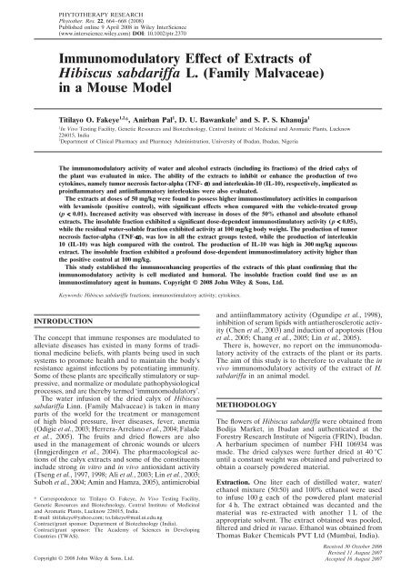
PHYTOTHERAPY RESEARCHPhytother. Res. 22, 664–668 (2008)664 Published online 9 April 2008 in Wiley InterScience T. O. FAKEYE ET AL.(www.interscience.wiley.com) DOI: 10.1002/ptr.2370<strong>Immunomodulatory</strong> Effect <strong>of</strong> Extracts <strong>of</strong><strong>Hibiscus</strong> sabdariffa L. (Family Malvaceae)in a Mouse ModelTitilayo O. Fakeye 1,2 *, Anirban Pal 1 , D. U. Bawankule 1 and S. P. S. Khanuja 11 In Vivo Testing Facility, Genetic Resources and Biotechnology, Central Institute <strong>of</strong> Medicinal and Aromatic Plants, Lucknow226015, India2 Department <strong>of</strong> Clinical Pharmacy and Pharmacy Administration, University <strong>of</strong> Ibadan, Ibadan, NigeriaThe immunomodulatory activity <strong>of</strong> water and alcohol <strong>extracts</strong> (including its fractions) <strong>of</strong> the dried calyx <strong>of</strong>the plant was evaluated in mice. The ability <strong>of</strong> the <strong>extracts</strong> to inhibit or enhance the production <strong>of</strong> twocytokines, namely tumor necrosis factor-alpha (TNF- α) and interleukin-10 (IL-10), respectively, implicated asproinflammatory and antiinflammatory interleukins were also evaluated.The <strong>extracts</strong> at doses <strong>of</strong> 50 mg/kg were found to possess higher immunostimulatory activities in comparisonwith levamisole (positive control), with significant <strong>effect</strong>s when compared with the vehicle-treated group(p < 0.01). Increased activity was observed with increase in doses <strong>of</strong> the 50% ethanol and absolute ethanol<strong>extracts</strong>. The insoluble fraction exhibited a significant dose-dependent immunostimulatory activity (p < 0.05),while the residual water-soluble fraction exhibited activity at 100 mg/kg body weight. The production <strong>of</strong> tumornecrosis factor-alpha (TNF-α), was low in all the extract groups tested, while the production <strong>of</strong> interleukin10 (IL-10) was high compared with the control. The production <strong>of</strong> IL-10 was high in 300 mg/kg aqueousextract. The insoluble fraction exhibited a pr<strong>of</strong>ound dose-dependent immunostimulatory activity higher thanthe positive control at 100 mg/kg.This study established the immunoenhancing properties <strong>of</strong> the <strong>extracts</strong> <strong>of</strong> this plant confirming that theimmunomodulatory activity is cell mediated and humoral. The insoluble fraction could find use as animmunostimulatory agent in humans. Copyright © 2008 John Wiley & Sons, Ltd.Keywords: <strong>Hibiscus</strong> sabdariffa fractions; immunostimulatory activity; cytokines.INTRODUCTIONThe concept that immune responses are modulated toalleviate diseases has existed in many forms <strong>of</strong> traditionalmedicine beliefs, with plants being used in suchsystems to promote health and to maintain the body’sresistance against infections by potentiating immunity.Some <strong>of</strong> these plants are specifically stimulatory or suppressive,and normalize or modulate pathophysiologicalprocesses, and are thereby termed ‘immunomodulatory’.The water infusion <strong>of</strong> the dried calyx <strong>of</strong> <strong>Hibiscus</strong>sabdariffa Linn. (Family Malvaceae) is taken in manyparts <strong>of</strong> the world for the treatment or management<strong>of</strong> high blood pressure, liver diseases, fever, anemia(Odigie et al., 2003; Herrera-Arrelano et al., 2004; Faladeet al., 2005). The fruits and dried flowers are alsoused in the management <strong>of</strong> chronic wounds or ulcers(Inngjerdingen et al., 2004). The pharmacological actions<strong>of</strong> the calyx <strong>extracts</strong> and some <strong>of</strong> the constituentsinclude strong in vitro and in vivo antioxidant activity(Tseng et al., 1997, 1998; Ali et al., 2003; Lin et al., 2003;Suboh et al., 2004; Amin and Hamza, 2005), antimicrobial* Correspondence to: Titilayo O. Fakeye, In Vivo Testing Facility,Genetic Resources and Biotechnology, Central Institute <strong>of</strong> Medicinaland Aromatic Plants, Lucknow 226015, India.E-mail: titifakeye@yahoo.com; to.fakeye@mail.ui.edu.ngContract/grant sponsor: Department <strong>of</strong> Biotechnology (India).Contract/grant sponsor: The Academy <strong>of</strong> Sciences in DevelopingCountries (TWAS).and antiinflammatory activity (Ogundipe et al., 1998),inhibition <strong>of</strong> serum lipids with antiatherosclerotic activity(Chen et al., 2003) and induction <strong>of</strong> apoptosis (Houet al., 2005; Chang et al., 2005; Lin et al., 2005).There is, however, no report on the immunomodulatoryactivity <strong>of</strong> the <strong>extracts</strong> <strong>of</strong> the plant or its parts.The aim <strong>of</strong> this study is to therefore to evaluate the invivo immunomodulatory activity <strong>of</strong> the extract <strong>of</strong> H.sabdariffa in an animal model.METHODOLOGYThe flowers <strong>of</strong> <strong>Hibiscus</strong> sabdariffa were obtained fromBodija Market, in Ibadan and authenticated at theForestry Research Institute <strong>of</strong> Nigeria (FRIN), Ibadan.A herbarium specimen <strong>of</strong> number FHI 106934 wasmade. The dried calyxes were further dried at 40 °Cuntil a constant weight was obtained and pulverized toobtain a coarsely powdered material.Extraction. One liter each <strong>of</strong> distilled water, water/ethanol mixture (50:50) and 100% ethanol were usedto infuse 100 g each <strong>of</strong> the powdered plant materialfor 4 h. The extract obtained was decanted and thematerial was re-extracted with another 1 L <strong>of</strong> theappropriate solvent. The extract obtained was pooled,filtered and dried in vacuo. Ethanol was obtained fromThomas Baker Chemicals PVT Ltd (Mumbai, India).Copyright © 2008 John Wiley & Sons, Ltd. Phytother. Received Res. 22, 30 664–668 October (2008) 2006Revised DOI: 11 August 10.1002/ptr 2007Copyright © 2008 John Wiley & Sons, Ltd.Accepted 16 August 2007
IMMUNOMODULATORY EFFECT OF HIBISCUS SABDARIFFA 665Extracts from the 50% ethanol/water mixture werefurther partitioned successively into dichloromethane(Merck KGaA, Darmstadt, Germany), ethyl acetate(Thomas Baker Chemicals PVT Ltd, Mumbai, India)and butanol (Thomas Baker Chemicals PVT Ltd,Mumbai, India).Anthocyanin determination. The anthocyanin content<strong>of</strong> the dried calyx was determined calculating forcyanindin-3-glucoside using a colorimetric method basedon the ability <strong>of</strong> anthocyanins to produce a color at pH1.0 that disappears at pH 4.5. This characteristic reactionis produced by a pH dependent structural transformation<strong>of</strong> the chromophore. The colored oxoniumion predominates at pH 1.0, while the non-coloredhemiketal is present at pH 4.5 allowing the accurateand fast determination <strong>of</strong> total anthocyanins, still withthe presence <strong>of</strong> polymeric pigments and other compounds(Wrolstad et al., 2005). Briefly, 1 g <strong>of</strong> the plantmaterial was extracted with the appropriate solvent anddiluted with buffer solutions at pH 1.00 and pH 4.5.The difference in the absorbance at 510 and 700 nm atthe different pH was used in calculating the monomericanthocyanin pigment present in 1 g <strong>of</strong> the plant materialusing the following equation:Total anthocyanins (mg/L)= A × MW × DF × ∑ × l (1)where A, absorbance =(A 510nm − A 700nm )pH 1.0 − (A 510nm − A 700nm )pH 4.5;MW, molecular weight; DF, dilution factor;∑ = molar extinction coefficient( L × mol −1 × cm −1 ).<strong>Immunomodulatory</strong> activity. This activity was evaluatedusing red blood cell-induced immunostimulationin a mouse model. Swiss albino mice weighing between18 g and 24 g consisting <strong>of</strong> five animals per group wereused for the study. Aqueous (A), 50% ethanol (AE) orabsolute ethanol (E) <strong>extracts</strong> were administered at 5,50 or 300 mg/kg body weight dose to albino mice withan oral gavage needle daily for 28 days. In a separateexperiment, the insoluble fraction (EAC) obtained fromthe 50% ethanol extract were administered at 50 and100 mg/kg and the residual water-soluble fraction at100 mg/kg body weight. Levamisole (0.6818 mg/kg) orallywas used as a positive control, while the vehicle-controlgroup was administered 0.5 mL <strong>of</strong> water daily. Thenegative control group was administered intraperitonealcyclophosphamide (200 mg/kg) on day 5.On day 7, 200 μL <strong>of</strong> 10% <strong>of</strong> New Zealand rabbitred blood cell (RRBC) suspension in normal salinewas administered intraperitoneally. This was repeated14 days later as a booster dose. The body weight <strong>of</strong>the animals was recorded weekly. On day 28, all theanimals were bled and the serum collected was storedat −20 °C for further studies.Haemagglutination test was performed by adding100 μL <strong>of</strong> rabbit RBC (6 × 10 3 cells per mL) to 25 μL <strong>of</strong>the serial two fold dilutions <strong>of</strong> the serum in Alsever’ssolution in U-bottom microtitre plates, shaken andallowed to stand for 4 h at 25 °C. Rabbit RBC-settingpatterns were then read. The haemagglutination (HA)titer was expressed as the reciprocal <strong>of</strong> the highestdilution <strong>of</strong> the serum showing definite agglutinationformation as opposed to smooth dot in the center <strong>of</strong>the well.Cytokine determination. Due to a prior study (Ogundipeet al., 1998) showing that extract <strong>of</strong> the dried calyxpossess antiinflammatory activity in carrageenaninducedrat paw model, two cytokines, namely tumornecrosis factor-α (TNF-α) and interleukin-10 (IL-10)implicated as proinflammatory and antiinflammatoryinterleukins, respectively, were evaluated using enzymelinkedimmunosorbant assay (ELISA) for the quantitativemeasurement <strong>of</strong> mouse TNF-α, and IL-10 (Endogen (R)Mouse ELISA kit, Pierce Biotechnology, Inc, Rockford).Sera from animals in the same group were pooled forthe assay.To 50 μL <strong>of</strong> pooled serum or standard concentrations<strong>of</strong> lyophilized recombinant mouse tissue necrosisfactor-alpha, biotin-labelled detecting antibody reagentwas added in tumor necrosis factor-alpha precoatedwells. Incubation was done for 2 h at 37 °C. Thereafter,a 100 μL dilution <strong>of</strong> 1:400 streptavidin conjugated withhighly purified horseradish peroxidase was added toeach well and further incubated for 30 min.For determination <strong>of</strong> IL-10, to each endogen mouseinterleukin-10 pre-coated well was added 50 μL assaybuffer. The same amount <strong>of</strong> standards (containingknown concentrations <strong>of</strong> lyophilized recombinant mouseIL-10) or samples from the pooled sera was added tothe wells and incubated at 37 °C for 3 h. After washingthe plates, 50 μL <strong>of</strong> biotin-labelled detecting antibodyreagent was added to each well, incubated for 1 h, afterwhich 100 μL <strong>of</strong> 1:400 diluted streptavidin-horseradishperoxidase was added and incubated for 30 min. Toboth IL-10 and TNF-α plates, 100 μL <strong>of</strong> premixed3,3′,5,5′-tetremethylbenzidine substrate solution wasadded to each well and developed in the dark for 30 min.The blue color reaction obtained was stopped with100 μL <strong>of</strong> 0.18 M sulphuric acid. The difference inabsorbance was measured using a spectrophotometerplate reader (Versamax, Molecular Devices, USA)at 450 nm and 550 nm. The difference in absorbancewas read <strong>of</strong>f the calibration curve obtained from thestandard concentrations. The concentration <strong>of</strong> eachcytokine was determined in picogram per milliliter<strong>of</strong> serum.Statistical analysis. The <strong>effect</strong>s <strong>of</strong> the <strong>extracts</strong> and fractionson haemagglutination titer and other parameterswere compared with the controls using the one-wayanalysis <strong>of</strong> variance with Dunnett’s post hoc test. Thelevel <strong>of</strong> significance was set at p < 0.05 using GraphPadInstat (R) statistical analytical s<strong>of</strong>tware.RESULTS AND DISCUSSIONThe yield <strong>of</strong> the <strong>extracts</strong> was found to be 18.54% withethanol (E), 15.18% with water (A) and 15.0% in 50%ethanol (AE). Ethanol was observed to extract the leastanthocyanin (as cyanidine-3-glucoside) <strong>of</strong> 1.23 mg/gplant material, while the highest was found with 50%ethanol (3.83 mg/g) and water (3.22 mg/g).The summary <strong>of</strong> the results for the immunomodulationtests are shown in Tables 1 and 2. The <strong>extracts</strong> didnot cause significant weight changes in the animals whencompared with the vehicle group. Cyclophosphamidecaused a reduction in the weight <strong>of</strong> the animals ascan be seen in Table 1. As expected, there was a greatCopyright © 2008 John Wiley & Sons, Ltd. Phytother. Res. 22, 664–668 (2008)DOI: 10.1002/ptr
666 T. O. FAKEYE ET AL.Table 1. The immunostimulatory <strong>effect</strong>s <strong>of</strong> the <strong>extracts</strong> <strong>of</strong> <strong>Hibiscus</strong> sabdariffa in a mouse model (mean ± SD, n = 5)Treatment groupParameterWeight changes RBC (10 6 ) WBC (10 3 ) <strong>of</strong> HA titer <strong>of</strong> TNF-α IL-10in animals in animals animals animals (pg/mL) (pg/mL)A 5 mg/kg 16.30 ± 2.99 7.67 ± 1.78 6.14 ± 2.25 384.00 ± 215.46 a 262 1315A 50 mg/kg 12.53 ± 7.50 8.32 ± 0.76 7.22 ± 2.46 768.00 ± 147.80 b 294 1151A 300 mg/kg 7.87 ± 5.79 7.96 ± 1.31 12.10 ± 4.05 a 384.00 ± 73.90 a 191 4321AE 5 mg/kg 3.75 ± 12.34 9.21 ± 1.76 7.93 ± 3.72 264.00 ± 98.09 a 207 1314AE 50 mg/kg 19.82 ± 7.30 7.54 ± 1.92 6.85 ± 4.06 640.00 ± 384.00 b 215 2086AE 300 mg/kg 16.61 ± 3.39 7.59 ± 0.77 8.99 ± 3.35 768.00 ± 147.80 b 814 2187E 5 mg/kg 14.88 ± 7.43 7.75 ± 1.47 7.41 ± 3.70 213.33 ± 42.67 a 396 3150E 50 mg/kg 16.27 ± 9.09 7.62 ± 0.49 5.63 ± 1.90 512.00 ± 258.00 a 302 1026E 300 mg/kg 2.86 ± 8.91 6.33 ± 0.86 6.27 ± 2.58 597.33 ± 198.12 b 246 3299Levamisole 20.99 ± 8.62 7.31 ± 0.50 11.29 ± 4.30 a 448.00 ± 73.90 a 396 2872Cyclophosphamide −9.83 ± 5.98 b 6.04 ± 0.33 5.24 ± 1.32 28.00 ± 4.00 1270 1006Vehicle group 10.16 ± 6.21 7.00 ± 0.72 6.71 ± 1.03 53.33 ± 37.33 1263 1138A, aqueous extract; AE, 50% ethanol extract; E, ethanol extract.Significant difference a p < 0.05; b p < 0.01 in comparison with the vehicle control, water.Table 2. The immunostimulatory activities <strong>of</strong> the fractions <strong>of</strong> 50% ethanol extract in a mouse model (mean ± SD, n = 5)TreatmentParameterWeight changes WBC (10 3 ) HA titer TNF-α IL-10in animals (%) <strong>of</strong> animals <strong>of</strong> animals (pg/mL) (pg/mL)EAC 50 mg/kg 8.57 ± 2.02 2.63 ± 0.44 b 144.00 ± 123.59 261 6 747EAC 100 mg/kg 5.20 ± 2.57 a 12.60 ± 1.35 b 928.67 ± 619.00 b 7 14 190RWSF 100 mg/kg −2.85 ± 2.84 b 8.80 ± 0.80 223.50 ± 32.50 233 5 219Levamisole 11.19 ± 2.04 8.48 ± 2.04 597.33 ± 225.97 240 9 631Cyclophosphamide 2.56 ± 2.44 4.33 ± 0.30 0.00 1108 3 902Vehicle group 16.31 ± 3.96 7.22 ± 0.25 0.00 1090 4 132EAC, ethylacetate-insoluble fraction; RWSF, residual water-soluble fraction.ap < 0.05; b p < 0.01 when compared with the vehicle-treated group.reduction in the leukocyte count <strong>of</strong> cyclophosphamidetreatedanimals, but not for the <strong>extracts</strong>.The results (Tables 1 and 2) implied that the mechanism<strong>of</strong> immunostimulation with <strong>extracts</strong> <strong>of</strong> <strong>Hibiscus</strong>sabdariffa may not be mainly through activity <strong>of</strong> thewhite blood cells. There seems to be an interplaybetween humoral (B-cells) and cell-mediated (T cells)immunity since the administration <strong>of</strong> the <strong>extracts</strong> andthe fractions led to changes in the production <strong>of</strong> twocytokines, TNF-α and IL-10 (cell mediated immunity)and antibodies titer (humoral immunity). However,the evaluation <strong>of</strong> immunoglobulin G and M, anotherhumoral immunity response parameter, could not bedetermined due to unavailability <strong>of</strong> the reagents. Thehaemagglutination, HA, titer <strong>of</strong> the <strong>extracts</strong> was foundto be significantly different when compared with thevehicle group. An increase in antibody production wasobserved with increase in doses <strong>of</strong> both the 50% ethanoland ethanol <strong>extracts</strong>. For the aqueous extract however,there was a decline <strong>of</strong> activity at 300 mg/kg. Failure tohave a dose-dependent, immunostimulatory activitycould have been due to the fact that with plant <strong>extracts</strong>,immune response is not always directly related withthe immunomodulator concentration. This phenomenonhas been noticed in other studies (Rezaeipoor et al.,2000; Tiwari et al., 2004), and has been proposed to bedue to different constituents present at different concentrationsin the fractions/<strong>extracts</strong> thereby affectingsaturation <strong>of</strong> cells responsible for the immune responsedifferently. It is not impossible that some <strong>of</strong> the constituentshave immunosuppressive activity while theothers have immunostimulant activity, an event usuallyobserved with <strong>extracts</strong> <strong>of</strong> plant origin. However, allthe <strong>extracts</strong> at different doses showed better immunostimulatoryactivity than observed either with the vehiclecontrol, water, or with the negative control, cyclophosphamide,with some having better activity than thepositive control, levamisole.The fractions, however, exhibited a different pr<strong>of</strong>ile.The insoluble fraction (EAC) exhibited a dose-dependentactivity. The group administered with 100 mg/kg bodyweight exhibited more than a 6-fold increase in immunostimulatoryactivity when compared with the 50 mg/kgdose using the HA titer as a response parameter(Table 2). The residual water-soluble fraction (RWSF)showed activity at 100 mg/kg body weight. There weresignificant changes in the white blood cell (WBC) pr<strong>of</strong>ile<strong>of</strong> the fractions. The WBC count was significantlyhigher for EAC 100 mg/kg (p < 0.01) and lower for theEAC 50 mg/kg (p < 0.05) than the control. The RWSFled to weight loss (p < 0.01), while the EAC dosesled to weight gain which was found to be significantlydifferent with the 100 mg/kg (p < 0.05).The <strong>extracts</strong> generally led to low TNF-α and highIL-10 production. It is not unusual to find both proandantiinflammatory types simultaneously producedCopyright © 2008 John Wiley & Sons, Ltd. Phytother. Res. 22, 664–668 (2008)DOI: 10.1002/ptr
IMMUNOMODULATORY EFFECT OF HIBISCUS SABDARIFFA 667in situ though with tightly controlled and balanced activity(Fujiwara and Kobayashi, 2005). Concentrations <strong>of</strong>the TNF-α, though not exhibiting dose-dependence inany <strong>of</strong> the extract group, were lowest in the aqueousextract at 300 mg/kg. With the ethanol extract, therewas a noticeable reduction in the amount <strong>of</strong> TNF-α asthe dose <strong>of</strong> the extract increases. The fractions alsoshowed a significant reduction in the production <strong>of</strong> TNFαwhen compared with the negative control and thevehicle control group. In none <strong>of</strong> the groups, however,was TNF-α concentration as high as in the negative orvehicle groups (Tables 1 and 2). Previous studies haveshown that the <strong>extracts</strong> and some <strong>of</strong> its constituentshave in vitro activity against some carcinoma cell lines(Lin et al., 2005; Hou et al., 2005) and in vivo activityagainst atherosclerosis in animal models (Chen et al.,2003). Previous studies have also implicated highlevels <strong>of</strong> TNF-α in clinical diagnoses <strong>of</strong> atherosclerosis(Bruunsgaard et al., 2000) suggesting that low TNF-αmay lead to reduction <strong>of</strong> calcification observed withatherosclerosis (Tintut et al., 2000), an activity that hasbeen confirmed with <strong>extracts</strong> <strong>of</strong> <strong>Hibiscus</strong> sabdariffa inan atherosclerotic animal model (Chen et al., 2003)though prolonged exposure to TNF-α may lead to someantiinflammatory <strong>effect</strong>s.Interleukin 10 (IL-10), usually referred to as an antiinflammatorycytokine, was high in the serum <strong>of</strong> animalson 300 mg/kg <strong>of</strong> <strong>extracts</strong> comparable to levamisole (thepositive control). Animals administered with A300 mg/kg had the highest concentrations bearing out the results<strong>of</strong> an earlier study by Ogundipe et al. (1998) that thewater extract <strong>of</strong> the dried calyx <strong>of</strong> H. sabdariffa hasantiinflammatory activity in carrageenan-induced ratpaw oedema. The insoluble fraction produced highamount <strong>of</strong> IL-10 in a dose-dependent manner, and thismay well be one <strong>of</strong> the fractions responsible for theimmunostimulatory activity. The residual water-solublefraction at 100 mg/kg body weight also caused an increasein IL-10 concentration at 100 mg/kg. IL-10 hasbeen known to down-regulate factors present in inflammationand tumors. An extract or chemical compoundthat could increase antiinflammatory interleukin, forexample, IL-10, and reduce the formation <strong>of</strong> a proinflammatoryfactor such as TNF-α may be found to be<strong>of</strong> great usefulness in preventing or reducing formation<strong>of</strong> atherosclerotic plaques, may also help in reduction<strong>of</strong> stress, and may also possess antitumour activity.The low level and high level <strong>of</strong> TNF-alpha and IL-10respectively confirmed that the <strong>extracts</strong> and the fractionstested, apart from having these biological properties,may also stimulate immunomodulation throughthe activities <strong>of</strong> cytoleukines.CONCLUSIONThe study revealed that, to some extent the <strong>extracts</strong>,and to a large extent, two fractions <strong>of</strong> the plant possessthe ability to stimulate the immune system in vivo.The activity may be as a result <strong>of</strong> interplay between theproduction <strong>of</strong> interleukin 10, inhibition <strong>of</strong> tumor necrosisfactor-alpha and the <strong>effect</strong> <strong>of</strong> B-cells responsible forantibody production.One <strong>of</strong> the fractions showed good possibilities <strong>of</strong>being developed into a drug entity that may be used tostimulate immunity as an adjunct to therapy in immunosuppresseddisease conditions. However, the extract, asit is taken in humans as a beverage may be <strong>of</strong> benefit inenhancing immunity. Further studies need to be doneto evaluate the exact mechanism <strong>of</strong> action.AcknowledgementsThis study was jointly sponsored by the Department <strong>of</strong> Biotechnology(India) and The Academy <strong>of</strong> Sciences in Developing Countries(TWAS) for the DBT/TWAS postdoctoral fellowship awarded tothe first author. The authors also acknowledge the support in form<strong>of</strong> facilities provided by the Central Institute <strong>of</strong> Medicinal andAromatic Plants, Lucknow, India.REFERENCESAli BH, Mousa HM, El-Mougy S. 2003. The <strong>effect</strong> <strong>of</strong> a waterextract and anthocyanins <strong>of</strong> <strong>Hibiscus</strong> sabdariffa L onparacetamol-induced hepatoxicity in rats. Phytother Res17: 56–59.Amin A, Hamza AA. 2005. Hepatoprotective <strong>effect</strong>s <strong>of</strong> <strong>Hibiscus</strong>,Rosmarinus and Salvia on azathioprine-induced toxicity inrats. Life Sci 77: 266–278.Bruunsgaard H, Skinhoj P, Pedersen AN, Schroll M,Pedersen BK. 2000. Ageing, tumor necrosis factor-alpha(TNF-alpha) and atherosclerosis. Clin Exp Immunol 121:255–260.Chang YC, Huang HP, Hsu JD, Yang SF, Wang CJ. 2005.<strong>Hibiscus</strong> anthocyanins rich extract-induced apoptotic celldeath in human promyelocytic leukemia cells. Toxicol ApplPharmacol 205: 201–212.Chen CC, Hsu JD, Wang SF et al. 2003. <strong>Hibiscus</strong> sabdariffaextract inhibits the development <strong>of</strong> atherosclerosis incholesterol-fed rabbits. J Agric Food Chem 51: 5472–5477.Falade OS, Otemuyiwa IO, Oladipo OO, Oyedapo OO, AkinpeluBA, Adewusi SRA. 2005. The chemical composition andmembrane stability activity <strong>of</strong> some herbs used in localtherapy for anaemia. J Ethnopharmacol 102: 15–22.Fujiwara N, Kobayashi K. 2005. Macrophages in inflammation.Curr Drug Targets Inflamm Allergy 4: 281–286.Herrera-Arellano A, Flores-Romero S, Chavez-Soto MA,Tortoriello J. 2004. Effectiveness and tolerability <strong>of</strong> a standardizedextract from <strong>Hibiscus</strong> sabdariffa in patients withmild to moderate hypertension: a controlled and randomizedclinical trial. Phytomedicine 11: 375–382.Hou DX, Tong X, Terahara N, Luo D, Fujii M. 2005. Delphinidin3-sambubioside, a <strong>Hibiscus</strong> anthocyanin, induces apoptosisin human leukemia cells through reactive oxygen speciesmediatedmitochondrial pathway. Arch Biochem Biophys440: 101–109.Inngjerdingen K, Nergård CS, Diallo D, Mounkoro PP, PaulsenBS. 2004. An ethnopharmacological survey <strong>of</strong> plants usedfor wound healing in Dogonland, Mali, West Africa.J Ethnopharmacol 92: 233–244.Lin HH, Huang HP, Huang CC, Chen JH, Wang CJ. 2005. <strong>Hibiscus</strong>polyphenol-rich extract induces apoptosis in human gastriccarcinoma cells via p53 phosphorylation and p38 MAPK/FasL cascade pathway. Mol Carcinogen 43: 86–89.Lin WL, Hsieh YJ, Chou FP, Wang CJ, Cheng MT, Tseng TH.2003. <strong>Hibiscus</strong> protocatechuic acid inhibits lipopolysaccharideinducedrat hepatic damage. Arch Toxicol 77: 42–47.Odigie IP, Ettarh RR, Adigun SA. 2003. Chronic administration<strong>of</strong> aqueous extract <strong>of</strong> <strong>Hibiscus</strong> sabdariffa attenuateshypertension and reverses cardiac hypertrophy in 2K-1Chypertensive rats. J Ethnopharmacol 86: 181–185.Copyright © 2008 John Wiley & Sons, Ltd. Phytother. Res. 22, 664–668 (2008)DOI: 10.1002/ptr
668 T. O. FAKEYE ET AL.Ogundipe OO, Moody JO, Oluwole FS, Fakeye TO. 1998. Antiinflammatoryand antimicrobial activities <strong>of</strong> selected NigerianPlant foods. In Proceedings <strong>of</strong> the 1st InternationalWorkshop on Herbal Medicinal Products. University <strong>of</strong>Ibadan: Ibadan, 138–146.Rezaeipoor R, Saeidnia S, Kamalinejad M. 2000. The <strong>effect</strong> <strong>of</strong>Plantago ovata on humoral immune responses in experimentalanimals. J Ethnopharmacol 72: 283–286.Suboh SM, Bilto YY, Aburjai TA. 2004. Protective <strong>effect</strong>s<strong>of</strong> selected medicinal plants against protein degradation,lipid peroxidation and deformability loss <strong>of</strong> oxidativelystressed human erythrocytes. Phytother Res 18: 280–284.Tintut Y, Patel J, Parhami F, Demer LL. 2000. Tumor necrosisfactor-alpha promotes in vitro calcification <strong>of</strong> vascular cellsvia the cAMP pathway. Circulation 102: 2636–2642.Tiwari U, Rastogi B, Singh P, Saraf DK, Vyas SP. 2004. <strong>Immunomodulatory</strong><strong>effect</strong>s <strong>of</strong> aqueous extract <strong>of</strong> Tridax procumbensin experimental animals. J Ethnopharmacol 92: 113–119.Tseng TH, Hsu JD, Lo MH et al. 1998. Inhibitory <strong>effect</strong> <strong>of</strong> <strong>Hibiscus</strong>protocatechuic acid on tumor promotion in mouse skin.Cancer Lett 126: 199–207.Tseng TH, Kao ES, Chu CY et al. 1997. Protective <strong>effect</strong>s <strong>of</strong>dried flower <strong>extracts</strong> <strong>of</strong> <strong>Hibiscus</strong> sabdariffa L. againstoxidative stress in rat primary hepatocytes. Food ChemToxicol 35: 1159–1164.Wrolstad RE, Durst RW, Lee J. 2005. Tracking color pigmentchanges in anthocyanin products. Trends Food Sci Technol16: 423–428.Copyright © 2008 John Wiley & Sons, Ltd. Phytother. Res. 22, 664–668 (2008)DOI: 10.1002/ptr


