Effect of ancylostomiasis on liver protein, amino ... - THE BIOSCAN
Effect of ancylostomiasis on liver protein, amino ... - THE BIOSCAN
Effect of ancylostomiasis on liver protein, amino ... - THE BIOSCAN
You also want an ePaper? Increase the reach of your titles
YUMPU automatically turns print PDFs into web optimized ePapers that Google loves.
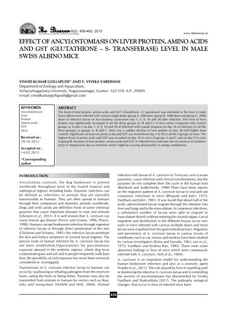
NSave Nature to Survive8(2): 459-462, 2013www.thebioscan.inEFFECT OF ANCYLOSTOMIASIS ON LIVER PROTEIN, AMINO ACIDSAND GST (GLUTATHIONE – S- TRANSFERASE) LEVEL IN MALESWISS ALBINO MICEVINOD KUMAR GOLLAPUDI* AND V. VIVEKA VARDHANIDepartment <str<strong>on</strong>g>of</str<strong>on</strong>g> Zoology and Aquaculture,AcharyaNagarjuna University, Nagarjunanagar, Guntur - 522 510. A.P., INDIAe-mail: vinodkumargollapudi@gmail.comKEYWORDSAncylostomiasisLiverProteinAmino acidsGSTMice.Received <strong>on</strong> :29.10.2012Accepted <strong>on</strong> :14.02.2013ABSTRACTThe level <str<strong>on</strong>g>of</str<strong>on</strong>g> total <strong>protein</strong>, <strong>amino</strong> acids and GST (Glutathi<strong>on</strong>e –S- transferase) was estimated in the <strong>liver</strong> <str<strong>on</strong>g>of</str<strong>on</strong>g> maleSwiss albino mice infected with various single doses (group A, 500 dose; group B, 1000 dose and group C, 2000dose) <str<strong>on</strong>g>of</str<strong>on</strong>g> infective larvae <str<strong>on</strong>g>of</str<strong>on</strong>g> Ancylostoma caninum<strong>on</strong> day 1, 4, 9, 16 and 30 after infecti<strong>on</strong>. The level <str<strong>on</strong>g>of</str<strong>on</strong>g> <strong>liver</strong><strong>protein</strong> was significantly increased in all the three groups (A, B and C) <str<strong>on</strong>g>of</str<strong>on</strong>g> mice when compared with c<strong>on</strong>trolgroups (a, b and c) <strong>on</strong> day 1, 4, 9, 16 and 30 <str<strong>on</strong>g>of</str<strong>on</strong>g> infecti<strong>on</strong> with a peak resp<strong>on</strong>se <strong>on</strong> day 16 <str<strong>on</strong>g>of</str<strong>on</strong>g> infecti<strong>on</strong> (in all thethree groups); in groups A, B and C, there was a sudden decline <str<strong>on</strong>g>of</str<strong>on</strong>g> <strong>liver</strong> <strong>protein</strong> <strong>on</strong> day 30 (still higher thanc<strong>on</strong>trol). Significant increase <str<strong>on</strong>g>of</str<strong>on</strong>g> <strong>amino</strong> acids and GST was found from day 1 to 30 in all the 3 groups <str<strong>on</strong>g>of</str<strong>on</strong>g> mice. Thehighest level <str<strong>on</strong>g>of</str<strong>on</strong>g> <strong>amino</strong> acids and GST was recorded <strong>on</strong> day 16 in mice <str<strong>on</strong>g>of</str<strong>on</strong>g> groups A and C and <strong>on</strong> day 9 in mice<str<strong>on</strong>g>of</str<strong>on</strong>g> group B. Increase <str<strong>on</strong>g>of</str<strong>on</strong>g> <strong>liver</strong> <strong>protein</strong>, <strong>amino</strong> acids and GST in infected mice indicates the occurrence <str<strong>on</strong>g>of</str<strong>on</strong>g> oxidativestress in hepatocytes due to infecti<strong>on</strong> which might be causing abnormality in energy metabolism.*Corresp<strong>on</strong>dingauthorINTRODUCTIONAncylostoma caninum, the dog hookworm is presentworldwide throughout most <str<strong>on</strong>g>of</str<strong>on</strong>g> the humid tropical andsubtropical regi<strong>on</strong>s including India. Zo<strong>on</strong>otic infecti<strong>on</strong>s canbe defined as infecti<strong>on</strong>s <str<strong>on</strong>g>of</str<strong>on</strong>g> animals that are naturallytransmissible to humans. They are <str<strong>on</strong>g>of</str<strong>on</strong>g>ten spread in humansthrough their compani<strong>on</strong> and domestic animals worldwide.Dogs and wild canids are definitive hosts <str<strong>on</strong>g>of</str<strong>on</strong>g> some intestinalparasites that cause important diseases in man and animals(Gholami et al., 2011). It is well known that A. caninum cancause human gut disease (Prociv and Croese, 1996; Prociv,1998). Humans can get hookworm infecti<strong>on</strong> through ingesti<strong>on</strong><str<strong>on</strong>g>of</str<strong>on</strong>g> infective larvae or through direct penetrati<strong>on</strong> <str<strong>on</strong>g>of</str<strong>on</strong>g> the skin(Glickman and Schantz, 1981); the infective larvae penetratethe skin and induce symptoms <str<strong>on</strong>g>of</str<strong>on</strong>g> visceral larval migrans. Theprecise route <str<strong>on</strong>g>of</str<strong>on</strong>g> human infecti<strong>on</strong> by A. caninum larvae hasnot been established.Opportunities for percutaneousexposure abound in the endemic regi<strong>on</strong>s, where dog fecesc<strong>on</strong>taminate grass and soil and in people frequently walk barefoot, the possibility <str<strong>on</strong>g>of</str<strong>on</strong>g> oral exposure has never been seriouslyc<strong>on</strong>sidered or investigated.Transmissi<strong>on</strong> <str<strong>on</strong>g>of</str<strong>on</strong>g> A. caninum infective larvae to humans canoccur by swallowing or inhaling pathogens from the reservoirhosts, eating the hosts or being bitten. Parasites may also betransmitted from animals to humans by vectors such as fleas,ticks and mosquitoes (Ostfeld and Holt, 2004). Humaninfecti<strong>on</strong> with larvae <str<strong>on</strong>g>of</str<strong>on</strong>g> A. caninum or Toxocara canis (canineparasites), cause infecti<strong>on</strong> and clinical manifestati<strong>on</strong>s, but theparasites do not complete their life cycle in the human host(Reinhard and Aufderheide, 1990).There have been reports<strong>on</strong> the migratory pattern <str<strong>on</strong>g>of</str<strong>on</strong>g> A. caninum larvae in oral and subcutaneous infecti<strong>on</strong>s in mice (Bhopale and Johri, 1975;Vardhani and Johri, 1981). It was found that about half <str<strong>on</strong>g>of</str<strong>on</strong>g> theorally administrated larvae migrate through the intestine into<strong>liver</strong> and lungs and to the musculature. In cutaneous infecti<strong>on</strong>s,a substantial number <str<strong>on</strong>g>of</str<strong>on</strong>g> larvae were able to migrate tomusculature directly without entering the visceral organs. Larvalmigrati<strong>on</strong> and distributi<strong>on</strong> in the different tissues occur veryearly in mice infected with various multiple doses and morelarvae were expelled from the gastrointestinal tract. Migrati<strong>on</strong>and persistence <str<strong>on</strong>g>of</str<strong>on</strong>g> A. caninum larvae in various tissues <str<strong>on</strong>g>of</str<strong>on</strong>g>vertebrates such as cat, mouse and m<strong>on</strong>key have been studiedby various investigators (K<strong>on</strong>o and Sawada, 1961; Lee et al.,1975; Vardhani and Krishna Rao, 1995). There were someabnormal findings in <strong>liver</strong> <str<strong>on</strong>g>of</str<strong>on</strong>g> mice which were cutaneouslyinfected with A. caninum. (Soh et al., 1969).A. caninum is an important model for understanding thehuman hookworm infecti<strong>on</strong> and also as a zo<strong>on</strong>otic agent(Franke et al., 2011). The role played by <strong>liver</strong> in expelling and/or destroying the infective A. caninum larvae and in c<strong>on</strong>trollingthe severity <str<strong>on</strong>g>of</str<strong>on</strong>g> <str<strong>on</strong>g>ancylostomiasis</str<strong>on</strong>g> has documented by VivekaVardhani and Shakunthala (2011). The pathophy siologicalchanges that occur in <strong>liver</strong> <str<strong>on</strong>g>of</str<strong>on</strong>g> infected mice have459
VINODKUMAR GOLLAPUDI AND V. VIVEKAVARDHANIreceived little attenti<strong>on</strong>. Hence, a new vista has been openedto investigate the level <str<strong>on</strong>g>of</str<strong>on</strong>g> <strong>protein</strong>, <strong>amino</strong> acids and GST in <strong>liver</strong><str<strong>on</strong>g>of</str<strong>on</strong>g> male Swiss albino mice infected with infective A. caninumlarvae.MATERIALS AND METHODSInfective A. caninum larvae were cultured following the method<str<strong>on</strong>g>of</str<strong>on</strong>g> Sen et al. (1965). Six weeks old male Swiss albino mice (25-31g) were employed in this work. Three experiments werec<strong>on</strong>ducted in the present study. Ten mice were infected orallywith a single dose i.e., 10 mice with 500 larvae/mouse (groupA), 10 mice with 1000 larvae/mouse (group B) and 10 micewith 2000 larvae/mouse (group C). Another three corresp<strong>on</strong>dinggroups (a, b and c) (10 mice in each) were kept asuninfected c<strong>on</strong>trols for comparis<strong>on</strong>. Two mice from each <str<strong>on</strong>g>of</str<strong>on</strong>g>the experimental and c<strong>on</strong>trol groups were necropsied <strong>on</strong> day1, 4, 9, 16 and 30 <str<strong>on</strong>g>of</str<strong>on</strong>g> experimental period. Tissues <str<strong>on</strong>g>of</str<strong>on</strong>g> <strong>liver</strong>were separated from normal and experimental groups <str<strong>on</strong>g>of</str<strong>on</strong>g> mice<strong>on</strong> day 1, 4, 9, 16 and 30 <str<strong>on</strong>g>of</str<strong>on</strong>g> infecti<strong>on</strong> and utilised for quantitativeestimati<strong>on</strong> <str<strong>on</strong>g>of</str<strong>on</strong>g> total <strong>protein</strong>, <strong>amino</strong> acids and GST followingthe method <str<strong>on</strong>g>of</str<strong>on</strong>g> Lowry et al. (1951), Moore and Stein (1948)and Habig et al. (1974) respectively.RESULTSResults are shown in Tables 1 to 3Protein c<strong>on</strong>tent in group AThe level <str<strong>on</strong>g>of</str<strong>on</strong>g> <strong>protein</strong> manifested in the low dose treated group(group A) was higher than the c<strong>on</strong>trol value throughout theexperimental period. The level <str<strong>on</strong>g>of</str<strong>on</strong>g> <strong>protein</strong> showed increase inan ascending fashi<strong>on</strong> from day 1 to 16 but declined <strong>on</strong> day30 (237.5µg/mg) - it was still higher than c<strong>on</strong>trols (189.08µg/mg). The level <str<strong>on</strong>g>of</str<strong>on</strong>g> <strong>protein</strong> was at peak <strong>on</strong> day 16 (291.71 µg/mg).Amino acids c<strong>on</strong>tent in group AThe level <str<strong>on</strong>g>of</str<strong>on</strong>g> <strong>amino</strong> acids found in experimental mice wasfound to be higher throughout the experimental tenure thanthe c<strong>on</strong>trols. In mice <str<strong>on</strong>g>of</str<strong>on</strong>g> group A, there is a standardizedelevati<strong>on</strong> <str<strong>on</strong>g>of</str<strong>on</strong>g> <strong>amino</strong> acids from day 1 to 30. A sudden rise wasnoticed <strong>on</strong> day 9 (806.0 µg/g) and further c<strong>on</strong>tinued in anexp<strong>on</strong>ential burst <str<strong>on</strong>g>of</str<strong>on</strong>g> <strong>amino</strong> acids by day 16 (977.0 µg/g). Thelevel <str<strong>on</strong>g>of</str<strong>on</strong>g> <strong>amino</strong> acids was at its pinnacle <strong>on</strong> day 16. But there isan abrupt fall <str<strong>on</strong>g>of</str<strong>on</strong>g> <strong>amino</strong> acids <strong>on</strong> the terminal day (30) <str<strong>on</strong>g>of</str<strong>on</strong>g>experiment (705.0 µg/g), but still it was higher than the normalvalues.GST activity in group AThe activity <str<strong>on</strong>g>of</str<strong>on</strong>g> GST in mice infected with low dose showedhigher value than the c<strong>on</strong>trols throughout the experimentalperiod. The level <str<strong>on</strong>g>of</str<strong>on</strong>g> GST in test mice recorded <strong>on</strong> day 1 (82units/mg <strong>protein</strong>) <str<strong>on</strong>g>of</str<strong>on</strong>g> infecti<strong>on</strong> was slightly higher than c<strong>on</strong>trols(79 units/mg <strong>protein</strong>). An accelerated increase <str<strong>on</strong>g>of</str<strong>on</strong>g> GST activitywas vividly manifested from day 4 (85.5 units/mg <strong>protein</strong>) to16 (96.5 units/mg <strong>protein</strong>). A slight depleted activity <str<strong>on</strong>g>of</str<strong>on</strong>g> GSTwas found <strong>on</strong> day 30 (89 units/mg <strong>protein</strong>). Nevertheless, thepenache <str<strong>on</strong>g>of</str<strong>on</strong>g> GST activity holds <strong>on</strong> day 16.Protein c<strong>on</strong>tent in group BExperimental mice <str<strong>on</strong>g>of</str<strong>on</strong>g> group B, (1000 larvae /mouse) showedenhanced <strong>protein</strong> values than the uninfected mice (group b)from day 1 to 30 <str<strong>on</strong>g>of</str<strong>on</strong>g> experimental period. Transcending andlogarithmic rise <str<strong>on</strong>g>of</str<strong>on</strong>g> <strong>protein</strong> was found from day 1 (234.69 µg/mg) to 16 (295.06 µg/mg). A steep fall in <strong>protein</strong> level wasobserved <strong>on</strong> the terminal day <str<strong>on</strong>g>of</str<strong>on</strong>g> experimental tenure i.e., day30 (267.72 µg/mg), but it was still higher than c<strong>on</strong>trols. Thelevel <str<strong>on</strong>g>of</str<strong>on</strong>g> <strong>protein</strong> in infected mice (group B) was at its epitome<strong>on</strong> day 16 (295.06 µg/mg) (the highest recorded value).Amino acids c<strong>on</strong>tent in group BThe level <str<strong>on</strong>g>of</str<strong>on</strong>g> <strong>amino</strong> acids found in mice <str<strong>on</strong>g>of</str<strong>on</strong>g> group B were higherthan c<strong>on</strong>trols from day 1 to 30 <str<strong>on</strong>g>of</str<strong>on</strong>g> infecti<strong>on</strong> period. The level <str<strong>on</strong>g>of</str<strong>on</strong>g><strong>amino</strong> acids showed a strategical enhancement from day 1(685.5 µg/g) to 9 (815.0 µg/g). But a steep fall was found <strong>on</strong>day 16 (768.5 µg/g), which further c<strong>on</strong>tinued till day 30 (703.5µg/g). There are two important points <str<strong>on</strong>g>of</str<strong>on</strong>g> interest- the level <str<strong>on</strong>g>of</str<strong>on</strong>g><strong>amino</strong> acids was higher and at its peak <strong>on</strong> day 9 (which is theinitial phase <str<strong>on</strong>g>of</str<strong>on</strong>g> the experimental period) and the level <str<strong>on</strong>g>of</str<strong>on</strong>g> <strong>amino</strong>acids was lower <strong>on</strong> day 4 (720 µg/g) and higher <strong>on</strong> day 16(768.5 µg/g).GST activityin group BThe experimental mice <str<strong>on</strong>g>of</str<strong>on</strong>g> group B which were parasitized bymedium dose (1000 L/mice) <str<strong>on</strong>g>of</str<strong>on</strong>g> A. caninum larvae showedheightened activity than the c<strong>on</strong>trols throughout theexperimental period. The level <str<strong>on</strong>g>of</str<strong>on</strong>g> GST showed heavy rise <strong>on</strong>day 9 (122.5 µg/mg) and 16 (119 units/mg <strong>protein</strong>) with apeak resp<strong>on</strong>se <strong>on</strong> day 9. But the GST activity slightly halted <strong>on</strong>the final day <str<strong>on</strong>g>of</str<strong>on</strong>g> experiment i.e., day 30 (103 units/mg <strong>protein</strong>),but it is higher than c<strong>on</strong>trol value.Protein c<strong>on</strong>tent in group CExperimental mice <str<strong>on</strong>g>of</str<strong>on</strong>g> group C showed higher <strong>protein</strong> c<strong>on</strong>tentin <strong>liver</strong> than the c<strong>on</strong>trols (group c) from day 1 to 30 <str<strong>on</strong>g>of</str<strong>on</strong>g>experimental period. The level <str<strong>on</strong>g>of</str<strong>on</strong>g> <strong>protein</strong> in experimental miceincreased from day 1 to 16. But it is slightly receded by day30; it was still above the normal value. An interesting facetnoticed in experimental mice <str<strong>on</strong>g>of</str<strong>on</strong>g> group C was the <strong>protein</strong> levelwhich stood nearly the same <strong>on</strong> day 9 (336.18 µg/mg) and 16(336.10 µg/mg); this proves that the enigmatic correlance <str<strong>on</strong>g>of</str<strong>on</strong>g><strong>protein</strong> metabolism in survivalence is in deep distress in cellularintegrity cast by parasitic infecti<strong>on</strong>s.Amino acids c<strong>on</strong>tent in group CThe level <str<strong>on</strong>g>of</str<strong>on</strong>g> <strong>amino</strong> acid in test mice <str<strong>on</strong>g>of</str<strong>on</strong>g> group C was found tobe higher than the normals from day 1 to 30 <str<strong>on</strong>g>of</str<strong>on</strong>g> infecti<strong>on</strong>period. In case <str<strong>on</strong>g>of</str<strong>on</strong>g> group C, the <strong>amino</strong> acid level tend to rise inexp<strong>on</strong>ential manner from day 1 (704 µg/g) to 9 (853 µg/g). Butit culminated in a steep declinati<strong>on</strong> <strong>on</strong> day 16 (809.05 µg/g)which further c<strong>on</strong>tinued till day 30 (762.5 µg/g). The <strong>amino</strong>acids level in test mice was at the pinnacle in <strong>on</strong> day 9.GST activity in group CThe activity <str<strong>on</strong>g>of</str<strong>on</strong>g> GST in infected mice (group C) showed highervalues than the c<strong>on</strong>trols. The experimental mice showedgradual increase from day 1 (91.5 units/mg <strong>protein</strong>) to 16(131 units/mg <strong>protein</strong>). There was a marked decrease by day30 (102.5 units/mg <strong>protein</strong>) but it is still higher than c<strong>on</strong>trolsand the recorded values <str<strong>on</strong>g>of</str<strong>on</strong>g> day 1 and 4 <str<strong>on</strong>g>of</str<strong>on</strong>g> infecti<strong>on</strong>. It isinteresting to note that the pace <str<strong>on</strong>g>of</str<strong>on</strong>g> GST is found to be nearer<strong>on</strong> day 9 (129 units/mg <strong>protein</strong>) and 16 (131 units/mg <strong>protein</strong>).460
EFFECT OF ANCYLOSTOMIASISTable 1: Protein (µg/mg) c<strong>on</strong>tent in the <strong>liver</strong> <str<strong>on</strong>g>of</str<strong>on</strong>g> infected (groups A, B and C) and uninfected (groups a, b and c) mice at different days <str<strong>on</strong>g>of</str<strong>on</strong>g>experiment. Values are expressed in the mean derived from 5 observati<strong>on</strong>sDays Experimental groups C<strong>on</strong>trol groups<str<strong>on</strong>g>of</str<strong>on</strong>g> Necropsy A B C A b c500 larvae/mouse 1000 larvae/mouse 2000 larvae/mouse1 200.67 234.69 279.69 189.09 189.09 189.094 222.37 260.75 299.67 189.07 189.06 189.089 263.68 280.64 336.18 189.05 189.09 189.0616 291.71 295.06 336.1 189.03 189.07 189.0130 237.5 267.72 315.31 189.08 189.04 189.04Table 2: Amino acids (µg/g), c<strong>on</strong>tent in the <strong>liver</strong> <str<strong>on</strong>g>of</str<strong>on</strong>g> infected (groups A, B and C) and uninfected (groups a, b and c) mice at different days <str<strong>on</strong>g>of</str<strong>on</strong>g>experiment. Values are expressed in the mean derived from 5 observati<strong>on</strong>sDays Experimental groups C<strong>on</strong>trol groups<str<strong>on</strong>g>of</str<strong>on</strong>g> necropsy A B C A b c500 larvae/mouse 1000 larvae/mouse 2000 larvae/mouse1 612 685.5 704 595 595 5954 681.5 720 789.5 596 596.5 596.59 806 815 853 595.5 595.5 595.516 977 768.5 809.5 596 596 59630 705 703.5 762.5 595 595.5 595Table 3: GST (units/mg <strong>protein</strong>) c<strong>on</strong>tent in the <strong>liver</strong> <str<strong>on</strong>g>of</str<strong>on</strong>g> infected (groups A, B and C) and uninfected (groups a, b and c) mice at different days<str<strong>on</strong>g>of</str<strong>on</strong>g> experiment. Values are expressed in the mean derived from 5 observati<strong>on</strong>sDays Experimental groups C<strong>on</strong>trol groups<str<strong>on</strong>g>of</str<strong>on</strong>g>necropsy A B C A b c500 larvae/mouse 1000 larvae/mouse 2000 larvae/mouse1 82 89 91.5 79 79 79.54 85.5 96.5 95 79.5 79.5 79.59 94.5 122.5 129 79 79 7916 96.5 119 131 79 79.5 79.530 89 103 102.5 80 80 80DISCUSSIONLiver plays a vital role in fighting infecti<strong>on</strong>s, particularly thosearising in the bowel. Hepatocytes play a significant role insynthesizing molecules that are utilized elsewhere to supporthomeostasis, in c<strong>on</strong>verting molecules <str<strong>on</strong>g>of</str<strong>on</strong>g> <strong>on</strong>e type to another,and in regulating energy balances. Amino acid metabolism,urea synthesis and <strong>protein</strong> metabolism occur in the <strong>liver</strong>.Evidence <str<strong>on</strong>g>of</str<strong>on</strong>g> the defects in each <str<strong>on</strong>g>of</str<strong>on</strong>g> these parameters may beobserved in hepatic diseases. These include abnormal plasmalevels <str<strong>on</strong>g>of</str<strong>on</strong>g> <strong>amino</strong> acids, <strong>protein</strong>s, urea and amm<strong>on</strong>ia.Experimental mice which received a single dose <str<strong>on</strong>g>of</str<strong>on</strong>g> 500 (groupA), 1000 (group B) and 2000 larvae (group C) showedremarkable changes in the quantitative values <str<strong>on</strong>g>of</str<strong>on</strong>g> <strong>protein</strong>, <strong>amino</strong>acids and GST. It is <str<strong>on</strong>g>of</str<strong>on</strong>g> interest to note that mice which received500 (group A), 1000 (group B) and 2000 (group C) dose <str<strong>on</strong>g>of</str<strong>on</strong>g>larvae showed much variati<strong>on</strong> in the biochemical assays (inthe level <str<strong>on</strong>g>of</str<strong>on</strong>g> <strong>protein</strong>, <strong>amino</strong> acids and GST). Increased <strong>protein</strong>and decreased RNA synthesis were found in the <strong>liver</strong> <str<strong>on</strong>g>of</str<strong>on</strong>g> guineapigs infected with Coxiella burneti (Mallavia and Paretsky, 1967;Thomps<strong>on</strong> and Paretsky, 1973).The increase <str<strong>on</strong>g>of</str<strong>on</strong>g> <strong>protein</strong> and <strong>amino</strong> acids in <strong>liver</strong> (in all the 3experimental groups) suggests that the abnormal physiologicalchanges were caused by the various single oral doses <str<strong>on</strong>g>of</str<strong>on</strong>g>infective larvae in the host system. It is clear that the <strong>liver</strong> hasunderg<strong>on</strong>e stress due to the oral dose <str<strong>on</strong>g>of</str<strong>on</strong>g> infective larvae. PerOl<str<strong>on</strong>g>of</str<strong>on</strong>g> et al. (1984) reported increased <strong>protein</strong> and <strong>amino</strong> acidsynthesis in <strong>liver</strong> <str<strong>on</strong>g>of</str<strong>on</strong>g> rats due to trauma and trauma complicatedby sepsis. It is known that oxygen required for energymetabolism in aerobic organisms may generate reactive oxygenand nitrogen radicals that impair a wide variety <str<strong>on</strong>g>of</str<strong>on</strong>g> biologicalmolecules like <strong>protein</strong>, DNA and lipid. The oxidative stress in<strong>liver</strong> is maximised due to the hepatocytes death and/or theabnormality in energy metabolism in <strong>liver</strong> as suggested byInoue et al. (2003). The increase or decrease <str<strong>on</strong>g>of</str<strong>on</strong>g> <strong>protein</strong> mightbe due to the modulati<strong>on</strong> in cell functi<strong>on</strong>s or damage in cellularc<strong>on</strong>stituents like <strong>protein</strong>s. Ames (1989), Stadtman (1992), Kasai(1997) and Inoue et al. (2003) also reported that in most <str<strong>on</strong>g>of</str<strong>on</strong>g>the mammalian tissues the Reactive Oxygen Species (ROS)react rapidly with a variety <str<strong>on</strong>g>of</str<strong>on</strong>g> molecules and thereby damagingthe cellular c<strong>on</strong>stituents such as lipids, <strong>protein</strong>s and DNA.Schwizer (1996), Beckman and Ames (1997) also opined thatlarge amount <str<strong>on</strong>g>of</str<strong>on</strong>g> reactive oxygen, nitrogen species generatedby activated lymphocytes, inflammati<strong>on</strong> and oxidative stressare the major causes that impair <strong>protein</strong>s and DNA in thetissues <str<strong>on</strong>g>of</str<strong>on</strong>g> mammals.The occurrence <str<strong>on</strong>g>of</str<strong>on</strong>g> intensive proteolytic changes in the <strong>liver</strong> <str<strong>on</strong>g>of</str<strong>on</strong>g>singly inflected animals c<strong>on</strong>tributes to the elevati<strong>on</strong> <str<strong>on</strong>g>of</str<strong>on</strong>g> <strong>amino</strong>acids. Sie (1985) also suggested that intensive proteolysis (dueto pathogenic stress) may lead to the enhanced level <str<strong>on</strong>g>of</str<strong>on</strong>g> <strong>amino</strong>acids. Dukan and Nystrom (1999) suggested that reactiveoxygen species/free radicals may react with <strong>protein</strong>s and/orDNA leading to their denaturati<strong>on</strong>. Flagg et al. (1994) alsosuggested that Mel<strong>on</strong>dialdehyde (<strong>on</strong>e <str<strong>on</strong>g>of</str<strong>on</strong>g> the end products <str<strong>on</strong>g>of</str<strong>on</strong>g>lipid preoxidati<strong>on</strong>) reacts with primary <strong>amino</strong> groups <str<strong>on</strong>g>of</str<strong>on</strong>g> <strong>protein</strong>sand with nucleic acid bases <str<strong>on</strong>g>of</str<strong>on</strong>g> DNA, resulting in tissue damage461
VINODKUMAR GOLLAPUDI AND V. VIVEKAVARDHANIand breakage <str<strong>on</strong>g>of</str<strong>on</strong>g> DNA and RNA. Mice infected withPlasmodium berghei showed alterati<strong>on</strong>s in hepatic histologyand phospho<strong>protein</strong> levels in mice (Maniam et al., 2012).ACKNOWLEDGEMENTSOne <str<strong>on</strong>g>of</str<strong>on</strong>g> the authors (VVV) is thankful to UGC for providingfinancial assistance in the form <str<strong>on</strong>g>of</str<strong>on</strong>g> MRP. The author (GVK) isindebted to UGC for being a partial benefactor in carrying outthe present investigati<strong>on</strong>s.REFERENCESAmes, B. 1989. Endogenous oxidative DNA damage, aging and cancer.Free Radic. Res. Commun. 7: 121-128.Beckman, B. and Ames, B. 1997. Oxidative decay <str<strong>on</strong>g>of</str<strong>on</strong>g> DNA. J. Biol.Chem. 272: 19633-19636.Bhopale, M. K. and Johri, G. N. 1975. Experimental infecti<strong>on</strong> <str<strong>on</strong>g>of</str<strong>on</strong>g>Ancylostoma caninumin mice. II. Migrati<strong>on</strong> and distributi<strong>on</strong> <str<strong>on</strong>g>of</str<strong>on</strong>g> larvaein tissues after oral infecti<strong>on</strong>. J. Helminthol. 49: 179-185.Dukan, S. and Nystrom, T. 1999. Oxidative stress defense anddeteriorati<strong>on</strong> <str<strong>on</strong>g>of</str<strong>on</strong>g> growth arrested Escherichia coli cells. J. Biol. Chem.274: 26027-26032.Flagg, E. W., Coates, R. J. and Greenberg, R. S. 1994. Epidemiologicstudies <str<strong>on</strong>g>of</str<strong>on</strong>g> antioxidants and cancer in humans.J.Am. Coll. Nutr. 14:419-427.Franke, E., Strube, C., Epe, C., Welz, C. and Schnieder, T. 2011.Larval migrati<strong>on</strong> in PERL chambers as an in vitro model forpercutaneous infecti<strong>on</strong> stimulates feeding in the canine hookwormAncylostoma caninum. Parasites and Vectors. 4: 7 (http://www.parasites and vectors.com/c<strong>on</strong>tent/4/1/7).Gholami, S., Daryani, A., Sharif, M., Amouei, A. and MobediI, I.2011. Seroepidemiological survey <str<strong>on</strong>g>of</str<strong>on</strong>g> helminthic parasites <str<strong>on</strong>g>of</str<strong>on</strong>g> straydogs in Sari City, Northern Iran. Pak. J. Bio. Sci. 14(2): 133-137.Glickman, L. T. and Schantz, P. M. 1981. Epidemiology andpathogenesis <str<strong>on</strong>g>of</str<strong>on</strong>g> zo<strong>on</strong>otic toxocariasis. Epidemiol. Rev. 3: 230-250.Habig, W. H., Pabst, M. J. and Jokoby, W. B. 1974. Glutathi<strong>on</strong>e S-transferase. The first enzyme step in mercapturic acid formati<strong>on</strong>. J.Biol. Chem. 249: 7130-7139.Inoue, M., Sato, E. F., Nishikawa, M., Park, A. M., Kira, Y., Imada, I.and Utsumi, K. 2003. Cross talk <str<strong>on</strong>g>of</str<strong>on</strong>g> nitric oxide, oxygen radicals andsuperoxide dismutase regulates the energy metabolism and cell deathand determines the fates <str<strong>on</strong>g>of</str<strong>on</strong>g> aerobic life. Antioxidants and RedoxSignal. 5(4): 475-484.Kasai, H. 1997. Analysis <str<strong>on</strong>g>of</str<strong>on</strong>g> a form <str<strong>on</strong>g>of</str<strong>on</strong>g> oxidative DNA damage, 8-hydroxy-2’-deoxyguanosine, as a marker <str<strong>on</strong>g>of</str<strong>on</strong>g> cellular oxidative stressduring carcino genesis. Mutat. Res. 387: 147-163.K<strong>on</strong>o, M. and Sawada, T. 1961. Studies <strong>on</strong> hookworm immunity(Ancylostoma cannum). I. Preliminary report <strong>on</strong> immunity acquiredby infecti<strong>on</strong> with larvae.kitakautoIgaku. II. 416-422 (in Japanese withEnglish Summary).Lee, K. T., Little, M. D. and Beaver, P. C. 1975. Intracellular (musclefiber)habitat <str<strong>on</strong>g>of</str<strong>on</strong>g> Ancylostoma caninum in some mammalian hosts.The J. Parasitol. 61(4): 589-598.Lowry, H., Rosebrough, N. I., Far, A. L. and Ranall, R. J. 1951.Protein measurement with Folinphenol reagent. J. Biol. Chem. 193:265-275.Mallavia, L.P. and Paretsky, D. (1967). Physiology <str<strong>on</strong>g>of</str<strong>on</strong>g> rickettsiae. VII.Amino acid incorporati<strong>on</strong> by Coxiella burnetiiand by infected hosts.J. Bacteriol. 93: 1479-1483.Maniam, P., Abidin Abu Hassan, Z., Noor, E. and MohdSidek, H.2012. Changes in hepatic phospho<strong>protein</strong> levels in mice infectedwith Plasmodium berghei. Sains Malaysiana. 41(6): 721-729.Moore, S. and Stein, W. H. 1948. Photometric ninhydrin method foruse in the chromatography <str<strong>on</strong>g>of</str<strong>on</strong>g> <strong>amino</strong> acids.J. Biol. Chem. 176: 367-388Ostfeld, R. S. and Holt, R. D. 2004. Are predators good for health?Evaluating evidence for top-down regulati<strong>on</strong> <str<strong>on</strong>g>of</str<strong>on</strong>g> zo<strong>on</strong>otic diseasereservoirs. Fr<strong>on</strong>t. Ecol. Envir<strong>on</strong>. 2: 13-20.Per Ol<str<strong>on</strong>g>of</str<strong>on</strong>g>, H., Rudolf, J., Lars, K., Peter, P. and Torsten, S. 1984.Changes <str<strong>on</strong>g>of</str<strong>on</strong>g> <strong>protein</strong> metabolism in <strong>liver</strong> and skeletal muscles followingtrauma complicated by sepsis. J. Trauma. 24(3): 224-228.Prociv, P. 1998. Zo<strong>on</strong>otic hookworm infecti<strong>on</strong>s (Ancylostomiasis)[chapter 61]. In: Palmer S.R., Lord Soulsby Simps<strong>on</strong>, DIH, editors.Zo<strong>on</strong>oses: biology, clinical practice and public health c<strong>on</strong>trol. Oxford:Oxford University Press. pp. 803-822.Prociv, P. and Croese, J. 1996. Human enteric infecti<strong>on</strong> withAncylostoma caninum, hookworms reappraised in the light <str<strong>on</strong>g>of</str<strong>on</strong>g> a “new”zo<strong>on</strong>oses. Acta Trop. 62(1): 23-44.Reinhard, K. J. and Aufderheide, A. C. 1990. Diphyllobothriasis inpre-Columbian Chile and Peru: adaptive radiati<strong>on</strong> <str<strong>on</strong>g>of</str<strong>on</strong>g> a helminthspecies to Native American populati<strong>on</strong>s. In: Papers <strong>on</strong> paleopathology,8 th European Members meeting, Cambridge, p.18.Schwizer, M. 1996. Oxidative stress during viral infecti<strong>on</strong>: A Review.Free Radic. Biol. Med. 21: 641-649.Sen, H. G., Joshi, U. N. and Seth, D. 1965. <str<strong>on</strong>g>Effect</str<strong>on</strong>g> <str<strong>on</strong>g>of</str<strong>on</strong>g> Cortis<strong>on</strong>e up<strong>on</strong>Ancylostoma caninum infecti<strong>on</strong> in albino mice. Trans. Roy. Soc.Trop. Med. and Hyg. 59: 684-689.Sie, H. 1985. Oxidative Stress, Academic Press, New York.Soh, C. T., Im-K-II. and Lim, H. C. 1969. Studies <strong>on</strong> the transmissibility<str<strong>on</strong>g>of</str<strong>on</strong>g> pathogenic-organisms to <strong>liver</strong> by larvae <str<strong>on</strong>g>of</str<strong>on</strong>g> <strong>liver</strong> fluke andhookworm.Y<strong>on</strong>sei Med. J. 10(2): 109-116.Stadtman, E.R. (1992). Protein oxidati<strong>on</strong> and Aging. Science. 257:1220-1224.Thomps<strong>on</strong>, H. A. and Paretsky, D. 1973. Rib<strong>on</strong>ucleic acid and <strong>protein</strong>synthesis in guinea pig <strong>liver</strong> during Q fever. Infect. Immun. 7(5): 718-724.Vardhani, V. V. and Johri, G. N. 1981. The migratory behaviour andsurvival pattern <str<strong>on</strong>g>of</str<strong>on</strong>g> Ancylostoma caninum larvae in an adaptivelyimmunized host. Internat. J. Parasitol. 11(2): 145-147.Vardhani, V. V. and Krishna Rao, B. V. 1995. The relati<strong>on</strong>ship betweenserum cholesterol and parasitism in mice. Pak. J. Zool. 27(4): 373-375.Viveka Vardhani, V. and Sakunthala, G. 2011. The specific role <str<strong>on</strong>g>of</str<strong>on</strong>g><strong>liver</strong> in expelling Ancylostoma caninum larvae from the host system.The Bioscan. 6(2): 255-256.462


