Diagnostic Criteria for Nonviable Pregnancy Early in the First Trimester
Diagnostic Criteria for Nonviable Pregnancy Early in the First Trimester
Diagnostic Criteria for Nonviable Pregnancy Early in the First Trimester

You also want an ePaper? Increase the reach of your titles
YUMPU automatically turns print PDFs into web optimized ePapers that Google loves.
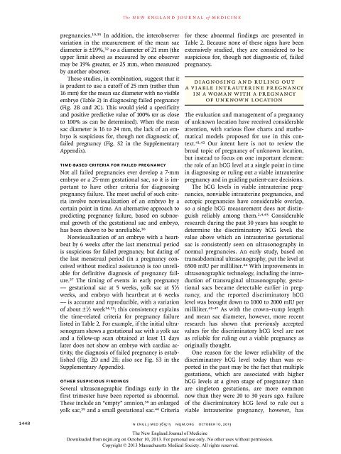
T h e n e w e ngl a nd j o u r na l o f m e dic i n epregnancies. 10,35 In addition, <strong>the</strong> <strong>in</strong>ter observervariation <strong>in</strong> <strong>the</strong> measurement of <strong>the</strong> mean sacdiameter is ±19%, 32 so a diameter of 21 mm (<strong>the</strong>upper limit above) as measured by one observermay be 19% greater, or 25 mm, when measuredby ano<strong>the</strong>r observer.These studies, <strong>in</strong> comb<strong>in</strong>ation, suggest that itis prudent to use a cutoff of 25 mm (ra<strong>the</strong>r than16 mm) <strong>for</strong> <strong>the</strong> mean sac diameter with no visibleembryo (Table 2) <strong>in</strong> diagnos<strong>in</strong>g failed pregnancy(Fig. 2B and 2C). This would yield a specificityand positive predictive value of 100% (or as closeto 100% as can be determ<strong>in</strong>ed). When <strong>the</strong> meansac diameter is 16 to 24 mm, <strong>the</strong> lack of an embryois suspicious <strong>for</strong>, though not diagnostic of,failed pregnancy (Fig. S2 <strong>in</strong> <strong>the</strong> SupplementaryAppendix).Time-based <strong>Criteria</strong> <strong>for</strong> Failed <strong>Pregnancy</strong>Not all failed pregnancies ever develop a 7-mmembryo or a 25-mm gestational sac, so it is importantto have o<strong>the</strong>r criteria <strong>for</strong> diagnos<strong>in</strong>gpregnancy failure. The most useful of such criteria<strong>in</strong>volve nonvisualization of an embryo by acerta<strong>in</strong> po<strong>in</strong>t <strong>in</strong> time. An alternative approach topredict<strong>in</strong>g pregnancy failure, based on subnormalgrowth of <strong>the</strong> gestational sac and embryo,has been shown to be unreliable. 36Nonvisualization of an embryo with a heartbeatby 6 weeks after <strong>the</strong> last menstrual periodis suspicious <strong>for</strong> failed pregnancy, but dat<strong>in</strong>g of<strong>the</strong> last menstrual period (<strong>in</strong> a pregnancy conceivedwithout medical assistance) is too unreliable<strong>for</strong> def<strong>in</strong>itive diagnosis of pregnancy failure.37 The tim<strong>in</strong>g of events <strong>in</strong> early pregnancy— gestational sac at 5 weeks, yolk sac at 5 1 ∕2weeks, and embryo with heartbeat at 6 weeks— is accurate and reproducible, with a variationof about ± 1 ∕2 week 14,15 ; this consistency expla<strong>in</strong>s<strong>the</strong> time-related criteria <strong>for</strong> pregnancy failurelisted <strong>in</strong> Table 2. For example, if <strong>the</strong> <strong>in</strong>itial ultrasonogramshows a gestational sac with a yolk sacand a follow-up scan obta<strong>in</strong>ed at least 11 dayslater does not show an embryo with cardiac activity,<strong>the</strong> diagnosis of failed pregnancy is established(Fig. 2D and 2E; also see Fig. S3 <strong>in</strong> <strong>the</strong>Supplementary Appendix).O<strong>the</strong>r Suspicious F<strong>in</strong>d<strong>in</strong>gsSeveral ultrasonographic f<strong>in</strong>d<strong>in</strong>gs early <strong>in</strong> <strong>the</strong>first trimester have been reported as abnormal.These <strong>in</strong>clude an “empty” amnion, 38 an enlargedyolk sac, 39 and a small gestational sac. 40 <strong>Criteria</strong><strong>for</strong> <strong>the</strong>se abnormal f<strong>in</strong>d<strong>in</strong>gs are presented <strong>in</strong>Table 2. Because none of <strong>the</strong>se signs have beenextensively studied, <strong>the</strong>y are considered to besuspicious <strong>for</strong>, though not diagnostic of, failedpregnancy.Di agnos<strong>in</strong>g a nd Rul<strong>in</strong>g Ou ta V iable Intr auter <strong>in</strong>e Pr egnancy<strong>in</strong> a Woman with a <strong>Pregnancy</strong>of Unknown LocationThe evaluation and management of a pregnancyof unknown location have received considerableattention, with various flow charts and ma<strong>the</strong>maticalmodels proposed <strong>for</strong> use <strong>in</strong> this context.41,42 Our <strong>in</strong>tent here is not to review <strong>the</strong>broad topic of pregnancy of unknown location,but <strong>in</strong>stead to focus on one important element:<strong>the</strong> role of an hCG level at a s<strong>in</strong>gle po<strong>in</strong>t <strong>in</strong> time<strong>in</strong> diagnos<strong>in</strong>g or rul<strong>in</strong>g out a viable <strong>in</strong>trauter<strong>in</strong>epregnancy and <strong>in</strong> guid<strong>in</strong>g patient-care decisions.The hCG levels <strong>in</strong> viable <strong>in</strong>trauter<strong>in</strong>e pregnancies,nonviable <strong>in</strong>trauter<strong>in</strong>e pregnancies, andectopic pregnancies have considerable overlap,so a s<strong>in</strong>gle hCG measurement does not dist<strong>in</strong>guishreliably among <strong>the</strong>m. 2,4,43 Considerableresearch dur<strong>in</strong>g <strong>the</strong> past 30 years has sought todeterm<strong>in</strong>e <strong>the</strong> discrim<strong>in</strong>atory hCG level: <strong>the</strong>value above which an <strong>in</strong>trauter<strong>in</strong>e gestationalsac is consistently seen on ultra sonography <strong>in</strong>normal pregnancies. An early study, based ontransabdom<strong>in</strong>al ultrasonography, put <strong>the</strong> level at6500 mIU per milliliter. 44 With improvements <strong>in</strong>ultrasonographic technology, <strong>in</strong>clud<strong>in</strong>g <strong>the</strong> <strong>in</strong>troductionof transvag<strong>in</strong>al ultrasonography, gestationalsacs became detectable earlier <strong>in</strong> pregnancy,and <strong>the</strong> reported discrim<strong>in</strong>atory hCGlevel was brought down to 1000 to 2000 mIU permilliliter. 45-47 As with <strong>the</strong> crown–rump lengthand mean sac diameter, however, more recentresearch has shown that previously acceptedvalues <strong>for</strong> <strong>the</strong> discrim<strong>in</strong>atory hCG level are notas reliable <strong>for</strong> rul<strong>in</strong>g out a viable pregnancy asorig<strong>in</strong>ally thought.One reason <strong>for</strong> <strong>the</strong> lower reliability of <strong>the</strong>discrim<strong>in</strong>atory hCG level today than was reported<strong>in</strong> <strong>the</strong> past may be <strong>the</strong> fact that multiplegestations, which are associated with higherhCG levels at a given stage of pregnancy thanare s<strong>in</strong>gleton gestations, are more commonnow than <strong>the</strong>y were 20 to 30 years ago. Failureof <strong>the</strong> discrim<strong>in</strong>atory hCG level to rule out aviable <strong>in</strong>trauter<strong>in</strong>e pregnancy, however, has1448n engl j med 369;15 nejm.org october 10, 2013The New England Journal of Medic<strong>in</strong>eDownloaded from nejm.org on October 10, 2013. For personal use only. No o<strong>the</strong>r uses without permission.Copyright © 2013 Massachusetts Medical Society. All rights reserved.