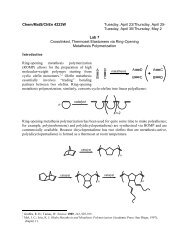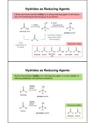
Chem/MatS/ChEn 4223W Tuesday, April 2/Thursday, April 4 ...
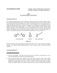
Chem/MatS/ChEn 4223W Tuesday, April 2/Thursday, April 4 ...
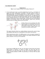
Chem/MatS/ChEn 4223W Tuesday, April 2/Thursday, April 4 ...

You also want an ePaper? Increase the reach of your titles
YUMPU automatically turns print PDFs into web optimized ePapers that Google loves.
understand how to control the temperature of the hot stage, and how to insert a sample intothe viewing area.You will be using a program called TSView to save images. Load a sample of crystallineimidazole onto the stage. Then, on the computer, make sure that the program is open, andshows an image. Focus the microscope while watching the computer screen. (Sometimes thefocal plane of the camera is slightly different from the focal plane of the eyepieces.) Noticethat the area imaged by the camera is smaller that the view in the eyepieces; as a result,sometimes it helps to use the eyepieces for rough positioning of features into the camera’sview. Rotate the sample stage to see the effect of polarizer and analyzer angle on the featuresof the crystal.In TSView, press the Snap button to take a picture. In the dialog box that pops up, type in thename you want to give the image file.With the heat stage temperature set near room temperature, place the stage micrometer in thestage, in the viewing column. Remove the analyzer so that you can see the micrometer. (Itisn’t polarizing, so you can’t see it if you don’t.) The micrometer slide has a set of ruled linesthat you will use to calibrate distances in your other images. Focus on these lines in theeyepieces, and then center and straighten them (horizontal/vertical) in the camera view. Takea picture.Sprinkle a small amount of your filtered PLLA onto the last third of a 3” x 1” microscopeslide, and then cover this with another slide. Be careful to keep the slides as free of dust aspossible; you can help this by wearing gloves while you prepare your samples, and bykeeping the box of unused slides closed.Set the hot stage to 120 °C. Wait until the stage reaches the set temperature beforecontinuing.Heat your sample slide on a hot plate, pre-set to ~190 °C, in order to melt the polymer solid.A minute or so after you see your material melt, press down on the hot cover slip withsomething other than your finger to spread the liquid material.Immediately slip your slide sandwich into the hot stage on the microscope. As your materialcools, move your sample around and try to find spherulitic nuclei in your sample using theeyepieces. As soon as you center a few spherulites nucleating in the TSView window, focusas best you can, and immediately take an image. Continue to take images in TSView every20 seconds or so, and record the run time of each image. If you don’t ever observespherulites, make another sample and try again. If you still don’t observe spherulites at thispoint, cool the stage to 90 °C (instead of 120 °C), make another sample, and try again. If youdon’t observe spherulites at this point, it may be because your polymer does not crystallize.Continue to collect images until the spherulites have grown into one another. Even if one sideof a spherulite has stopped growing, you can always use a different direction to determine theradius, so keep imaging until nothing is growing.Repeat these instructions for all of the group’s samples. (You may want to use differentfilename prefixes for your different runs.)Once you are finished, save your files to a flash drive.