15. Abnormalities of the amniotic fluid volume
15. Abnormalities of the amniotic fluid volume
15. Abnormalities of the amniotic fluid volume

You also want an ePaper? Increase the reach of your titles
YUMPU automatically turns print PDFs into web optimized ePapers that Google loves.
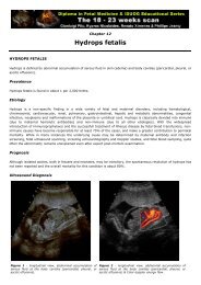
Chapter 14<strong>Abnormalities</strong> <strong>of</strong> <strong>the</strong> <strong>amniotic</strong> <strong>fluid</strong> <strong>volume</strong>Amniotic <strong>fluid</strong> is produced by fetal urination but in <strong>the</strong> first 16 weeks <strong>of</strong> gestation additional sources include <strong>the</strong>placenta, <strong>amniotic</strong> membranes, umbilical cord and fetal skin. Removal <strong>of</strong> <strong>amniotic</strong> <strong>fluid</strong> is by fetal swallowing.Ultrasonographically, <strong>the</strong> diagnosis <strong>of</strong> polyhydramnios or oligohydramnios is made when <strong>the</strong>re is excessive or virtualabsence <strong>of</strong> echo-free spaces around <strong>the</strong> fetus.OLIGOHYDRAMNIOS/ANHYDRAMNIOSOligohydramnios means reduced <strong>amniotic</strong> <strong>fluid</strong> and anhydramnios means absence <strong>of</strong> <strong>amniotic</strong> <strong>fluid</strong>.PrevalenceOligohydramnios in <strong>the</strong> second trimester is found in about 1 per 500 pregnancies.EtiologyOligohydramnios in <strong>the</strong> second trimester is usually <strong>the</strong> result <strong>of</strong> preterm premature rupture <strong>of</strong> <strong>the</strong> membranes,uteroplacental insufficiency and urinary tract malformations (bilateral renal agenesis, multicystic or polycystickidneys, or urethral obstruction).DiagnosisThe diagnosis <strong>of</strong> oligohydramnios is usually made subjectively.Quantitative criteria include:(a) <strong>the</strong> largest single pocket <strong>of</strong> <strong>amniotic</strong> <strong>fluid</strong> being 1 cm or less, or(b) <strong>amniotic</strong> <strong>fluid</strong> index (<strong>the</strong> sum <strong>of</strong> <strong>the</strong> vertical measurements <strong>of</strong> <strong>the</strong> largest pockets <strong>of</strong> <strong>amniotic</strong> <strong>fluid</strong> in <strong>the</strong> fourquadrants <strong>of</strong> <strong>the</strong> uterus) <strong>of</strong> less than 5 cm.Anecoic areas simulating pockets <strong>of</strong> <strong>amniotic</strong> <strong>fluid</strong>Color Doppler energy at <strong>the</strong> umbilical cordIn <strong>the</strong> absence <strong>of</strong> <strong>the</strong> "accoustic window" normally provided by <strong>the</strong> <strong>amniotic</strong> <strong>fluid</strong>, and <strong>the</strong> "undesirable" postures<strong>of</strong>ten adopted by <strong>the</strong>se fetuses, confident exclusion <strong>of</strong> fetal defects may be impossible. Never<strong>the</strong>less, <strong>the</strong> detection <strong>of</strong>a dilated blader in urethral obstruction and enlarged echogenic or multicystic kidneys in renal disease should berelatively easy. The main difficulty is in <strong>the</strong> differential diagnosis <strong>of</strong> renal agenesis. In cases <strong>of</strong> preterm prelabour
upture <strong>of</strong> <strong>the</strong> membranes, detailed questioning <strong>of</strong> <strong>the</strong> mo<strong>the</strong>r may reveal a history <strong>of</strong> chronic leakage <strong>of</strong> <strong>amniotic</strong><strong>fluid</strong>. Fur<strong>the</strong>rmore, in uteroplacental insufficiency, Doppler blood flow studies will <strong>of</strong>ten demomstrate high impedanceto flow in <strong>the</strong> placental circulation and redistribution in <strong>the</strong> fetal circulation. In <strong>the</strong> remaining cases, intra-<strong>amniotic</strong>instillation <strong>of</strong> normal saline may help improve ultrasonographic examination and lead to <strong>the</strong> diagnosis <strong>of</strong> fetalabnormalities like renal agenesis.PrognosisBilateral renal agenesis, multicystic or polycystic kidneys are lethal abnormalities, usually in <strong>the</strong> neonatal period dueto pulmonary hypoplasia. Preterm rupture <strong>of</strong> membranes at 20 weeks or earlier is associated with a poor prognosis;about 40% miscarry within five days <strong>of</strong> membrane rupture due to chorioamnionitis and in <strong>the</strong> remaining 60% <strong>of</strong>pregnancies more than 50% <strong>of</strong> neonates die due to pulmonary hypoplasia. Uteroplacental insufficiency resulting inoligohydramnios at 18-23 weeks is very severe and <strong>the</strong> most likely outcome is intrauterine death.POLYHYDRAMNIOSPolyhydramnios means increased or excessive <strong>amniotic</strong> <strong>fluid</strong> <strong>volume</strong>.PrevalencePolyhydramnios in <strong>the</strong> second trimester is found in about 1 per 200 pregnancies.EtiologyThere are essentially two major causes <strong>of</strong> polyhydramnios; reduced fetal swallowing or absorption <strong>of</strong> <strong>amniotic</strong> <strong>fluid</strong>and increased fetal urination. Reduced fetal swallowing may be due to craniospinal defects (such as anencephaly),facial tumours, gastrointestinal obstruction (such as esophageal atresia, duodenal atresia and small bowelobstruction), compressive pulmonary disorders (such as pleural effusions, diaphragmatic hernia or cystic adenomatoidmalformation <strong>of</strong> <strong>the</strong> lungs), narrow thoracic cage (due to skeletal dysplasias), and fetal akinesia deformationsequence (due to neuromascular impairement <strong>of</strong> fetal swallowing). Increased fetal urination is observed in maternaldiabetes mellitus and maternal uremia (increased glucose and urea cause osmotic diuresis), hyperdynamic fetalcirculation due to fetal anemia (due to red cell isoimmunization or congenital infection) or fetal and placental tumoursor cutaneous arteriovenous malformations (such as sacrococcygeal teratoma, placental chorioangioma), or twin-totwin transfusion syndrome.DiagnosisThe diagnosis <strong>of</strong> polyhydramnios is usually made subjectively. Quantitatively, polyhydramnios is defined as an<strong>amniotic</strong> <strong>fluid</strong> index (<strong>the</strong> sum <strong>of</strong> <strong>the</strong> vertical measurements <strong>of</strong> <strong>the</strong> largest pockets <strong>of</strong> <strong>amniotic</strong> <strong>fluid</strong> in <strong>the</strong> fourquadrants <strong>of</strong> <strong>the</strong> uterus) <strong>of</strong> 20 cm or more. Alternatively, <strong>the</strong> vertical measurement <strong>of</strong> <strong>the</strong> largest single pocket <strong>of</strong><strong>amniotic</strong> <strong>fluid</strong> free <strong>of</strong> fetal parts is used to classify polyhydramnios into mild (8-11 cm), moderate (12-15 cm) andsevere (16 cm or more). Although 80% <strong>of</strong> cases with mild polyhydramnios are considered to be idiopathic, in <strong>the</strong>majority <strong>of</strong> cases with moderate or severe polyhydramnios <strong>the</strong>re are maternal or fetal disorders. In most casespolyhydramnios develops late in <strong>the</strong> second or in <strong>the</strong> third trimester <strong>of</strong> pregnancy. Acute polyhydramnios at 18-23weeks is mainly seen in association with twin-to-twin transfusion syndrome. Testing for maternal diabetes, detailedsonographic examination for anomalies, and fetal karyotyping should constitute <strong>the</strong> cornerstones <strong>of</strong> <strong>the</strong> diagnosticprotocol in <strong>the</strong> investigation <strong>of</strong> <strong>the</strong>se cases.
Moderate polyhydramniosPolyhydramnios - Diaphragmatic HerniaPrenatal <strong>the</strong>rapyThe aim is to reduce <strong>the</strong> risk <strong>of</strong> very premature delivery and <strong>the</strong> maternal discomfort that <strong>of</strong>ten accompanies severepolyhydramnios. Treatment will obviously depend on <strong>the</strong> diagnosis, and will include better glycemic control <strong>of</strong>maternal diabetes mellitus, antiarrhythmic medication for fetal hydrops due to dysrrhythmias, thoraco<strong>amniotic</strong>shunting for fetal pulmonary cysts or pleural effusions. For <strong>the</strong> o<strong>the</strong>r cases, polyhydramnios may be treated byrepeated amniocenteses every few days and drainage <strong>of</strong> large <strong>volume</strong>s <strong>of</strong> <strong>amniotic</strong> <strong>fluid</strong>. However, <strong>the</strong> procedureitself may precipitate premature labour. An alternative and effective method <strong>of</strong> treatment is maternal administration<strong>of</strong> indomethacin; however, this drug may cause fetal ductul constriction and close monitoring by serial fetalechocardiographic studies is necessary. In twin-to-twin transfusion syndrome, presenting with acute polyhydramniosat 18-23 weeks <strong>of</strong> gestation, endocopic laser occlusion <strong>of</strong> placental anastomoses or serial amniodrainage may becarried out.PrognosisThis depends on <strong>the</strong> cause <strong>of</strong> polyhydramnios.Diagnosis <strong>of</strong> Fetal <strong>Abnormalities</strong> - The 18-23 weeks scanCopyright 2002 © by <strong>the</strong> authors, ISUOG & Fetal Medicine Foundation, London