CHAPTER 4: SCREENING FOR CERVICAL CANCER
CHAPTER 4: SCREENING FOR CERVICAL CANCER
CHAPTER 4: SCREENING FOR CERVICAL CANCER

Create successful ePaper yourself
Turn your PDF publications into a flip-book with our unique Google optimized e-Paper software.
794<strong>CHAPTER</strong> 4: <strong>SCREENING</strong> <strong>FOR</strong> <strong>CERVICAL</strong> <strong>CANCER</strong>
Chapter 4: Screening for Cervical Cancer 81<strong>CHAPTER</strong> 4: <strong>SCREENING</strong> <strong>FOR</strong> <strong>CERVICAL</strong> <strong>CANCER</strong>Key points4• Screening is testing of all women at risk of cervical cancer, most of whom will bewithout symptoms.• Screening aims to detect precancerous changes, which, if not treated, may leadto cancer.• Screening is only effective if there is a well organized system for follow-up andtreatment.• Women who are found to have abnormalities on screening need follow-up,diagnosis and possibly treatment, in order to prevent the development of canceror to treat cancer at an early stage.• Several tests can be used in screening for cervical cancer. The Pap smear(cytology) is the only test that has been used in large populations and that hasbeen shown to reduce cervical cancer incidence and mortality. Other tests (VIA,VILI, HPV) show promise but there is as yet no comparable evidence on theireffectiveness. Large studies are still under way.• Regardless of the test used, the key to an effective programme is to reach thelargest proportion of women at risk with quality screening and treatment.• Organized screening programmes designed and managed at the central level toreach most women at risk are preferable to opportunistic screening.Chapter 4: Screening for Cervical CancerABOUT THIS <strong>CHAPTER</strong>This chapter provides detailed information on screening, and explains why organizedscreening is superior to opportunistic screening. It describes available screening testsand their comparative advantages and disadvantages.ROLE OF THE HEALTH CARE PROVIDERThe health care provider is a central figure in any coordinated public health effort toscreen women for cervical cancer. Such an effort may include the ministry of health,programme planners, managers, laboratory technicians, health professionals andcommunity workers.The role of health care providers is to ensure that:• Women who come for screening receive appropriate information and counselling.• National guidelines on cervical cancer screening and treatment are followed.• Screening is well organized and no opportunity to screen targeted women attendingservices is missed.
82Chapter 4: Screening for Cervical Cancer4Chapter 4: Screening for Cervical Cancer• Each woman who comes for screening understands what is involved and givesinformed consent for screening and follow-up.• The screening test, treatment and referral are performed competently; patients areproperly assessed and infection control measures are strictly adhered to.• Women screened are informed of their test results, especially if they are inadequateor positive (abnormal).• Any sexual and reproductive health problems identified by either the patient or theprovider are managed appropriately.• Appropriate and confidential records are kept in the facility; the records may begiven to the woman herself.• Women who need repeat screening, further testing, referral, or care after treatmentare followed up appropriately.These responsibilities are further explained in this chapter.STORY 5Pratibha is a 37-year-old woman livingin Maharashtra, India. One day, when shereturned home from fetching water, shefound two women health workers talkingwith her husband. The health workers askedher many questions, such as how old shewas, when she married, and how manychildren she had. Then they told her aboutcervical cancer and about an opportunity forher to be screened in the village. Pratibha asked why she was selectedfor this and she was relieved to learn that all women over 30 years old inthe village were being visited and invited to attend the screening clinic.One of the advantages of attending this programme was that testing andtreatment (if needed) were free. Almost all the women invited attendedthe clinic, including Pratibha. The test was fast and painless, as she hadbeen told it would be. After the examination, the health worker emphasizedthat she should return in two weeks to get the test results. WhenPratibha returned, she was told that her test was normal and that it wouldbe important for her to repeat the test every 3 years.5 Adapted from: Alliance for Cervical Cancer Prevention. Women’s stories, women’s lives:experiences with cervical cancer screening and treatment. Seattle, WA, ACCP, 2004.
Chapter 4: Screening for Cervical Cancer 83<strong>SCREENING</strong> PROGRAMMESWhat is screening?Screening is a public health intervention used on a population at risk, or targetpopulation. Screening is not undertaken to diagnose a disease, but to identifyindividuals with a high probability of having or of developing a disease. Women targetedfor screening for cervical cancer may actually feel perfectly healthy and may see noreason to visit a health facility.Not all diseases can be screened for. The following criteria should be met by anydisease that is the object of a screening programme:• The disease must have serious public health consequences.• The disease must have a detectable preclinical stage (without symptoms).• The screening test must be simple, non-invasive, sensitive, specific, inexpensive andacceptable to the target audience.• Treatment at the preclinical stage must favourably influence the long-term courseand prognosis of the disease.• Any further testing and treatment needed must be available, accessible andaffordable for those who have a positive screening test.Cervical cancer meets these criteria.4Chapter 4: Screening for Cervical CancerScreening programmes will only be successful if the following elements are present:• high coverage 6 (80%) of the population at risk of the disease;• appropriate follow-up and management for those who are positive on screening.Efforts to increase coverage will be wasted if those who test positive are notfollowed up correctly;• effective links between programme components (e.g. from screening to diagnosisand treatment);• high quality of coverage, screening tests, diagnosis, treatment, and follow-up;• adequate resources.Cervical cancer screening aims to test the largest possible proportion of women at riskand to ensure appropriate follow-up for those who have a positive or abnormal testresult. Such women will need diagnostic testing and follow-up or treatment. Colposcopyand biopsy are often used to reach a specific diagnosis of the extent of the abnormalityin women with a positive screening test (see Chapter 5).6 “Coverage” is the proportion of women in the target age group who are screened at therecommended intervals during a given time period. The number of screening tests done is notcoverage, since this number may include women outside the target age, and women screenedmore often than recommended.
84Chapter 4: Screening for Cervical CancerOrganized and opportunistic cervical cancer screening4Chapter 4: Screening for Cervical CancerOrganized screeningOrganized screening is designed to reach the highest possible number of women atgreatest risk of cervical cancer with existing resources. It is usually planned at thenational or regional level. An organized screening programme should specify:• the target population;• screening intervals;• coverage goals;• a mechanism for inviting women to attend screening services;• the screening test or tests to be used;• the strategies to ensure that all women found positive on screening are informed oftheir result;• a mechanism for referring women for diagnosis and treatment;• treatment recommendations;• indicators for monitoring and evaluating the screening programme.Opportunistic screeningOpportunistic screening is screening done independently of an organized or populationbasedprogramme, on women who are visiting health services for other reasons.Screening may be recommended by a provider during a consultation, or requested by awoman. Opportunistic screening tends to reach younger women at lower risk, who areattending antenatal, child health and family planning services.It is generally accepted that organized screening is more cost-effective thanopportunistic screening, making better use of available resources and ensuring thatthe greatest number of women will benefit. However, both organized and opportunisticscreening can fail because of poor quality-control, low coverage of the population atrisk, overscreening of low-risk populations, and high loss to follow-up.Benefits and risks of screeningThe benefits and risks of screening should be discussed with women as part of generalhealth education and before obtaining informed consent. The benefits of screeninghave been described in previous chapters. However, as with all large efforts directedtowards healthy populations, screening for cervical cancer has the potential to produceundesirable outcomes, such as:• psychological consequences – anxiety and fear about being tested for cancer;• a mistaken belief that a positive test is a cancer diagnosis;
Chapter 4: Screening for Cervical Cancer 85• false positive test results (abnormalities reported in women whose cervix is normal),which may lead to unnecessary interventions and anxiety;• false negative test results (a normal screening test in women with cervicalabnormalities);• identification of other illnesses, for which treatment may not be available.Following the recommendations in this Guide will, in general, help to minimize theseundesirable outcomes.Target groups and frequency of screeningDecisions on the target age group and frequency of screening are usually made at thenational level, on the basis of local prevalence and incidence of cervical cancer, relatedfactors such as HIV prevalence, and availability of resources and infrastructure.All existing data on recommended ages and frequency of screening are derived fromexperience in cytology programmes. To date, there are no comparable data fromprogrammes using HPV-based and visual screening methods.When deciding on target age group and screening frequency, planners should take intoaccount the following:• HPV infection is very common in young women, but most infections are transient.• Only a small percentage of all HPV infections will lead to invasive cancer.• Cervical cancer usually develops slowly, taking 10–20 years from early precancer toinvasive cancer.• Cervical cancer is rare before the age of 30 years. Screening younger women willdetect many lesions that will never develop into cancer, will lead to considerableovertreatment, and is not cost-effective.• Screening every three years is nearly as effective as yearly screening. If resourcesare limited, screening every 5–10 years – or even just once between the ages of 35and 45 years – will significantly reduce deaths from cervical cancer.4Chapter 4: Screening for Cervical Cancer
86Chapter 4: Screening for Cervical Cancer4Chapter 4: Screening for Cervical CancerRecommended target ages and frequency of cervicalcancer screening• New programmes should start by screening women aged 30 years or more,and include younger women only when the higher-risk group has been covered.Existing organized programmes should not include women less than 25 years ofage in their target populations.• If a woman can be screened only once in her lifetime, the best age is between 35and 45 years.• For women over 50 years, a five-year screening interval is appropriate.• In the age group 25–49 years, a three-year interval can be considered if resourcesare available.• Annual screening is not recommended at any age.• Screening is not necessary for women over 65 years, provided the last twoprevious smears were negative.Special considerationsBefore embarking on a widespread screening programme, national planners shouldensure that the services needed to manage newly identified cancer cases are in place.To treat invasive cancer effectively, specialized facilities are needed; these must be inplace before a screening programme is put into effect (see Chapter 6).If a population has not previously been screened, many cases of pre-existing cancer indifferent stages will be detected in a new screening programme. Women whose diseaseis very advanced, or for whom treatment is impossible for any reason, should receivepalliative care (see Chapter 7).Screening in settings with high HIV prevalenceIn settings with high HIV prevalence, screening for cervical cancer is particularlyimportant. HIV-positive women have more persistent HPV infections, and a higherincidence of cervical precancer and, in some settings, invasive cervical cancer. WhereHIV is endemic, screening results may be positive in up to 15–20% of the targetpopulation. Cytology screening is equally effective in HIV-positive and HIV-negativewomen. Although HIV-infected women are at greater risk of precancer and cancer,screening, follow-up and treatment may not be a priority for the women themselves,who have competing health or social needs. All women, regardless of their HIV status,
Chapter 4: Screening for Cervical Cancer 87should be encouraged to be screened for cervical cancer, provided that they haveaccess to affordable services. Care should be taken not to link a positive cervicalcancer screening test to HIV testing. However, a woman with precancer may benefitfrom knowing her HIV status, especially if antiretroviral treatment (ART) is available.Screening criteria for women with known HIV infection should be developed at thenational level with these issues in mind.RecommendationWomen should be offered the same cervical cancer screening options irrespectiveof their HIV status.Screening of pregnant womenNot screening for cervical cancer during pregnancy is sometimes seen as a missedopportunity. Visits for antenatal care may be a good occasion for screening. However,integrating screening into routine antenatal care is not the best option for the followingreasons:• Most pregnant women are younger than the target group.• In some cultures, pregnant women may be reluctant to undergo a gynaecologicalexamination.• During pregnancy, interpretation of screening tests, such as cytological tests, is moredifficult.• Regression of CIN during pregnancy is minimal, but there is a significant rate ofspontaneous regression postpartum.• A biopsy for diagnosis should be taken from a pregnant woman only if invasivecancer cannot be ruled out.• Treatment of preinvasive disease is contraindicated during pregnancy.4Chapter 4: Screening for Cervical CancerWomen in the target age group who attend antenatal services should be advised toreturn for screening 12 weeks after giving birth. However, if a cervical abnormality isnoted on speculum examination, or if the provider feels there is a risk that the womanwill not return, she should be offered screening during the visit. In addition, the providercan suggest that the woman should encourage other women in the target age group inher extended family to be screened.
88Chapter 4: Screening for Cervical Cancer4Chapter 4: Screening for Cervical CancerScreening family planning clientsOpportunistic cervical cancer screening is often integrated into family planning services.Family planning counselling provides a good opportunity to discuss the benefits of cervicalcancer screening and a gynaecological examination is often more easily accepted duringa reproductive health consultation. Screening should be encouraged and performed onclients of family planning services within the target age group. Contraceptive users do notneed to be screened more often than other women, regardless of the method they use.Screening women with a reproductive tract or sexually transmitted infection (RTI/STI)Women in the target age group who present to health facilities with complaints suggestiveof RTI/STI should be examined. They should be screened for cervical cancer only if there isno visible acute infection. If the speculum examination reveals evidence of acute infection,appropriate treatment should be given and cervical cancer screening should be deferreduntil after the infection has resolved.Health education and counselling on RTI/STI should include information on HPV infection,its relation to cervical cancer, and the protection offered by safer sex behaviours, includingcondom use. Male partners too should be treated, and counselled on cervical cancerprevention. STI services aimed primarily at men should include information on HPV andcervical cancer prevention.Other opportunities for cervical cancer screeningWomen at the end of their reproductive years are at greatest risk of cervical cancer,particularly if they have never been screened. They tend to use reproductive healthservices less often than younger women, but may use other health services, e.g. formanagement of hypertension, heart disease, diabetes or infectious diseases. In addition,women in the target age group may come to a health facility with a child or relative whoneeds services. All women in the target age group who visit a facility for any reasonshould receive information and be encouraged to come for screening (see also Chapter3). General medical services at primary, secondary and tertiary levels can provide cervicalcancer screening for such women, using on-site, trained providers. If this is not possible,women should be given health education and referred to a convenient screening clinic.No missed opportunitiesCervical cancer screening programmes should also try to reach all women in thetarget age group who have contact with the health system for any reason.
Chapter 4: Screening for Cervical Cancer 89Choice of screening test to be usedThe choice of screening test or tests to be used is usually made at the national orregional level. Nevertheless, providers should have some basic knowledge of all theavailable screening tests.Decisions on the test or tests to be used may be based on:• the organization of the health system;• the funds available;• the number and type of health workers;• the availability of laboratory services and transport;• the availability and cost of the various screening tests.The test used may also be determined based on the physical proximity of services towomen; for example, it might be decided to use the Pap smear (which requires womento return for their test results) in urban areas and visual inspection with acetic acid (VIA)(for which results are immediately available) in more inaccessible rural areas in thesame country.The most extensive and long-term experience in cervical cancer screening is withcytology, which has been used in numerous countries since the 1950s. Cytology-basedscreening and treatment programmes have reduced cervical cancer incidence andmortality by as much as 80% in Canada, the USA and some Nordic countries, and by50–60% in other European countries.It has been difficult to replicate this success in low-resource settings, because of theinherent requirements of a cytology-based programme. These include highly trainedpersonnel, well equipped laboratories, transport of specimens, and an effective systemfor collecting information and following up patients. In addition, the demands of othercompeting health needs often result in a lack of resources or political will to makecervical cancer screening a priority.Because of the problems of implementing quality cytology-based screening, alternativemethods, such as visual inspection, have been developed. These methods have shownpromise in controlled research settings but have not yet been widely implemented.Their ultimate impact on cervical cancer incidence and mortality will not be known untillarge ongoing population-based studies are completed. HPV-based tests are now alsocommercially available, but have disadvantages, including the need for sophisticatedlaboratory facilities and high cost.4Chapter 4: Screening for Cervical Cancer
90Chapter 4: Screening for Cervical Cancer4Chapter 4: Screening for Cervical CancerEthical issuesDecisions on how best to use scarce resources have to weigh the extent of disabilityand death caused by different diseases, and the efficacy, cost and impact of diagnosingand treating them. While decisions about priorities are usually made at national level,providers should understand the reasons for the decisions, so that they are motivated toimplement them and can explain them to their patients (see Chapter 1). If well plannedand integrated into other sexual and reproductive health activities, screening forcervical cancer has the potential to both strengthen the health care system and improvethe health of women, particularly women over childbearing age, whose health is oftenrelatively neglected.Before a screening programme is implemented, the following elements should beconsidered to ensure an ethical and equitable approach:• Screening should be accessible to all women in the target group, including thepoorest, most vulnerable, and hardest to reach.• Patients, providers and communities should receive health education to ensureinformed decision-making on screening and treatment.• Patient record systems should ensure confidentiality.• Diagnostic tests, follow-up, and treatment should be available and accessible.• Providers should have clear guidelines on follow-up and management of womenwith positive screening results.• A referral system should be in place for other health problems, includinggynaecological disorders, discovered during the screening process.Informed choice and Informed consent 7Informed choice and informed consent are based on the ethical principles of autonomyand respect for the individual. In many cultures, the notion of consent may be acollective decision-making process involving others, such as partner,family, and village leaders. Accurate information provided through healthPS6education and counselling can ensure that women and their extendedfamilies understand the facts about cervical cancer, who is at risk, how Informed consentscreening can reduce risk, and any potential harm related to screening.Before consenting to screening, women should be given information on the specifictest to be used, the meaning and consequences of a positive test, and the availabilityof treatment. In addition, when results are not available immediately (as they are with7 Note: informed consent is not equivalent to informed choice. Consent refers to the explicitpermission given by a person for a procedure or test, once she (or he) has received sufficientinformation to make a rational personal (informed) choice.
Chapter 4: Screening for Cervical Cancer 91visual screening methods), informed consent should include explicit permission to becontacted at home or at work. Respect for autonomy requires that the choice to bescreened is voluntary and free of coercion.4Client assessmentAll clients attending for screening should have a basic assessmentbefore proceeding to the screening test. This assessment should includeinformation and counselling, informed consent, a social and clinicalhistory, and a physical examination.The history can provide useful information for guiding decisions about managementor additional examinations or tests that might benefit the patient. Because of thestigma associated with genital problems, women are often reluctant to talk abouttheir concerns or symptoms and signs. To establish and maintain trust and respect,confidentiality and privacy must be explicitly guaranteed to each woman who presentsfor screening before she is asked about her history.For cervical cancer screening, the essential components of the pelvicexamination are visual inspection of the external genitals and aPS7speculum examination. Providers should explain what is being done ateach step during the examination; if an abnormality is noted, the provider Pelvic examshould inform the woman without alarming her. Having female providersperform the physical examination, if possible, can greatly reduce reluctance to beexamined and can play a major role in making screening acceptable. When the provideris a man, the woman may request that a female companion or clinic attendant is in theroom.PS4CounsellingChapter 4: Screening for Cervical CancerSexual and reproductive health problems detected during history-takingand examinationAn integrated approach to management of sexual and reproductive health problemsduring screening can help improve the health of women, especially older women.The provider should pay particular attention to signs and symptoms suggestive ofcancer, STI, or other diseases detected during history-taking and pelvic examination. Inaddition, women should be offered an opportunity to raise personal concerns regardingsexual and reproductive health issues. Women with abnormal findings can be treated orreferred for further investigation, as appropriate.Infection prevention in cervical cancer screeningIn screening, as in all clinical activities, scrupulous attention should be given to infectionprevention. Pathogens, including HIV, can be transmitted if guidelines on handwashing,handling of instruments, and disposal of used supplies, including gloves, are neglected.
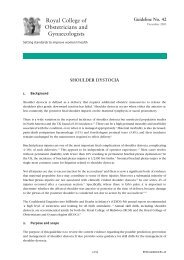
92Chapter 4: Screening for Cervical Cancer4Chapter 4: Screening for Cervical CancerUniversal precautions (see Annex 1) against spreading infection shouldAnnexbe used with all patients, whether they appear sick or well, and whether 1their HIV or other infection status is known or not. In this way, providersprotect both their patients and themselves. Providers should use only Infection preventionuncontaminated instruments, and should wear latex gloves on bothhands when performing speculum or bimanual examinations and taking specimens,and when performing procedures such as cryotherapy.<strong>SCREENING</strong> TESTSA good screening test should be:• accurate;• reproducible;• inexpensive;• easy to perform and easy to follow up;• acceptable;• safe.The following tests meet the above criteria to a greater or lesser extent:• cytology: conventional (Pap smear) and liquid-based;• HPV DNA test;• visual inspection: with acetic acid (VIA) or Lugol’s iodine (VILI).The performance of each test is described below. The strengths and limitations ofthe different tests are summarized in Table 4.1. Measurement and interpretation ofperformance characteristics are outlined in Annex 3.Annex 3CytologyTest’s performanceConventional Pap smearIn the Pap smear test, a sample of cells is taken from the transformationPS8zone of the cervix using an extended-tip wooden spatula or brush; usinga cotton swab is no longer recommended. The entire transformationPap smearzone should be sampled since this is where almost all high-gradelesions develop. The sample is then smeared onto a glass slide andimmediately fixed with a solution to preserve the cells. The slide isAnnex 2sent to a cytology laboratory where it is stained and examined usinga microscope to determine whether the cells are normal (Figure 4.1) Bethesda systemand to classify them appropriately, using the Bethesda classification(see Annex 2). The results of the Pap smear are then reported to the clinic where the
Chapter 4: Screening for Cervical Cancer 93specimen was taken. Health workers are responsible for ensuring that the woman isinformed of her result and that she receives appropriate follow-up as outlined in Annex4a. The Pap test takes less than 5 minutes to perform, is not painful,and can be done in an outpatient examination room. It is advisable toAnnexpostpone taking a Pap smear if the woman is menstruating actively, has 4aa clinically evident acute inflammation, or is pregnant. A satisfactoryAnnex 4asmear requires adequate numbers of well preserved squamousepithelial cells and an adequate endocervical/transformation zonecomponent. Each smear should be legibly labelled.Figure 4.1 Graphic representation of normal and abnormal epithelial cellsNormal squamouscell4Chapter 4: Screening for Cervical CancerHigh gradelesionThe accuracy of cytological testing depends on the quality of the services, includingsampling practices (taking and fixing the smears), and preparation and interpretation ofsmears in the laboratory. Under the best conditions in developed countries or researchsettings, conventional cytology can detect up to 84% of precancer and cancer. However,under poor conditions its sensitivity can be as low as 38%. The specificity of the test isusually over 90%.Liquid-based cytology (LBC)This refinement of conventional cytology was introduced in the mid-1990s and isincreasingly used in high-resource settings. Instead of smearing cervical cells on aslide, the provider transfers the specimen from a brush to a preservative solution. Thespecimen is sent to a laboratory where the slide is prepared. LBC is more expensivethan conventional cytology and laboratory staff need to be specially trained. However, itappears to have a number of advantages over conventional methods.• The specimens obtained are more representative of the areas sampled with fewerfalse negatives.• There are fewer unsatisfactory specimens.• Each specimen requires a shorter interpretation time, leading to increased efficiencyand cost-effectiveness.• The material collected can also be tested for HPV DNA.
94Chapter 4: Screening for Cervical Cancer4Although, as yet, no randomized controlled trial comparing LBC with conventional Papsmear has been published, several studies have shown that LBC is more sensitive thanPap smear and has almost the same specificity.Chapter 4: Screening for Cervical CancerProvidersAfter a short training course, any provider who knows how to do a speculumexamination (nurse, auxiliary or assistant nurse, midwife, clinical officer, medical doctor)can take a Pap smear.IndicationsThe following groups of women should be offered screening:• Any woman between the ages of 25 and 65 years, who has never had a Pap smearbefore or who had one 3 or more years ago (or according to national guidelines).• Women whose previous Pap smear was reported as inadequate or showed a mildabnormality.• Women who have abnormal bleeding, bleeding after intercourse or after themenopause, or other abnormal symptoms.• Women who have been found to have abnormalities on their cervix.Interpretation of smearsSmears are read in a laboratory by trained cytotechnicians, under the supervision of apathologist, who has final responsibility for the reported results. Correct interpretationof slides is crucial to a successful programme. To maintain proficiency and avoidfatigue, cytotechnicians should spend a maximum of 5 hours a day at the microscopeand should review a minimum of 3000 slides per year. Quality assurance is crucialand should be established in all cytology laboratories. The two most commonly usedmethods are rapid review of all negative slides, and full rescreening of a 10% randomsample of slides originally reported as negative. In both methods, the review is doneby another cytotechnician, with confirmation of abnormal smears by the supervisingpathologist. Current evidence shows that, of the two methods, rapid review of allnegative smears is more effective and more cost-effective. Laboratories should beequipped to read a minimum of 15 000 smears annually. 8 Therefore, cytology servicesshould not be decentralized to primary health care clinics or to small laboratories.Reliable transport of slides and test results to and from the laboratory is essential.8 Detailed information on cytology laboratories is beyond the scope of this Guide. Further informationcan be found in the references listed under “Additional resources” at the end of this chapter.
Chapter 4: Screening for Cervical Cancer 95The speed with which results are sent to the health facility is an important ele-ment of the quality of the laboratory service and the quality of care, and greatlyaffects women’s satisfaction with the service.4RECOMMENDATIONCytology is recommended for large-scale cervical cancer screening programmes,if sufficient resources exist.HPV DNA-based screening methodsNew screening procedures are based on the detection of high-risk HPVPS9DNA in vaginal or cervical smears. A sample of cells is collected fromthe cervix or vagina using a swab or small brush, and placed in a smallHPV testcontainer with a preservative solution. The specimen can be collectedby a health care provider or by the woman herself, inserting a swab deep into thevagina. Studies comparing the two collection methods have shown that self-collectionis less sensitive than provider-collection. In either case, the specimen containers aretransported to a laboratory where they are processed. HPV DNA-based tests currentlyrequire sophisticated and expensive laboratory equipment, although work is under wayto develop a more affordable and less complicated test that can be carried out in lowerlevelsettings. Detection of high-risk HPV does not necessarily mean that precanceror cancer is present; it indicates simply that there is an HPV infection. As mentionedearlier, HPV infections are extremely common in women under 35 years, and most ofthem resolve spontaneously. When detection of HPV is used as a primary screening test,the sensitivity for detection of precancer and cancer varies from 50% to 95%, with moststudies reporting high sensitivity of 85% or more. The specificity ranges from 50% to95%, with an average of 84%. In women aged 35 years or older, HPV DNA tests performbetter because in these women a positive test is more likely to be due to a persistentinfection than in younger women. The average sensitivity and specificity in this groupare 89% and 90%, respectively. The combination of cytology and HPV testing has veryhigh sensitivity and negative predictive values approaching 100% (see Annex 3). Itmight therefore be possible to increase the interval between screeningsfor women who are negative on both tests. However, performing the Annextwo tests together is expensive. The high cost, and the need for both a 3molecular laboratory and reliable methods of transport, present majorAnnex 3challenges, and the feasibility of HPV testing has not been demonstratedin low-resource settings. A new, faster, highly sensitive and less costlytest for HPV is under development but is not yet available.Chapter 4: Screening for Cervical Cancer
96Chapter 4: Screening for Cervical Cancer4Chapter 4: Screening for Cervical CancerProvidersHPV DNA testing can be done by trained providers at any level of the health caresystem, provided that there is an appropriate laboratory within a reasonable distance,and that reliable transport is available for specimens. Clinic needs for HPV testing arethe same as for Pap smears and visual methods.IndicationsHPV is not generally used on its own as the primary screening test. It is mainly used incombination with cytology to improve the sensitivity of the screening or as a triage toolto assess which women with borderline Pap results need to be referred for colposcopy.The main indication is a Pap result of “atypical cells of undetermined significance”(ASC-US). Of the women with this lesion, only those who test positive for high-risk HPVwill need to be referred for colposcopy and biopsy, significantly reducing the number ofcolposcopies.Laboratory facilitiesThe HPV laboratory requires a special clean room to avoid contamination, and highlytrained technicians. It also requires equipment and reagents as specified by themanufacturers of the test.RECOMMENDATIONHPV DNA tests as primary screening methods, at this time, are recommended foruse only in pilot projects or other closely monitored settings. They can be used inconjunction with cytological or other screening tests, where sufficient resourcesexist. HPV DNA-based screening should not begin before 30 years of age.Visual methodsTwo visual methods are available:• visual inspection with acetic acid (VIA);• visual inspection with Lugol’s iodine (VILI).PS10VIA and VILIAbnormalities are identified by inspection of the cervix without magnification, afterapplication of dilute acetic acid (vinegar) (in VIA) or Lugol’s iodine (in VILI). When vinegaris applied to abnormal cervical tissue, it temporarily turns white (acetowhite) allowingthe provider to make an immediate assessment of a positive (abnormal) or negative
Chapter 4: Screening for Cervical Cancer 97(normal) result. If iodine is applied to the cervix, precancerous and cancerous lesionsappear well-defined, thick, and mustard or saffron-yellow in colour, while squamousepithelium stains brown or black, and columnar epithelium retains its normal pinkcolour.Because they do not rely on laboratory services, VIA and VILI are promising alternativesto cytology where resources are limited. They are currently being tested in large,cross-sectional, randomized controlled trials in developing countries. Until data fromthese studies are available, VIA and VILI are recommended by WHO only for use inpilot settings, because the impact on cervical cancer incidence and mortality is stillunproven. In research settings, VIA has been shown to have an average sensitivity fordetection of precancer and cancer of almost 77%, and a range of 56% to 94%.The specificity ranges from 74% to 94% with an average of 86%. Low-levelmagnification does not improve the performance of VIA over and above that of nakedeye visualization. One study has shown that VILI can detect 92% of women withprecancer or cancer, a sensitivity considerably higher than that of either VIA or cytology.Its ability to identify women without disease is similar to that of VIA (85%), and lowerthan that of Pap smears. One study showed that VILI had a higher reproducibility thanVIA. VIA and VILI can be performed in clinics and other outpatient facilities. They areboth short procedures and cause no pain. Assessment is immediate, and no specimenis required.4Chapter 4: Screening for Cervical CancerAdvantages• VIA and VILI are relatively simple and can be taught to nurses, nurse-midwives andother health workers.• Assessment is immediate and no transport, or laboratory equipment or personnel, isneeded.• The tests are likely to be less costly than other approaches in routine use.• Results are available immediately, eliminating the need for multiple visits in mostcases, and reducing loss to follow-up.• They could potentially be used in an approach based on screening and treatingwomen in a single visit (see Chapter 5).Disadvantages• Because of the low positive predictive value of the test (see Annex3), a considerable number of women who test positive do nothave disease, resulting in excessive diagnosis and treatment, andunnecessary anxiety.• Visual tests cannot be relied on in postmenopausal women, because thetransformation zone of these women is often inside the cervical canal.Annex3Annex 3
98Chapter 4: Screening for Cervical Cancer4• There is no permanent record of the test that can be reviewed later.• VIA has mostly been evaluated as a once-in-a-lifetime screening test, and itsperformance in periodic screening has not been assessed.Chapter 4: Screening for Cervical CancerProvidersTrained nurses, nurse-midwives, nurse assistants, physicians and other health workerswith adequate and ongoing support and supervision can perform VIA. Training takes5–10 days using a competency-based approach. To maintain quality services, it isimportant that an experienced provider conducts regular assessments. Studies showthat immediately after training, providers have more false positive results. Thesedecrease in a few months as the providers gain experience.IndicationsIf adopted by a programme as a screening method, VIA and VILI are indicated for allwomen in the target age group specified in national guidelines, provided that:• They are premenopausal. Visual methods are not recommended for postmenopausalwomen, because the transition zone in these women is most often inside theendocervical canal and not visible on speculum inspection.• Both squamocolumnar junctions (i.e. the entire transformation zone) are visible.If the patient does not meet the above indications and no alternative screening methodis available in the particular clinical setting, she should be referred for a Pap smear.RECOMMENDATIONVisual screening methods (VIA and VILI), at this time, are recommended for use onlyin pilot projects or other closely monitored settings. These methods should not berecommended for postmenopausal women.
Chapter 4: Screening for Cervical Cancer 99Table 4.1 Summary of characteristics of screening methods for cervical cancerTest Procedure Strengths Limitations StatusConventionalcytology(Pap smear)Sample ofcervical cellstaken by providerand examinedby trainedcytotechnicians ina laboratory• History of long use• Widely accepted• Permanent recordof test• Training andmechanisms forquality controlestablished• Modestinvestments inexistingprogrammes canimprove services• High specificity• Results notimmediatelyavailable• Systems neededto ensure timelycommunicationof test resultsand follow-up ofwomen• Transport requiredfor specimen tolaboratory and forresults to clinic• Requires laboratoryquality assurance• Moderatesensitivity• Availablein manycountriessince the1950s• Cytologybasedprogrammeshave reducedcancermortality indevelopedcountries4Chapter 4: Screening for Cervical CancerLiquid-basedcytology (LBC)Sample of cervicalcells is obtainedwith a small brush,immersed inspecial liquid andsent to laboratoryfor processing andscreening• Fewer inadequateor unsatisfactorysamples requiringpatient call-backand rescreening• Oncecytotechniciansare proficient, LBCsamples take lesstime to review• Samples can beused for moleculartesting (such asfor HPV)• Results notimmediatelyavailable• Supplies andlaboratoryfacilities moreexpensive thanfor conventionalcytology• No controlledstudies, to date,comparingsensitivity andspecificity withconventionalcytologySelected asscreeningmethod in somedevelopedcountries(e.g. UnitedKingdom)continued next page
100Chapter 4: Screening for Cervical Cancer4Chapter 4: Screening for Cervical CancerContinued from page 99Table 4.1 Summary of characteristics of screening methods for cervical cancerTest Procedure Strengths Limitations StatusHPV DNAtestingMoleculartesting forHPV – swabtaken byprovideror womanherselfand sent tolaboratory• Collection ofspecimen simple• Automatedprocessing• Can be combinedwith Pap smearto increase thesensitivity, but thisincreases also thecost• A negative testmeans no HPV andrelated morbidityis present• The assay resultis a permanentrecord• High specificityin women overage 35• Results notimmediatelyavailable• High unit cost• Complex laboratoryrequirements andspecimen transport• Low specificityin young womenleading toovertreatment• Storage of reagentsproblematic• Commerciallyavailable and usedin some developedcountries inaddition to cytology• Lower-cost tests indevelopmentVisualmethods(VIA andVILI)Trainedproviderexaminescervix afterstaining withvinegar (inVIA) andwith Lugol’siodine (inVILI)• Relatively simpleand inexpensive• Results availableimmediately• Can be performedby wide range ofpersonnel aftershort training• Low level ofinfrastructurerequired• Can be combinedwith offer ofimmediatetreatment insingle-visitapproach• High providervariability• Lower specificityresulting in highreferral rate andovertreatment• No permanentrecord of test• Not appropriate forpostmenopausalwomen• Lack ofstandardization• Frequent retrainingneeded• Limited evidenceavailable• Only recommendedat this time for usein demonstrationprojects• Large randomizedcontrolled trialsunder way todetermine effect oncancer incidenceand mortality
Chapter 4: Screening for Cervical Cancer 101FOLLOW-UPFollow-up and management of women with an abnormal (positive) testScreening by itself will not prevent a single case of cervical cancer. An effective systemfor follow-up and treatment of women who test positive is perhaps the most importantcomponent of a successful cervical cancer prevention programme.Ideally, all women should receive the results of their test, whether negative or positive.In practice, resources will sometimes be too limited to allow this.At the very least, women whose test result is positive or abnormalAnnex4must be informed of the result and of what follow-up is needed.Follow-up should be in line with national protocols or based on the Flowchart screeningrecommendations found in Annex 4.Follow-up is essential for the woman’s welfare and for the success of the programmeand every effort should be made to contact women with positive test results.The following actions will help ensure that women with an abnormal screening test canbe reached for follow-up:• The woman’s address, or other information on how she can be reached, should benoted at the time of screening (with her consent).• During counselling and after screening, providers need to emphasize the importanceof coming back for results and follow-up care.• Every clinic should have a directory of all women with abnormal test results, with anindication of whether they have received the results and been followed up. Clinicsshould designate someone to ensure that follow-up is done.4Chapter 4: Screening for Cervical CancerFor women who do not return spontaneously as advised, providers can:• send a letter by mail;• telephone women at home or at work;• ask community health workers to contact women directly at home.Health care managers and providers can develop other locally appropriate approachesto reach women with abnormal screening tests.Health facilities need to make every effort to find women with abnormal results ifthey do not return for scheduled appointments.
102Chapter 4: Screening for Cervical Cancer4Chapter 4: Screening for Cervical CancerRecord-keepingRecords should be compatible throughout a country, so that all the data collected by thecervical cancer control programme can be compared. The information system shouldinclude every woman’s clinical record, appointments scheduled, and those kept ormissed. This can be a simple paper record or can be computer-based. A logbook can beused to register women screened and record their test results. If women need to returnlater for their results, a system must be in place to ensure that thosewith abnormal results are notified and that women who are hard toAnnex7locate are traced. Sample forms for follow-up can be found in Annex 7.Documents
Chapter 4: Screening for Cervical Cancer 103screening activities at different levels of the health systemIn the communityAt the health centre• Educate and inform the community, promote the screeningprogramme, and encourage women to attend.• Refer appropriate women for screening.• Assist women to attend screening clinics.• Assist in follow-up of women with a positive screening to ensurethat they return to the clinic for management.• Screen, using methods specified by national guidelines andintegrating screening into other services.• Train, support and supervise CHWs.• Work with CHWs to educate women, and recruit them forscreening.• Participate in campaigns to bring women at high risk for testing.• Provide counselling and health education in the clinic andcommunity.• Inform and counsel women with positive screening test results,and advise them on needed follow-up, diagnosis and treatment.• Implement an accurate patient information system, to allowproper tracking and follow-up of women after treatment.4Chapter 4: Screening for Cervical CancerAt the district hospital• Carry out screening activities as per national programme.• Inform and counsel women with positive screening test results,and advise them on needed follow-up, diagnosis and treatment.• Train, support and supervise providers at health centre level.• Manage referral systems with lower and higher levels of thehealth system.At the central hospital• Carry out screening in outpatient clinics where women are seen.• Maintain central cytology, pathology, and molecular laboratories,as feasible.• Interpret screening and histopathology results and ensure thatresults reach the screening site.• Train medical personnel, and support and supervise providers inlower-level health facilities.• Manage referral and links with lower levels of the health system.
104Chapter 4: Screening for Cervical CancerCounselling messages4Chapter 4: Screening for Cervical CancerWomen who have just had a screening test need to be told:• if anything abnormal was noted;• when the results will be available;• the date of the next appointment.Women returning for test results should be counselled on:• the result of the test and what it means;• if normal, when they need to return for repeat screening;• if inadequate or not normal, what follow-up is needed;• where and when to go for follow-up.
Chapter 4: Screening for Cervical Cancer 105ADDITIONAL RESOURCES• ACCP. Planning and implementing cervical cancer prevention programs: a manual formanagers. Seattle WA, Alliance for Cervical Cancer Prevention, 2004.• Arbyn M. A technical guideline: collection of adequate Pap smears of the uterinecervix. Brussels, Scientific Institute of Public Health, 2001.• Cervical cancer prevention: guidelines for low-resource settings. Baltimore, MD,JHPIEGO Corporation, 2001.• CHIP. Implementing cervical screening in South Africa. Volume I. A guide forprogramme managers. New York, Cervical Health Implementation Project, SouthAfrica, University of Cape Town, University of the Witwatersrand, EngenderHealth,2004.• IARC. A practical manual on visual screening for cervical neoplasia. Lyon, IARCPress,2003.• IARC. Cervix cancer screening. Lyon, IARC Press, 2005 (IARC Handbooks of CancerPrevention, Volume 10).• Infection prevention: a reference booklet for health care professionals. New York,EngenderHealth, 2001.• Infection prevention curriculum: a training course for health care providers and otherstaff at hospitals and clinics. New York, EngenderHealth, 1999.• Miller AB. Cervical cancer screening programmes, managerial guidelines. Geneva,WHO, 1992.• PATH. Planning appropriate cervical cancer prevention programs. Seattle, WA,Program for Appropriate Technology in Health, 2000.• PATH VIA/VILI curriculum. Course in visual methods for cervical cancer screening. In:Tsu V et al., Western Kenya Cervical Cancer Prevention Project Final Report. Seattle,WA, Program for Appropriate Technology in Health, 2004 (Annex 10).• Salas Diehl I, Prado Buzeta R, Muñoz Magna R. Manual de Procedimientos deLaboratorio de Citología. Washington, DC, Organización Panamericana de la Salud,2002.• WHO. Cervical cancer screening in developing countries. Report of a WHOConsultation. Geneva, 2002.4Chapter 4: Screening for Cervical Cancer
106
PS 6: Obtaining Informed Consent107PRACTICE SHEET 6: OBTAINING IN<strong>FOR</strong>MED CONSENTWhat is informed consent?Women must give informed consent before being screened for cervical cancer. Thismeans that she should understand what is to take place, including the potential risksand complications of both proceeding and not proceeding, and has given permissionfor the procedure. It should be made clear to the woman that there will be no punitiveaction if she refuses the procedure.When asking for informed consent:• Give the woman all essential information on what you are about to do and requesther consent before starting any examination or procedure. It is unethical to ask forinformed consent retroactively.• If there is a possibility that she might need to be contacted at home or at work(e.g. to give test results or remind her to return for an appointment), obtainconsent for doing so.• Family members should be included in the discussion only if the woman has givenexplicit permission.• Keep medical terminology to a minimum. Explain any technical words that have nolocal equivalent.• You may find it helpful to draw or use pictures to illustrate your explanations.• Be clear and direct; do not use words the patient will not understand, or which arevague, such as “growth” or “neoplasm”.• Do not confuse the woman by saying too much, but cover all the important issues.• Allow some time for the woman to take in what you have said. Then let her askquestions. When all the questions have been addressed, ask the woman for herformal consent.• It might be culturally important to include others, such as the woman’s partner,in the decision-making process; however, you should ensure that the woman’swishes are respected.PS 6Practice Sheet 6: Obtaining Informed ConsentExplaining practices and proceduresYou will find explanations for patients included in each chapter of this Guide andin the practice sheets. You may adapt these to individual situations to help explainprocedures in terms the patient and her family understand.
108 PS 6: Obtaining Informed ConsentPS 6Practice Sheet 6: Obtaining Informed ConsentSTEPS <strong>FOR</strong> OBTAINING IN<strong>FOR</strong>MED CONSENTPreparation1. Ensure privacy and explain that confidentiality is always respectedin your facility.PS42. Follow your facility’s regulations on obtaining informed consent.Counselling3. Apply general rules on counselling and good communication. Listencarefully and address the woman’s concerns; give her the time she needs tounderstand and to make a decision.4. Ask her if she would like to have family members present or if she would like todiscuss the decision with family members at home. Do not pressure her to make adecision before she is ready.Process5. Give all the necessary information on the test, procedure or treatment you arerecommending and any available alternatives. Use the explanations for patientsincluded in this Guide, adapted to your facility and the individual situation, to helpexplain procedures such as cryotherapy, surgery, and radiotherapy. Include thefollowing information:• purpose of the procedure;• possible benefits;• risks of doing what you suggest and of not doing it;• need for anaesthesia or hospitalization;• potential side-effects and complications and what to do if any of them occur;• recovery time;• cost;• chance of success or failure.6. Ask the woman if she has any questions, and answer them.7. Check that the patient has understood. You can do this by asking her to repeatpoints that may be difficult or important, or by using other words to reiterate themost important issues, such as: “Did you understand that you should not haveintercourse for 4 weeks after this procedure? How do you think your husband willfeel about that?”8. Correct any misunderstanding.9. Keep a written record, either on a consent form or in the medical record(according to your facility’s guidelines), that:• you confirmed her understanding of the information;• her decision to undergo a test or treatment (or to refuse it) was voluntary.
PS 7: Taking a History and Performing a Pelvic Examination109PRACTICE SHEET 7: TAKING A HISTORY AND PER<strong>FOR</strong>MINGA PELVIC EXAMINATION 9PS 7Cervical cancer screening includes taking a history, to assess if the woman hasspecific risk factors or suggestive symptoms. Most screening tests involve aspeculum examination.The following equipment and supplies should be available:• clinical chart and pencil;• drawings of pelvic organs, if possible;• soap and water for washing hands;• light source to examine the cervix;• examination table covered by clean paper or cloth;• disposable or high-level disinfected examination gloves;• specula of different sizes, high-level disinfected (need not to be sterile);• small container of warm water to lubricate and warm the speculum;• 0.5% chlorine solution for decontaminating instruments and gloves.HISTORYAsk the patient about:• her age, education, number of pregnancies, births and living children, lastmenstrual period, menstrual pattern, previous and present contraception;• previous cervical cancer screening tests, their dates and results;• medical history including any medications or drug allergies;• social history, including factors that may increase her risk of cervical cancer;• sexual history including age of sexual initiation and of first pregnancy, number ofpartners, previous STIs, and any behaviours that may suggest an increased risk ofcervical cancer;• any symptoms and signs of cervical cancer and other illnesses.Practice Sheet 7: Taking a History and Performing a Pelvic Examination9 Adapted from: Burns A et al. Where women have no doctor. Berkeley, CA, HesperianFoundation, 1997; and WHO. Sexually transmitted and other reproductive tract infections: aguide to essential practice. Geneva, 2005.
110PS 7: Taking a History and Performing a Pelvic ExaminationPS 7Practice Sheet 7: Taking a History and Performing a Pelvic ExaminationPER<strong>FOR</strong>MING A PELVIC EXAMINATIONAfter taking a history, perform a pelvic examination. There are three components tothe female genital examination:• an external genital examination;• a speculum examination;• a bimanual examination.Before the examination1. Have all necessary equipment and supplies ready. Ensure the speculum used isat a comfortable temperature.2. If tests or interventions are planned (e.g. a Pap smear), tell the woman what theyare, what they are for, and when you expect to have the results.3. Ask the woman if she has any questions, and answer them truthfully.4. Explain what the pelvic examination consists of and show the woman aspeculum.5. Ask the woman to empty her bladder (urinate) and have her undress from thewaist down. Be particularly sensitive to her sense of modesty about uncoveringnormally clothed areas, or if the examination is perceived to be invasive.6. Position the woman on the examination table.Examination of the external genital area7. Using a gloved hand to gently touch the woman, look for redness, lumps,swelling, unusual discharge, sores, tears and scars around the genitals and inbetween the skin folds of the vulva. These can be signs of a sexually transmittedinfection.
PS 7: Taking a History and Performing a Pelvic Examination111The speculum examination8. Hold the speculum blades togethersideways and slip them into thevagina. Be careful not to press on theurethra or clitoris because these areasare very sensitive. When the speculumis halfway in, turn it so the handle isdown. Gently open the blades and lookfor the cervix. Move the speculumslowly and gently until you can seethe entire cervix. Tighten the screw(or otherwise lock the speculum in theopen position) so it will stay in place.9. Check the cervix, which should look pink, round and smooth. There may be smallyellowish cysts, areas of redness around the opening (cervical os) or a clearmucoid discharge; these are normal findings.10. Look for any abnormalities, such as:a. Vaginal discharge and redness of the vaginal walls, which are common signsof vaginitis. If the discharge is white and curd-like, there is probably a yeastinfection.b. Ulcers, sores or blisters. Genital ulcers may be caused by syphilis, chancroid,herpes virus or, in some cases, cancer. Sores and blisters are usually caused byherpes virus.c. Easy bleeding when the cervix is touched with a swab, or a mucopurulentdischarge, which are signs of a cervical infection.d. An abnormal growth or tumour, which might be cervical cancer.cervix11. Gently pull the speculum towards you until the blades are clear of the cervix, closethe blades and remove the speculum.PS 7Practice Sheet 7: Taking a History and Performing a Pelvic Examination
112PS 7: Taking a History and Performing a Pelvic ExaminationPS 7The bimanual examinationThe bimanual examination allows you to feel the reproductive organs inside theabdomen.Practice Sheet 7: Taking a History and Performing a Pelvic Examination12. Test for cervical motion tenderness. Put the pointing and the middle finger ofyour gloved hand in the woman’s vagina. Turn the palm of your hand up. Feelthe cervix to see if it is firm and round. Then put one finger on either side of thecervix and move the cervix gently while watching the woman’s facial expression.If this causes pain (you may see the woman grimace), this indicates cervicalmotion tenderness, and she may have an infection of the womb, tubes orovaries (pelvic inflammatory disease or PID). If her cervix feels soft, she may bepregnant.13. Feel the womb by gently pushing on her lower abdomen with your other hand.This moves the womb, tubes and ovaries closer to the fingers inside her vagina.The womb may be tipped forwards or backwards. When you find the womb, feelfor its size and shape. It should feel firm, smooth and smaller than a lemon.• If the womb feels soft and large, the woman is probably pregnant.• If it feels lumpy and hard, she may have a fibroid or other growth.• If it hurts her when you touch it, she may have an infection.• If it does not move freely, she may have scars from an old infection.
PS 7: Taking a History and Performing a Pelvic Examination11314. Feel the tubes and ovaries. If these are normal, they will be hard to feel. If you feelany lumps that are bigger than an almond or that cause severe pain, she may havean infection or other condition needing urgent treatment. If she has a painful lump,and her period is late, she may have an ectopic pregnancy; in this case, she needsmedical help right away.15. Move your finger to feel the inside of the vagina. Make sure there are no unusuallumps, tears or sores.16. Ask the woman to cough or push down as if she were passing stool. Look to seeif something bulges out of the vagina. If it does, she may have a fallen womb orfallen bladder (prolapse).After the examination17. Place used equipment and gloves in decontamination solution.18. Wash your hands with soap and water.19. Record all findings on the woman’s chart.20. Tell the woman if her examination was normal or if you noted anything unusual orabnormal, and explain what any abnormality you noted might mean.21. If you noted any signs that might indicate a sexually transmitted infection, treat thewoman and her partner immediately, according to national or WHO guidelines. 10Provide condoms and teach them how to use them. If you found an acute cervicalinfection or PID, provide treatment as outlined in Annex 8.22. If you found something that needs urgent treatment or that cannotbe handled at your centre (e.g. ectopic pregnancy, prolapse,cervical tumour), refer the woman to a higher level of care.23. Give her a date to return for follow-up if necessary.Annex8Annex 8PS 7Practice Sheet 7: Taking a History and Performing a Pelvic Examination10 WHO. Sexually transmitted and other reproductive tract infections: a guide to essential practice.Geneva, 2005.
114PS 7: Taking a History and Performing a Pelvic ExaminationPS 7Practice Sheet 7: Taking a History and Performing a Pelvic Examination
PS 8: Taking a Pap Smear115PRACTICE SHEET 8: TAKING A PAP SMEARIn a Pap smear test, a sample of cells is taken from the uterine cervix using a spatulaor brush (see figure PS8.1), smeared onto a slide, and examined under a microscopefor abnormal cells (precancer or cancer). When a Pap smear shows abnormalepithelial cells, it is reported as positive. Most women with a positive Pap smear needmore tests to confirm the diagnosis and to determine whether treatment is needed. 11The following materials and equipment are needed for taking a conventional Papsmear:• soap and water for washing hands;• a light source to examine the cervix;• an examination table covered by clean paper or cloth;• a speculum, high-level disinfected (it need not be sterile);• disposable or high-level disinfected examination gloves;• an extended-tip wooden or plastic spatula (or another device for sampling);• a glass slide with frosted edge and pencil for labelling;• fixative solution;• recording form;• small container of warm water to lubricate and warm the speculum;• 0.5% chlorine solution for decontaminating instruments and gloves.PS 8PS 8: Taking a Pap SmearFigure PS8.1 Devices for Pap smear sampling(a) Wooden spatula(b) Endocervical brush(c) Plastic brush / broom11 When the Pap smear reports ASC-US or LSIL, only persistent lesions (reported on two Papsmears within 6 months to 1 year) should be investigated further.
116PS 8: Taking a Pap SmearPS 8PS 8: Taking a Pap SmearTAKING A PAP SMEARNote the following:• It is best not to take a smear from women who are actively menstruating or havesymptoms of an acute infection. Slight bleeding is acceptable.• Pregnancy is not an ideal time for a Pap smear, because it can give misleadingresults. However, if the woman is in the target age group and it is likely that shewill not return after giving birth, proceed with the smear.Use Practice Sheet 4 to give counselling before doing any examination,test or procedure. Counselling steps specific to taking smears are PS4included in the steps below.CounsellingPreparation1. Explain the procedure, what the test results mean, and whyit is important to return for the test results and act on themappropriately. Ensure that the woman has understood and obtaininformed consent.2. Do a speculum examination as described in Practice Sheet 7.PS6Informed consentPS7PS7 Pelvic examTaking the smear with a wooden spatula3. Insert the long tip of the spatula into the os, and rotate it through a full circle(360 degrees).Figure PS8.2 Taking a sample of cervical cells with a wooden spatula4. Smear both sides of the spatula onto the glass slide with one or two carefulswipes. If you see any abnormalities outside the area sampled, take a separatespecimen and smear it on another slide.
PS 8: Taking a Pap Smear1175. Immediately fix each slide. Either use spray fixative, at a right angle to, and adistance of 20 cm from, the slide, or immerse the slide in a container of 95%ethanol for at least 5 minutes.PS 8Figure PS8.3 Fixing a conventional Pap smear20 cmPS 8: Taking a Pap SmearIf the slide is not fixed immediately, the cells will dry and become misshapen; it willthen not be possible to read the slide accurately in the laboratory.6. Gently close and remove the speculum.7. Place all used instruments in decontamination solution.After taking the smear8. Label the frosted edge of each slide carefully with the woman’s name and clinicrecord number, and the date.9. On the patient record, note and illustrate any features you have noted: visibilityof the transformation zone, inflammation, ulcers or other lesions, abnormaldischarge. Note whether other samples were taken, for example Pap smear ofother areas, any STI tests and, if the woman has been referred elsewhere, towhom and when.10. Ask the woman if she has any questions.11. Tell her when and how she will receive the test results and stress the importanceof returning for her results. Ideally, results should be sent to the clinic within 2 or3 weeks. It is not acceptable for the laboratory to take more than 1 month beforereporting back.
118PS 8: Taking a Pap SmearPS 8PS 8: Taking a Pap Smear12. If you saw something for which you wish to refer the woman to a higher level,explain why, where and when she must go, and whom to see; stress theimportance of keeping this appointment.13. Suggest to the woman that she encourage family members and friends in thetarget age group to come in for a Pap smear.Follow-up14. When the woman returns, give her the test results, explain what they mean, andadvise what needs to be done.• If the test is negative (normal), tell her to have another test within 3 years (oras per national guidelines).• In the other cases, use the flowchart in Annex 4a to advise the woman onhow she should be followed up.Annex4aFlowchart PAP15. If the woman does not return, and her smear was abnormal orinadequate, try to contact her. A sample letter to send to suchpatients is given in Annex 7. Other strategies to ensure return aredescribed in Chapter 4.Annex 7DocumentsYour task is not completed until each woman has been told her test results,or at least those women with abnormal test results.
PS 9: Collecting Samples for HPV DNA Testing 119PRACTICE SHEET 9: COLLECTING SAMPLES <strong>FOR</strong> HPV DNATESTINGPS 9For HPV DNA testing, secretions are collected from the cervix or vagina using a swabor small brush, and placed in a special liquid to be sent to the laboratory. There theycan be tested for HPV infection, which can stimulate changes in the cells covering thecervix. The test does not diagnose cervical precancer or cancer.The following materials and supplies are needed to collect samples for HPV testing:• soap and water for washing hands;• a light source to examine the cervix;• an examination table covered by clean paper or cloth;• a speculum, high-level disinfected (it need not be sterile);• disposable or high-level disinfected examination gloves;• small brush or soft swab;• small container with preservative solution;• recording form;• small container of warm water to lubricate and warm the speculum;• 0.5% chlorine solution for decontaminating instruments and gloves.Practice Sheet 9: Collecting Samples for HPV DNA TestingTAKING A SAMPLE <strong>FOR</strong> HPV TESTINGNote the following:• It is best not to take a sample from women who are actively menstruating. Slightbleeding is acceptable.• HPV testing, if available, is most useful when done in conjunction with acytological test, in women aged 35 years or older.Use Practice Sheet 4 to give counselling before doing any examination,test or procedure. Counselling steps specific to the HPV test are includedin the steps below.PS4Counselling
120 PS 9: Collecting Samples for HPV DNA TestingPS 9Practice Sheet 9: Collecting Samples for HPV DNA TestingPreparation1. Explain what an HPV test is and what a positive test means. Ensure that thewoman has understood and obtain informed consent.2. Do a speculum examination as described in Practice Sheet 7.Taking the sample3. Take a smear from the top of the vagina and the cervical os using a brush orswab.4. Place the brush or swab in a special container with preservative solution.5. Gently close and remove the speculum.6. Place all used instruments in decontamination solution.7. Label the container with the woman’s name and clinic record number, and thedate.After taking the specimen8. Tell the patient about anything unusual you noted.9. Record your observations and the taking of the sample on the patient chart.10. Tell the woman when she should return for the test results.PS7PS7 Pelvic exam11. If you saw something for which you wish to refer the woman to a higher level,explain why, where and when she must go, and whom to see; stress theimportance of keeping this appointment.Alternative method: self-collection1. Explain to the woman how to collect her own specimen, as per instructions ofthe manufacturer of the test kit.2. Provide her with swabs and a vessel with preservative solution.3. She can collect the specimen in the clinic, if there is a private area, or at home.4. If she collects the specimen at home, she should bring it to the clinic as soon aspossible, and in any case within the time specified by the manufacturer of thetest kit.5. Send the specimen to the special laboratory for examination.
PS 9: Collecting Samples for HPV DNA Testing 121Follow-up12. When the woman returns, whether the specimen was collected by herself or by theprovider, give her the test result, explain what it means, and if necessary adviseher on any additional tests or treatment needed.13. If the test was used as a primary screening tool, women with a positive test shouldbe referred for colposcopy. If the test was done in conjunction with a Pap smear,whose result was ASC-US, only women positive for high-risk HPV need to bereferred for colposcopy and biopsy.14. Be prepared to respond to questions concerning the implications ofa positive HPV test.PS2FAQsPS 9Practice Sheet 9: Collecting Samples for HPV DNA Testing
122 PS 9: Collecting Samples for HPV DNA TestingPS 9Practice Sheet 9: Collecting Samples for HPV DNA Testing
PS 10: Visual Screening Methods 123PRACTICE SHEET 10: VISUAL <strong>SCREENING</strong> METHODSIn a visual test, the provider applies acetic acid (in VIA) or Lugol’s iodine solution (inVILI) to the cervix, and then looks to see if there is any staining. A VIA test is positiveif there are raised and thickened white plaques or acetowhite epithelium; a VILI testis positive if there are mustard or saffron-yellow coloured areas, usually near theSCJ. Either test is suspicious for cancer if a cauliflower-like fungating mass or ulceris noted on the cervix. Visual screening results are negative if the cervical lining issmooth, uniform and featureless; it should be pink with acetic acid and dark brown orblack with Lugol’s iodine.The following materials and equipment are needed for visual methods:• soap and water for washing hands;• a bright light source to examine the cervix;• a speculum, high-level disinfected (it need not be sterile);• disposable or high-level disinfected examination gloves (need not be sterile);• examination table covered by clean paper or cloth;• cotton-tipped swabs;• dilute acetic acid solution (3–5%) or white vinegar;• Lugol’s iodine solution;• 0.5% chlorine solution for decontaminating instruments and gloves;• recording form.PS 10Practice Sheet 10: Visual Screening MethodsPER<strong>FOR</strong>MING VISUAL <strong>SCREENING</strong> TESTSNote the following:• Visual methods are not recommended for use in postmenopausal women, becausetheir transition zone is most often inside the endocervical canal and not visible onspeculum inspection.Preparation1. Explain the procedure, how it is done, and what a positive testmeans. Ensure that the woman has understood and obtaininformed consent.2. Do a speculum examination as described in Practice Sheet 7.PS6Informed consentPS7PS7 Pelvic exam
124PS 10: Visual Screening MethodsPS 10Practice Sheet 10: Visual Screening MethodsPerforming the test3. Adjust the light source in order to get the best view of the cervix.4. Use a cotton swab to remove any discharge, blood or mucus from the cervix.5. Identify the SCJ, and the area around it.6. Apply acetic acid or Lugol’s iodine to the cervix; wait a minute or two to allow colourchanges to develop. Observe any changes in the appearance of the cervix. Give specialattention to abnormalities close to the transformation zone.7. Inspect the SCJ carefully and be sure you can see all of it. Report if the cervix bleedseasily. Look for any raised and thickened white plaques or acetowhite epithelium if youused acetic acid or saffron-yellow coloured areas after application of Lugol’s iodine.Remove any blood or debris appearing during the inspection.8. Use a fresh swab to remove any remaining acetic acid or iodine solution from the cervixand vagina.9. Gently remove the speculum.After screening10. Record your observations and test result. Draw a map of any abnormal findingson the record form.Figure PS10.1 VIA results recorded on labelled drawingOutline of squamocolumnar junction (SCJ)White epitheliumActual cervical os11. Discuss the results of the screening test with the patient. If the test is negative,tell her that she should have another test in three years. If the testis positive or cancer is suspected, tell her what the recommendednext steps are (see Annex 4a for standard approach and Annex 4bfor the screen-and-treat approach). If she needs to be referred forfurther testing or treatment, make arrangements and provide herwith all necessary forms and instructions before she leaves. If youcan make the appointment immediately, do so.Annex4a & 4bAnnex 4a & 4b
5<strong>CHAPTER</strong> 5: DIAGNOSIS AND MANAGEMENTOF PRE<strong>CANCER</strong>
Chapter 5: Diagnosis and Management of Precancer 127<strong>CHAPTER</strong> 5: DIAGNOSIS AND MANAGEMENTOF PRE<strong>CANCER</strong>Key points• Further investigations are needed in all women with a positive or abnormalscreening test, in order to make a definitive diagnosis.• The standard method for diagnosis of cervical precancerous lesions ishistopathological examination of tissue obtained through biopsy guided bycolposcopy.• The “screen-and-treat” approach involves providing treatment on the basis of apositive screen test, without further diagnostic testing. This is a new approach andthe long-term impact on cancer incidence has yet to be evaluated.• It is essential that precancerous lesions graded CIN 2 or 3 are treated. CIN 1 lesionsare more likely to resolve spontaneously, but should be treated if it is likely that thewoman will not return for follow-up, and in other special circumstances.• Outpatient treatments, such as cryotherapy and loop electrosurgical excisionprocedure (LEEP), are preferable to more invasive treatments (such as cold knifeconization), which require anaesthesia and often hospitalization, and have morecomplications.• Cold knife conization is appropriate when the eligibility criteria for cryotherapy andLEEP are not met.• Hysterectomy should not be used to treat precancer, unless there are othercompelling reasons to remove the uterus. A desire for surgical sterilization is not anacceptable reason.5Chapter 5: Diagnosis and Management of PrecancerABOUT THIS <strong>CHAPTER</strong>This chapter describes diagnostic and treatment procedures for precancer – colposcopyand biopsy, cryotherapy, loop electrosurgical excision procedure and cold knifeconization – and discusses their indications, advantages and disadvantages. It alsooutlines the “screen-and-treat” approach.ROLE OF THE PROVIDERThe health care provider is responsible for ensuring that all women with abnormalscreening tests receive the follow-up and treatment they need. They should explainto women with a positive screening test what follow-up is indicated, managing cases
128 Chapter 5: Diagnosis and Management of Precancer5Chapter 5: Diagnosis and Management of Precancerlocally where possible, or referring them to a higher-level facility. They also need tocounsel women who undergo diagnostic and treatment procedures on the importanceof abstaining from sexual intercourse, or using condoms correctly and consistently, forsome time afterwards.STORYMaria is a 60–year-old Nicaraguan mother with12 children, who has been married to the sameman for 45 years. The teacher at her literacy classtold her about a clinic to be held in her village totest women for cervical cancer, and advised her toattend. At the clinic, she had a Pap smear. When shereturned for her test results, she was told she had aHSIL, a condition that needed to be treated becauseotherwise it could get worse and become cancer. She was referred to thedistrict hospital, where a doctor looked inside her vagina with a colposcopeand took a biopsy from the abnormal area. The biopsy confirmedthat she had a precancerous lesion and she was treated with cryotherapy.The doctor explained the importance of regular examinations after treatment,as sometimes a few abnormal cells remain and continue to progresstowards cancer. But Maria was leaving the country and did not return formany months. When she came back, she was told that the health workerhad come to visit her and had left a message that it was very important forher to attend the follow-up visit. She finally attended the clinic 18 monthsafter treatment. The doctor in the hospital repeated the colposcopy, whichrevealed that there was again a suspicious lesion. The biopsy confirmed aCIN 3 lesion, needing further treatment. Maria was admitted to the hospitalfor a cold knife conization under anaesthesia; she was operated on earlyin the morning and discharged the same day. The entire abnormal areawas removed, and she has had normal follow-up tests since then.
Chapter 5: Diagnosis and Management of Precancer 129MANAGEMENT OPTIONS <strong>FOR</strong> PRE<strong>CANCER</strong>Standard practice for diagnosis: colposcopy and biopsyBiopsy performed with the aid of a colposcope is the standard method for diagnosis ofcervical precancer and preclinical invasive cancer. For satisfactory biopsy, the entiretransformation zone must be visible to allow the degree of abnormality to be assessedand to identify areas for biopsy. If the SCJ or the transformation zone is partially orentirely inside the cervical canal, an endocervical speculum examination should bedone to visualize any lesions in their entirety, and an endocervical curettage (ECC)done to obtain a sample for histopathological examination. If precancer is diagnosed, itshould be treated using cryotherapy, LEEP or cold knife conization.Barriers to colposcopy and biopsy servicesIdeally, colposcopy and biopsy should be used to manage women with a positivescreening test, but there are frequently barriers to the establishment of these services:• Colposcopes are sophisticated, relatively expensive instruments.• Specialized training and experience are required to maintain proficiency.• Biopsy samples need to be transported to a histopathology service, which may bedifficult in low-resource settings.5Chapter 5: Diagnosis and Management of PrecancerAlternative approaches to diagnosis and treatment“Screen-and-treat” approachAnnexIn this approach, treatment decisions are based on the results of the 4bscreening test, without a prior diagnostic test. Most screen-positivewomen can be treated with cryotherapy at primary health care level Screen and treatat the time of screening; this could reduce loss to follow-up and havean impact on cervical cancer control. However, tissue will not be available for laterexamination. This approach is discussed in more detail in Annex 4b.Colposcopy-based “see-and-treat” approachTo address the issue of potential overtreatment with the screen-and-treat approach, anintermediate approach can be used. Patients with a positive screen (on Pap smear, VIA,VILI, or HPV) can be examined with a colposcope. If a precancerous lesion is detected,it can be treated immediately. If cryotherapy is the chosen treatment, colposcopicallydirectedbiopsies can be taken before treatment to confirm the diagnosis following theprocedure. If LEEP is used, tissue will be available as a result of the procedure. Thisapproach is contingent on the availability of equipment and trained and experiencedproviders.
130 Chapter 5: Diagnosis and Management of PrecancerDIAGNOSIS5Chapter 5: Diagnosis and Management of PrecancerColposcopy, biopsy and endocervical curettageColposcopyColposcopy is the examination of the cervix, vagina and vulva with a colposcope,which provides illumination and magnification, allowing the cellular patterns inthe epithelial layer and surrounding blood vessels to be examined. Applicationof dilute acetic acid 12 will highlight abnormal areas, which can then be biopsied.Used as a diagnostic tool on patients with a positive screen test, colposcopy hasa high sensitivity (around 85%) and a specificity of about 70% for the detection ofprecancer and cancer.Colposcopy is used to:• visually evaluate precancerous and cancerous lesions;• help define the extent of lesions;• guide biopsies of areas that appear abnormal;• assist treatment with cryotherapy or LEEP.Colposcopy should not be used as a screening tool.PS11ColposcopyRECOMMENDATIONColposcopy is recommended only as a diagnostic tool and should be performed byproperly trained and skilled providers.BiopsyBiopsy is the removal of small areas of the cervix for histopathological diagnosis.It should be done only with colposcopic assistance. With a punch biopsy forceps(Figure 5.1), one or more small pieces of tissue (1–3 mm across) are removed from theabnormal areas of the cervix identified by colposcopy. Bleeding is usually minimal. Thesamples are placed in a preservative, such as formalin, and the container labelled. Thisis then sent to a laboratory for precise histopathological diagnosis of the abnormalities,whether they are precancer or cancer, and their severity and extent, so that treatmentcan be tailored to each case.12 Staining with Lugol’s iodine, although still used, is not recommended for routine use because it canpotentially produce artefacts in the biopsy specimen.
Chapter 5: Diagnosis and Management of Precancer 131Figure 5.1 Cervical punch biopsy forceps5Endocervical curettageIf a woman has a positive Pap test, but no abnormal areas are observed withcolposcopy, there may be a lesion in the cervical canal. In this case, the endocervixcan be examined with a special speculum and a sample of cells can be obtained withan endocervical curette for microscopic diagnosis. Endocervical curettage is a simpleprocedure, in which some of the surface cells are gently scraped from the cervicalcanal. The cells are then sent to a laboratory for examination. The procedure takes onlya few minutes.Colposcopy, biopsy and endocervical curettage are almost painless (although they maycause brief cramping) and do not require anaesthesia. After a biopsy or endocervicalcurettage, the woman should abstain from sexual intercourse until she has no moredischarge or bleeding; this usually means a couple of days. If this is not possible, sheshould use condoms.Chapter 5: Diagnosis and Management of PrecancerProvidersIf a colposcope, biopsy forceps and a endocervical curette are available, colposcopy,biopsy and endocervical curettage can be performed at primary care level by trainedand skilled physicians, nurses and other health care providers. More commonly, theyare performed as outpatient procedures at secondary level (district hospital).Indications for colposcopy and biopsyColposcopy and biopsy should be performed:• on women with an abnormal screening test;• if suspicious lesions are seen on the cervix on speculum examination;• to map abnormalities before cryotherapy or LEEP.
132 Chapter 5: Diagnosis and Management of Precancer5Chapter 5: Diagnosis and Management of PrecancerIndications for endocervical curettageEndocervical curettage should be performed in the following circumstances:• The patient has a positive Pap smear, but no abnormality is seen with colposcopy.There may be a precancer or cancer hidden inside the cervical canal, which can bedetected by examining tissue obtained by curettage.• The Pap smear revealed a glandular lesion. These usually arise from the columnarepithelium inside the canal. In this case, endocervical curettage must be performedregardless of the colposcopy findings.• Colposcopy was unsatisfactory because the transformation zone was not seen in itsentirety.Special considerations• The entire transformation zone is not visible. In this case, the colposcopy isunsatisfactory and an endocervical curettage should be done. If this is not possible,women should be referred for LEEP or cold knife conization. This is especiallyimportant if the screening test revealed a high-grade lesion.• The woman is pregnant. As discussed in Chapter 4, pregnancy is not the ideal timeto perform a screening test. However, if a test is done and is abnormal, or if a lesionis noted on speculum examination, the patient should be referred for colposcopy.Taking biopsies during pregnancy can be associated with significant bleeding.Therefore, if there is no colposcopic indication of invasive cancer, the patientcan be given an appointment to return at 12 weeks postpartum for colposcopicre-evaluation and possible biopsy. If cancer is suspected, she should be referredimmediately to a specialist.• The woman is postmenopausal. In many postmenopausal women, the entiretransformation zone is not visible. If an adequate endocervical curettage is notpossible, a cold knife conization should be done.• The woman is HIV-positive. Management of abnormalities, including colposcopyand biopsy, should not be modified on the basis of a woman’s HIV status. During thehealing process after any procedure, seropositive women might have increased virusshedding and, if re-exposed, might be more likely to acquire an additional virus load.Abstinence from intercourse until healing has occurred is most important.
Chapter 5: Diagnosis and Management of Precancer 133Follow-upThe patient should be asked to return in 2–3 weeks for the results of the biopsy.Treatment options, according to the severity and extent of the abnormality, should thenbe discussed with her. Women who do not return as requested should be contacted,given their results and advised about what treatment they need (see Chapter 4 forstrategies to ensure that women receive the information they need).TREATMENT OF PRE<strong>CANCER</strong>Patient management depends on the results of the colposcopy,biopsy and endocervical curettage, and should be in linewith national guidelines. The flowchart in Annex 5 indicatesmanagement options.Annex 5Flowchart precancerPrinciples of treatmentIn most cases, precancerous lesions can be treated on an outpatient basis usingrelatively non-invasive procedures, such as cryotherapy or LEEP. For lesions thatcannot be treated in this way, inpatient methods such as cold knife conization can beused. Hysterectomy, a highly invasive procedure with a risk of complications, suchas infection, haemorrhage and injury to adjacent organs, should not be used to treatprecancer, unless there are other reasons to remove the uterus. Desire for permanentcontraception on the part of the patient is not an acceptable concurrent reason forhysterectomy.5Chapter 5: Diagnosis and Management of PrecancerRECOMMENDATIONPrecancer should be treated on an outpatient basis whenever possible. Bothcryotherapy and LEEP may be suitable for this purpose, depending on eligibilitycriteria and available resources.
134 Chapter 5: Diagnosis and Management of Precancer5Chapter 5: Diagnosis and Management of PrecancerIndications for treatmentAll biopsy-confirmed CIN 2 and 3 lesions should be treated, because the majority ofthem persist and may eventually progress to invasive cancer. CIN 1 is more likely toresolve spontaneously; these patients can be followed up with colposcopy and cytologyevery 6 months until the lesion regresses to normal, or there is evidence of progressionof the abnormality. If progression is noted, or in cases where follow-up is problematic,as well as in older women in whom spontaneous regression is less likely, immediatetreatment should be considered.Special considerations• Pregnancy. Women known or suspected to be pregnant should not be treatedfor precancer; they should be advised to return at 12 weeks postpartum forfurther evaluation. If invasive cancer is suspected, the patient should be referredimmediately to a specialist (see Chapter 6).• The woman is menstruating. Women who present for treatment duringmenstruation can be treated if the bleeding is slight. It is advisable to delay theprocedure if menstruation is heavy and interferes with visualization of the extent ofthe lesion.• The woman has a cervical infection or pelvic inflammatory disease (PID).– A cervical infection with no evidence of PID (diagnosed clinically during speculumexamination or with laboratory tests) can be treated with antibiotics concurrentlywith cryotherapy. If LEEP or cold knife conization is to be used, the infection mustbe treated before the procedure.– If PID is suspected, a full course of appropriate antibiotic treatment should becompleted prior to any treatment.– Whenever a woman is treated for a cervical infection, with or without PID, herpartner also needs to be fully treated to prevent reinfection. Until both have beenfully treated, they should be advised to abstain from sexual intercourse or usecondoms. Condoms and instructions on their use need to be provided to all suchpatients.• The woman is HIV-infected. HIV-positive women should be managed in the samemanner as uninfected women. However, HIV-positive women are known to havehigher rates of persistence, progression and recurrence of disease after treatment.Women with HIV infection should therefore be monitored every 6 months aftertreatment, and promptly re-treated if persistent, progressive or recurrent high-gradelesions are detected.
Chapter 5: Diagnosis and Management of Precancer 135At present there is no clear evidence on whether treatment with highly activeantiretroviral drugs modifies regression or progression of cervical precancer and cancer.Before any treatment, HIV-positive women should receive counselling to ensure thatthey understand the need for close follow-up, and the possibility of need for repeattreatments, as well as the potential for increased transmission and acquisition of STIsand HIV during healing. Abstinence from sexual intercourse is the best protectionfollowing treatment; if this is not feasible, condoms should be used consistently andcorrectly.RECOMMENDATIONWomen should be offered the same treatment options irrespective of their HIVstatus.Treatment methodsTreatment methods may be ablative (destroying abnormal tissues by heating orfreezing) or excisional (surgically removing abnormal tissues). The main disadvantageof ablative methods is that, unless a biopsy is taken before treatment, there is no tissuespecimen for histological examination and confirmation of the lesion.5Chapter 5: Diagnosis and Management of PrecancerThe choice of treatment will depend on:• the training and experience of the provider;• the cost;• the advantages and disadvantages of each method;• the location and extent of the lesion.Cryotherapy and LEEP are the recommended outpatient treatment options. Cryotherapyis the easiest and least costly treatment method for precancer. However, LEEP is thetreatment of choice when the lesion is too large for the cryoprobe or involves theendocervical canal, or when a histological specimen is needed. The two methods havecomparable effectiveness (see Table 5.1). Cold knife conization should be done whenthe eligibility criteria for outpatient methods are not fulfilled, or when such methods arenot available.Regardless of the treatment method to be used, the patient mustreceive full information on what will be done. Informed consent must beobtained before the procedure is undertaken.PS6Informed consent
136 Chapter 5: Diagnosis and Management of Precancer5Chapter 5: Diagnosis and Management of PrecancerCryotherapyCryotherapy eliminates precancerous areas on the cervix by freezing them. Thisrelatively simple procedure takes about 15 minutes and can be performed onan outpatient basis. It involves applying a highly cooled metal disc(cryoprobe) to the cervix, and freezing its surface using carbon dioxidePS12(CO2) or nitrous oxide (N2O) gas. The cryoprobe is applied to the cervixtwice, for three minutes each time, with a 5-minute thaw in between Cryotherapy(double-freeze technique). A continuous supply of carbon dioxide ornitrous oxide is required. The more expensive, bone-dry medical grade of gas ispreferred, but industrial-grade gas can be used if that is what is locally availableand affordable. Cryotherapy is highly effective for the treatment of small lesions, butfor larger lesions the cure rate is below 80%. Because the area of the cervix that isfrozen has very few nerve endings, cryosurgery is generally associated only with somecramping or mild pain. It can, therefore, be done without anaesthesia.ProvidersCryotherapy can be performed at all levels of the health care system by a variety oftrained providers (doctors, nurses, midwives) skilled in pelvic examination, and trainedin cryotherapy as an outpatient procedure.Indications and exclusion criteriaEligibility criteria• Positive screening test for cervicalprecancer• Lesion small enough to be covered bythe cryoprobe with no more than2 mm beyond its edges• The lesion and all edges fully visiblewith no extension into the endocervixor onto the vaginal wallExclusion criteria• Evidence or suspicion of invasivedisease or glandular dysplasia• The lesion extends more than 2 mmbeyond the cryoprobe edge• Pregnancy• PID (until treated)• Active menstruation
Chapter 5: Diagnosis and Management of Precancer 137Loop electrosurgical excision procedure (LEEP)LEEP, also called large loop excision of the transformation zone (LLETZ), is theremoval of abnormal areas from the cervix using a thin heated wire. It requires anelectrosurgical unit that produces a constant low voltage and transmitsit to a wire loop device, which is used to remove the abnormal tissue.The loops are of very fine stainless steel or tungsten wire and come PS13in different sizes and shapes. The loop cuts and coagulates at thesame time. LEEP aims to remove both the lesion and the entireLEEPtransformation zone. The tissue removed can be sent for examinationto the histopathology laboratory, allowing the extent of the lesion to be assessed. Thus,LEEP serves a double purpose: it treats the lesion, and at the same time, produces aspecimen for pathological examination. The procedure also has the advantage that itcan be performed under local anaesthesia on an outpatient basis. It is successful ineradicating precancer in more than 90% of cases. Treatment failure (i.e. persistentlesions at 6 or 12 months follow-up) is seen in less than 10% of women.ProvidersLEEP is a relatively simple surgical procedure, but it should be performed only by a welltrained provider with demonstrated competence in the procedure and in recognizingand managing intraoperative and postoperative complications, such as haemorrhage.LEEP is best carried out in facilities where back-up is available for management ofpotential problems. In most resource-poor countries, this will limit LEEP to second-level(district hospital) facilities.5Chapter 5: Diagnosis and Management of PrecancerIndications and exclusion criteriaEligibility criteria• A positive diagnostic test forprecancer• Lesion extending less than1 cm into the endocervicalcanalExclusion criteria• Suspicion of invasive cancer or glandulardysplasia• Lesion extending more than 1 cm into theendocervical canal, or whose distal or upperextent is not visible (these lesions are treated bycold knife conization)• Cervical infection or PID (until treated orresolved)• Pregnancy or delivery within the last 12 weeks• Bleeding disorders
138 Chapter 5: Diagnosis and Management of Precancer5Chapter 5: Diagnosis and Management of PrecancerCold knife conizationCold knife conization is the removal of a cone-shaped area from the cervix, includingportions of the outer (ectocervix) and inner cervix (endocervix) (Figure 5.2). Conization isrecommended for the treatment of dysplasia when outpatient treatmentis not feasible or not accessible, and to rule out invasive cervical cancer.PS14It is a rather extensive operation, involving removal of a large area ofthe cervix with a scalpel, and is usually done under general or regionalConization(spinal or epidural) anaesthesia. It takes less than one hour. The patientmay be discharged from hospital the same or the next day. Because ofpossible side-effects, cold knife conization should be reserved for cases that cannot beresolved with cryotherapy or LEEP excision. The extent of the conization will depend onthe size of the lesion and the likelihood of finding invasive cancer. The woman’s desireto have more children also has to be taken into account, as conization may result incervical stenosis or incompetence in a few women. The tissue removed is sent to thepathology laboratory for histological diagnosis and to ensure that the abnormal tissuehas been completely removed.Fig 5.2 Area of the cervix removed in conizationCone biopsyProvidersCold knife conization should be performed only by providers with surgical skills, in anequipped surgical facility. Providers are usually gynaecologists or surgeons trained toperform the procedure and to recognize and manage complications.
Chapter 5: Diagnosis and Management of Precancer 139Indications and exclusion criteriaEligibility criteriaExclusion criteria5• Screen or diagnostic testsuspicious for microinvasive cancer• Endocervical glandular neoplasia• Abnormal endocervical curettage• Positive screen showing need forexcisional procedure and outpatientprocedures, such as LEEP, are notfeasible• No contraindications to anaesthesia• Untreated cervicitis or PID• Pregnancy or childbirth within the past12 weeks• Obvious invasive cancerManagement of complicationsAfter cold knife conization, bleeding is the most common complication; it can occurimmediately (primary bleeding) or up to 14 days after the procedure(secondary bleeding). In either case, the patient needs to return to theAnnexsurgical facility. Secondary haemorrhage is usually related to local8infection and, along with measures to stop the bleeding, treatmentInfection treatmentwith antibiotics should be prescribed.Chapter 5: Diagnosis and Management of Precancer
140 Chapter 5: Diagnosis and Management of PrecancerTable: 5.1 Comparison of cryotherapy, LEEP and cold knife conization5Cryotherapy LEEP Cold Knife ConizationChapter 5: Diagnosis and Management of PrecancerAdvantages • High cure rate (86–95%)for small lesions• Equipment simple andrelatively inexpensive• Can be performed bytrained and competentphysicians and nonphysicians.Trainingtakes a few days• Can be performed as anoutpatient procedure in aprimary care setting• Fast (about 15 minutesfor double-freezemethod)• Anaesthesia not required• Electricity not required• Complications and sideeffectsrare• High cure rate(91–98%)• Reliable histologyspecimen obtained,which allows invasivedisease to be ruled out• Few complications• Can be performed onan outpatient basis ata secondary level• Fast (5–10 min) andtechnically simple toperform• In a see-and-treatapproach, diagnosisand treatment can beoffered at the sametime, maximizingtreatment coverage• Highly effective (curerate 90–94%)• A single surgicalspecimen, without“burnt” edges, isremoved, whichfacilitates theevaluation of themargins for completeexcision of thediseased areaDisadvantages• Less effective for largerlesions (cure rates< 80% at one year)• No tissue sampleavailable for histologicalexamination• Needs continuous supplyof carbon dioxide ornitrous oxide• Causes prolonged andprofuse watery discharge• Requires intensivetraining• Postoperative bleedingin less than 2% oftreated women• More sophisticatedequipment needed• Requires electricity• Requires localanaesthesia• Requireshospitalization and anoperating theatre• Requires spinal orgeneral anaesthesia• Requires highly skilledpersonnel• Complications mayoccur, includingbleeding, infection,stenosis and cervicalincompetence withpossible decreasedfertilityThe “screen-and-treat” approachIf there is no capacity for tissue diagnosis with colposcopy and histology, treatmentbased on screening alone may be appropriate, especially in limited-resource settings.Screening tests for the screen-and-treat approach can include visual tests, HPV or
Chapter 5: Diagnosis and Management of Precancer 141cytological tests. With screening tests that provide immediate results, such as VIA andVILI, screening and treatment can be provided during a single hospital visit. However, asecond visit might be needed in the following circumstances:• The patient is menstruating heavily, is pregnant or needs treatment for PID.• The therapy available is not appropriate for the lesion.• Treatment is not available at the same site and the patient needs to be referred toanother facility.• The client prefers to discuss the treatment with her partner before proceeding.• The client needs further evaluation.Studies and pilot projects using the screen-and-treat approach have mainly focusedon the use of visual tests for screening and cryotherapy for treatment, because of theadvantages of a single-visit approach that can be decentralized to primary care level.A flowchart for this approach is given in Annex 4b. It is important tonote that the impact of the screen-and-treat approach on the incidenceAnnexand mortality of invasive cervical cancer is not yet known. Therefore, 4bif this approach is implemented in countries, careful monitoring andScreen and treatevaluation must be carried out.5Chapter 5: Diagnosis and Management of PrecancerAdvantages and limitations of the screen-and-treat approachAdvantages• Infrastructure and equipment aresimpler and less costly, and providerlevel lower• Single-visit approach reduces lossto follow-up and treatment, resultingin a reduced burden of tracking andcontacting women• Lowers burden for women byreducing the number of visits• Highly acceptable to women andprovidersLimitations• Impact on cervical cancer incidenceand mortality not yet known• Important ethical and resource useconcerns, including overtreatment andundertreatment 13• No specimen available for laterevaluation, unless biopsy taken beforetreatment13 Overtreatment is treatment of women who do not have disease. If specificity of VIA is 85%, about15% of women screened would be treated on the basis of false positive results, wasting resourcesand increasing exposure to potential risks and side-effects. Undertreatment occurs if women withinvasive disease or disease within the endocervical canal are treated with cryotherapy.
142 Chapter 5: Diagnosis and Management of Precancer5Chapter 5: Diagnosis and Management of PrecancerFOLLOW-UP AFTER TREATMENTWomen should return for a follow-up visit 2–6 weeks after treatment. The visit shouldinclude the following:• gynaecological examination to ensure the cervix is healing well;• counselling to emphasize the need for regular follow-up;• discussion of results of histopathology (in the case of LEEP and conization).If the entire lesion was removed, the patient should return for further follow-up visits at6 and 12 months. In cases of positive margins (for precancer) after LEEP or cold knifeconization, the patient should be advised that she will need close follow-up and mightneed further treatment.Follow-up visits after 6 and 12 months should include the following:• A screening test and, if possible, colposcopy and directed biopsy of any persistentlesions.• If no abnormalities are seen on the first two follow-up visits, patients treated forCIN 1 or CIN 2 can be referred back to the screening programme.Patients treated for CIN 3 should be rescreened every year for 5 Annex 5years, and then referred back to the screening programme(see Annex 5).Flowchart precancer• If the lesion progresses or persists, re-treatment is needed.
Chapter 5: Diagnosis and Management of Precancer 143DIAGNOSIS AND TREATMENT ACTIVITIES AT DIFFERENT LEVELSIn the communityAt the health centre• Support women who have been treated, by encouragingabstinence from intercourse or condom use, helping withremoval of vaginal packing, enquiring about and acting onsymptoms of complications.• Provide condoms to all women. Train them in consistent andcorrect use.• Contact the health centre if the patient has questions that youcannot answer, or if you are concerned about her status.• Keep records and visit women to remind them when they haveto return to the health centre for follow-up.• Track women who do not return for follow-up, on request ofproviders at the health centre.• Perform colposcopy, biopsy and cryotherapy (if providers havenecessary training and equipment).• Refer women who need further care to the district hospital.• Provide routine and emergency follow-up care for womentreated in the health centre and district hospital.• Maintain communication with the district hospital and withCHWs.• Train, supervise, and support CHWs doing home visits, andprovide supplies.• With CHWs, track women who do not return to the centre in atimely manner.5Chapter 5: Diagnosis and Management of PrecancerAt the district hospital• Manage women referred by the health centre (for diagnosisand treatment) and advise women on follow-up.• Refer women with invasive disease and complicationsrequiring higher expertise to the central hospital.• Assist in the training and supervision of CHWs and healthcentre staff.• Maintain two-way communication with health centre staff.At the central hospital• Maintain quality services in the histopathology laboratory.• Manage women who are referred by the lower levels.• Train and supervise workers at lower levels.• Maintain communication with lower levels about referredwomen, their management and follow–up.
144 Chapter 5: Diagnosis and Management of PrecancerCounselling messages5Chapter 5: Diagnosis and Management of PrecancerFor women who will be managed at your level:• Explain management options.• Explain procedures that they are likely to need and where they take place.• Obtain informed consent.• Explain what follow-up is needed.For women who are referred to a different level for diagnosis, treatment orcomplications:• Explain why you are referring her, and when and where she must go.• Tell her that she can come to see you if she has questions and concerns.• Educate her about self-care, and symptoms of complications, and advise herwhat to do if she experiences any symptoms.Advise all women to use condoms, train women (and their partners) in how to usethem, and provide them with condoms.
Chapter 5: Diagnosis and Management of Precancer 145ADDITIONAL RESOURCES• ACCP. Effectiveness, safety, and acceptability of cryotherapy. A systematic literaturereview. Seattle, WA, Alliance for Cervical Cancer Prevention, 2003 (Cervical CancerPrevention Issues in Depth, No.1).• McIntosh N, Blumenthal P, Blouse A, eds. Cervical cancer prevention: guidelines forlow-resource settings. Baltimore, MD, JHPIEGO, 2001.• Sellors JW, Sankaranarayanan R. Colposcopy and treatment of cervical intraepithelialneoplasia: a beginners’ manual. Lyon, IARCPress, 2003.• WHO. Sexually transmitted and other reproductive tract infections. A guide toessential practice. Geneva, 2005.• Wright TC, Richart RM, Ferenczy, A. Electrosurgery for HPV-related diseases ofthe lower genital tract. A practical handbook for diagnosis and treatment by loopelectrosurgical excision and fulguration procedures. Quebec, Arthur Vision Inc., 1992.5Chapter 5: Diagnosis and Management of Precancer
146
PS 11: Colposcopy, Punch Biopsy and Endocervical Curettage 147PRACTICE SHEET 11: COLPOSCOPY, PUNCH BIOPSYAND ENDO<strong>CERVICAL</strong> CURETTAGEPS 11What are colposcopy and biopsy?Colposcopy is the use of a colposcope (Figure PS11.1) – an instrument that providesmagnification and a strong light – to look at the cervix. Biopsy involves taking a smalltissue sample from the abnormal areas of the cervix using a biopsy forceps. Biopsymay cause mild discomfort or cramping. An endocervical curettage (ECC) can also beperformed to obtain a sample of cells from inside the cervical canal. This can causecramping, but not severe pain, and occasionally may trigger a vasovagal reaction. 14The following equipment and supplies are needed for colposcopy, biopsy and ECC:• vaginal speculum, high-level disinfected,and sterile endocervical speculum;• normal saline solution;• 3–5% acetic acid;• colposcope;• Monsel’s paste;• punch biopsy forceps;• endocervical curette;• ring forceps;• cotton swabs;• specimen bottles with 10% formalin;• pencil and labels.Figure PS11.1 ColposcopePractice Sheet 11: Colposcopy, Punch Biopsy and Endocervical CurettageFor basic equipment to perform a pelvicexamination refer to PS7.14 Occasionally, when an ECC is being performed, the patient may experience a vasovagalreaction, which is usually self-limiting. If it persists, elevate the patient’s legs and lower herhead.
148 PS 11: Colposcopy, Punch Biopsy and Endocervical CurettagePS 11Practice Sheet 11: Colposcopy, Punch Biopsy and Endocervical CurettagePER<strong>FOR</strong>MING COLPOSCOPY, BIOPSY AND ECCPreparation1. Explain the procedure, what the tests may show, and why it is important toreturn for further management as requested. Ensure that the patient hasunderstood and obtain informed consent.2. Show the patient the colposcope and explain how you will use it to examine her.3. Prepare the patient for a gynaecological examination, and do a speculumexamination (see Practice Sheet 7).4. Make sure the posterior fornix (vaginal space surrounding the ectocervix) is dry.Procedure5. Tell the patient what you will do at every step, and warn her before you doanything that might cause cramps or pain.6. Inspect the cervix at low-power magnification (5x to 10x), looking for anyobvious areas of abnormality (e.g. leukoplakia, condylomata). Identify thetransformation zone and the original and new squamocolumnar junctions.If advisable, or if the entire SCJ is not visible, you can inspect the cervicalcanal using an endocervical speculum. If the entire SCJ is still not visible,the colposcopic procedure is termed inadequate or unsatisfactory and anendocervical curettage should be done (see Step 12).7. Apply saline to the cervix. Inspect the cervix with a green filter and 15xmagnification, noting any abnormal vascular patterns.8. After telling the patient that she might feel a mild stinging sensation, applyacetic acid. 15 Wait one or two minutes to allow colour changes to develop.Observe any changes in the appearance of the cervix. Give special attention toabnormalities close to the SCJ.9. Integrate the findings of the saline test and the acetic acid test to make acolposocpic assessment.10. Tell the woman that you will take a biopsy of her cervix, which may cause somecramping.15 Sometimes Lugol’s iodine is applied after the acetic acid, to help in identifying the lesion.However, it is not always possible in resource-poor settings. Moreover, the routine use of Lugol’siodine is not recommended because high concentrations can cause histological artefacts in thebiopsy specimen.
PS 11: Colposcopy, Punch Biopsy and Endocervical Curettage 14911. Take cervical biopsies of the most abnormal areas, and place tissues in separatelabelled bottles containing formalin.12. If necessary, perform an endocervical curettage. Hold the curette like a pen andscrape the endocervical canal in short firm strokes until it is completely sampled.Keep the curette inside the canal during the entire procedure. At the end, removethe curette, place the curettings on gauze or brown paper, and immediately immersein 10% formalin.13. If active bleeding is noted, apply Monsel’s paste to the bleedingareas.14. Withdraw the colposcope and gently remove the speculum.After the procedure15. Explain what you saw and, if you took biopsies and endocervical curettings, whatthese may reveal.16. Advise the woman how to take care of herself when she goes home:a. She should abstain from sexual intercourse until she has no moredischarge or bleeding. If this is not possible, she should use condoms.b. She should not insert anything in the vagina for 3 or 4 days.c. Tell her the signs and symptoms of complications: activebleeding, serious cramping or lower abdominal pain, pus-likedischarge, fever. If she experiences any of these, she needs toreturn to the centre or go to hospital.17. Provide condoms and teach her how to use them.18. Give a specific date for the return visit. Laboratory reports should be available within2–3 weeks, so a follow-up visit should be planned 2–3 weeks after the colposcopy.19. Explain when the results will be available, and the importance of returning to theclinic for them.20. Document the findings. Use appropriate forms to record the colposcopic assessment.21. Send labelled biopsies and curetted tissue to the laboratory.Annex 9Monsel’s paste22. If you noted something you cannot handle, refer the woman immediately to a higherlevel for further examinations or tests.PS5Condom usePS 11Practice Sheet 11: Colposcopy, Punch Biopsy and Endocervical Curettage
150 PS 11: Colposcopy, Punch Biopsy and Endocervical CurettagePS 11Practice Sheet 11: Colposcopy, Punch Biopsy and Endocervical CurettageFollow-up (2-3 weeks after the colposcopy)23. Explain what is in the laboratory report.24. Advise the patient what follow-up she needs, on the basis of the results. Usenational guidelines or, if not available, the flowchart in Annex5, to advise the woman of her diagnosis and recommended Annex 5treatment plan.25. Do a pelvic examination and check for healing.26. Refer her for needed therapy or make an appointment for the next visit.Flowchart precancerYour job is not done until you have reviewed the histopathological report withthe patient and have a treatment plan in place.
PS 12: Cryotherapy 151PRACTICE SHEET 12: CRYOTHERAPYCryotherapy is the freezing of the abnormal areas of the cervix by the application ofa very cold disc to them. It takes only a few minutes and usually only causes somecramping.The following materials and equipment are needed for cryotherapy:• speculum, high-level disinfected (it need not be sterile);• disposable or high-level disinfected examination gloves (need not be sterile);• cotton swabs for wiping the cervix;• normal saline solution;• colposcope, if used in the particular venue;• cryosurgery unit with adequate gas supply (Figure PS12.1).PS 12Practice Sheet 12: CryotherapyFor basic equipment to perform a pelvic examination refer to PS7.Figure PS12.1 Cryotherapy equipment components107 1 3 286 4 5 8 91. Probe2. Trigger3. Handle grip (fibreglass)4. Yoke5. Inlet of gas from cylinder6. Tightening knob7. Pressure gauge showing cylinder pressure8. Silencer (outlet)9. Gas-conveying tube10. Probe tipSource: Sellors JW, Sankaranarayanan R. Colposcopy and treatment of cervical intraepitheilalneoplasia: a beginners’ manual. Lyon, IARCPress, 2002.
152PS 12: CryotherapyPS 12Practice Sheet 12: CryotherapyPER<strong>FOR</strong>MING CRYOTHERAPYBefore the procedure1. Explain the procedure, and why it is important to return forfurther management as requested. Ensure that the woman hasunderstood and obtain informed consent.2. Show her the cryotherapy equipment and explain how you willuse it to freeze the abnormal areas on the cervix.3. Prepare the patient for a gynaecological examination, and perform a speculumexamination (see Practice Sheet 7).4. If there is no evidence of infection, proceed with cryotherapy.PS6&7PS6 Informed consentPS7 Pelvic exam5. If there is a cervical infection, provide treatment as described in Annex 8. Youmay proceed with the cryotherapy, or you may give the patient an appointmentto return once the infection is cured.Procedure6. Wipe the cervix with a saline-soaked cotton swab and wait a few minutes.7. Apply acetic acid to outline the abnormality and wait a further few minutes.8. Tell the woman she might feel some discomfort or cramping while you arefreezing the cervix. 169. Wipe the cryoprobe surface with saline to ensure optimum effectiveness.10. Apply the cryoprobe tip in the centre of the os and make sure the probeadequately covers the lesion (Figure PS12.2). If the lesion extends more than 2mm beyond the probe, discontinue the procedure. Explain to the woman why youare doing this and what needs to be done for her as an alternative.11. Ensure that the vaginal wall is not in contact with the cryoprobe or you maycause a freezing injury to the vagina.12. Set the timer and release the gas trigger to cool the probe.13. You will observe the ice forming on the tip of the cryoprobe and on the cervix(Figure PS12.2). When the frozen area extends 4–5 mm beyond the edge of thecryoprobe, freezing is adequate.16 In some cases, the patient may have a vasovagal reaction, with fainting and plummeting bloodpressure. If this happens, stop the treatment immediately and raise the patient’s legs as much aspossible.
PS 12: Cryotherapy 153Figure PS12.2 Position of cryoprobe on the cervix and ice formingPS 12cervical canalcervix14. Allow two cycles of freezing and thawing: 3 minutes freezing, followed by 5minutes thawing, followed by a further 3 minutes freezing.15. Once the second freezing is complete, allow time for thawing before attemptingto remove the probe from the cervix. Removing it before it is fully thawed willpull tissue off the cervix.16. Gently rotate the probe on the cervix to remove it. The area you have frozen willappear white.17. Examine the cervix for bleeding. If bleeding is noted, applyMonsel’s paste.18. Do not pack the vagina.19. Remove the speculum.cryotipfrozen areacryoprobeAnnex 9Monsel’s pastePractice Sheet 12: CryotherapyAfter the procedure20. Provide a sanitary pad.21. Instruct the woman to abstain from intercourse and not to use vaginal tamponsfor 4 weeks, until the discharge stops completely. This to avoid infection.22. Provide condoms for use if she cannot abstain from intercourseas instructed. Teach her how to use them.23. Invite her to return in 2–6 weeks to be checked for healing,and again in 6 months for a repeat Pap smear and possiblecolposcopy.PS5Condom use
154PS 12: CryotherapyPS 12Practice Sheet 12: Cryotherapy24. Inform her of possible complications and ask her to return immediately if she notes:a. fever with temperature higher than 38 °C or shaking chills;b. severe lower abdominal pain;c. foul-smelling or pus-like discharge;d. bleeding for more than two days or bleeding with clots.25. Clean and disinfect the cryoprobe and decontaminate the cryogun, tubing, pressuregauge and gas tank: 17a. Decontaminate the cryotherapy unit, hose and regulator by wiping them withalcohol.b. Wash the cryotip and the plastic sleeve with soap and water until visibly clean.c. Rinse the cryotip and plastic sleeve thoroughly with clean water.d. High-level disinfect (HLD) the cryotip and plastic sleeve by one of the followingmethods:• boil in water for 20 minutes; or• steam for 20 minutes; or• soak in chemical disinfectant (0.1% chlorine solution or 2–4% glutaral) for20 minutes and then rinse with boiled water.e. It is critical that the hollow part of the cryotip is completely dry when next used,otherwise the water will freeze and the probe could crack or the treatment notwork.f. Either use a rubber cap to seal off the hollow part of the cryoprobe duringprocessing, or thoroughly dry the cryoprobe before it is reused.g. If none of the high-level disinfection options are available, the cryotip and sleevemay be disinfected by soaking in 70–90% ethanol or isopropanol for 20 minutes.Allow to air-dry and then reassemble.Follow-up26. Perform a pelvic examination to check for healing 2–6 weeksafter the cryotherapy.27. At 6 and 12 months, do a Pap test and a colposcopy and take abiopsy if necessary. Follow up as described in Annex 5.Annex 5Flowchart precancer17 Some cryoguns get blocked by ice. This can be avoided by pushing the defrost button every 20seconds to clean the tube. Alternatively, use the cryotherapy gas conditioner developed by PATH.
PS 13: Loop Electrosurgical Excision Procedure (LEEP) 155PRACTICE SHEET 13: LOOP ELECTROSURGICAL EXCISIONPROCEDURE (LEEP)PS 13LEEP is the removal of abnormal areas from the cervix, using a thin wire heated withelectricity. It is successful in curing precancer in 9 out of 10 women.The following equipment and supplies are needed for LEEP:• reliable power supply;• electrosurgical generator and electrode handle;• colposcope;• non-conducting speculum, preferably with side retractors;• return electrode;• wire electrodes of several sizes (Figure PS13.1);• coagulating/ball electrode;• smoke evacuator;• forceps;• local anaesthetic: 1% or 2% lidocaine, with or without 1:100 000epinephrine;• 5-ml syringes with long 27-gauge needle;• bottles with normal saline and with 5% acetic acid;• Monsel’s paste;• large swabs;• needles and suture material;• specimen containers with 10% formalin.For basic equipment to perform a pelvic examination refer to PS7.Practice Sheet 13: Loop Electrosurgical Excision Procedure (LEEP)Figure PS13.1 Different types and sizes of electrodes(a) Ball electrode(b) Square loop electrode(c) Semicircular loop electrodea b c
156 PS 13: Loop Electrosurgical Excision Procedure (LEEP)PS 13Practice Sheet 13: Loop Electrosurgical Excision Procedure (LEEP)PER<strong>FOR</strong>MING LEEPBefore the procedure1. Explain the procedure and why it is important to return for further management asrequested. Ensure that the woman has understood and obtain informed consent.2. Prepare the patient for a gynaecological examination.3. Attach a return electrode to the inner thigh.4. Insert a non-conducting speculum with an electrically insulating coating, or aspeculum covered with a latex condom.5. Look at the cervix, and note any abnormalities, such as discharge from the os,inflammation, bleeding or lesions. Record the findings.6. If there is no evidence of infection, proceed. If you note signs of infection, suspendthe procedure and treat the patient and her partner completely before making asecond attempt.During LEEP 187. Before each step, tell the woman what you will do and what she may feel.8. Wipe the cervix with a saline-soaked cotton swab.9. Apply 5% acetic acid and examine with the colposcope to determine the locationand extent of the lesion.10. Inject 3–5 ml of local anaesthetic (1% or 2% lidocaine with 1:100 0000epinephrine (to control bleeding)), using a long 27-gauge needle, just beneaththe cervical epithelium at the 12 o’clock, 3 o’clock, 6 o’clock and 9 o’clockpositions (in patients with cardiac problems, use lidocaine without epinephrine).11. Select the appropriate electrode to remove the entire abnormal area in a singlepass: for small low-grade lesions in nulliparous women, use an electrode 1.5 cmwide by 0.5 cm deep; for larger lesions and multiparous women, use one 2.0 cmwide by 0.8 cm deep.12. Turn the vacuum suction on and activate the generator.13. Excise the lesion: push the electrode perpendicularly into the tissue to a depthof 4–5 mm and draw it laterally across the cervix to the other side, producinga dome-shaped circle of tissue with the canal in the centre. Do not insert theelectrode deeper than 5 mm at the 3 o’clock and 9 o’clock positions, becausethis can damage the uterine arteries.18 In some cases, the patient may have a vasovagal reaction, with fainting and plummetingblood pressure. If this happens, stop the treatment immediately and raise the patient’s legs asmuch as possible.
PS 13: Loop Electrosurgical Excision Procedure (LEEP) 157Figure PS13.2 LEEP of an ectocervical lesion with one pass: excision of the lesionwith wire electrode and fulguration with ball electrodePS 1314. Additional passes with the loop can be made to excise residual tissue.15. Pick up all excised tissues with the forceps, and place in a labelled bottle withformalin to send to the histopathology laboratory.16. Perform an endocervical curettage and place the tissue in a separate bottle withformalin.17. Fulgurate any bleeding tissue in the crater base using a ball electrode andcoagulation current.18. Apply Monsel’s paste to the crater base to prevent further bleeding and removethe speculum.After the procedure19. Provide a sanitary pad.Annex 9Monsel’s paste20. Instruct the patient to abstain from sexual intercourse for a minimum of 4weeks, and until the bleeding stops completely. This to avoid infection and heavybleeding.21. Provide condoms for use if she cannot abstain as instructed. Teach her how touse them.PS5Practice Sheet 13: Loop Electrosurgical Excision Procedure (LEEP)Condom use
158 PS 13: Loop Electrosurgical Excision Procedure (LEEP)PS 13Practice Sheet 13: Loop Electrosurgical Excision Procedure (LEEP)22. Tell her she may have some mild to moderate pain for a couple of days; she cantake ibuprofen or paracetamol.23. Explain that she may have very light bleeding and that she will notice blood-tingeddischarge for one month or more. She can use sanitary pads but not tampons forthis.24. Advise her how to take care of herself when she goes home:a. She should rest and avoid heavy work for several days.b. She should not put anything in the vagina.25. Inform her of possible complications and ask her to return immediately if shenotes:a. fever with temperature higher than 38 °C or shaking chills;b. severe lower abdominal pain;c. foul-smelling or pus-like discharge;d. heavy bleeding or bleeding with clots.26. Answer her questions.27. Recommend that she should return to the health centre in 2–6 weeks to bechecked for healing and to receive the laboratory report.28. Agree a follow-up date with her.
PS 13: Loop Electrosurgical Excision Procedure (LEEP) 159Management of complications of LEEPProblemTreatmentPS 13Bleeding during theprocedure: can be diffuseor arterialBleeding after theprocedure (happens inless than 2% of cases)Infection after theprocedure: pus-likedischarge, pain, feverFor diffuse bleeding: use a combination of pressure andcoagulation with ball electrode.For arterial bleeding: place ball electrode in firm contact withthe source and use coagulation current.Remove blood clot, clean with 5% acetic acid, identifybleeding area, anaesthetize with lidocaine and epinephrine.If bleeding is not heavy, apply Monsel’s paste. If bleedingis heavy, fulgurate using either a 5-mm ball electrode or amacroneedle electrode and the coagulation current.Treat with antibiotics: for example,• cefixime 400 mg, orally, single dose, plus• doxycyclin 100 mg orally twice a day for 14 days, plus• metronidazole 400–500 mg, orally, twice daily for 14 daysAt the first follow-up visit (2–6 weeks)29. Ask how she is feeling and if she has had any unexpected problems since theLEEP.30. Review the pathology report and advise next steps based on it.31. Examine her to check healing.32. Make an appointment for the next visit.Practice Sheet 13: Loop Electrosurgical Excision Procedure (LEEP)At 6 months and 12 months33. Do a Pap test and a colposcopy, and take a biopsy if necessary. Follow up asdescribed in Annex 5.Annex 5Flowchart precancer
160 PS 13: Loop Electrosurgical Excision Procedure (LEEP)PS 13Practice Sheet 13: Loop Electrosurgical Excision Procedure (LEEP)
PS 14: Cold Knife Conization 161PRACTICE SHEET 14: COLD KNIFE CONIZATIONCold knife conization is the surgical removal of a cone-shaped area of the cervix.It should be done by a specialist, and the patient should be given anaesthesia orsedation. This Practice Sheet is included to allow a first- or second-level health careprovider to explain to a patient, before she goes to hospital, how the procedure will beperformed, and to help her recover once she returns home.EXPLAINING THE PROCEDUREGive the woman as much information as you can on the procedure, the anaesthesia,and the possible side-effects and complications of surgery. The description below willhelp you answer any questions she may have.Before the woman goes to hospital1. The hospital staff will give her instructions for preparation: what clothing to takewith her and any medicines she needs to take beforehand. She will be told not toeat or drink anything in the 8 hours before surgery, and to bathe before going tohospital.PS 14Practice Sheet 14: Cold Knife ConizationThe operation2. General or regional anaesthesia will be used for the operation.3. The surgeon will insert a speculum to visualize the cervix.4. An iodine solution will be applied to highlight the abnormal areas, and the cervixwill be examined with a colposcope.5. A substance to reduce risk of heavy bleeding will be injected into the cervix. Orthe surgeon may suture the small arteries supplying the area to be removed.
162 PS 14: Cold Knife ConizationPS 14Practice Sheet 14: Cold Knife Conization6. A cone-shaped area of the cervix, including the endocervical canal, will beremoved using a special knife (Figure PS14.1). The removed tissue will be placedin a jar with formalin and sent to the laboratory, with the findings recorded on theappropriate histology form.Figure PS14.1 Removal of a cone-shaped area of the cervix7. After the cone is removed, the base of the crater (the area of the cervix afterexcision) will be cauterized using ball cautery.8. Any active bleeding will be stemmed by applying pressure using cotton balls,and by applying Monsel’s paste or by cauterizing using ball cautery.9. A gauze pack may be placed in the vagina to apply pressure andcontrol the bleeding, but this will not be done if Monsel’s pastehas been used.Annex 9Monsel’s pasteJust after the operation10. After the operation, the patient will be monitored by the hospital staff in therecovery room. Once she wakes up, she will be moved to a regular bed torecover fully.11. If she feels well, has no significant bleeding, and lives near the hospital, she willbe discharged after a few hours. If she is not able to go home the same day, shewill be discharged the next day, provided there are no complications.12. The woman and her partner will be instructed to abstain from sexual intercoursefor 6 weeks after the operation, so that the raw area of the cervix has a chanceto heal.
PS 14: Cold Knife Conization 163At the first follow-up visit (2–6 weeks)13. A speculum examination will be done to determine if the wound has fully healed.14. The laboratory results will be discussed and the next steps planned.15. The patient will be advised to return in 6 months and 12 months for assessment.At 6 months and 12 months16. A Pap test and colposcopy will be done, and a biopsy if necessary. The patient willthen be followed up as described in Annex 5.Annex 5FOLLOW-UP AT HOMEBefore she leaves hospital, the woman will be given counselling on how to take care ofherself, and what symptoms of complications to look for. You can help her by reinforcingthis advice.1. If gauze packing was left in the vagina, it must be removed within 6-12 hours toavoid infection. If there is a local health care provider who knows how to do this,he or she can assist the woman.2. Relative rest for a few days is recommended. The patient should avoid heavy workfor the first three weeks. Normal daily activities can be performed, such as lighthousework, bathing, showering, and eating.3. If the patient has discomfort (not severe pain), she may take paracetamol.Flowchart precancer4. She will have a hidden wound in the vagina, which needs at least 4–6 weeks toheal. To prevent infection and allow proper healing, she should not put anythinginto the vagina for that time, including fingers or tampons, and she should notdouche or have sexual intercourse (although she can be intimate in other ways). Ifshe is unable to abstain from intercourse, provide condoms and teach her (and herpartner) how to use them.5. Make sure she knows the symptoms of complications (see next page) and instructher to go to the health centre or hospital immediately if any of them occur.6. She should have been given an appointment for a check-up in 2–6 weeks todiscuss the results of the tissue examination and to be examined by the surgeon.Encourage her to keep this appointment.PS 14Practice Sheet 14: Cold Knife Conization
164 PS 14: Cold Knife ConizationPS 14Practice Sheet 14: Cold Knife ConizationComplication Symptoms TreatmentInfectionPain in the lowerabdomenFoul-smelling yellowdischarge from vagina• Provide treatment for PIDHaemorrhage Heavy vaginal bleeding • Speculum examination,remove blood clot,identify bleeding areas• Fulgurate/cauterizebleeding area using ballelectrode• Apply Monsel’s paste orpack with ribbon gauzeAnnex 8Infection treatment