Journal 1pages FINAL 34- - National Board Of Examination
Journal 1pages FINAL 34- - National Board Of Examination
Journal 1pages FINAL 34- - National Board Of Examination
You also want an ePaper? Increase the reach of your titles
YUMPU automatically turns print PDFs into web optimized ePapers that Google loves.
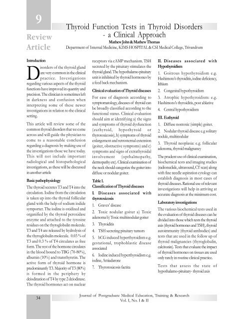
9ReviewArticleThyroid Function Tests in Thyroid Disorders- a Clinical ApproachMathew John & Mathew ThomasDepartment of Internal Medicine, KIMS HOSPITAL & CSI Medical College, TrivandrumIntroductionDisorders of the thyroid glandare very common in the clinicalpractice. Investigationsregarding various aspects of the thyroidfunctions have improved in quantity andprecision. The clinician is sometimes leftin darkness and confusion wheninterpreting some of these newerinvestigations in relation to the clinicalsetting.This article will review some of thecommon thyroid disorders that we comeacross and will guide the physician tocome to a reasonable conclusionregarding a diagnosis by making use ofthe investigations those we have today.This will not include importantradiological and histopathologicalinvestigations, as these will be discussedin another articleBasic pathophysiologyThe thyroid secretes T3 and T4 into thecirculation. Iodine from the circulationis taken up into the thyroid folliculargland with the help of sodium iodidesymporter. The iodine is oxidized andorganified by the thyroid peroxidaseenzyme and attached to the tyrosineresidues on the thyroglobulin molecule.T3 and T4 are released by hydrolysis ofthe thyroglobulin molecule. 0.03 % ofT3 and 0.3 % of T4 circulates as freeform. The rest of the hormone circulatesin the blood bound to TBG (70-80%),albumin (10%) and transthyretin. Theactive form of thyroid hormone ispredominantly T3. Majority of T3 (80%)is formed in the periphery bydeiodination of T4 by type 2 deiodinase.The thyroid hormones act on nuclearreceptors via cAMP mechanism. TSHsecreted by the pituitary stimulates thethyroid gland. The hypothalamo-pituitaryunit is inhibited by thyroid hormones bya feed back mechanism.Clinical evaluation of Thyroid diseasesFor ease of diagnosis according tosymptomatology, diseases of thyroid canbe broadly classified according to thefunctional status. Clinical evaluationshould aim at identifying a) the signsand symptoms of thyroid dysfunction(euthyroid, hypothyroid orthyrotoxicosis), b) symptoms of thyroidenlargement and retrosternal extension(goiter, obstructive symptoms) and c)symptoms and signs of extrathyroidalinvolvement (opthalmopathy,dermopathy etc). Clinical examination ofthe neck should categorize the goiter intodiffuse or nodular goiter.Table 1.Classification of Thyroid diseasesI. Diseases associated withthyrotoxicosis1. Graves’ disease2. Toxic nodular goiter a) Toxicadenoma b) Toxic multinodular goiter3. Thyroiditis4. TSH secreting pituitary tumors5. hCG induced hyperthyroidism e.g.gestational, trophoblastic diseaseassociated6. Iodine induced hyperthyroidism e.g.iodine, Amiadarone7. Thyrotoxicosis factitaII. Diseases associated withHypothyroidism1. Goitrous hypothyroidism e.g.Hashimoto’s thyroiditis, iodine deficiency,lithium2. Congenital hypothyroidism3. Atrophic hypothyroidism: e.g.Hashimoto’s thyroiditis, post ablative4. Central hypothyroidismIII. Euthyroid1. Diffuse nontoxic (simple) goiter.2. Nodular thyroid disease e.g solitarynodule, multinodular3. Thyroid neoplasia: e.g. follicularadenoma, thyroid malignancyThe prudent use of clinical examination,biochemical tests and imaging studies(radionuclide, ultrasound, CT scan) alongwith fine needle aspiration cytology canestablish diagnosis in most cases ofthyroid diseases. Rational use of relevantinvestigations will help in arriving ataccurate diagnosis at the minimum cost.Laboratory investigationsThe various biochemical tests used inthe evaluation of thyroid diseases can bedivided into those which tests the thyroidaxis (thyroid hormones and TSH), thyroidautoimmunity (thyroid antibodies) andtests that are used in the follow up ofthyroid malignancies (thyroglobulin,calcitonin). Tests that evaluate the impactof thyroid hormones on tissues are usedonly rarely in routine clinical practice.Tests that assess the state ofhypothalamo-pituitary- thyroid axis<strong>34</strong><strong>Journal</strong> of Postgraduate Medical Education, Training & ResearchVol. I, No. I & II
TSH (Thyrotropin)TSH estimation forms the mostimportant part of a thyroid function test.The rate of thyrotropin secretion isexquisitely sensitive to plasma level offree thyroid hormones. The normal rangeof TSH is between 0.5 and 5 mU/L.This will vary slightly between labsdepending on TSH referencepreparations and the assay used. TSHassays over time has been classified into“generations” with respect to sensitivity.Each successive generation offers about10-fold improvement in sensitivity. Thelower limits of detectability of theseassays are given in the table 2.Table 2: Generations of TSH assaysGene- Sensitivity Methods/rationComments1 1 mU/L Radioimmunoassay.Cannot differentiateeuthyroid fromhyperthyroidism2 0.1 mU/L Immunometricassays. Candifferentiateeuthyroid fromhyperthyroidpatients3 0.01mU/L Immunometricassays. Candifferentiateeuthyroid fromhyperthyroidpatients4 0.001mU/L Chemiluniscenceassays. May beuseful todifferentiate NTIfromhyperthyroidismA minimally suitable assay should be ableto quantitate concentrations of TSH of0.1 mU/L with a coefficient of variationof less than 20 %, thus falling intosecond and third generation category.Currently most laboratories useimmunometric assay technology. In thisassay, a TSH molecule in the test serumis used as a link between a TSH antibodybound to an inert surface (e.g. coatedtube) and a second antibody directedagainst a second TSH epitope that islabeled with a detectable marker. (I 125or chemiluminescence). This techniqueis more specific, sensitive and rapid thanradioimmunoassay. Rarely HAMA(Heterophilic antimouse IgG antibodies)present in the sera of patients maysubstitute for TSH and cause falsely highvalues.In patients with thyrotoxicosis, TSH issuppressed. (less than 0.1 mU/L).Patients with TSH values between thelower limit of normal and 0.1 mU/L arerelatively asymptomatic (subclinicalhyperthyroidism). Patients with primaryhypothyroidism have serum TSHconcentration that range from minimallyelevated to very high values. In patientswith central hypothyroidism, TSH canbe normal, low (not suppressed) or evenelevated. In those patients of central withelevated TSH, the molecules havereduced biological activity.TRH stimulation test was previouslyused in clinical practice. Since the adventof sensitive TSH assays, TRHstimulation test has been given up. TRHexaggerates the normal behavior of basalTSH.Serum Thyroid hormonesQuantitation of the circulating thyroidhormone concentration is essential toconfirm that the thyroid statusabnormality suggested by an abnormalTSH result is accurate and document itsseverity. This can be done by quantitatingfree or total thyroid hormones.In vitro, the interaction between thethyroid hormone and their bindingproteins conforms to a reversible bindingequilibrium that can be expressed byconventional equilibrium equations.(Table 3) The free fraction of T4 isinversely proportional to theconcentration of unoccupied TBGbinding sites. Estimates of free T4concentration in serum is generated bydirect or indirect assay. In normal serum,the free T4 is approximately 0.02 % oftotal T4 and that of free T3 is 0.3 % oftotal T3.Table 3: interaction between thyroidhormone and binding proteinKT4 +TBGT4.TBGT4 - concentration of free T4TBG - unoccupied binding proteinK - equilibrium association constantT4 .TBG - T4 bound to TBGBecause T4 is the major secretory productof the thyroid gland and correlates mostclosely with serum TSH, in mostsituations, a serum free T4 along withTSH is all that is required to ascertainthe state of thyroid secretion or supply.A) Free Thyroid hormonesCurrently more labs are adapting to freeT4 concentration assays for clinical use.However, there is a bewildering array ofmethods used to quantitate free T4 orT3 in whole serum that involveautomation. Many of these commercialFT4 assays give misleading results inpatients with abnormal binding proteins.This includes critical illness conditionswhere there are changes in bindingprotein concentration, albuminconcentration and increase in fatty acids.The clinician should be wary if the FT4report by any method does not agreewith the clinical status and TSH. Themethods of free hormone assays aregiven in table 4<strong>Journal</strong> of Postgraduate Medical Education, Training & ResearchVol. I, No. I & II35
Other antibodies associated with thyroid,directed against Na- I symporter, CA 2(second colloid antigen), thyroidhormones, Megalin etc have limitedclinical use.Table 5: Normal range of ThyroidhormonesT3: 70-190 ng/dlT4: 5-11 mcg/dlFT3: 0.2-0.5 ng/dlFT4: 0.7-2.1 ng/dlTSH: 0.5-5.0 mU/LNB: These ranges vary betweenlaboratories depending on referencepreparations and assays.Rational use of Thyroidfunction testsA) Diagnosing Thyroid diseaseInitial investigations of any patient withsuspected thyroid disease should be animmunometric TSH assay. If thephysician has a reasonable suspicion offunctional thyroid disease, FT4 or T4should be included along with TSH.There is rarely a reason to measure totalT3 in the initial evaluation unless thepatient is taking triiodotyronine. Allpatients with abnormal TSH (elevatedor suppressed) should have a FT4 or T4measured.In conditions with elevated TBG (e.gpregnancy, estrogen replacement wheretotal T4 is often elevated) free T4 shouldbe measured rather than total T4. T3should be measured for confirmingdiagnosis in certain clinical situations1. Suspected T3 toxicosis (suspected ina patient with suppressed TSH andnormal FT4)2. In nonthyroidal illness (NTI, Sickeuthyroid syndrome): this would showlow T3 levels invariably3. Amiadarone induced thyroid functionabnormalities: elevated T4, low/normalT3 and normal TSH.Despite reaching a diagnosis in the aboveconditions, treatment seldom changes.Thus T3 measurements have limited rolein routine practice. Primary thyroiddiseases show concordant changes in FT4and TSH i.e. as FT4 increases, TSHdecreases and vice versa. Discordantthyroid functions are seen in pituitarydiseases. (Table 6)Table 6: Thyroid function tests indifferent conditionsDisease FT4 TSHPrimary Low HighhypothyroidismThyrotoxicosis High SuppressedCentral Low Low (normalHypothyroidism or high)TSH producing High InappropriatelypituitaryHighadenomaAnti TPO antibodies can be used toconfirm a diagnosis of autoimmunethyroiditis and postpartum thyroiditis.Patients with subclinical hypothyroidismand positive anti TPO antibody havehigher probability of progressing tohypothyroidism compared to those withnegative antibody titre. Hence, antibodymeasurements will form decision-makingcriteria in patients with subclinicalhypothyroidism. Since the sensitivity ofanti TPO antibodies is higher than antiTg antibodies, estimation of anti Tgantibody has limited role in routineclinical practice.However, quantitation of antibodiesagainst Tg proves to be extremelyimportant in the context of Tgestimations in view of possibleinterference with assays. The use of Tgin clinical practice is for detection andfollows up of DTC. In a rare patientwith low uptake thyrotoxicosis in whomexogenous L-T4 intake is suspected,thyroglobulin levels can be used todifferentiate it from thyroiditis: low infactitious thyrotoxicosis and elevated inthyroiditis.B) Monitoring therapyIn patients with primary hypothyroidism,TSH should be the monitoring parameter.In those with central hypothyroidism,thyroxine dose adjustments should beguided by monitoring free T4. Inhyperthyroid patients treated withantithyroid drugs, recovery of suppressedTSH takes 6-9 months. Till TSHrecovery takes place, monitoring shouldbe undertaken with free T4measurements. In patients withthyrotoxicosis in pregnancy, free T4monitoring should be done to keep levelsat upper quartile of the normal range.Confusing Thyroid function testsAt times, interpreting thyroid functiontests can turn out to be a nightmareeven for the best of physicians. A poorlymaintained quality control in the hormoneassay laboratory contributes to significantnumber of these weird thyroid functiontests. A few things need to be kept inmind when trying to interpret weird tests1. Making sure that the test report iscorrect. This can be done by recheckingthe reports in the same lab or differentlab/method. Assay artifacts with free T4can be confirmed with total T4 as theseare subjected to less assay errors. Mildelevations of TSH should be recheckedbefore planning more expensiveinvestigations like scans and consideringtreatment.2. Re-examine the patient and look forsigns of thyroid disease. If the functionalstatus of the patient (hypo/hyper orreports that show normal TSH with lowFT4 levels, drug interference (e.g.phenytoin) and severe systemic illnessshould be considered. A meticulousclinical history and examination can sort<strong>Journal</strong> of Postgraduate Medical Education, Training & ResearchVol. I, No. I & II37
Imaging of ThyroidVenkatesh K, Vikram Gulati, Dayananda L & Srikanth MoorthyDepartment of Radiology, Amrita Institute of Medical Sciences & Research Centre, Elamakkara, Kochi10ReviewArticleIntroductionThe thyroid gland is located in thelower anterior neck in theinfrahyoid compartment. Thegland is made up of two lobes locatedlateral to the trachea joined at the midlineby a thin bridge of tissue called isthmus.The pyramidal lobe is an accessory lobeoriginates from the isthmus or medialaspect of either lobe and extendssuperiorly along the course of distalthyroglossal duct. Imaging of thyroidgland can be done by USG, CT and MRIwhich provides anatomical informationwhile nuclear scintrigraphy providesfunctional information.Imaging modalitiesUltrasoundReal time ultrasound of the thyroid glandis usually performed with high resolutionlinear array transducers ranging from 7.5-10 MHz 1 . These allow excellentvisualization of both superficial portionsof gland and the deep structuresposteriorly to the level of spine.Ultrasound is an accurate method to usein calculating thyroid volume. Tissueharmonic imaging techniques are usedas an adjunct to conventionalsonography to improve lesion detectionand characterization .The thyroid glandis the most richly vascularised organ ofall superficially located normal structuresof the body. As a result colour and powerdoppler provides useful diagnosticinformation in thyroid disease.Ultrasound also provides guidance forperforming FNA biopsy. Evaluation ofretrotracheal and mediastinal regions isdifficult by ultrasound because ofacoustic shadowing from overlying airor bone 2 . Another limitation is that USGis inferior to CT and MRI in identifyinglymphadenopathy 3 .The normal thyroid parenchyma ishomogenous in appearance with greaterechogenicity than the adjacent strapmuscles. It is limited by a thin highlyreflective capsule. The dimensions ofnormal thyroid vary with the size ofsubject. The anteroposterior thickness isconsidered the most reliable index ofthyroid size. When it is larger than 2cmenlargement can be confidentlydiagnosed. Arterial and venous brancheswithin the thyroid parenchyma are clearlyvisible with current high sensitivity colourdoppler machines 4 . Normal peak systolicvelocities reach 20-40cm/s in the majorthyroid arteries and up to 15-30cm/s inthe intra parenchymal arteries.Computed tomographyThe thyroid gland is well seen on CTdue to its higher attenuation than softtissue caused by the physiologically highiodine content of the gland. CT isoccasionally used to demonstrate theextent of local invasion or local recurrenceof thyroid malignancy and to detect thepresence of retrosternal and retrotrachealextension of thyroid enlargement.Iodinated contrast may provide additionalinformation about lesion within thethyroid but the contrast will alterradioactive iodine uptake for about sixweeks following the study. Nuclearimaging should be performed prior toCT. On non contrast CT , normal thyroidgland has a density of approximately 80-100 HU because of its iodine content.Intravenous injection of iodinatedcontrast material results in diffuseincrease in density of the gland.Magnetic resonance imagingMRI is best performed with a dedicatedsurface coil centered over thyroid gland.MR imaging provides excellent depictionof internal parenchyma of the thyroidgland as well as relationship withadjacent structures in the neck andthoracic inlet. Like CT, MRI is useful instaging of a known thyroid malignanttumor, to identify local invasion , regionallymph node metastases and to detectthe recurrence following thyroidectomy.Using dynamic contrast enhancementcharacteristics thyroid lesions can beclassified as benign, indeterminate ormalignant. Rapid enhancement is seen in90% of malignant lesions while steadyor gradual enhancement is seen in 70%of benign cases. Normal thyroid glandis homogenously hyperintense to neckmusculature on both T1 and T2 weightedimages. The gland enhances diffusely andhomogeneously following contrastadministration.Plain filmPlain X-ray has a limited role in the thyroiddisorders. It may be useful in theassessment of retrosternal extension andtracheal displacement.Thyroid disordersThyroid disorders are further classifiedinto congenital and acquired disorders.CongenitalCongenital disease of thyroid includesagenesis, hemi-agenesis and varyingdegrees of hypoplasia and ectopias.Ultrasonography can easily demonstrateall of them except ectopias which arebest diagnosed by isotope scan.<strong>Journal</strong> of Postgraduate Medical Education, Training & ResearchVol. I, No. I & II39
Nodular disease of ThyroidOne of the most common clinicalproblem is patients presenting with asolitary palpable thyroid nodule. Themajority of thyroid nodules are benigndue to cyst, thyroiditis, adenomas orcolloid nodules. Though thyroidcarcinoma is uncommon it also veryoften presents as solitary thyroid nodule 5 .The challenge is to distinguish the fewclinically significant malignant nodulesfrom the many benign ones and toidentify the patients who are candidatesfor surgical excision.Role of Ultrasound in Thyroid nodulesGoitrousnonNodulesAdenomaThyroid nodule is defined as any discretelesion that is sonographicallydistinguishable from adjacentparenchyma.High resolution sonography has fourmajor applications in thyroid disease:1. Detection of thyroid and other neckmasses.2. Differentiation of benign frommalignant masses based on theirsonographic appearance3. Guidance for FNA/biopsy. FNAbiopsy is the most effective method fordiagnosing malignancy in thyroid nodule.4. Guidance for percutaneous treatmentof non functioning and hyperfunctioningbenign thyroid nodules and of lymphnode metastasis from thyroid carcinoma.For the differentiation of benign versusmalignant thyroid nodules sonographyhas sensitivity rates ranging from 63%-94%,specificity from 61%-95% andover all accuracy of about 80%-94% 6 .Currently no single sonographiccriterion distinguishes benign thyroidnodules from malignant thyroid noduleswith complete reliability.Non GoitrousCarcinomaFollicular Non Follicular PapillaryFollicularAnaplasticFeatures almost unique for benigngoitrous nodules are• Completely cystic appearance• Moving comet tail artifacts• Widespread cystic changes inisoechoic or highly echogenic nodules.• Hyperechoic nodules.• Thin uniform thickness perilesionalhypoechoic halo• Well defined and regular margins• Perilesional egg shell like or coarsecalcification• Doppler signal of flow around thelesion.If most of these signs are present in athyroid nodule the diagnosis of benigndisease is highly reliable and no cytologicalassessment is needed. Conversely theultrasound signs for malignancy are:• Hypoechoic nodule• Irregular margins• Thick irregular halo• Doppler signal of flow within thelesion• Microcalcifiactions• Hypervascularity• Invasion of vessels and adjacentstructures• Vessel encasementThe feature with the highestsensitivity is solid composition;however, this feature has a fairly lowpositive predictive value. The featurewith the highest positive predictivevalue is the presence ofmicrocalcifications. However thisfeature has low sensitivity. Acombination of these featuresimproves the positive predictive valueof US to some extent.The detection of additional occultnodules in patients with clinicallysolitary lesion though pathognomonicfor benign goitre, it does not totallyrule out malignancy 7 . In as many as33%-64% of patients with thyroid cancerat least one associated benign nodulewas found either surgically or atultrasound. When multinodularity isdetected each nodule should be carefullystudied. If all of them show the samecharacteristic pattern for goitrous nodules, cytological assessment is not strictlyneeded and the patient can be followedwith periodic scans. Nodules withsuspicious ultrasound criteria shouldunder go further assessment with FNA/biopsy.40<strong>Journal</strong> of Postgraduate Medical Education, Training & ResearchVol. I, No. I & II
Abnormal cervical lymph nodesUS diagnosis of the abnormallymphnodes depends on the size, shape,vascularity and internal architecture 8 . TheUS features associated with highest riskof cancer includes heterogenousechotexture, calcifications and cystic areaswithin the lymph node. A rounded lymphnode or one causing a mass effect is alsoat elevated risk of being malignant. Ingeneral, size is a less reliable criterion formalignancy in a lymphnode althoughchance of malignancy increases as thesize of lymphnode increases. Lymphnodeshould be considered suspicious if itmeasure more than 7 mm in short axis 8 .Benign neoplasmsThyroid adenomaThyroid adenomas are true neoplasms.They are usually solitary and nonfunctioning. They are usually less than 3cm in diameter , have well definedmargins and may involute, become cysticor may develop internal hemorrhage ,necrosis , calcifications or fibrosis 9 . Mostthyroid adenomas are hyperechoic or isoechoic with a hypoechoic peripheralhalo 10 . On CT scan adenoma may appearsolid or cystic when it has degenerated.FNA cannot generally distinguishfollicular adenoma from follicularcarcinoma as they have similar cytologicalfeatures. Only histopathologicalexamination can differentiate them basedon vascular and capsular invasion.Malignant neoplasmsPapillary carcinomaPapillary carcinoma is the most commontype of thyroid cancer. It accounts forabout 75- 90% of all cases. It mostcommonly occurs in female adolescentsand young adults. Sonographically 90%of papillary carcinoma are hypoechoic.Fine punctate, intralesionalmicrocalcifications are consideredcharacteristic of papillary carcinoma.Typically a centrally vascularised lesionwith a chaotic vascular pattern is seen.Cervical lymphnode metastasis seen inupto 50% of cases in the lower deepjugular chain. Metastatic nodes typicallyshow microcalcification and can be cystic.Medullary, Follicular and Anaplasticcarcinomas combined represent only10%-25% of all thyroid carcinomas.Incidentally detected nodulesThe goal of investigation should be toavoid extensive and costly evaluation inthe majority with benign disease, withoutmissing the minority of patients whohave clinically significant thyroid cancer.Nodules more than 1.5cm in diameterand having Sonographic feature ofmalignancy should need FNAC.USG guided FNA/biopsySonographically guided percutaneousneedle biopsy of cervical masses hasbecome an important technique in manyclinical situations. It allows continuousreal time visualization of the needle, acrucial requirement for the biopsy ofsmall lesions. Palpable thyroid nodulesgenerally undergo biopsy without imageguidance. The 25-gauge needles aregenerally sufficient to yield adequatecytologic specimens; however, in selectedcases core biopsy with an 18-to 20-gaugeneedle may be helpful. Fine-needleaspiration can be done by usingnonaspiration-capillary action techniqueor suction aspiration technique .Indications for image guided FNA/biopsy• Inconclusive physical examinationwhen a nodule is suggested but cannotbe palpated with certainty• Patient who is at high risk fordeveloping thyroid cancer and who hasnormal gland by physical examination butin whom sonography demonstrates anodule• Patients who have had a previousnon diagnostic or inconclusive biopsyperformed under direct palpationThe diagnostic accuracy is very high, withrates of sensitivity of approximately 85%and specificity 99% in centers with alarge experience with these procedures.Diffuse Thyroid diseaseDiagnosis of these conditions is usuallymade on the basis of clinical andlaboratory findings and or occasion byFNA. Sonography is seldom indicated.High-resolution sonography is useful torule out the possibility of a mass in thelarge lobe if the underlying diffuse diseasecauses asymmetric enlargement.Grave’s diseaseOn US the parenchyma is diffuselyhypoechoic and inhomogenous. Withcolour doppler hypervascular patternreferred to as ‘thyroid inferno ‘is seenindicating an acute stage of the process.Spectral doppler shows peak systolicvelocities to exceed 70 cm/s. There isno correlation between the hyperfunctionand the extent of hypervascularity orblood flow velocities. But with doppleranalysis therapeutic response in patientswith Graves disease can be monitored 11 .Chronic autoimmune lymphocytic(Hashimoto’s) thyroiditisThe thyroid gland often enlarged in sizeand is diffusely hypoechoic with coarseheterogenous echogenicity. Multiplediscrete hypoechoic micronodules from1-6 mm in diameter (‘micronodulation’)are strongly suggestive of chronicthyroiditis 12 . These nodules aresurrounded by multiple linear echogenicfibrous septations. With colour dopplerthe vascularity is normal or decreased inmost patients.References1. Gooding GAW. Sonography ofthyroid and parathyroid. RCNA 1993;31(5): 967-989.2. Hopkins CR, Reading CC. Thyroidand parathyroid imaging. Seminars in US,CT and MRI 1995; 16: 279-295<strong>Journal</strong> of Postgraduate Medical Education, Training & ResearchVol. I, No. I & II41
3. Loevener LA. Imaging of thyroidgland. Seminars in US, CT and MRI1996; 17: 539-562.4. Solbiati L, Charboneau JW, JamesEM, Hay ID. The thyroid gland. In:Rumack CM, Wilson SR, CharboneauJW,eds. Diagnostic ultrasound. St Louis:Mosby, 2004: 735-770.5. Hussain HK, Britton KE,Grossman AB, Reznek RH. Thyroidcancer. In: Husband JE, Reznek RH,eds. Imaging in oncology. ISIS Medicalmedia, Oxford, 1998: 481-514.6. Koike E, Noguchi S, Yamishita Het al: Ultrasonographic characteristics ofthyroid nodules: Prediction ofmalignancy. Arch Surg 2001; 136: 3<strong>34</strong>-337.7. Solbiati L, Volterrani L, Rizzatto Get al. The thyroid gland with low uptakelesions: Evaluation by ultrasound.Radiology 1985; 155: 187-191.8. Ying M, Ahuja A, Metreweli C.Diagnostic accuracy of sonographiccritera for evaluation of cervicallymphadenopathy. J Ultrasound M,ed1998; 17: 437-445.9. Yousem DM. Parathyroid andthyroid imaging. Neuroimaging Clinicsof North America 1996; 6(2): 435-459.10. Gritzmann N, Koischwitz D,Rettenbacher T. Sonography of thyroidand parathyroid glandsw. RCNA 2000;38(5): 1131-1145.11. Castagnone D, Rivolta B, RescallisS, et al. Colour doppler sonography inGraves disease: Value in assessing activityof disease and predicting outcome. AmJ Roentgenol 1996; 66: 203-207.12. Yeh HC, Futteweit W, Gilbert P.Minonodulation: Ultrasonographic signof Hashimoto’s thyroiditis. J UltrasoundMed 1996; 15: 813-819.DNB (Rural Surgery)<strong>National</strong> <strong>Board</strong> of <strong>Examination</strong>s haslaunched DNB (Rural Surgery),on pilot basis from June 2006. Theconcept of rural surgery has evolvedin India over the past decade basedon the ground reality of surgicalpractice of surgeons practicingoutside high-tech institutions in ourcountry. In India, 400 million peoplehave no access to basic surgical care,termed by the WHO as “essentialsurgical care”. The aim of this coursewould be to create a cadre of basicmultipurpose surgeons, who wouldacquire the expertise to provide basicand emergency and lifesaving surgicalcare to rural population of our country.They can form the back bone of healthcare delivery system and can play avital role in fulfilling the Rural HealthMission announced by theGovernment of India.Goal: After qualifying the finalexaminations the candidate should beable to function as a consultant(specialist) in Rural Surgery ( multiplesurgical disciplines) within theconstraints of limited resources.Objectives: At the end of thetraining period, the candidate shouldbe able to acquire followingcompetencies:•Basic & general surgery withemphasis on open surgeries.•Basic orthopaedics includingtrauma care.•Obstetrics and Gynaecology.•Basics of anesthesia, ultrasoundand X-Ray.•Emergency careTraining for DNB in Rural surgery willtake place in two kinds of hospitals:1. Multi specialty hospital which willbe called as Nodal Rural SurgicalTraining Center: Two years oftraining will take place here. Thisinstitute will take primary responsibilityfor the candidate in terms of-Organizing and scheduling the trainingprogram for the entire 3 years inconsultation with the peripheralinstitutes. A co-ordinator from boththe institutions will be appointed tolook after the total training of thecandidates; Providing hands onexperience to the candidate thusimparting practical surgical skills.Candidate should eventually be ableto perform procedures independentlyand not merely be a first assistant;Placement of the candidate to aperipheral rural surgical centre wherethe candidate is regularly monitoredfor skills training and for preparationof the dissertation.2. Peripheral Rural SurgicalCentre: One year of training will takeplace here.This will train the candidateto work in resource limited situationsand develop his/her capability to learnto innovate and manage a rural surgicalpractice; This is also the setting inwhich the candidate will write up adissertation based on a topic which isrelevant to the rural surgical practice.The candidates will be posted inperipheral Rural Surgical Center for3-4 months in first, second and thirdyears of trainingEligibility criteria for theCandidates1. Essential- Any medical graduatewith MBBS qualification, who hascompleted internship and is registeredwith MCI/State Medical Council canregister with the AccreditedInstitutions for 3 years of training.2. Desirable- One year experienceafter completing internship in aperipheral/rural set up. In servicecandidates from Defence, Central/State Government, Railways, Publicsector institutions may also be givenpreference.42<strong>Journal</strong> of Postgraduate Medical Education, Training & ResearchVol. I, No. I & II
Management of Common MedicalDiseases of ThyroidAlka GaneshDepartment of Medicine, Christian Medical College, Vellore11ReviewArticleIntroductionTreatment modalities of thecommon medical thyroiddiseases have not changed in thelast several decades. However, with theadvent of sensitive assays for hormonallevels, there has been a greater emphasison early treatment, as well as on moreaccurate dosing to attain as near normallevels as possible. This refers particularlyto the new generation TSH tests whichcan distinguish between normal andsubnormal levels.A goiter whatever it’s etiology is a highlyvisible deformity, and therefore, adistressing cosmetic problem. Thyroiddisease is a common problem in thecommunity. It is important to understandthe rationale of treatment modalities andto have a proper understanding ofeffectiveness and adverse effects, so thatthe best possible advice can be offeredto the patient.This article will deal with therapy ofsimple goiter, replacement therapy ofhypothyroidism, and the management ofthyrotoxicosis. Childhood thyroiddiseases and surgical diseases will not bediscussed.Simple goiter 1Diffuse enlargement of the thyroidoccurring in individuals who are clinicallyand biochemically euthyroid, in theabsence of thyroid anti-bodies, is usuallydue to iodine deficiency, but may also bepuberty- related, or due to goitrogens inthe food. In these cases the thyroidtissue is being stimulated but is able toproduce enough hormone and thus doesnot result in elevations of TSH, andthyroid hormone levels are in the normalrange. Thyroid antibodies are negative.Reassurance is all that is necessary.Alternatively, therapy with thyroxine canbe initiated, aimed at suppressing furtherstimulation of the gland hopefullyachieving regression in size. A dose of100mcg levothroxine is given with theaim of suppressing the TSH level to thelow normal range of 0.5 to 1 iU/ml. Forthe elderly, the starting dose should be50 mcg/ day. Regression of thyroidtissue can be expected in soft, smallglands which have been present for shortperiods of time. Long-standing largeglands usually have significant fibrosisand do not regress. Surgery can be offeredfor cosmetic reasons.Hypothyroidism 2Overt hypothyroidism is easily recognizedif it is suspected and tested for. Themost common causes are auto-immunethyroid disease, post-surgical or postradio-iodine ablation. Secondary thyroidfailure due to pituitary or hypothalamicdisease is less common.Replacement with thyroxine in the formof levothyroxine is the most widely usedtherapy. The initial dose should be 50mcg daily, and needs to be increased to100 mcgs daily, then 150 mcgs daily atthree to four week intervals. Sincethyroxine has a long half-life of one week,doses should not be adjusted until aminimum of 3 to 4 half-lives has elapsedto allow a steady –state to be attained.In post-surgical cases where thehypothyroidism has occurred rapidly, thepatient can be started initially with 100mcg, and then titrated upwards rapidly.Once daily administration on an emptystomach is advisable, and other drugsshould be taken separately.Assessment of adequate replacementAfter 3 months, the TSH level shouldachieve a level of 1-2 mU/ ml. If theTSH level continues to be above 5 mU/ml, the dose of levothyroxine should beincreased by 25 mcg, assuming that noncompliancehas been ruled out. If theTSH value is suppressed to below 0.1mU/L. and or the T4 level is above thenormal range, it indicates excessivereplacement. It is important to not toover-treat the patient. It has been shownthat over-treatment can cause increasedbone resorption, and arrhythmias. In theelderly who have ischemic heart disease,replacement has to be quite cautious toprevent exacerbation of angina. In thesepatients a low initial dose of 12.5-50mcg/day is advised.Non-compliancePatients do not feel unwell when theymiss thyroxine for a few days, and henceare often erratic with their medications.This can cause problems in evaluatingadequacy of therapy. An irregular patientmay often become very regular justbefore a clinic visit. This results in ahigh TSH, but normal T4 levels. Thetests can be repeated after a couple ofmonths of compliance.Combined therapy with Thyroxine andTri-iodothyronineSome researchers have studied the effectsof hormone replacement withcombinations of T4 and T3. It has beenfound that supra-physiologic levels ofT3 are achieved in a few hours. Thiscauses palpitations, and other thyrotoxic<strong>Journal</strong> of Postgraduate Medical Education, Training & ResearchVol. I, No. I & II43
symptoms which are likely to bedetrimental. The level of T3 in tissuesand blood is normally a result ofconversion of T4 to T3 in extra-thyroidaltissue, as per physiologic requirements.Thus in illness, or fasting, T3 levelsdecrease, but exogenous administrationof T3 upsets this fine physiologicallycontrolled balance. Hence, administrationof tri-iodothronine for replacementpurposes is not recommended by mostexperts. Another disadvantage of triiodothyronineis that it needs to be giventhrice daily because of it’s short half-life.Subclinical hypothyroidismSubclinical hypothyroidism refers topatients who have normal T4 and T3but the TSH level is slightly elevatedabove 5 iU/L. This occurs spontaneouslyin 3% of adults and 10% of postmenopausalwomen. It can also occur asa result of surgical or radio-active iodinetreatment for hyperthyoidism.It is still controversial whether subclinicalhypothyroidism should be treatedor not. Those who advocate treatmentargue that certain target organs arecompromised if left untreated. Amongthese is cited left ventricular dysfunction,reduced hearing, and hyperlipidemia.There are also claims of increasedvulnerability to ischemic heart disease.However, a recent study has shown thatthere is no increased morbidity ormortality in women with subclinicalhypothyroidism who are not giventhyroxine replacement. Many cliniciansfavour treatment with thyroxine becausepatients feel better, and because there isa 5% rate of progression to overthypothyroidism every year. The chanceof this happening are greater in autoimmunethyroiditis.If it is decided to treat patients withsubclinical hypothyroidism, it is prudentto start with small doses of 50 mcgdaily of thyroxine, & check levels after 3months.If treatment is started in patients wherethe TSH is only marginally elevated ( e.gless than 10 mU/L), there is no goiter,and thyroid antibodies ( microsomal )are absent, it is often the case that 3 or6 months, the biochemical parametersindicate thyrotoxicosis. It is possible thatthese patients had transienthypothyroidism( see below), or nonthyroidalillness was the cause for elevatedTSH. Therapy should be stopped in thesecases.Transient hypothyroidismThere are certain situations under whichhypothyroid state occurs transiently. Thishappens during recovery phase of subacutethyroiditis ( de Quervains ), postpartumthyroiditis, and even in somecases of chronic auto-immune thyroiditis.Biochemical features of hypothyroidism( elevated TSH and and low T4 ), maybe seen for up to 6 months after surgicalor radio-active iodine treatment forGraves’ disease. Hence in thesementioned situations, it is prudent towait for 6 months before diagnosingpermanent hypothyroidism.In the eventthat replacement thyroxine has alreadybeen started immediately after the abovetherapy, careful follow-up at 6 months iswarranted to determine continued use.Sick euthyroid syndrome 1Severe non-thyroidal illnesses give riseto low T3 and or low T4 levels, as alsoabnormal TSH levels. Theseabnormalities revert once the illnessresolves.It is therefore not advisable totest thyroid functions during severeillness unless specifically indicated.Most experts advise that thyroxinereplacement should not be given, butrather to repeat the levels after a fewweeks when the patient is better. Ifhowever there is a history of thyroiddisease in the past, then this can be takeninto account when deciding on treatment.Thyrotoxicosis 3The main causes of thyrotoxicosis areGraves disease, toxic multinodular goiterand functioning benign adenoma of thethyroid. Other causes include thyroiditis,drug-induced and pituitary adenomas.The three modalities of therapy are antithyroiddrugs, radio-iodine and surgery.The choice of therapy, or combinationof therapies depends on the underlyingdisease, patient preference, and priorresponse to a treatment regime.Anti-thyroid drugsThese are favoured for Graves diseaseas there is an expectation of remissionof the auto-immune process after 18 to24 months. It is specially useful inpregnancy, and in patients who are youngand have small glands. Anti-thyroiddrugs are not recommended in toxicgoiters or adenomas, because there islittle scope for spontaneous remission.Drugs are preferred over radio-iodinewhen patients have Gravesophthalmopathy, as the latter isassociated with worsening of eye disease.Drugs are also used prior to surgery orradio-iodine to prevent thyroid storm.The antithyroid drugs arepropylthiouracil, methimazole, andcarbimazole. Carbimazole is completelyconverted to methimazole in the body.The drugs are actively concentrated bythe thyroid gland against a concentrationgradient.They prevent T4 formation byinhibiting thyroid peroxidase-mediatediodination of tyrosine residues.Propylthiouracil has an additional effectof preventing the peripheral conversionof T4 to T3.The drugs are also consideredto have immunosuppressive actions onthe gland.Methimazole and carbimazole can be usedonce daily, whereas Propylthiouracil needsthrice daily dosing.44<strong>Journal</strong> of Postgraduate Medical Education, Training & ResearchVol. I, No. I & II
Dosing scheduleThe drugs can be used in two principalways; 1) Block and replace schedule, or2) titration method.Block and replace regime: A larger doseof anti-thyroid drugs such asCarbimazole 40mgs to 60 mgs, alongwith thyroxine 100-200mcg daily. Thishas the advantage of being able to givea relatively higher dose without causingsymptomatic hypothyroidism. Thisregime is preferred in patients who haveunstable hyperthyroidism, in whom asmall variation in dose causes markedfluctuation in T4 levels.Titration regime: Lower doses such as10 to 30 mgs of carbimazole are givenand biochemical testing is doneperiodically every 4 to 6 weeks, and thedose of drugs is lowered (or raised) tomake the patient euthyroid. This usuallytakes about three months, and amaintenance dose of carbimazole 5 to10 mgs, is needed for 18 to 24 months.Studies have shown no superiority ofone regimen over the other. The adverseeffects of anti-thyroid drugs are slightlyhigher with the block and replace therapyand therefore it is less popular than thetitration regime.Follow-up on treatmentAfter initiation of treatment, T4 levelsare done every 4 to 6 weeks and dosesare adjusted accordingly. TSH levels arenot used to assess remission ofthyrotoxicosis as the level is suppressedeven after thyroid function normalizes.TSH level is however useful to correlateif the patient appears clinicallyhypothyroid. Occasionally the T4 levelsappear normal, but the patient is clinicallythyrotoxic. This is due to elevated T3levels and should be tested for. The antithyroiddrug doses should be increasedtill T3 levels come down. After euthyroidstatus has been achieved then for theduration of treatment (18 to 24months),three monthly testing is sufficient.The relapse rate of thyrotoxicosis afterdrug therapy is as high as 50 to 68%.Relapse usually occurs within the first 3months of stopping treatment, but canoccur even after several years. Relapsesare often treated with a second cause ofdrugs, or more commonly with radioiodineor surgery.Side-effectsAgranulocytosis is the most feared fatalside-effect. It has been reported in .35%with both drugs. Most cases occur within90 days of treatment. Patients shouldbe advised to report as soon as theyhave sore throat and fever. Routine WBCcounts are not advocated.Because the onset of agranulocytosis isvery rapid. Most cases respond to drugomission, but a few fatalities haveoccurred. Other side effects are jaundice,vasculitis, and lupus-like syndrome.Pruritis and rash are known.Adjunctive therapyBeta-blockers: Non-selective betablockers, particularly propranolol are veryuseful in symptomatic therapy of tremor,anxiety and palpitations by directblockage of the adrenergic system. Thedoses usually needed are 20 to 40 mgsin divided doses. They also prevent someperipheral conversion of T4 to T3, andare reputed to decrease gland vascularityprior to surgery.Inorganic iodine: This is given as Lugol’siodine ( 5% iodine and 10% potassiumiodide in water) .1 ml to .3 ml thricedaily. Alternatively Potassium iodide 60mgs tds. Iodine helps to prevent therelease of thyroid hormone from thegland for a few days to a few weeks, butthe effect is lost beyond that. This therapyis used as preparatory to surgery.Radio-active iodineThis is the preferred method of treatmentof Graves thyrotoxicosis, in manycountries around the world. In India,radio-iodine is available and inexpensive,but may not be accessible outside thetertiary care centers. The advantage ofradio-iodine therapy is that relapse rateis very low, and follow-up is simple. It isabsolutely contra-indicated in pregnancy(see below). Since radio-iodine worksslowly, a patient will usually need to betreated with anti-thyroid drugs for a fewweeks or months prior to treatment tomake him/her euthyroid. The drugs areusually withdrawn for a few days beforeradio-iodine is administered and resumeda few days later in order to ensure thatentrapment and incorporation of iodineby the gland is not hindered.In spite of decades of usage of radioiodinethere is still debate about the mostappropriate dose. Since delayedhypothyroidism is almost certain, as alate side-effect, efforts have been madeto tailor the dose to the gland size anduptake characteristics of the gland. Theusual dose delivered for Graves diseaseis 5 to 10 mCiSide-effects of radio-iodine1. The most predictable side-effect ishypothyroidism which occurs anytimefrom the first year onwards to 25 yearsafter radio-iodine ablation.2. Radio-iodine has been found toworsen ophthalmopathy, and is thereforeavoided in such cases.3. Although thyroid cancer has beendescribed occasionally in a few patients,several large studies have disproved acause and effect relationship.4. Teratogenesis is a concern.However, so far there has been noevidence for increased incidence ofcongenital anomalies in children born tomothers who have had radio-iodine.<strong>Journal</strong> of Postgraduate Medical Education, Training & ResearchVol. I, No. I & II45
5. If radio-iodine is administered to apregnant lady by 10 th week of gestationat which time the fetal thyroid hasdeveloped, it will undergo completeablation resulting in congenitalhypothyroidism.Surgical treatmentSub-total thyroidectomy is the surgicalprocedure for Graves disease. It is rarelya primary treatment. It is reserved forpatients who have relapsed on drugs andare not eligible for radio-iodine(pregnancy), or refuse it. Surgery ispreferred in patients who have largeglands with pressure symptoms.Surgery should always be preceded byanti-thyroid drugs to produce a euthyroidstate, and reduce the risk of thyroid storm.Additional several measures are alsotaken to reduce the vascularity of thegland. Propranolol has been used aloneor in combination with potassium iodide.Complications of surgery are:1. Laryngeal nerve damage.2. Hypoparathyroidism.3. Bleeding into the neck.These complications are minimal inexpert hands, and mortality is almostzero.Post-operative Thyroid functionRelapse of thyrotoxicosis occurs in 10%of patients, and usually occurs in thefirst five years after surgery. Howeverrelapses as late as 25 years have alsobeen reported.Post-operative hypothyroidism is also afeature. As mentioned above it can occurtransiently upto six months postoperatively.Toxic adenomas & toxic multinodulargoitresRadio-iodine is the preferred treatmentfor these cases, but the doses used aremuch higher than what is needed forGraves disease, and are in the range of10 to 50 mCi. Surgical treatment isindicated if goiters are large with pressuresymptoms, or if the patient refuses radioiodine.Antithyroid drugs are not usedbecause the hyperthyroidism is permanentwith no chance of remissions, thusindefinite drug therapy would be requiredwith problems of follow-up and sideeffects.Thyroiditis 3This should be suspected in patients whohave pain in the thyroid region, havealmost no enlargement of the gland, andhave very low radio-iodine uptake. Thethyrotoxicosis is transient and so notreatment is warranted, except perhapswith propranolol for a few weeks forsymptomatic relief. NSAID’S andglucocorticoids may be needed to reducepain; thereafter hypothyroidism may setin and replacement therapy may be neededtransiently or permanently. Post-partumthyroiditis should be treated similarly.Hyperthyroidism in pregnancy 3, 4Antithyroid drugs are the treatment ofchoice. Propylthiouracil is preferredbecause it not crosses the placenta lessthan do carbimazole and methimazole.The lowest possible dose of the drug ispreferred, so as to prevent as littletransplacental passage of the drugs aspossible, thereby minimizing fetalhypothyroidism, and goiter formation. Ifthe dose is too low, uncontrolled maternalthyrotoxicosis will not only causematernal distress but may initiate earlylabour and thus harm the fetus. To walkthis tightrope successfully, frequentthyroid function tests are required, oncein every 2 weeks throughout pregnancy.Thyroxine does not cross the placenta,so it is better avoided in the mother inwhom an inadvertently excessive doeswill necessitate an increased dose of antithyroiddrugs, which in turn will hurt thefetus, as explained above. Breast-feedingis safe on anti-thyroid drugs.Teratogenecity of anti-thyroid drugs 4Very rare instances of “methimazoleembryopathy” in which the fetus haschoanal atresia, or esophageal atresia,have been reported. However, otherstudies have shown this to occurirrespective of drug intake.Drug-induced Thyroid disease 1Lithium, and amiadarone are importantdrug-related causes of thyroid disorders.Amiadarone toxicity can cause eitherhypothyroidism or hyperthyroidism. Itcontains 39% iodine by weight.Hypothyroidism is treated bylevothyroxine replacement withoutdiscontinuing amiadarone.Amiadarone –induced hyperthyroidism ismore complex to treat. If it occurs inpatients with pre-existing goiter orpreclinical Graves disease(Type 1), thenhigh doses of anti-thyroid drugs iseffective. In type 2 disease (where nopre-existing thyroid disease is present),oral contrast agents such as sodiumipodate 500 mgs/day can begiven.Lithium, potassium perchlorate andgluco-corticoids are other forms oftherapy. If at all possible, amiadaroneshould be discontinued.Thyrotoxic crisis 3This is a medical emergency presentingas marked tachycardia, fever, agitation,and has a high mortality if untreated.The drugs of choice are, glucocorticoids,high dose propranolol ( 2 to 5 mgs IVevery 4 hours; 320 to 480 mgs orallydaily); potassium iodide orally to blockrelease of thyroid hormones;propylthiouracil100mgs every 6 hours;radiographic contrast ipodate sodium 1gm per day by mouth. The iodine releasedfrom this compound has anti-thyroideffects. The agent also preventsperipheral conversion of T4 to T3.46<strong>Journal</strong> of Postgraduate Medical Education, Training & ResearchVol. I, No. I & II
ConclusionHypothyroidism as well ashyperthyroidism can be effectively treatedwith the available therapeutic agents. Subclinicaldisease is considered by manyexperts to be grounds for treatment.Pregnancy, co-morbid illness, age andenvironmental factors need to be takeninto account when treatment is beingplanned. Accurate dosing is required inorder to prevent over- or undertreatment.Bibliography1. J. Jameson, A. Weetman.Disorders of the thyroid gland.Harrison’s Principles of InternalMedicine.McGraw Hill 16 th Ed: 2104-2126.2. A. Toft. Thyroxine therapy.NEJM 1994;vol 331: 174-1803. J. Franklyn. Management ofhyperthyroidism. NEJM. 1994, vol 330,1731-17384. D. Cooper. Antithyroid drugs.NEJM. 2005, 352: 905-917<strong>National</strong> <strong>Board</strong> of<strong>Examination</strong>’s CMEprogrammes go high techon 22 nd July 2006The first Interactive CMEsession for DNB candidatesusing Indira Gandhi <strong>National</strong> OpenUniversity satellite infrastructurewas held on 22 nd July 2006. The<strong>Board</strong> in collaboration with theSchool of Health Sciences, IndiraGandhi <strong>National</strong> Open Universityhas planned five such sessions forAugust 2006 and gradually thefrequency will be increased. Thesesessions will also be recorded andDVDs will be made available toaccredited hospitals and DNBcandidates. The transmission willbe available at C- BAND (GD1-GD4), 4165 MHz, HorizontalPolarization, Transponder C-12 onINSAT 3C, Symbol Rate: 26,000SPS, FEC: ½ and also at DTH (KUBAND) GD-1, NSS 6, Down linkfrequency: 12427.5 MHz, SymbolRate: 21, 0937, Polarizationhorizontal, FEC: ¾. All the<strong>National</strong> <strong>Board</strong> of <strong>Examination</strong>s’accredited hospitals may kindlyensure that all the DNB candidatesmust attend these sessions. TheHospitals, which have not yetinstalled the reception equipmentsare requested to contact theRegional Director IGNOU in theirrespective states (city) and requesthim/her for making the necessaryarrangements for DNB candidatesat their centers for reception ofsatellite transmission on thespecified dates and time. Thedetails are on the <strong>National</strong> <strong>Board</strong>of <strong>Examination</strong>s website: www//natboard.nic.in<strong>Journal</strong> of Postgraduate Medical Education, Training & ResearchVol. I, No. I & II47
12ReviewArticleSurgical Management ofCommon Diseases of ThyroidVikram Kate & N. AnanthakrishnanDepartment of Surgery, Jawaharlal Institute of Postgraduate Medical Education and Research, PondicherrySolitary thyroid nodule, multinodular goiter, thyroid cancer, thyroiditis, Graves’ disease and Plummer’sdisease (multinodular toxic goiter) arethe common disorders of thyroid whichwe come across needing surgicalmanagement. Although the surgicalprocedures described for the abovementioned conditions are fairlystandardized, controversy rages about theextent of surgery for benign andmalignant thyroid disease. Thiscontroversy balances the completeextirpation of the disease with theincidence of two primary potentialcomplications of thyroidectomy in theform of recurrent laryngeal nerve (RLN)injury and hypoparathyroidism. Thecommonly performed operations onthyroid include lobectomy orhemithyroidectomy, subtotalthyroidectomy, near total thyroidectomyand total thyroidectomy. The extent ofsurgery in a benign solitary nodule islobectomy or hemithyroidectomy of theaffected lobe. In presence of a malignantsolitary nodule the procedure of choiceis total or near thyroidectomy. There aresome reports that the extent of surgeryin well differentiated cancers is determinedby the high or low risk factors associatedwith the malignancy. In high risk patientstotal or near total thyroidectomy isjustified whereas in patients with “lowrisk” a lobectomy can be done withoutcompromising the results. The surgicalprocedure for non-toxic and toxic MNGis same in the form of subtotalthyroidectomy or total thyroidectomy.Most clinicians currently recommend neartotal or total thyroidectomy for all clinicalcancers. Conventionally a functional(sparing sternocleidomastoid muscle,internal jugular vein, spinal accessorynerve) or modified radical neck dissectionis done when the metastases areconfirmed in cervical nodes.Postoperative therapy following surgeryfor well differentiated thyroid cancerincludes radioactive iodine ablation ofthyroid remnant and distant metastases.Thyroid stimulating hormonesuppression is also done by givingsuppressive doses of thyroxinepostoperatively. The procedure of choicein familial and sporadic medullary thyroidcarcinoma is near total or totalthyroidectomy with central compartmentlymph node dissection. Anaplasticcarcinoma most of the time atpresentation is inoperable. Surgery maybe needed to palliate airway or esophagealobstruction. In Graves’ disease althoughmedical therapy is commonly used,surgical intervention is necessary whenthere is a possibility of a coincidentcarcinoma, large bulky glands, patientpreference to avoid I 131 or antithyroidmedications and young females in thereproductive age groups where I 131therapy may not be advisable. Total ornear total thyroidectomy is the surgeryof choice. Other options include subtotalthyroidectomy or Hartley-Dunhillprocedure where one lobe is completelyresected and partial resection ofcontralateral lobe is done. Subtotalthyroidectomy is the common procedureperformed for all types of thyroiditis. InRiedel’s thyroiditis this may be difficultdue to extensive fibrosis and destructionof normal tissue planes; hence wedgeresection of isthmus can provide reliefof the pressure symptoms insymptomatic cases. Minimally invasive,video-assisted thyroidectomy (MIVAT)is characterized by a unique central accessand external retraction. Pathologiestreated are mainly nodular goiter, smalldifferentiated carcinoma without lymphnode involvement. Nowadays thisminimally invasive surgery, in selectedpatients, clearly demonstrates excellentresults regarding patient cure rate andcomfort, with shorter hospital stay,reduced postoperative pain and mostattractive cosmetic results.IntroductionSolitary thyroid nodule, multinodulargoiter, thyroid cancer, thyroiditis, Graves’disease and Plummer’s disease(multinodular toxic goiter) are commondisorders of thyroid needing surgicalmanagement. Although the surgicalprocedures described for the abovementioned conditions are fairlystandardized, controversy still existsabout the extent of surgery for benignand malignant thyroid disease. Thiscontroversy balances the completeextirpation of the disease with theincidence of two primary potentialcomplications of thyroidectomy in theform of recurrent laryngeal nerve (RLN)injury and hypoparathyroidism. Thecommonly performed operations onthyroid include lobectomy orhemithyroidectomy, subtotalthyroidectomy, near total thyroidectomyand total thyroidectomy. Lobectomy orhemithyroidectomy is removal of onelobe with the isthmus and subtotalthyroidectomy is excision of both lobesleaving behind 2 to 4 grams of thyroidtissue in the tracheoesophageal groove48<strong>Journal</strong> of Postgraduate Medical Education, Training & ResearchVol. I, No. I & II
on either side. Near total thyroidectomyis complete removal of one lobe withthe excision of the contralateral lobeleaving behind a small rim of thyroidtissue on the contralateral side to protectthe parathyroids and the RLN. Totalthyroidectomy is the complete removalof all macroscopic thyroid tissue. Thisreview includes the surgical options,controversies and recent developmentsin the surgical treatment of commonthyroid disorders.Surgical treatment of thyroiddisordersSolitary thyroid noduleThe common disorders of thyroid whichcan present with a solitary nodule includea dominant nodule of a multinodulargoiter, thyroid adenoma; thyroid cancerespecially well differentiated type,thyroiditis and rarely a thyroid cyst. Acolloid nodule can be euthyroid or a toxicnodule. Conventionally the indicationsfor surgery in a fine needle aspirationcytology (FNAC) reported benign solitarynodule comprises of extreme age(younger than 20 or older than 45), malesex, pain, pressure symptoms, largenodule (> 4 cm), rapid growth, historyof radiation to neck, family history and atoxic nodule when medical or radioiodinetherapy has failed.The extent of surgery in a benign solitarynodule is lobectomy orhemithyroidectomy of the affected lobe 1 .This comprises of removal of the lobewith the isthmus. If the nodule is limitedto the isthmus only then anisthmusectomy is the surgical procedureof choice when benign. When there is asuspicion regarding malignancy, a frozensection is performed on the excised lobeand further procedure is done based onthe report. In cases of follicularneoplasm reported on FNAC even afrozen section may not be able to ruleout malignancy. In these cases it isnecessary to await the paraffin sectionreport and perform completionthyroidectomy the excised specimen isreported as malignant.In the presence of a malignant solitarynodule the procedure of choice is totalor near thyroidectomy. In low volumecenters near total thyroidectomy ispreferable to reduce the risks of injuryto recurrent laryngeal nerve andhypoparathyroidism. There are somereports that the extent of surgery in welldifferentiated cancers is determined bythe high or low risk factors associatedwith the malignancy 2,3 . The patientswith “high risk” factors are extremes ofage, lesions greater than 4 cm,extrathyroidal spread, regional or distantmetastases or high grade tumors. In thesepatients total or near total thyroidectomyis justified whereas in patients with “lowrisk” hemithyroidectomy and a smalladjoining part of the opposite lobe at itsjunction with the isthmus can be donewithout compromising the results. Weare performing a total thyroidectomy atour centre for a malignant solitary nodule.Multinodular goiterMultinodular goiter (MNG) is thecommonest thyroid disorder encounteredin most of the hospitals in our country.MNG can be toxic or non-toxic, the latterbeing more common. The common causeis due to iodine deficiency 4 . Therapy forMNG can be medical or surgical. Surgicaltherapy in a euthyroid MNG is indicatedin a large MNG, when patient haspressure symptoms due to long standinggoiter, there is suspicion of malignancy,substernal extension or for cosmeticreasons. In toxic MNG, surgery isindicated when medical treatment hasfailed, it is very large or there is failure ofradioiodine therapy. Surgery in toxicMNG is also performed for theindications mentioned for non-toxicMNG.The surgical procedure for non-toxic andtoxic MNG is same in the form ofsubtotal thyroidectomy or totalthyroidectomy. The patient has to be wellprepared before attempting surgery in apatient with toxic MNG. This includespreoperative cardiac evaluation,antithyroid drugs, beta blockers andpotassium iodide to make the patienteuthyroid before surgery. Control oftoxicity prior to surgery reduces thechances of precipitating postoperativethyroid crisis which can sometimes befatal. Although many centers may prefersubtotal thyroidectomy for a non-toxicgoiter that leaves behind less than 2 gmof thyroid tissue on either side, for atoxic MNG total thyroidectomy isrecommended. As the treatment ofrecurrent thyrotoxicosis followingsubtotal thyroidectomy is difficult, totalthyroidectomy is commonly performedfor this condition. The surgicaltechniques in patients with toxic or nontoxicMNG should allow for the bestchance for removal of the abnormalthyroid tissue with the least morbidity.This depends on the volume of thyroidsurgery being performed at the centerand the surgeon’s experience 5-7 . Mulleret al have reported in a large retrospectivereview that the complication ratesbetween subtotal and total thyroidectomyare similar 8 . In this review, thecomplications following totalthyroidectomy were a wound infectionrate of 0.9%, a secondary hemorrhagerate of 0.6%, 8% transient recurrentnerve palsy rate, 0.9% permanent nervepalsy rate, 28% rate of immediatehypocalcemia and 0.9% patients ofpermanent hypocalcemia. Thecorresponding complications in patientswith subtotal thyroidectomy were, 1.6%wound infection rate, 1.8% secondaryhemorrhage rate, 0.7 % permanentrecurrent nerve palsy rate and 0.7% rateof permanent hypocalcemia 8 .<strong>Journal</strong> of Postgraduate Medical Education, Training & ResearchVol. I, No. I & II49
Postoperatively, following subtotalthyroidectomy for non-toxic MNG, thepatient can be put on thyroxine to providethyroid replacement and reduce thechances of recurrent nodular formation 9 .Although we follow this practiceroutinely, a benefit in outcome followingpostoperative thyroxine therapy is notproved yet 10. Percutaneous ultrasoundguided ethanol injection is also reportedas an alternative therapy in patients withtoxic MNG when other non-surgicalmethods have failed and surgery iscontraindicated due to co-morbidreasons 11 . Hyperthyroidism resolves in42% of the lesions at 3 months and66% at 1 year. The chances of controlof toxicity are more in nodules less than3 cm. Weekly 2 to 4 cc of 95% ethanolis injected in the nodules till euthyroidstatus is achieved 11 . We have no personalexperience of this procedure.Thyroid cancerThyroid cancer can be broadly dividedinto well differentiated thyroid cancer(WDTC), medullary thyroid carcinoma(MTC) and other rare tumours of thethyroid. WDTC constitutes about 85 to90% of all thyroid cancers, MTC about8 to 10% and the remaining form therare tumors of the thyroid 12 .Well differentiated Thyroid cancerWell differentiated thyroid cancer includespapillary thyroid carcinoma (PTC),follicular thyroid carcinoma (FTC) andHurthle cell carcinoma. Papillarycarcinoma predominantly represents thisgroup. Surgery is the first line therapyfor the treatment of well differentiatedthyroid cancer. However, the extent ofresection for WDTC remainscontroversial. Conventionally theoperation of choice is near total or totalthyroidectomy. As mentioned earlier forsolitary nodule morbidity following totalthyroidectomy will be less at high volumecenters for thyroid surgery. Hence neartotal thyroidectomy should be done atlow volume centers. The protagonistsof total or near total thyroidectomy arguethat as there is high risk for contralaterallobe malignancy (more common withPTC), enhanced postoperative I 131ablation, better postoperative surveillanceby clinical exam and thyroid scan and asthis facilitates use of thyroglobulin (Tg)as a tumor marker; near total or totalthyroidectomy is the procedure of choicefor WDTC. Studies have also shownreduced recurrence and improved survivalfollowing total thyroidectomy whencompared to lobectomy for an ipsilateraldisease 13,14 . The opponents of totalthyroidectomy in early stage tumorsbelieve that the increased risk of RLNinjury and hypocalcemia are not justifiedby an improvement in disease control orsurvival rates over those attained byunilateral thyroid lobectomy 15 . Althoughthe extent of surgery is still controversial,most clinicians currently recommend neartotal or total thyroidectomy for all clinicalcancers 16 .Most WDTC are cured by initialoperation. Sometimes these cancersmight be advanced with extrathyroidalspread and distant metastases whenradioiodine therapy and external beamradiotherapy (EBRT) with adjuvantchemotherapy (doxorubicin) can be given.Lymph node metastases are more oftenseen in patients with PTC. Elective neckdissection is not indicated in WDTCalthough papillary carcinomas can haveoccult metastases in as many as 90% ofelective neck specimens as the impactof such finding on survival is debatable17,18. Hence, conventionally a functional(sparing sternocleidomastoid muscle,internal jugular vein, spinal accessorynerve) or modified radical neck dissectionis done when the metastases areconfirmed in cervical nodes. UsuallyLevel I (submental and submandibular)nodes rarely get involved with thyroidmetastases and hence are not includedin the dissection. The block dissectionincludes Level II to VI lymph nodes.The risk of bilateral disease is greatestwith male sex, advanced tumor stage,bulky ipsilateral nodal disease,involvement of the thyroid isthmus, andextrathyroidal extension 19 . Contralateralneck dissection is indicated in theseconditions.Postoperative therapy following surgeryfor WDTC includes radioactive iodine(RAI) ablation of the thyroid remnantand distant metastases. Thyroidstimulating hormone (TSH) suppressionis also achieved by giving suppressivedoses of thyroxine postoperatively asWDTC is a TSH dependent tumor. TheTSH levels should be near zero for anadequate suppressive therapy. Theadvantages of postoperative RAI includethe ablation of micrometastases andresidual thyroid tissue. The latterfacilitates optimal follow up as inpresence of normal thyroid tissue RIAtrapping by metastases may not occurand their identification becomes difficultby I 131 total body scan (TBS). Theabsence of thyroglobulin production bythe thyroid tissue helps in increasing theaccuracy of thyroglobulin tumor markerfor detecting recurrence. The need forRAI ablation as a routine in youngerpatients with smaller lesions confined tothe thyroid gland remains controversial20.RAI administration can producecomplications that include radiationthyroiditis (when a large thyroid remnantis present), dysphagia, sialoadenitis,glossodynia, tumor edema andhemorrhage and pulmonary fibrosis inpatients with extensive lung metastases.A high level of TSH is necessary for anoptimum I 131 TBS. To achieve thisthyroxine hormone replacement therapyis withdrawn before TBS. Patients receivea dose of 2 to 5 mCi for a TBS. The50<strong>Journal</strong> of Postgraduate Medical Education, Training & ResearchVol. I, No. I & II
scanning is performed 2 to 3 days afterI 131 administration. The serumconcentration of TSH should be between25 to 30 mIU/L for an optimum scan 21 .As the production of Tg is alsodependent on TSH ideally this shouldalso be done when the TSH level is high.This standard method of increasing thelevel of TSH by producinghypothyroidism can lead to physicaldisturbances, psychological alterations,and disruption of patient’s family, socialand working life 22.Recent development is the use of analternative method in the form ofrecombinant human TSH (rhTSH) 23,24 .The rhTSH allows the quicker clearanceof RAI with the potential for fewer sideeffects and patients no longer need toexperience symptoms of prolongedhypothyroidism before TBS as patientscontinue on their replacement therapybefore the scan. RhTSH is administeredas a single intramuscular dose of 0.9 mgon two consecutive days. It has beenreported that when a combination of RAITBS and serum Tg was performed afterrhTSH stimulation, the assays togetherdetected the remnant thyroid tissue orcancer within thyroid bed in 93% ofpatients and in 100% of patients withmetastatic disease 25 . The generally usedcut-off for the Tg level is 2 ng/ml.Mazzaferri et al reported in a recent reviewthat Tg levels and RAI scan are used incombination for detection ofrecurrence 26 . If Tg is detectable, neckultrasound and chest radiographs areadvised, and RAI therapy or surgery isconsidered. If Tg is undetectable thenrhTSH stimulation is used to assess theTg levels. In patients with greater than2ng/ml assessment is done for locationof recurrence. In patients with elevatedTg levels and negative RAI TBS, apositron emission tomography (PET)scan is done to detect the recurrence 26 .The prognosis is excellent in patientswith WDTC with the 10 year survivalrate for papillary cancer being 95% and90% for follicular cancer 20 . Althoughthe overall prognosis is excellent there isa small subgroup of patients with highrisk factors who have lower survival.Medullary Thyroid cancerMedullary thyroid cancer is relatively rarewhen compared to WDTC, however, themortality due to this disease is highercompared to WDTC 27 . MTC arises fromthe parafollicular C cells which areneuroendocrine in origin. Surgery is themainstay of therapy in MTC like otherthyroid malignancies. The high incidenceof multifocal disease, the lack of efficientadjuvant therapies, and a good responseto complete surgical extirpation arejustifications for a more radical approach.Familial MTC is almost always multifocaland bilateral 28 . 67% of sporadic caseshave bilateral disease 29 . The procedureof choice in familial and sporadic MTCis total thyroidectomy with centralcompartment lymph node dissection.Central compartment dissection includesremoval of pretracheal and paratracheallymph nodes from the level of the hyoidbone above down to the level of thesuprasternal notch and laterally to thecarotid sheaths. These nodes are sentfor frozen section and if found positivefor malignant cells, a complete functionalneck dissection is done form Level II toLevel VI. The involvement of superiormediastinal nodes (Level VII) is higherwith MTC when compared to otherthyroid cancers and these nodes shouldbe included in dissection if involved.Radiation therapy is used as an adjuvanttherapy when patient has significantextrathyroidal spread after removal ofgross disease 30 . Serum calcitonin andcarcinoembryonic antigen (CEA) are usedas tumor markers for MTC.The overall survival for patients withMTC is 72% at 5 years and 56% at 10years 31 . Familial MTC has betterprognosis than sporadic MTC. Higherlevels of Calcitonin content, DNAeuploidy and the absence of capsularinvolvement are associated with betterprognosis 32 .Anaplastic carcinoma and lymphoma ofThyroidAnaplastic carcinoma is a rare tumor ofthe thyroid and accounts for less than2% of all thyroid cancers 33. Althoughvery rare this tumor is very aggressiveand rapidly invades the surroundingtissues leading to an overall survival ofless than 6 months in the majority ofthe cases 33 . Most of the time atpresentation this tumor is inoperable.Surgery may be needed to palliate airwayor esophageal obstruction. Rarely whensurgical resection of the tumor is possiblea multimodality approach may be triedwith radiation and chemotherapy. Inspiteof these efforts the prognosis is badwith a patient survival of only a fewmonths.Primary thyroid lymphoma is very rareoccurring in less than 5% of thyroidcancers <strong>34</strong> . Primary thyroid lymphoma isassociated with Hashimoto’s thyroiditisin 85% of patients <strong>34</strong> . Surgical therapyis limited to patients with localized MALTlymphomas where complete excision ispossible. Otherwise radiation orchemotherapy is the treatment of choicefor therapy 35 .Graves’ diseaseGraves’ disease is an autoimmune diseasecharacterized by hyperthyroidism, goiterand ophthalmopathy 36 . Sometimespatients might present withdermatopathy or acropachy 37 . Althoughmedical therapy is commonly used fortreating this condition, sometimessurgical intervention is necessary whenthere is a possibility of a coincidental<strong>Journal</strong> of Postgraduate Medical Education, Training & ResearchVol. I, No. I & II51
carcinoma, patients with large bulkyglands, patient preference to avoid I 131or antithyroid medications and youngfemales in the reproductive age groupswhere I 131 therapy may not be advisable38. In patients with severeophthalmopathy I 131 administration maylead to complications, hence surgerymight be preferable 39 . Total or near totalthyroidectomy is the surgery of choice.Other options include subtotalthyroidectomy or Hartley-Dunhillprocedure where one lobe is completelyresected and partial resection ofcontralateral lobe is done 40 . Surgeryshould always be done after a goodpreoperative preparation of the patient.ThyroiditisThyroiditis is a group of inflammatorythyroid disorders which includesHashimoto’s thyroiditis, de Quervians’sdisease, Riedel’s thyroiditis and rarelyinfectious and amiodarone inducedthyroiditis. Therapy is usually medical andsurgery is indicated only for cosmeticreasons, suspicion of malignancy orpressure symptoms. Pressure symptomsare common with Riedel’s thyroiditiswhere fibrosis is predominant. Subtotalthyroidectomy is the common procedureperformed for all types of thyroiditis. InRiedel’s thyroiditis this may be difficultdue to extensive fibrosis and destructionof normal tissue planes; hence wedgeresection of isthmus can provide reliefof the pressure symptoms insymptomatic cases 41 . Glucocorticoidsand tamoxifen have also been used withgood results for therapy of Riedel’sthyroiditis 41 .Minimally invasive video-assistedthyroidectomy (MIVAT) was first usedin Pisa in 1998 42 . The technique ischaracterized by a unique central accessand external retraction. Although thereis some controversy about the validityand indications of this procedure andother minimally invasive thyroidectomytechniques, MIVAT looks promising.The advantages of MIVAT are similarto other minimimally invasiveprocedures. Less trauma, better postoperativecourse, early discharge fromhospital and improved cosmetic resultsare seen with MIVAT. Minimally assistedvideo-assisted thyroidectomy is a gaslessprocedure performed under endoscopicvision through a single 1.5-2.0-cm skinincision, using a technique very similarto conventional surgery. This isconventionally performed for smallthyroid nodules usually less than 35 mmor thyroid volume less than30 ml andwhen no previous conventional necksurgery has been done 43 .The minimallyinvasive approach wound is much shorter(1.5 cm for small nodules, up to 2-3 cmfor the largest ones). Patients alsoexperience much less pain after MIVATsurgery than after conventionalthyroidectomy. This is due to lessdissection and destruction of tissues. Theusual indication is a nodular goiter. Theonly kind of thyroid cancer which maybe approached with MIVAT is a smalldifferentiated carcinoma without lymphnode involvement 44 . Nowadays thisminimally invasive surgery, in selectedpatients, clearly demonstrates excellentresults regarding patient cure rate andcomfort, with a shorter hospital stay,reduced postoperative pain and moreattractive cosmetic results.References1. Gharib H, Mazzaferri EL. Thyroxinsuppressive therapy in patients withnodular thyroid disease. Ann Intern Med1998; 128: 386-94.2. Shaha AR. Controversies in themanagement of thyroid nodule.Laryngoscope 2000; 110: 183-93.3. Castro MR, Gharib H. Continuingcontroversies in the management ofthyroid nodules. Ann Intern Med 2005;142: 926-31.4. Matovinovic J. Endemic goiter andcretinism at the dawn of thirdmillennium. Annu Rev Nutr 1983; 3:<strong>34</strong>1-12.5. Bononi M, de Cesare A, Atella F,Angelini M, Fierro A, Fiori E et al.Surgical treatment of multinodular goiter:incidence of the lesions of the recurrentnerves after total thyroidectomy. Int Surg2000; 85: 190-3.6. Delbridge L, Guinea AI, Reeve TS.Total thyroidectomy for bilateral benignmultinodular goiter: effect of changingpractice. Arch Surg 1999; 1<strong>34</strong>: 1389-93.7. Pappalardo G, Guadalaxara A,Frattaroli FM, Illomei G, Falaschi P. Totalcompared with subtotal thyroidectomyin benign nodular disease: personal seriesand review of published reports Eur JSug 1998; 164: 501-6.8. Muller PE, Kabus S, Robens E,Spelsberg F. Indications, risks, andacceptance of total thyroidectomy formultinodular benign goiter. Surg Today2001; 31: 958-62.9. Rotondi M, Amato G, Del BuonoA, Mazziotti G, Manganella G, Biondi Bet al. Postintervention serum TSH levelsmay be useful to differentiate patientswho should undergo levothyroxinesuppressive therapy after thyroid surgeryfor multinodular goiter in a region withmoderate iodine deficiency. Thyroid 2000;10: 1081-60.10. Hegedus L, Nygaard B, Hansen JM.Is routine thyroxine treatment to hinderpostoperative recurrence of nontoxicgoiter justified? J Clin Endocrinol Metab1999; 84: 756-60.11. Freitas JE. Therapeutic options inthe management of toxic and non-toxicnodular goiter. Semin Nucl Med 2000;30: 88-97.52<strong>Journal</strong> of Postgraduate Medical Education, Training & ResearchVol. I, No. I & II
12. Udelsman R, Chen H. The currentmanagement of thyroid cancer. Adv Surg1999; 33: 1-27.13. DeGroot LJ, Kaplan EL, Straus FH,Shukla MS. Does the method ofmanagement of papillary thyroidcarcinoma make a difference in outcome?World J Surg 1994; 18: 123-130.14. Duren M, Yavuz N, Bukey Y,Ozyegin MA, Gundogdu G, Acbay Oet al. Impact of initial surgical treatmenton survival of patients with differentiatedthyroid cancer: experience of endocrinesurgery center in an iodine-deficientregion. World J Surg 2000; 24: 1290-4.15. Orgen FP. Under mostcircumstances, thyroid lobectomy isappropriate for low risk patients withpapillary cancer of the thyroid. ArchOtolaryngol Head Neck Surg 2001; 127:461-2.16. Demeure MJ, Clark Oh. Surgery inthe treatment of thyroid cancer.Endocrinol Metab Clin North Am 1990;19: 663-83.17. Sivanandan R, Soo KC. Pattern ofcervical lymph node metastases frompapillary cancer of the thyroid. Br J Surg2001; 88: 1241-4.18. Shaha AR. Management of the neckin thyroid cancer. Otolaryngol Clin NorthAm 1998; 31: 823-31.19. Ohshima A, Yamashita H, NoguchiS, Uchino S, Watanabe S, Ohshima A etal. Indications for bilateral modifiedradical neck dissection in patients withpapillary carcinoma of thyroid. Arch Surg2000; 135: 1194-99.20. Mazzaferri EI, Robyn J. Postsurgicalmanagement of differentiated thyroidcarcinoma. Otolaryngol Clin North Am1996; 29: 637-62.21. Thyroid Carcinoma Task Force.AACE/AAES medical /surgicalguidelines for clinical practice:management of thyroid carcinoma.American Association of ClinicalEndocrinologists. American College ofEndocrinology. Endocr Pract 2001; 7:202-20.22. Dow KH, Ferrell BR, Anello C.Quality of life changes in patients withthyroid cancer after withdrawal of thyroidhormone therapy. Thyroid 1997; 7:613-19.23. Robbins RJ, Tuttle RM, SonenbergM, Shaha A, Sharaf R, Robbins H et al.Radioiodine ablation of thyroid remnantsafter preparation with recombinanthuman thyrotropin. Thyroid 2001; 11:865-9.24. Gourgiotis L, Skarulis MC. Clinicaluses of recombinant human thyrotropin.Expert Opin Pharmacother 2004; 5:2503-14.25. Haugen BR, Pacini F, Reiners C,Schlumberger M, Ladenson PW, ShermanSI et al. A comparison of recombinanthuman thyrotropin and thyroid hormonewithdrawal for the detection of thyroidremnant or cancer. J Clin EndocrinolMetab 1999; 84: 3877-85.26. Mazzaferri EL, Robbins RJ, SpencerCA, Braverman LE, Pacini F, WartofskyL et al. A consensus report of the roleof serum thyroglobulin as a monitoringmethod for low0risk patients withpapillary thyroid carcinoma. J ClinEndocrinol Metab 2003; 88: 1433-41.27. Hundahl SA, Fleming ID, FremgenAM, Menck HR. A <strong>National</strong> CancerData Base report on 53, 856 cases ofthyroid carcinoma treated in the U.S.,1985-1995. Cancer 1998; 83: 2638-48.28. Phay JE, Moley JF, Lairmore TC.Multiple endocrine neoplasias. SeminSurg Oncol 2000; 18: 324-32.29. Kebebew E, Ituarte PH, SipersteinAE, Duh OY, Clark OH. Medullarythyroid carcinoma: clinical characteristics,treatment, prognostic factors andcomparison of staging systems. Cancer2000; 88: 1139-48.30. Brierley J, Tsang R, Simpson WJ,Gospodarowicz M, Sutcliffe S, PanzarellaT. Medullary thyroid cancer: analyses ofsurvival and prognostic factors and therole of radiation therapy in local control.Thyroid 1996; 6: 305-10.31. Hyer SL, Vini L, A’Hern R, HarmerC. Medullary thyroid cancer: multivariateanalysis of prognostic factors influencingsurvival. Eur J Surg Oncol 2000; 26:686-90.32. Bergholm U, Bergstrom R, EkbomA. Long term follow-up of patients withmedullary carcinoma of the thyroid.Cancer 1997; 79: 132-8.33. Giuffrida D, Gharib H. Anaplasticthyroid carcinoma: current diagnosis andtreatment. Ann Onc 2000; 11: 1083-9.<strong>34</strong>. Derringer GA, Thompson LD,Frommelt RA, Bijwaard KE, Heffess CS,Abbondanzo SL. Malignant lymphomaof the thyroid gland: a clinicopathologicstudy of 108 cases. Am J Surg Pathol2000; 24: 623-39.35. Ansell SM, Grant CS, HabermannTM. Primary thyroid lymphoma. SeminOncol 1999; 26: 316-23.36. Dabon-Almirante CLM, Surks MI.Clinical and laboratory diagnosis ofthyrotoxicosis. Endocrinol Metab ClinNorth Am 1998; 27: 25-35-23.37. Schwartz KM. Dermopathy ofGraves’ disease (pretibial myxedema):long term outcome. J Clin EndocrinolMetab 2002; 87: 438-46.38. Allsanea O, Clark OH. Treatmentof Graves’ disease: the advantages ofsurgery. Endocrinol Metab Clin NorthAm 2000; 30: 321.<strong>Journal</strong> of Postgraduate Medical Education, Training & ResearchVol. I, No. I & II53
39. Fatourechi V. Medical treatment ofGraves’ ophthalmopathy: Ophthalm ClinNorth Am 2000; 13: 683-91.40. Boger MS, Perrier ND. Advantagesand disadvantages of surgical therapyand optimal extent of thyroidectomy forthe treatment of hyperthyroidism. SurgClin North Am 2004; 84: 849-74.41. De Toma G. Therapeutic approachin Riedel’s thyroiditis. Ann Ital Chir 2000;71: <strong>34</strong>9-53.42. Miccoli P, Materazzi G. Minimallyinvasive, video-assisted thyroidectomy(MIVAT). Surg Clin North Am 2004;84: 735-41.43. Lombardi CP, Raffaelli M, Princi P,De Crea C, Bellantone R. Video-assistedThyroidectomy: Report on the Experienceof a Single Center in More than FourHundred Cases. World J Surg. 2006;30:794-800.44. Ruggieri M, Straniero A, MascaroA, Genderini M, D’Armiento M,Gargiulo P et al. The minimally invasiveopen video-assisted approach in surgicalthyroid diseases. BMC Surg 2005; 5: 9.Six monthly appraisal ofDNB candidates andaccredited hospitals- forfurther improving the DNBtraining programmesThe purpose of introducing sixmonthly appraisals of NBEaccredited hospitals/institutions isto further improve the quality oftraining, assess the traininginfrastructure for the DNB candidatesand also assist the local institutionsto develop in to a center of academicexcellence. This would further addvalue to the services being renderedin these accredited hospitals/institutions. Please do not think thatthis assessment has negativeconnotation. Please plan theappraisal in such a way as tominimally affect the routine workingof the department. The <strong>Board</strong> expectsthe local appraiser to be a postgraduate in the speciality withteaching and research experience.He/She should have enough time andexpertise to carry out the followingactivities in the allotted hospitals/Institutions:i. He/she should participate inthesis protocol/progress presentation& discussion; assist the DNBcandidates in their thesis work bygiving them suggestions andmonitoring their progress. He/sheshould give specific remarks toimprove the Thesis work afterreviewing the objectives,methodology (sample size, samplingtechnique, data collection tools etc.),data analysis plan and statisticaltests, results and discussion planetc. of thesis of each candidate. Theseremarks should also becommunicated in writing to thesupervisor and the concernedcandidate by the appraiser and a copybe sent to <strong>National</strong> <strong>Board</strong> of<strong>Examination</strong>s.ii. He /she is expected to examinethe log book maintained by thecandidates and give specific remarksto improve the log book maintenanceafter reviewing the contents of thelog book ( name of procedure, detailsof the case, salient findings, remarksof the supervisor for the improvementof the candidate etc). These remarksshould also be communicated inwriting to the supervisor and theconcerned candidate by the appraiserand a copy be sent to <strong>National</strong> <strong>Board</strong>of <strong>Examination</strong>s.iii. He/ should prepare question papercontaining ten short structuredquestions in the speciality on thetopics covered during the precedingsix months and evaluate the answersheets. He/she will maintain totalconfidentiality in these activities.The arrangements for six monthlytheory and practical examination willbe made by local accreditedhospitals/institutions.iv. He/she will formally conductpractical examination (On the topics/areas covered in preceding sixmonths). The practical will havelong case, short cases; ward round,spots and viva voce as per the DNBformat.v. He/she will communicate theresult of assessment to theconcerned candidates along withdetailed feed back on theirperformance. He/she will givedetailed suggestions to eachcandidate in writing for improving his/her performance. He/she will act ascounselor and give specific remarksfor improving the overallperformance level of the candidate.These remarks should also becommunicated in writing to thesupervisor and the concernedcandidate by the appraiser and a copybe sent to <strong>National</strong> <strong>Board</strong> of<strong>Examination</strong>s.vi. He/she will prepare the<strong>Examination</strong> worksheet for eachcandidate and submit the same tothe concerned hospital for recordswith a copy of the same to the<strong>National</strong> <strong>Board</strong> of <strong>Examination</strong>s. He/she will submit the report to theExecutive Director, NBE, on the givenformat.vii.He/she will also send six monthlyreport on the infrastructure, patientload and manpower in the concernedspeciality of the accredited hospital,to the Executive Director, <strong>National</strong><strong>Board</strong> of <strong>Examination</strong>s, Ring Road,Ansari Nagar, New Delhi-110029.54<strong>Journal</strong> of Postgraduate Medical Education, Training & ResearchVol. I, No. I & II
Thyroid Gland:Anaesthetic ImplicationsRavinder Kumar BatraDepartment of Anaesthesiology, All India Institute of Medical Sciences, Ansari Nagar, New Delhi13ReviewArticleThyroid gland is one of theimportant endocrine glands inthe body which secretesthyroxine (T 4) and 3,5, 3’ triiodothyronine(T 3), and these are themajor regulators of the cellular metabolicactivity. Thyroid hormones carry outvariety of actions by regulating thesynthesis and activity of various proteinsfor proper cardiac, pulmonary, andneurological function during both healthand illness. Thyroid hormones increasecarbohydrate and fat metabolism and areimportant factor in determining growthand metabolic rate.PhysiologyDietary iodine is absorbed by the gastrointestinaltract, converted to iodine ion,and actively transported into the thyroidgland. Iodide is oxidized back into T 3and T 4(organification, mono-iodotyrisineor di-iodotyrosine and is coupledenzymatically by thyroid peroxidases) –which are bound to proteins and storedwithin the thyroid gland. Production ofthyroid secretions is maintained bysecretion of thyroid stimulatingHormone ( TSH ) in the pituitary, whichin turn is regulated by secretion ofthyrotropin- releasing hormones (TRH)in the hypothalamus. Secretion of TSHand TRH appears to be negativelyregulated by T 4and T 3. Many believe T 3mediates all effects of thyroid hormoneand T 4functions as prohormone. Thethyroid gland releases more T 4than T 3,the later is more potent and less proteinbound. Under normal circumstances 80-85% of T 3is produced outside thethyroid gland. The half-life of T 3is 24 to30 hours.Thyroid hormones create their effectsthrough several mechanisms. Binding ofT 3to high-affinity nuclear receptors (TRaand TRß) and subsequent activation ofDNA-directed mRNA synthesis mayaccount for the anabolic growth anddevelopment effects, plus somecalorigenic effect.Although thyroid hormone is importantto many aspects of growth and function,the anaesthesiologist most oftenconcerned with cardiovascularmanifestations of the disease. Thyroidhormones affect tissue responses tosympathetic stimuli and increase theintrinsic contractile state of cardiacmuscle. ß-adrenergic receptors areincreased in numbers and cardiac a-adrenergic receptors are decreased bythyroid hormones.Thyroid diseases that have anaestheticimplications include hypothyroidism,hyperthyroidism and conditions requiringthyroidectomy. The patients with wellcontrolledhypo- or hyperthyroidism donot present much difficulty for theanaesthetists. However, patients withuncontrolled myxoedema, or those withuncontrolled hyperthyroidism presentingas an emergency, are at considerable risk.HypothyroidismThe incidence of hypothyroidism iniodine-sufficient areas is five per thousandand that for subclinical form is 15 per1000 and it depends on the level of iodinein the diet 1 . It is caused by autoimmunedisease (Hashimoto’s thyroiditis),thyroidectomy, radioactive iodine,antithyroid medications, iodine deficiency,or failure of the hypothalamic-pituitaryaxis( secondary hypothyroidism).Clinical Features: The sign andsymptoms of hypothyroidism result fromdepression of myocardial function,decreased spontaneous ventilation,abnormal baroreceptor function, andreduced plasma volume. These are weightgain, cold intolerance, constipation,hypoactive reflexes, dull facial expression,depression, muscle fatigue, lethargy, slowmental functions, slow movements, slowheart rate, decreased cardiac output andmyocardial contractility, increasedperipheral resistance, low voltage ECG(accumulation of cholesterol- richpericardial fluid, CHF, angina, ventilatoryresponse to decrease O 2and increaseCO 2is blunted, anaemia, coagulopathy,hypothermia, sleep apnoea, impaired renalfree water clearance, and hyponatremia,decreased GIT motility 2 . Pleural,abdominal and pericardial effusions arealso common. Stress response bluntedand adrenal depression may occur.Hypoglycemia, and difficulty inintubation because of large tongue(Amyloidosis) are other potentialproblems in hypothyroid patients.Diagnosis is confirmed by thyroidfunction tests.Myxoedema coma results from extremehypothyroidism and is characterized byimpaired mental function,hypoventilation, hypothermia,hyponatremia, . It is more common inelderly patients and is precipitated byinfection, surgery or trauma.The hypothyroid patients should berendered euthyroid before surgery byreplacement therapy. However mild tomoderate hypothyroid does not appearto be an absolute contraindication tosurgery. Hypothyroid patients withsymptomatic coronary artery disease maybenefit from delay in thyroid surgery untilafter coronary artery surgery.<strong>Journal</strong> of Postgraduate Medical Education, Training & ResearchVol. I, No. I & II55
Thyroxine has a half life of 7 days and itwill not have any effect for sometimeafter administration. The half life of T 3is 1.5 days. The combinations of T 3andT 4is recommended in the managementof preoperative myxoedamatous coma.A loading dose of T 3or T 4( 300-500µgIV over 5-10 min initially and 100 µgIV daily of levothyroxine in patientswithout heart disease) 3 . The ECG mustbe monitored during therapy to detectmyocardial ischaemia or arrhythmias.Hydrocortisone 100mg intravenously 6-8 hourly is routinely given in cases ofadrenal suppression. Fluids andelectrolyte therapy, ventilatory supportand external warming may be required insome patients.Regional anaesthesia is the technique ofchoice wherever possible. The presenceof hypo metabolic state warrants thecareful peri-operative cardiovascularmonitoring and judicious use ofanaesthetic drugs. Hypothermia shouldbe prevented and hydrocortisone covershould be given during peri-operativeperiod.HyperthyroidismThe prevalence of overt hyperthyroidismin iodine-sufficient areas is 2 per 1000and that of sub-clinical hyperthyroidismis 6 per 1000 1 . The causes of excesshypothyroidism are Graves disease, toxicmultinodular goitre, thyroiditis, TSHsecreting pituitary tumours, functioningthyroid adenomas, or overdosage ofthyroid replacement therapy or may occurafter iodide exposure (angiographiccontrast media) in patients withchronically low iodide intake (Jod-Baselow phenomenon). The classicalfeatures of hyperthyroidism are hyperactive reflexes, tachycardia, increasedsystolic and decreased diastolic bloodpressure, weight loss, heat intolerance,warm moist skin, muscle weakness,diaorrhea, nervousness, hypercalcemia,menstrual abnormalities, osteopenia,thrombocytopenia, mild anaemia,ophthalmopathy (exopthalmos) andtremors. Atrial fibrillation, mitral valveprolapse, congestive heart failure andischaemic heart disease are thecardiovascular effects, which areimportant to the anaesthetist.Thyroid function testsThere will be elevated levels of T3, T4and decreased level of TSH.In an attempt to prevent thyroid storm,patients should be euthyroid beforesurgery. This is achieved by the use ofcarbimazole, methimazole orpropylthiouracil 4 . These drugs block thesynthesis of thyroxine but take 6-8weeks to act. ß- blockers (propanolol)are used to ameliorate the effects ofthyrotoxicosis. These impair peripheralconversion of T 4to T 3over 1-2 weeksand decrease heart rate, heat intolerance,anxiety and tremors. Propanolol andiodides (2 – 5 drops every 8 hours) aloneis more quicker than traditional approach(7-14 days versus 2-6 weeks). It shrinksthe thyroid gland and treats symptomsbut may not correct abnormalities in leftventricular function. Inorganic iodidesinhibits iodide organification and thyroidhormone release – the Wolf-Chaikoffeffect. Antithyroid drugs should bestarted before iodide treatment becauseof the possibility of worsening thethyrotoxicosis. Iopanoic acid (radiocontrast agent) decreases peripheralconversion of T 4to T 3and releasesiodine that inhibits synthesis and is usefulin emergency preparation.Dexamethasone (8-12mg) is used in themanagement of severe thyrotoxicosisbecause it decreases secretion of thyroidhormone and peripheral conversion ofT 4to T 3.Anaesthetic drugs may be affected bythe hypermetabolic state ofhyperthyroidism. The clearance anddistribution volume of propofol havebeen seen to be increased.Thyroid crises (Thyroid Storm)Thyroid crises are seen in uncontrolledhyperthyroidism patients as a result of atrigger such as surgery, infection or traumabut are rare now due to widespread useof antithyroid drugs and beta blockers.It is characterized by hyperpyrexia,tachycardia, altered consciousness(agitation, delirium and coma) andhypotension. The differential diagnosisof thyroid crises is malignanthyperthermia and pheochromocytoma.Dantrolene sodium has successfully beenused in the management of thyroid crises.Magnesium sulfate would also seem tobe, theoretically, a useful drug as itreduces the incidence and severity ofdysrhythmias caused by cathecholamines.The treatment consists of hydration,cooling, an esmolol infusion (50-500µg/kg titrated) or intravenous propanolol(0.5mg increments until heart rate is
symptoms, retrosternal goiter,hyperthyroidism unresponsive to medicaltreatment, recurrent hyperthyroidism, andcosmetic reasons are the indications forthyroidectomy. It is also indicated if thereis any evidence of superimposedlymphoma in patients with a small goiterand Hashimoto’s disease.Preoperative assessmentA thorough preoperative check up shouldbe done in these patients to rule out anyevidence of hypo- or hyperthyroidism.Evidence of other medical conditionssuch as cardiovascular, respiratory andassociated endocrine disorder should belooked into. There can be association ofpheochromocytoma in patients withmedullary cancer. History of respiratorydifficulties like dyspnoea, may beassociated with dysphagia, specially inpatients with goitre. Signs of vena cavaobstruction may be present in patientswith retrosternal extension. So the airwayassessment should be done carefully toplan the anaesthetic management.InvestigationsThe investigations include thyroidfunction tests, haemoglobin, white celland platelet count, urea and electrolytes,serum calcium, chest x-ray, x-ray neckantero-posterior and lateral view (forcompression and deviation of trachea)and indirect laryngoscopy forpreoperative vocal cord dysfunction. Itis also useful, since the need of fibreopticlaryngoscope may be required for difficultintubation cases. Although not doneroutinely Computerized tomography(CT) and magnetic resonance imaging(MRI) may provide excellent views ofretrosternal goitre ,trachea and larynxrespectively. Respiratory functions testsare of debatable value and are notroutinely recommended.Preoperative preparation and medication:All the medication which the patient istaking should be continued on the dayof surgery ( both antithyroid, beta blockersor replacement therapy as the case maybe). Benzodiazepines are a good choicefor preoperative sedation in hyperthyroidpatients, but care should be taken inpatients with compromised airways,where as in hypothyroid patients sedationshould be avoided as they are more proneto drug induced respiratory depression.Consideration should be given topremedicating hypothyroid patients withhistamine H 2antagonists andmetoclopramide because of their delayedgastric-emptying times.Intra-operative monitoring:Cardiovascular functions (Pulse, BP),temperature, oxygen saturation, end-tidalcarbon dioxide, and urine output areimportant parameters to be monitoredintraoperatively.Anaesthesia techniqueAfter a careful preoperative assessment,the anaesthetist can plan the anaesthesiatechnique for a particular patient anddiscuss with the him the same. Awakefibreoptic intubation should be themethod of choice if there is any concernof loss of airway during intubation inpatients with inability to visualize vocalcord by indirect laryngoscopy. Difficultintubation cart (different sizes ofendotracheal tube, gum elastic bougie,McCoy blade, straight blade, laryngealmask airway (LMA), intubatingfiberscope, transtracheal jet ventilation)should always be kept ready for thesepatients. The anaesthetist should be wellversed with technique of awake fibreopticintubation. It has been reported that 6%of tracheal intubations for thyroidsurgery would be difficult. Induction ofanaesthesia with sevoflurane has gainedpopularity in these patients, especially inanxious and patients with small goitre,where the anaesthetist believes thatairway will not be lost after induction ofanaesthesia.General anaesthesia with trachealintubation and muscle relaxant is the mostpopular technique. However, the casescan be done without the use of musclerelaxants under adequate depth ofanaesthesia using inhalational agents.LMA has been used with spontaneousventilation and intermittent positivepressure ventilation in thyroid surgerybut this technique is contraindicated inpatients with tracheal narrowing and/ordeviation.There are risks that the LMA will bedisplaced during surgery andlaryngospasm occurs in relation tosurgical manipulation.Anaesthesia can be induced withthiopentone in both hypo or hyperthyroidpatients, when no difficulty withintubation is suspected. Thiopentone isan induction agent of choice in thyrotoxicpatients since it possesses someantithyroid property at high doses.Thyrotoxic patients can be chronicallyhypovolemic and vasodilated and careshould be taken during induction ofanaesthesia. Ketamine, pancuronium,indirect-acting agonists and other drugsthat stimulate the sympathetic nervoussystem are best avoided in thyrotoxicpatients where as ketamine is oftenrecommended in patients withhypothyroidism. Hypothyroid patientsare more susceptible to the hypotensiveeffect of anaesthetic agents because oftheir diminished cardiac output, bluntedbaroreceptor reflexes, and decreasedintravascular volume. Neuromuscularblocking agents (NMBAs) should beadministered with care, becausethyrotoxicosis is associated withincreased incidence of myopathies andmyasthenia gravis.The different tubes used are PVC,reinforced, and north-pole RAE tubes.Care should be taken that the trachealtube should not kink during surgery,especially when it attains bodytemperature. After intubation, theposition of tracheal tube should bechecked, the tube secured and eyes areprotected. Both arms are placed on sidesand a 15 -25 0 head up tilt is given to<strong>Journal</strong> of Postgraduate Medical Education, Training & ResearchVol. I, No. I & II57
prevent venous congestion. Although itcan increase the risks of venous airembolism. Slight head extension willallow the surgeon excellent access to thethyroid gland. Postoperatively the vocalcords are checked. A fibreopticendoscope may be used to view vocalcords atraumatically. However routinepostoperative visualization of the vocalcord is not warranted. Patient is extubatedafter return of laryngeal reflexes. Thepatient is carefully observed for anyrespiratory obstruction and cervicalhaematoma.Endoscopic removal of thyroid is beingpracticed with good results. Theadvantages for this approach is minimalincision, less pain, less bleeding and earlydischarge.Regional anaesthesia: Thyroidectomy canbe performed under bilateral deep andsuperficial cervical plexus blocks in highrisks patients. I have successfully donethe thyroidectomy under regionalanaesthesia in a case of thyroidmalignancy with multiple metastasis inspine and lungs.Postoperative complications1. Thyroid storm: It manifest between6-24 hours after surgery but can occurintra-operatively also mimickingmalignant hyperthermia (MH),pheochromocytoma, or neurologicmalignant syndrome. Unlike MH,however it is not associated with musclerigidity, elevated creatine kinase or amarked degree of metabolic andrespiratory acidosis.2. Bleeding is potentially catastrophicwhen one is operating on the neck. Ihave experienced a patient developingsevere bleeding leading into respiratoryobstruction and hypotension minutesafter the extubation. Sutures should beopened immediately to relieve pressureon the airway and hemostasis should bedone at the earliest. Intubation is requiredimmediately and it may be difficult.Although due to meticulous hemostasis,this complication is rare but shouldalways be kept in mind.3. Extubation problems: coughing,oxygen desaturation, laryngospasm, andrespiratory obstruction. These can beprevented by extubation under deepanaesthesia or administration of shortacting narcotics or intravenous or topicallignocaine. Respiratory obstruction maybe caused by laryngeal and pharyngealoedema as a result of venous andlymphatic obstruction by the haematoma.4. Recurrent Nerve damage: It canoccur due to ischaemia, contusion,traction, entrapment and actualtransaction. There is greater incidence ofnerve damage during surgery for thyroidcancer. Intraoperatively the damage canbe prevented by careful dissection andstimulation and identification of the nerve.Unilateral vocal cord paralysis leads toglottic incompetence, hoarseness,breathlessness, ineffective cough andaspiration. Bilateral vocal cord paralysiswill lead to stridor (aphonia) requiringintubation.5. Laryngeal oedema has been seenafter thyroidectomy in hypothyroidism,thyroid lymphoma, tracheal manipulationduring surgery and after large haematoma.6. Tracheomalacia: It results fromprolonged compression of trachea (neglected goitre) and malignancyinvolving infiltration of trachea. It is lifethreatening complication and requiringre-intubation or tracheostomy.Tracheomalacia can be anticipated byabsence of leak around the deflated cuffof tube.7. Hypocalcaemia: It can result afterexcision of large multinodular goitre andunintentional parathyroidectomy aftertotal thyroidectomy. It manifests between24 to 96 hours post-operatively andrequire calcium supplementation.8. Delayed recovery: Recovery fromanaesthesia may be delayed inhypothyroid patients due to hypothermia,respiratory depression,, or slowedbiotransformation. These patients mayrequire prolonged mechanical ventilation.9. Others: Wound complication, postoperative nausea and vomiting, postoperativepain. These should be identifiedand tackled accordingly.References1. Lind P, Langsteger W, Molner M etal.Epidemiology of throid disease iniodine sufficiency. Thyroid 1998; 8:1179-83.2. Stathatos N, Wartofsky L.Perioperative management of patientwith hypothyroidism. Endocrinol MetabClin of North America. 2003;32:5033. Karga HJ, Papapetrou PD,Karpathiose et al. L-thyroxine therapyattenuates the decline in serum triiodothyroxinein non thyroidal illnessinduced hysterectomy. Metabolism2003;52(10):1307-1312.4. Langley RW, Burch HB. Perioperativemanagement of patient withhyperthyroidism. Endocrinol Metab Clinof North America. 2003;32:519.5. Loh KC. Amiodarone-inducedthyroid disorders: A clinical review.Postgrad Med J. 2000; 76:133-140.6. Williams M, Lo Gerfo P.Thyroidectomy using local anaesthesiain critically ill patients with amiodaroneinducedthyrotoxicosis: A review anddescription of the technique. Thyroid2000;12:523-525.DNB(Family Medicine, New Rules)Family Medicine is defined as aspecialty of medicine which isconcerned with providingcomprehensive care to individuals andfamilies by integrating biomedical,behavioral and social sciences. As anacademic discipline, it includescomprehensive health care services,58<strong>Journal</strong> of Postgraduate Medical Education, Training & ResearchVol. I, No. I & II
education and research. A familydoctor provides primary and continuingcare to the entire family within thecommunities; addresses physical,psychological and social problems; andcoordinates comprehensive health careservices with other specialists, asneeded. The practitioners in familymedicine can play an important role inproviding healthcare services to thesuffering humanity. The Generalpractitioner’s responsibility in Medicareincludes management of emergencies,treatment of problems relating tovarious medical and surgical specialties,care of entire family in its environment,appropriate referrals and follow up. Heis the first level contact for the patientsand his family. In a country with largepopulation spread over to rural sector,the need for adequately trained,properly qualified, competent generalpractitioners is acutely felt. NBE is keento encourage Family Medicine as aspecialty programme since it servesthe need of the society by providingcomprehensive and continuing care ofthe patients in their own family settings.Objectives of DNB (FamilyMedicine) Programme1) The present undergraduate medicalcurriculum and the internship periodare inadequate to turn out well trained,safe and competent medicalprofessionals to serve the CommunityNeeds.2) Preventive, Promotive andRehabilitative aspects which form anintegral part of healthy living has lostits focus with most of the medicalpractitioners.3) More than 80 percent of ourpopulation are either urban poor or Ruralbased. They are unable to get accessto medical care facilities from existinghospitals.4) To practice holistic medicine thetreating physician should understandthe social, cultural and economicconditions of the family.5) Family physician needs to makeoptimal use of funds and Judiciousselection of Investigations.6) Family Physicians form thebackbone of any health delivery system.Management of emergencies andappropriate two way referral to thespecialist and back for follow up willfacilitate patient care.Which are the Institutionseligible to apply?1) All hospitals attached toGovernment and Private MedicalColleges2) All Government hospitals includingGeneral Hospitals, District hospitals,ESI, Defence, Railways, etc3) Multispeciality hospitals alreadyaccredited by the NBE (Single specialityHospitals not eligible)4) Public Sector Hospitals,Corporation, Port Trust & Missionhospitals and multi speciality privateHospitalsInspection FeeNo Inspection fee for above categories1, 2 & 3.For category 4, Inspection fee isRs.10,000/- only.What are the minimumrequirements for eligibility1. The hospital should have full timeconsultants with Postgraduatequalifications MD/MS or DNB orequivalent in the speciality of InternalMedicine, General Surgery, Obst &Gyn. and Pediatrics.Full time or part time or visitingconsultants can provide training in otherspecialities.2. The hospital should have aminimum number of 50 beds (for 2candidates) and 100 beds (for 4candidates)3. The hospital should have causality/emergency medicine department with24 hours service including availabilityof Anesthetists and Blood transfusionservices.4. The hospital should haveRadiological, Imaging and LaboratoryInvestigation facilities viz Biochemistry,Microbiology, Pathology, etc.5. Facilities for teaching in smallgroups, seminars and bed side clinics.6. Library with standard text books andjournals and access to internet.Eligibility criteria for theCandidates1. Any Medical Graduate with MBBSqualification, who has completedInternship and registered with MCI/StateMedical Council (age limit upto 50years).2. Any medical graduate holding P.G.diploma qualification from Indianuniversities and Foreign Medicalgraduates who have passed screeningtest conducted by the NBE andRegistered with MCI/State MedicalCouncil.3. In service candidates from Defence/Central/State Govt. or Railways andPublic sector institutions.How to apply1. The Institutions which are keen onstarting the programme shall fill in theforms available at NBE <strong>Of</strong>fice ordownloaded from NBE Website –Complete details should be providedabout the Faculty members andavailability of ancillary services.2. The accredited institutions shallsend the information about thecandidates selected for Registrationwith NBE.Evaluation1. There will be no entrance test tojoin for the course. The Institution shallselect suitable candidates with aptitudefor general practice, their concern andcompassion to live within thecommunity to ensure healthy living.2. The candidate will be evaluated forvarious technical skills, medical ethicsand communication skill at the end of12-18 months.3. The candidate shall maintain a LogBook recording learning schedulemanagement of emergencies,complications and also submit a thesison common subject relevant to generalmedical practice.4. The candidate will be awardedcredit hours for attending variousacademic programmes/seminars/professional conferences conducted byIMA/Professional bodies and others.<strong>Journal</strong> of Postgraduate Medical Education, Training & ResearchVol. I, No. I & II59
14ReviewArticleKey Issues Related to the Management ofPregnant Mother with Common Disease ofThyroid & Related Recent AdvancesRaksha Arora & Sadhna SharmaDepartment of OBG, Maulana AzadMedical College, New DelhiIntroductionThyroid disease is relativelycommon in women ofreproductive age group andtherefore commonly in pregnancy.Disorders of thyroid hormoneproduction and their treatment can affectfertility, maternal well being, fetal growthand development.Fetal and neonatal thyroid disease mayoccur as a consequence of maternalthyroid dysfunction or independently, butin both cases the diagnosis andmanagement can be challenging. Asregards with thyroid hormone levels inpregnancy – the pregnancy is a state ofthyroid hyperstimulation, therefore ofchanges of thyroid hormone values.Pregnancy has an effect on thyroideconomy with significant changes iniodine metabolism, serum thyroid bindingproteins and the development ofmaternal goiter especially in iodinedeficient areas. Pregnancy is alsoaccompanied by immunologic changes,mainly characterized by a shift from a Thelper-1 lymphocyte to a Th2 lymphocytestate 1 Fetal brain development dependson T transport into the fetus which inturn depends on sufficient maternal iodinesupply.Common Thyroid disease duringpregnancy are Hypothyroidism andHyperthyroidism and PostpartumThyroiditis.Hypothyroidism in pregnancyHypothyroidism was reported to occurin 0.05% of pregnancies 2 , however,population-screening studies havesuggested a higher incidence. In a U.S.study, 49 of 2000 women who had theirserum TSH levels checked between 15and 18 weeks of gestation had levelsthat were elevated to 6 mU/L or greater.<strong>Of</strong> these 49 women, 58% had positivethyroid antibodies compared with an 11%rate in the group that was euthyroid 3 .In a more recent study that examinedmid trimester TSH levels in pregnantwomen, only 75 of the 25, 215 women(0.30%) had elevated TSH levels 4 . Whencombined with sub clinicalhypothyroidism much higher incidence(2.5%) has been reported by recent study. 1The urge to determine the true incidenceof hypothyroidism in pregnancy is drivenby the knowledge that these women haveincreased rates of miscarriage,preeclampsia, placental abortion, growthrestriction, prematurity and stillbirths andtheir fetuses are at risk for impairedneurologic development.Causes of HypothyroidismThe most common cause ofhypothyroidism is iodine deficiency. Oneto 1.5 billion people are at risk; 500million live in areas of overt iodinedeficiency. Because the transplacentalpassage of maternal T 4 is necessary forfetal brain development early in the firsttrimester before the development of thefetal thyroid gland, lack of iodine duringthis time may lead to impaired neurologicdevelopment. Moreover, even when thefetal thyroid gland has developed, if thereis no iodine substrate for the gland touse, then the fetus is unable to synthesize11s own thyroid hormones. The resultof severe iodine deficiency (intake of20-25 µg/dg) is endemic cretinism. Theseinfants are characterized by severe mentalretardation, deafness, muteness, andpyramidal or extrapyramidal syndromes;the most common cause of mentalretardation worldwide is iodine deficiency.Other common causes ofhypothyroidism are primary thyroid failure(autoimmune), iatrogenic and congenitalhypothyroidism. Diagnosis of primarythyroid failure is made by elevated TSHand low FT4 and FT3 concentrations.In two studies 3, 4 sub clinicalhypothyroidism was found in over 2%of pregnancies, with apparently twothirdsof these resolving spontaneouslywithin 10 weeks of diagnosis. 6 There isa suggestion that screening for thyroidfunction in early pregnancy andlevothyroxin intervention therapy formaternal subclinical hypothyroidismshould be considered but evidence is stillawaited. 1Treatment of HypothyroidismTreatment is with thyroxine replacementand should be initiated as soon asdiagnosis is made. The starting dose ofthyroxinsis 0.1 mg/d to 1.15 mg/d. thedosage is adjusted every 4 weeks to keepTSH at the lower end of the normalwomen who are enthyroid or onthyroxine at the beginning of pregnancyshould have their TSH and free T 4 levelschecked every 8 weeks. T 4 requirementsmost likely will increase as the pregnancyprogresses 7 . This increase in requirementcan be secondary to the increaseddemand for T4 during pregnancy as wellas its inadequate intestinal absorption thatis caused by ferrous sulfate. Thereforeferrous sulfate and thyroxine dosagesshould be spaced at 4 hours apart. Somehave found that thyroxine requirementsin the first trimester of pregnancy increaseby 50-1 00%. 8, 9 . This increasedthyroxine requirement is usually sustained60<strong>Journal</strong> of Postgraduate Medical Education, Training & ResearchVol. I, No. I & II
throughout pregnancy. Some hypothesisethat this increased requirement, even inthe third trimester, may be due toincreased transfer ofT4 to the fetus,increased maternal clearance, placentalcatabolism and maternal weight gain 10 .Others claim that many women couldgo through pregnancy on the sameprepregnancy thyroxine dose and anyincrease in pregnancy is a reflection ofinadequate treatment prepregnancy. 11Roti et al. 12 and Mandel et al. 9 haverecommended that thyroid function testsare performed in each trimester as mildhypothyroidism may be asymptomaticand could have potentially deleteriousconsequences. Using these results withappropriate reference ranges, togetherwith maternal signs and symptoms as aguide, the dose of thyroxine can beadjusted accordingly. Following a doseadjustment, the thyroid function shouldbe checked again in a month’s time.With maternal throxine therapy, the fetusis not at risk of thyrotoxicosis (as theplacenta metabolises most of thethyroxine presented to it) and breastfeedingis safe. Pregnant women onadequate replacement ITom the start ofpregnancy should expect a goodobstetric outcome.However, they are still at increased riskof postpartum thyroiditis regardless ofcontrol during pregnancy. 2Obstetric outcome in HypothyroidismInadequate thyroxine replacement, on theother hand, can lead to maternal and fetalcomplications. Interestingly, women whoare hypothyroid at the start of pregnancyare more at risk of preeclampsia andpossibly fetal distress in labour, despitesubsequent euthyroidism withtreatment during pregnancy. 13,14Hypothyroidism at term is also associatedwith an increased risk of pregnancyinduced hypertension with thesubsequent need for premature delivery,placental abruption, postpartumhaemorrhage and cardiac dysfunction. 15Increased risks of fetal distress in labour,low birth weight, stillbirths and reducedintellect in the offspring have all beenreported in maternal hypothyroidism. 16There does not appear to be an increasedrisk of major congenital anomalies15, 14,associated with hypothyroidism.17, 18The majority of infants are healthyand are without any evidence of thyroiddysfunction. In general, the poorer thecontrol of hypothyroidism the more atrisk the pregnancy.Maternal HyperthyroidismThe incidence of thyrotoxicosis inpregnancy is 0.05-0.2%, with over 90%due to Graves’ disease. 12,18,19 Thisautoimmune condition is caused byantibodies that can stimulated the TSHreceptor (TSAbs) resulting in thyroidhyperactivity and glandular enlargement.Pregnant women can tolerate mildthyrotoxicosis quite well. Weight lossdespite increased food intake, tremor,anxiety, and lid lag are all characteristicof thyrotoxicosis whilst pretibialmyxoedema and exophthalmos areextrathyroidal manifestations of Graves’disease. A goiter associated withhyperthyroidism can further expand inpregnancy and may cause retrodsternalobstruction resulting in enhancedanaesthetic risks. 20Causes of HyperthyroidismGraves’ disease is the most commoncause of hyperthyroidism duringpregnancy and accounts for 95% of thecase 21 other causes include gestationaltrophoblastic disease, noduler goiter orsolitry toxic adenoma, viral thyroiditisand tumours of pituitary gland or ovary(stuma ovarii) women can also presentwith transient hyperthyroidism as seenin cases of hyperemesis gravidarum andgestational transient thyrotoxicity.Treatment of Hyperthyroidism andThyrotoxicosisHyperthyroidism requires amultidisciplinary management. Treatmentwith antithyroid drugs is a compromisebetween the risk of uncontrolledmaternal hypothyroidism and the risk offetal hypothyroidism 22.Propylthiouracil (PTU) and carbimazoleare very effective drugs which arecommonly used. PTU is started atdosages of 100-150 mg every 8 hours(total daily dose should be 300-450 mg,depending on severity of disease) freeT4 levels should be monitored monthly.The dosage should be tapered once themother is enthyroid.Carbimazole and propylthiouracil (PTU)inhibit thyroid hormone synthesis andare equally effective in pregnancy. Theaim of treatment is to maintain thematernal serum FT4 at the upper endofthe normal range using as Iowa doseof the drug as possible in order tominimize the sideeffects on the fetus. 23Both carbimazole and PTU can cross theplacenta. As clearance of these drugs isvery slow in the fetus, they tend toaccumulate with the risk of fetalhypothyroidism and goitrogenesis. Thereported incidence of goiter in neonatesis 10% following in utero exposure toeither drug. 24, 25 Therefore, block andreplace regimens (combined carbimazoleor PTU and thyroxine) have no place inpregnancy because inadequate amountsof thyroxine are transferred to the fetusto comensate for the effect of theantithyroid drug. Neither carbimazole norPTU is teratogenic.Overall 2% of patients takingcarbimazole or PTU suffer side-effects,the most serious of which isagranulocytosis, a rapidly developingidiosyncratic phenomenon which occursin 0.3% of patients on PTU It can alsooccur with high doses of carbimazole. 23Hepatitis and vasculitis are alsorecognized side-effects of PTU A drug<strong>Journal</strong> of Postgraduate Medical Education, Training & ResearchVol. I, No. I & II61
ash or urticaria occurs in 1-5% of patientson either drug. Carbimazole, which ismore widely used in the UK duringpregnancy, has a’ionger half-life so patientcompliance is improved with lessfTequent dosing. There are fewer majortoxic side effects with carbimazole and itis cheaper. 24 Women who are alreadystable on one drug need not switch overto the other during pregnancy.Obstetric outcome in ThyrotoxicosisIf thyrotoxicosis is well controlled, a goodobstetric outcome can be expected. Thereare reports of increased obstetriccomplications in uncontrolledthyrotoxicosis, especially in the secondhalf of pregnancy, like preterm labour,intra-uterine death, intra-uterine growthrestriction, low birth weight, preeclampsia,placental abruption, andmaternal infection. 28, 29 Overall, there isincreased perinatal and neonatal mortalityassociated with maternal thyrotoxicosis.Postpartum ThyroiditisBiochemical evidence of postpartumthyroid dysfunction has a world-wideprevalence of about 5%, usuallydeveloping 1-8 months postpartum.There is wide geographical variation inprevalence rates. Amongst the highestreported is 16.7% in Mid-Glamorgan(UK), but most of these women wereasymptomatic with only biochemicalevidence of the condition. About 70%of women who are positive for thyroidanti-microsomal antibodies in earlypregnancy 31 and about 25% of womenwith type I diabetes mellitus developpostpartum thyroiditis. Women with afamily history of autoimmunehypothyroidism are also at increased riskof this condition.The recent findings have beensummarized by Lao TT 321. Thyroid disorders constitute thecommonest group of pregestationalendocrine disorders found in pregnantwomen.2. In mother’s taking antithyroid drugs,breast feeding is considered safe.3. The relatively high prevalence ofhypothyroidism, especially subclinicalhypothyroidism, the significance ofscreening and treatment, and the rolesof iodine insufficiency and thyroidantibodies on the outcome of pregnancyand long term neurological developmentof the offspring have been documented4. In hypothyroid women, the dose ofthyroxine replacement often needs to beadjusted from as early as the firsttrimester to maintain an adequatecirculating thyroxine concentration.5. Apart from overt hyperthyroid andhypothyroidism diagnosed before andduring pregnancy, biochemicalabnormalities in clinically euthyroidwomen can affect both obstetricoutcome and long term neurologicaldevelopment of the child.6. Screening for thyroid function andautoimmunity and timely and appropriatetreatment, will improve pregnancyoutcome.7. The thyroid function of infants bornto mothers with thyroid disorders shouldalso be assessed as serial monitoring andtreatment may be necessary.References1. Lazarus JH. Thyroid disordersassociated with pregnancy: etiology,diagnosis and management. TreatEndocrinol 2005;4(1): 31-41.2. Montoro MN. Management ofhypothyroidism during pregnancy. ClinObstet Gynecol 1997;40:65-803. Klein RZ, Haddow JE, Faix JD.Prevalence of thyroid deficiency inpregnant women. ClinEndocrinoI1991;35:41-8.4. Haddow JE, Palomaki GE, AllanWC, et al. Maternal thyroid deficiencyduring pregnancy and subsequentneuropsychological development of thechild. N Engl J Med 1999;<strong>34</strong>1 :549-55.5. Lejeune B, Lemone M, Kinthaer Jetal. The epidemiology of autoimmune andfunctional thyroid disorders in pregnancy.J Endocrinol Invest 1992; 15 (Suppl 2)776. Kamijo K, Saito T, Sato M et al.Transient subclinical hypothyroidism inearly pregnancy. Endocrinol Jpn1998;37:397-403.7. Donna Neale, Gerard Burrow.Thyroid Disease in pregnancy, ObstetGynecol. Clin N. Am 2004;31: 893-905.8. Toft AD. Thyroxine therapy. N EnglJ Med 1994;331: 174-1809. Mandel SJ, Larsen PR, Seely EW,Brent GA. Increased need for thyroxineduring pregnancy in women with primaryhypothyroidism. N Engl J Med1990;323:91-96.10. Burrow GN, Fisher DA, LarsenPRo Maternal and fetal thyroid function.N Engl J Med 1994;331 :1072-1078.11. Girling JC, de Swiet M. Thyroxinedosage during pregnancy in women withprimary hypothyroidism. Br J ObstetGynecoI1992;99: 368-370.12. Roti E, Minelli R, Salvi M.Management of hyperthyroidism andhypothyroidism in pregnant woman. JClin Endocrinol Metab 1996;81: 1679-168213. Buchshee k, Kriplani A, Kapil A etal. hypothyroidism complicatingpregnancy. Aust N A J ObstetGynaecoI1992;32:240-24214. Wasserstrum N, Anania CA.Perinatal consequences of maternalhypothyroidism in early pregnancy andinadequate replacement. ClinEndocrinoI1995;42:353-35815. Davis LE, Leveno KJ, CUhninghamFG. Hypothyroidism complicatingpregnancy. Obstet GynecoI1988;72:108-112.62<strong>Journal</strong> of Postgraduate Medical Education, Training & ResearchVol. I, No. I & II
16. Leung AS, Millar LK, Koonings PPet al. Perinatal outcome in hypothyroidpregnancies. Obstet Gyneco11993;81:<strong>34</strong>9-3517. Montro M, Collea JV, Frasier SD,Mestman JH. Successful outcome ofpregnancy in women withhypothyroidism. Ann Intern Med 1981;94:31-<strong>34</strong>.18. ACOG Technical Bulletin Number181-June 1993. Thyroid disease inpregnancy. Int J Gynecol Obstet1993;43:82-88.19. Girling JC. Thyroid disease andpregnancy. Br J Hosp Med 1996;56:316-320 20.20. Nelson- Piercy C. Thyroid disease.In:. Handbook of Obstetric Medicine.Oxford: ISIS Medical Media, 1997;80-94.21. Eeker JL, Muse TJ. Thyroid functionand disease in pregnancy. CurrProbobstet Gynecol fertile 2000;23: 109-22.22. Rodin P, Coutant R, Vasseur C,Bourdelot A, Laboureak S, Rohmenr V.Thyroid dysfunction in pregnancy. PrevPrat 2005 Jan 31;55 (2):174-179.23. Franklyn JA. The management ofhyperthyroidism. N Engl J Med1994;330:1731-1738.24. Burrow GN. Neonatal goiter aftermaternal propylthiouracil therapy. J ClinEndocrinol 1965;25:403-411.25. Cooper DS. Antithyroid drugs. NEngl J Med 1984;311:1353-1362.26. Momotani N, !to K, Hamada N etal. Maternal hyperthyroidism andcongenital malformation in the off-spring.Clin Endocrinol 1984;20:695-700.27. Van Dijke CP, Heydendael RJ,deKleine MJ. Methimazole, Carbimazoleand congenital skin defects. Ann InternMed 1987;106:60-61.28. Mestman JH. Hyperthyroidism inpregnancy. Clin ObstetGynecoI1997;40:45-64.29. Millar LK, Wing DA, Leung AS.Low birth weight and pre-eclampsia inpregnancy complicated byhyperthyroidism. Am J ObstetGynecoI1994;84:946-949.30. Amino N, Tada H, Hidaka Y.Autoimmune thyroid disease andpregnancy. J Endocrinol Invest 1996;19:59-70.31. Lao TT, Thyroid disorders inpregnancy, curropin Obstet Gynecol 2005Apr; 17(2): 123 127.Post Doctoral FellowshipCourses In Sub-specialties<strong>National</strong> <strong>Board</strong> of <strong>Examination</strong>soffers courses in post doctoralfellowships(duration is two years)in highly specialized areas toprovide opportunities with in thecountry for development of Technicalexpert manpower at the selectedcenters of excellence.Objectives1) To provide highest quality ofspecialty services comparable toany country in the World.2) To recognize ‘Centres ofExcellence’ and to identify expertsin various sub-specialties ofmedicine, surgery and allied fields3) To create a forum for high-levelscientific Interaction between expertgroups.4) To facilitate and encourage ouryoung postgraduates aspiring forhigher level of specialization withinour own country.5) To prevent brain drain of qualifiedmedical Professionals leaving ourcountry in search of greater jobopportunities and greener pastures.6) To improve and promote theexisting medical institutions in Indiato perform high quality work andprofessionalism in various fields.7) To promote Medical Researchand Innovations adaptable to thesocio-economic and culturalbackground of our country.8) To conduct evaluation forassessment of training programmesfor exit examination beforecertification as specialist to practicethe subspeciality by the <strong>National</strong><strong>Board</strong> of <strong>Examination</strong>s.NBE-post doctoral fellowshipcourses in sub specialities• Critical Care Medicine• Accident, Trauma Care andEmergency Medicine• High Risk Pregnancy andPerinatology• Reproductive Medicine• Cardiac Anaesthesia• Interventional Cardiology• Paediatric Cardiology• Vitreo Retinal diseases• Paediatric Ophthalmology• Minimal Access Surgery• Hand and Microsurgery• Peripheral Vascular Surgery• Spinal Surgery• Paediatric OrthopedicsThe following courses arebeing introduced in future• Paediatric Haemato oncology• Paediatric Intensive care• Paediatric Cardio Vascularsurgery• Paediatric Urology• Advanced Endoscopic &Laparoscopic Urology• Organ Transplantation• Human Genetics• Laboratory medicine• Diabetology• Rheumatology• Haematology<strong>Journal</strong> of Postgraduate Medical Education, Training & ResearchVol. I, No. I & II63
15ReviewArticleIntroductionFor many years the line ofmanagement of thyroidcarcinomas has beencontroversial. However, inspite of thesecontroversies a consensus has emergedin the line of management and treatmentof this entity in recent years. Fortreatment purposes the thyroid cancersare divided into Differentiated cancersof thyroid and other group comprisedby rest of malignancies seen in this organmainly depending upon thehistopathological characteristics and theirnatural history & clinical behavior.The treatment modalities for cancerThyroid include surgery, thyroid hormonetherapy, Radioactive Iodine therapy (RAI-I 131 , External Beam Radiation Therapy(EBRT) and Chemotherapy. Themanagement of Paediatric thyroid cancershas been described by Herzog B 1 . Inmajority of thyroid cancers specially indifferentiated thyroid cancers surgeryfollowed by radioactive Iodine 131(RAI)and suppressive therapy of thyroidhormones has been accepted as the idealmode of treatment 2, 3 . Except for theconditions that prohibit surgery, thesurgical treatment is the starting pointof treatment and management.The 10 year survival of differentiatedthyroid cancers which have beenoptimally managed remains higher than80-90% 4, 5, 6, 7, 8 . The poor outcome tothe treatment in thyroid cancer ispredicted depending upon various factorsviz., presence of distant metastasis atpresentation, age greater than 50 years,tumour size, nodal involvement,histopathological type, marked vascularRole of Radiotherapy inCancers of ThyroidRajeev K. SeamDepartment of Radiationtherapy & Oncology, Regional Cancer CentreIndira Gandhi Medical College, Shimla, Himachal Pradeshinvasion, high histologic grade etc.External beam radiotherapy in cancerThyroidThe vast majority of thyroid cancersreceive Radioactive iodine therapy I 131as the main form of radiation treatment.External Beam Radio therapy (EBRT)has a modest role in this entity. Thyroidcancers are moderately radiosensitiveexcept for the rare varieties like Medullarycarcinoma, Anaplastic carcinoma thyroidand Lymphomas. The response toradiotherapy is usually slow andregression is seen to continue upto ayear after radiotherapy. For residual,recurrent and nodal involvement, EBRThas been suggested to play an importantrole in preventing distant metastasis 9,10 .However, radio-resistance of thyroid cellsand lack of benefit of EBRT have alsobeen reported by Samaan et al 11 .Conventional radiation-therapy mayprove to be detrimental to the successof radioactive iodine therapy in thyroidcancer and should not precede thetherapeutic efforts with RAI. The roleof EBRT in thyroid cancers is now welldefined.It is mainly indicated in Anaplasticcarcinoma, Medullary cancer thyroid,Lymphomas and the differentiatedthyroid cancer which are non functionalor which do not take up radioactiveiodine or lack the ability to concentrateradioactive iodine. The main indicationsof EBRT can be summarised as in thefollowing table:Indication of EBRT in Thyroid cancer1. Preoperative in borderline inoperablecase2. Large bulky, nonfunctional thyroidcancers3. Inoperable thyroid cancers4. P.O. Residual or Relapsed thyroidcancer after max. RAI therapy5. Macroscopic or Microscopic residualnon functional differentiated cancerthyroid6. P.O or as Primary radical treatmentof Anaplastic cancer, Medullarycancer, or lymphoma7. Palliative in Stage IV disease with orwithout CCT or RAI8. Palliative in Bone mets, SVCS, Brain,Hepatic Metastasis with or withoutRAI.In differentiated cancer Thyroid, EBRTis indicated only when there is noconcentration of RAI or the tumour isunresponsive to RAI I 131 therapy.Medullary and Anaplastic cancers do notgenerally concentrate I 131 . PostoperativeEBRT is indicated in these typeof thyroid cancers as the incidence oflymph node involvement and spread toadjacent organs and distant organs isquiet common. Even if optimal surgeryhas been done, there are high chances ofloco-regional failure or distant metastasis.A dose of 50 Gy to 60Gy is usuallyrequired for achieving good local regionalcontrol. Thus EBRT is mainly indicatedin Anaplastic cancer thyroid, Medullarycancer thyroid, Lymphoma of thyroid and64<strong>Journal</strong> of Postgraduate Medical Education, Training & ResearchVol. I, No. I & II
in non functional differentiated cancersof thyroid.EBRT in differentiated cancer ThyroidPost-operative adjuvant EBRT iseffective in differentiated non functionalthyroid cancers However, it is indicatedfor poor prognostic group of thesepatients. Indications for post operativeEBRT could be residual diseasemicroscopic/macroscopic positive,advance age at diagnosis, extrathyroidalinvasion, high grade cancer, retrosternalthyroid extension, high number of lymphnode involvement.Sheline etal in 1966, reported the use ofadjuvant P.O EBRT in thyroid cancersand their study suggested a positive roleof EBRT in controlling the residual orgross disease in differentiated thyroidcancers although the dose of EBRT wassuboptimal 50 Gy, by present standards. 12Tubiana & colleagues also advocatedpost operative EBRT for patients withmicroscopic or macroscopic residualdisease. Since 1956,their practice hasbeen to deliver 50Gy/25Fr/5weeks tothe neck with boost of 5 to 10 Gy toresidual disease with cobalt-60teletherapy. The spinal cord dose limitedto 42 Gy. They observed a 5 yr. survivalrate of 94% (62 of 66) for patients withcomplete surgery and 78% ( 76 of 97 )patients with incomplete surgery. 13Farahate and coworkers evaluated therole of adjuvant EBRT in 238 patientswith differentiated thyroid cancers afterprimary optimal treatment of surgery ,RAI therapy and TSH suppressivetherapy with thyroid hormones. Analysisof 99 patients showed that adjuvantEBRT improves the recurrence freesurvival in patients older than 40 yearswith invasive papillary thyroid cancer andlymph node involvement. 14At Princess Margaret Hospital theprescription of irradiation is 40 Gy givenin 15 fractions in 3 weeks for microscopicdisease. A boost of 10 Gy in 5 fractionsin 1 week delivered to areas of grosstumour 15 .Pre operative EBRT can be used forborderline inoperable differentiatedthyroid cancers to make them operableor should be approached with curativeintent specially in the patients wheresurgery is contraindicated because of comorbidconditions.Inoperable, bulky disease should beapproached with curative intent.Inoperable, papillary thyroid cancertreated with local EBRT has regressedmarkedly or disappeared with patientsurviving as long as 25 years. Thetreatment field shall encompass the entirethyroid tumour, neck and superiormediastinum. A tumour dose of 65-70Gy in 7-8 weeks is recommended. 16The tumour volume for post operativeEBRT should include the thyroid bedwith 2-3 cm. margin around the primaryand draining lymph nodes. Aim of EBRTshould be homogenous dose distributionand with due care of the spinal cordtolerance as it is very close to the skinsurface in the region of thyroid. A directfield with electron beam with 12-16 MeVenergy is preferable, though a multifieldwedge pair set up with cobalt-60 can beother option. The recent techniques of3-D Conformal Radiotherapy (CRT ) andIntensity modulated Radiation therapy (IMRT ) may help in achieving betterresults with minimum possible toxicityand late complications otherwise reportedby conventional EBRT in thesemalignancies.EBRT in medullary cancer ThyroidThe role and effectiveness of EBRT inmedullary ca. thyroid is still controversial,but it can be used in its treatment aspost operative adjuvant for anymicroscopic residual or gross residualdisease. 17,18,19,20,21,22 T heradiosenstivity of this type of thyroidcancer falls between that of differentiatedand anaplastic cancers. Therecommended dose is 60 Gy in 6-7weeks. In in-operable disease, the doserequired might be higher 65-70 Gy andshrinking field may be appropriate todecrease the acute and late complicationsor recent techniques of 3-D CRT orIMRT may be the answer for deliveringhigh precise dose required .The results of treatment of Medullarythyroid carcinomas with EBRT or CCTare disappointing. 23,24 EBRT leads toincreased toxicity and moreover the localrecurrence rate has also not shown anyimprovement. A study reported fromFrance of 59 patients reported localrecurrence within the radiotherapy fieldin 30% of patients 25 . In medullarycarcinoma thyroid , a modified mantlefrom the level of chin to the 4 th Dorsalvertebra and laterally covering the wholeextent of the neck should be used withspecial attention to the radiation dose tospinal cord.EBRT in anaplastic Thyroid cancerIt is one of the most aggressive cancersof thyroid and lacks the ability toconcentrate radioactive I 131 . A distinctpathological type of this variety smallcell anaplastic carcinoma of thyroid(Insular carcinoma) has shown the abilityto concentrate radioactive I 131 .Aggressive local therapy is indicated inall patients who can tolerate it and inwhom it is technically feasible.EBRT has been used in its treatment asprimary modality with or without surgeryand with or without CCT. EBRT hasalso been used with limited success totreat Anaplastic thyroid carcinoma. It isconsidered to be not much sensitive toradiotherapy.One study from London reported 10 of<strong>Journal</strong> of Postgraduate Medical Education, Training & ResearchVol. I, No. I & II65
17 patients with an objective responseto accelerated radiotherapy (3 CR and 7PR) but the toxicity was considerable 26 .In mid 1980’s Kim & Leeper reportedimproved response to a combination ofEBRT and relative low dose doxorubicinas an apparent synergistic agent achievingresponse in 84% of 19 patients but witha median survival of 12 months only. 27Early diagnosis with aggressive surgicaltherapy supplemented by EBRT anddoxorubicin based CCT is the mostappropriate treatment for patients withAnaplastic thyroid cancer.Tallroth and associates have reported theSwedish experience of anaplastic giantcell cancer at the Radiumhemmet usingthree-drug combination of bleomycin, 5-flourouracil & cyclophosphamide withconventional radiation therapy andaccelerated radiotherapy with equivocalresults. 28EBRT in Thyroid LymphomaThe optimal treatment of thyroidlymphoma has evolved with the successof combination chemotherapy used innon-Hodgkin’s Lymphoma. Role ofsurgery in the thyroid lymphoma is justto obtain adequate tissue for diagnosisby large needle or core needles biopsy.The primary treatment should be EBRTplus combination chemotherapy. 29Some people still advocate surgery forthe primary disease plus EBRT especiallyin patients where there is no evidence ofextra-thyroidal disease. 30 Any extensionof the disease by direct extension orlymph node involvement should beconsidered as systemic disease.The dose of EBRT in lymphomas isfrom 30 – 45 Gy over 3 to 4 weeks withCCT.For very bulky disease higher doseor boost radiotherapy can be considered.EBRT in metastatic cancer ThyroidIn advanced disease with metastasis,EBRT has an important and establishedrole for symptom control and achievinggood palliation. EBRT with or withoutRAI and combination chemotherapy hasbeen utilized for the purpose in differentjudicious combinations. A fractionateddose of 30-45 Gy over 2-4 weeks isjustified in view of expected long termsurvival in advanced cancer thyroid. Anaccelerated high dose palliative EBRTcan also be prescribed in individualsituations. Palliative EBRT is used forbone, brain, hepatic and soft tissuemetastases impending pathologicalfractures, neurological complications andin cases of SVCS .EBRT in secondary ThyroidmalignanciesThe incidence of involvement of thyroidgland by secondary metastasis from othersites is fewer than 1% in clinical situationand its incidence in various autopsy basedseries ranges from 2-25% . The mostcommon malignancies, which metastasizeto thyroid gland, are cancer of lung andbreast. Each accounts for about 25% ofthem 31 .Melanoma, renal cell carcinoma andgastrointestinal malignancies account forapproximately 10%. EBRT can be utilizedin some of these patients to palliate thelocal symptoms in individual situation.Future directions in the management ofcancer ThyroidThe recent explosion of knowledgeregarding the molecular and cellularpathogenesis of cancer has led to thedevelopment of range of targetedtherapies and these are being evaluatedin clinical practice of various cancersincluding thyroid cancers. These holdpromise and can be advocated onexperimental basis in the patients withlife threatening disease unresponsive tothe available treatmentmodalities.Targeted therapies can beOncogene inhibitor(tyrosine kinaseinhibitors ,RAS,RAF and MEK kinase),Modulators of growth or apoptosis (Coxinhibitors,retinoids),Angiogenesis(VEGF) inhibitors,Immunomodulators & gene therapy 32 .Long term high dose octreotide treatmentin patients of medullary carcinoma thyroidhas shown beneficial effects in tumourbearing somatotastin receptors. 33References1. Harzog B. Thyroid gland diseases andtumors ; Surgical aspects.Prog.Ped.Surg.183;16;752. Beierwaltes WH,RabbaniR,Duochowski C,Lloyd RV,EyreP,MalletteS. Ananalysis of “Ablationof thyroid remanants” with I-131 in 511patients from 1947-1984;experience atUniversity of Michigan.J. Nucl. Med1984.;25;1287-933. Goolden AWG.The indications forablating normal thyroid tissue with I-131 in differentiated thyroid cancer.ClinEndocrino 1985;23;81-6,4. MazzaferiEL,YoungRL,Papillarythyroid carcinoma ;A 10 year follow upreport of the impact of therapy in 576patients .Am J Med 1981;70;511-5185.Livalsic VA. Well differentiated thyroidcarcinoma: review article. Clin Oncol1996;8:281-288.6.Beierwaltes WH.The treatment ofthyroid carcinoma with radioactive iodine.Semin Nucl Med 1978; 8:79.7.Maxon HR, Thomas SR ,Hertzbrg VSet al.Relation between radiation effectivedose and oucome of radioiodine therapyfor thyroid cancer.N Engl J Med1983;309:9378. Mazzaferri EL , Jhiang SM. Long termimpact of initial surgical and medicaltherapy on papillary and follicular thyroid66<strong>Journal</strong> of Postgraduate Medical Education, Training & ResearchVol. I, No. I & II
cancer.Am J Med 1994;97:418.9. O’Connel MEA,A’Hern RP, HarmerCL, Results of external beamradiotherapy in differentiated thyroidcarcinoma: a retrospective study fromRoyal Marsden Hospital . Eur J Cancer1994 ;30A:733-9,10.Simpson WJ,Panzarella T, CarruthersJS,Gospodarowicz MK,SutcliffeSB.Papillery and follicular thyroidcancer:impact of treatment in 1578patients.Int J Radiat Oncol Biol Phys 198814;1063-75.11. Samaan NA,Schultz PN, HickeyRC,Haynie TP,Johnston DA,OrdonezNG. Well differentiated thyroidcarcinoma and the results of variousnodalities of treatment. A retospectivereview of 1599 patients . J Clin EndocrinolMetab 1992;75:714-20.12 ShelineGE,Galante M, Lindsurg S.Radiationtherapy in the control ofpersistent thyroid cancer.Am J RoentgenolRad Ther Nucl Med 1966;97;923-93013.Venkatesh YSS,Ordonez NG,SchultzPN,et al.Anaplastic carcinoma of thyroid.A clinicopathologic study of 121 cases.Cancer 1990 ;66:321-330.14.Farahati J, Reiners C, Stuschke M, etal Differentiated thyroidcancer.Cancer1996 ;77:172-80.15.Simpson W,Sutcliffe S,GospodarowiczM.The Thyroid.In Moss W,Cox J.Radiation Oncology rationale,technique,results.St.Louis;CV Mosby 1989.16. .Perry W. Grigsby The Thyroid inCarlos A.Parez.Luther W.Bradey,EdwardC.Halperin,Rupert K.Schmidt-Ullrich.Principles and paractice of RadiationOncology Pub Lippincott Williams andWilkins 2004 .IV Edition p1190.17.Chung CT,SagermanRH,RyooMC.External irradiation formalignant thyroid tumors.Radiology1980:136:753-75618. Greenfield LD,George FW III,et al.The role of radiationtherapy in medullarythyroid cancer. Int J Radiat Oncol Biol Phys1979;(Suppl 1):8119. Greenfield LD,Ucmakli A, GeorgeFW III, et al.The role of radiationtherapyin the treatment of medullary thyroidcancer.Contemp Surg 1981;18:59.20. Halnan KF, The non surgicaltreatment of thyroid cancer.Br J Surg1975;62:76921.Lynn J,Gamvros OI,TaylorS.Medullary carcinoma of thyroid.World JSurg 1981;5:27-32.22.McDayJB,Danoff BF.External beamradiotherapy in the management of locallyinvasive carcinoma of thyroid.Int J RadiatOncol Biol Phys 1978;4(Suppl 2):22623. Schulmberger M, AbdelmoumeneN,Delisle NJ,Couette JE,Grouped’Ekudedes tumours a calcitonine(GETC).Treatment of advancedmedullary thyroid cancer with analternating combination of 5-FUstreptozocinand 5-FU-Dacarbazine.Br JCancer 1995;71:36324.Orlandi F,Cardei P,Berruti A. et al.Chemotherapy with dacarbazine and 5-Flourouracil in advanced medullarythyroid cancer. Ann Oncol 1994;5:763.25.Nguyen TD,Chassard JL,Lagarde P,et al .Results of post operativeradiationtherapy in medullary carcinomaof thyroid cancer by operative resection.Ann Surg 1998;227(6):88726.Mitchell G,Huddart R, Harmer C.Phase II evaluation of high doseaccelerated radiotherapy foranaplastin thyroid carcinoma.RadiotherOncol 1999;50(1):33.27.Kim JH, Leeper RD, Treatment ofanaplastic giant and spindle cellcarcinoma of the thyroid gland withcombination Adriamycin and radiationtherapy; a new approach.Cancer1983;52:954.28.Tallroth E, Wallin G, Lundell G.et alMultimodality treatment of anaplasticgiant cell thyroid carcinoma .Cancer1987;60:142829 Skarsgard ED,Connors JM,RobinsRE. A current analysis of primarylymphoma of thyroid. Arch Surg1991;126:19930. Freidberg MH, Coburn MC, ManchikJM. Role of surgeryin stage I E nonHodgkin’s Lymphoma of thyroid.Surgery1994;116:1061.31.Haugen BRE, Nawaz S, Cohn A. etal . Secondary malignancy of thyroidgland a case report and review ofliterature .Thyroid 1994;4(3):297.32.Cooper D.S,Doherty GM,HaugenBR,et al Management guidelines forpatients with throid nodules anddifferentiated throid cancer. Thyroid2006;16 (2):1-24.33.Mahler C,Verhelst J,DeLonguevilleM,Harris A.Long termtreatment of metastatic medullarycarcinoma with somatostatin analogueoctreotide .Clin Endocrinol 1990;33:261-269.Special placement inhospitals for the DNBcandidates who failrepeatedlyAs a part of the student supportservices for DNB candidates,the <strong>Board</strong> has identified certainhospitals in major cities, wherecandidates can be placed as observerfor participation in academicactivities. The candidates who wishto utilize these facilities should senttheir request addressed to theExecutive Director, <strong>National</strong> <strong>Board</strong>of <strong>Examination</strong>s.<strong>Journal</strong> of Postgraduate Medical Education, Training & ResearchVol. I, No. I & II67
16RecentAdvanceWireless Capsule EndoscopyVishalkumar Shelat & Garvi ShelatCaithness General Hospital, UKHistoryIddan G, Meron G, Glukhovsky A andSwain P published a short paperdescribing miniaturised cameracapsule form of gastrointestinalendoscopy in May 2000. 1 This wasapproved by FDA on 1 st August 2001. InOctober 2003, FDA approved its use inchildren aged 10-18 years. This novelmarvel is available in nearly thirty threecountries worldwide including India.Why need for this device?Small bowel endoscopy is limited by itslength and also by distances fromaccessible orifices. Prior to thedevelopment of capsuleendoscopy, the non surgicalevaluation of patients withobscure gastrointestinalbleeding was limited to pushenteroscopy, Sonde enteroscopy,small bowel follow through X-ray series, small bowel MRenteroclysis, technetium 99mlabelled red blood cell scan andangiography. Twenty sevenpercent of patients with obscureGI bleeding have been shown tohave small bowel lesions. 2Wireless capsule endoscopy is aboon for detecting pathology ofsmall bowel that is difficult byradiology. The data aboutusefulness of this device in successfullylocating pathologies are encouraging. 2however, at present the exact role is yetto be established.The capsule endoscopy system3,4It has three components:(1) The wireless capsule endoscope –disposable biocompatible plasticcapsule weighing 3.7gm and measuring26mm in length with 11mm diameter.The capsule is disposable, resistant togut digestion and is propelled byperistalsis. The contents includecomplimentary metal oxide silicon(CMOS) chip camera, a lens, 4 white lightemitting diode (LED) illuminationsources, 2 silver oxide batteries and aUHF band radio telemetry transmitter.Image features include a 140 degrees fieldof view, 1:8 magnification, 1 to 30mmdepth of view and a minimum size ofdetection of about 0.1mm. The capsuleis in ready to use pack and is activated onremoval from a magnetic holder. The timeelapsed between capsule activation andingestion should not exceed 1 minute.The activated capsule has image accrualand transmission at frequency of 2 framesper second until the battery expires after6-82) Receiver/recorder unit – patientswear an antenna array consisting of 8leads (just similar to 12 leads of ECG)that are connected by wires to therecording unit. Patient also wearsshoulder supported belt pack holding apower supply of 5 nickel 1.2V batteriesand a 305GB hard drive recording device.The antenna array and battery pack can beworn under normal clothing. The patientshould be instructed to never remove thebelt during the examination.(3)Computer workstation –Data are downloaded from beltpack recorder to computerworkstation over 2-3 hourswhere it becomes available tophysician as a digital video. Thesoftware allows the viewer towatch video and to captureindividual frames as video clips.Enhanced software’s aredeveloped that allow capsulelocalisation in correlation tovideo images and highlightingthe images from suspectedbleeding sites.The newly developed capsulethat acquires images from bothends shows high correlationwith the endoscopic views foroesophageal disorders. 5The procedurePatients are instructed overnight fastingand no formal bowel preparation ismandatory. The physician may considerprescribing simethicone to enhanceviewing. Water intake can begin 2 hours68<strong>Journal</strong> of Postgraduate Medical Education, Training & ResearchVol. I, No. I & II
after capsule swallow and food ingestionafter 4 hours. 7-8 hours after swallow, theexamination can be considered completehowever transit time may vary for hypohypermotility states and patient shouldbe instructed of the repeat procedure ifnecessary. Patient can be ambulatory butshould avoid exercise during these 7-8hours and also should check a blinkinglight on the belt pack for confirmingsignal reception. Electromagnetic fieldscan interfere with signal perception andMRI should not be performed whilecapsule is still in-situ however, patientcan use cellular phones, faxes andcomputers.Indications(1) Obscure gastrointestinal bleeding(after negative upper/lower endoscopypush enteroscopy and small bowelradiography) is the most importantindication to wireless capsule endoscopy.(2) Surveillance in patients withpoylposis syndromes.(3) Chronic gastrointestinal bloodloss.(4) Recurrent bleeding in patients withnegative results of endoscopicexaminations(5) Evaluation of malabsorptive,inflammatory or infiltrative conditionsthat are incompletely characterised bystandard studies.(6) Small bowel transplantation(7) Chronic diarrhoea of unknowncause.Studies thus far indicate that it is moresensitive than other non-surgicalprocedures for diagnosing the etiologyof obscure GI bleeding. Lewis B andSwain P 6 in a blinded analysis forevaluation of patients with suspectedsmall intestinal bleeding found capsuleendoscopy to be superior to enteroscopyin locating the bleeding source and alsopatients preferred capsule endoscopy.ContraindicationsIntestinal obstruction is the absolutecontraindication.(1) Known or suspectedgastrointestinal obstruction/pseudoobstruction and strictures orfistulas.(2) Cardiac pacemakers or implantedelectromechanical devices.(3) Swallowing disorders(4) Zenkers diverticulumA recent case report documents 3hour lossof capsule recording in a patient with anabdominal pacemaker while the capsulewas in proximity to the pulse generator,but no adverse pacing events werenoted. 7, 8 Safety of the device is notestablished in pregnancy, children < 10years age and patients with significantgastrointestinal diverticular disease.Drawbacks(1) No therapeutic potential.(2) Difficulty in identifying preciselocation of pathology.(3) Expensive and insurance companyreimbursement is poor.The wireless capsule views the bowel ina functioning semi collapsed state and sopart of the bowel mucosa may not bevisualised even if the capsule passedthrough it. There is a learning curve toanalyse the villus based capsule images,which are very different from the standardendoscopic images. Manufacturers aredeveloping technology for identifyingthe anatomy, sampling the luminalcontents, biopsy of the mucosa,controlling the movement of the deviceand even disintegrable capsule (for usein patients with obstruction).ComplicationsCan obstruct the bowel at site of strictureor stenosis or get lodged in thediverticula. Endoscopic retrieval ofentrapped capsules has been requiredfrom the cricopharyngeus 9 and anappendicular stump 10 . Surgical retrievalhas been required for impaction at thestricture site in small bowel. 11A capsule endoscope which autodisintegrates after 100 hours (if notalready expelled) has been designed forpatients with stenoses or strictures.The current statusHartmann D, Schmidt H et al 12 in aprospective study of 47 patientsconcluded that capsule endoscopyshowed source of bleeding in 74.4% ofall patients and that the method is moreeffective in patients with ongoingbleeding. The also report overallsensitivity, specificity, and positive andnegative predictive values of 95%, 75%,95% and 86% respectively. Golder S K,Schreyer A G et al 13 in a study of 36patients concluded that wireless capsuleendoscopy revealed significant moreinflammatory lesions in the proximaland middle part of the small bowel incomparison to MR enteroclysis where asin patients with obscure GI bleedingwireless capsule endoscopy was superior.Maieron A, Hubner D et al 14 in amulticenter retrospective evaluation ofcapsule endoscopy in 191 patientsconcluded that visualisation of entiresmall bowel was adequate in 78.4%examinations, relevant pathologicalfindings were identified in 56.2% ofpatients with obscure GI bleeding and themost common finding wasangiodysplasia [39.7%]. Sriram P V, RaoG V and Reddy D N 15 in a study of 24patients from India also explain thatcapsule endoscopy has a high diagnosticyield for small bowel mucosal disease(like tuberculosis, Crohn’s disease) and<strong>Journal</strong> of Postgraduate Medical Education, Training & ResearchVol. I, No. I & II69
is very limited in use for undiagnosedabdominal pain. Many studies done showthat although the diagnostic yield ishigh, the proportion of patients in whomthe management changes is less.The futureProlonging the battery life can help forvisualising the colon taking in mind theneed for colonic preparation and delay incecal transit. Insertion devices aredesigned to allow for capsule placementin the duodenum or stomach for cases ofpyloric stenosis or esophagealstructuring. 8, 16 Capsules have been sewninto the wall of gastrointestinal tract forprolonged viewing. Magnetic devices aredesigned to allow retrieval of capsuleslodged in intestine. 8, 16 Smaller devicesintended for paediatric age groups arealso under development.References1. Iddan G, Meron G, Gluckhowsky A,Swain P. Wireless capsule endoscopy.Nature 2000; 405:417.2. Appleyard M, fireman Z,Glukhovsky A et al. A randomised trialcomparing wireless capsule endoscopywith push enteroscopy for the detectionof small bowel lesions.Gastroenterology 2000; 119(6):1431-8.3. Adler D G, Gostout C J. Wirelesscapsule endoscopy – state of the art.Hospital physician.2003 May: 14-22.Available on www.turner-white.com.4. American Society forGastrointestinal endoscopy: Membersite available on www.asge.org/nspages/practise/patientcare/technology/wireless.cfm.5. Eliakim R, Yassin K, Shlomi I. Anovel diagnostic tool for detectingoesophageal pathology: the Pill Camoesophageal video capsule. AlimentPharmacol Ther 2004;20:1083-1089.6. Lewis B, swain P. capsule endoscopyin the evaluation of patients withsuspected small intestinal bleeding, ablinded analysis: the results of the firstclinical trial. Gastrointestinal Endoscopy2001;53:AB70.7. Guyomar Y, Vandeville L, Heuls S.Interference between pacemaker andvideo capsule endoscopy. Pacing ClinElectrophysiol 2004;27:1329-1330.8. Melmed G Y, Lo S K. Capsuleendoscopy: practical implications. ClinGastroenterol Hepatol. 2005May;3(5):411-22.PMID:15880309.9. Leighton JA, Sharma VK, Yousfi M,Musil D, McWane T, Fleischer DE. Videocapsule endoscopy (VCE): newinformation and limitations defined[abstract]. Gastrointest Endosc 2002; 55:AB1<strong>34</strong>.10. Van Gossum A, Deviere J. Wirelessendoscope: methodological features[abstract]. Gastrointest Endosc 2002; 55:AB13511. Bhinder F, Schneider DR, Farris K,Wolff R, Mitty R, Lopez M, et al. NSAIDassociated small intestinal ulcers andstrictures: diagnosis by video capsuleendoscopy [abstract]. Gastroenterology2002; 122: A<strong>34</strong>512. Hartmann D, Schmidt H et al. Aprospective two-centre study comparingwireless capsule endoscopy withintraoperative enteroscopy in patientswith obscure GI bleeding. GastrointestEndoscopy. 2005.Jun; 61(7):826-32.13. Golder S K, Schreyer A G et al.Comparison of capsule endoscopy andMR enteroclysis in suspected smallbowel disease. Int J Colorectal dis. 2005Apr 22; [Epub ahead of print].14. Maieron A, Hubner D et al.Multicenter retrospective evaluation ofcapsule endoscopy in clinical routine.Endoscopy.2004 Oct; 36(10): 864-8.15. Sriram P V, Rao G V, Reddy D N.Wireless capsule endoscopy: experiencein a tropical country. J GastroenterolHepatol. 2004 Jan; 19(1):63-7.16. Fritscher-Ravens A, Mills T, MosseA. A study of devices for insertion andretrieval of wireless capsule endoscopes.Gastrointest Endosc 2003; 57: AB163.Objective StructuredClinical <strong>Examination</strong>(OSCE)The assessment of Postgraduatemedical students hasalways been a cause of concernfor medical teachers. As aconsequence of this concern,the <strong>Board</strong> had tried and testeddifferent modalities ofassessments. In order toovercome the limitations in thetraditional methods of clinicalevaluation, the <strong>Board</strong> hasintroduced Objective StructureClinical <strong>Examination</strong> (OSCE) inthe disciplines of ENT,Ophthalmology & Pediatrics.OSCE in the specialties ofOrthopedics, Anesthesia,Psychiatry, Dermatology wouldbe soon introduced in DNBexaminations.70<strong>Journal</strong> of Postgraduate Medical Education, Training & ResearchVol. I, No. I & II
Multifocal IOLsL.S. JhalaAlaka Nayan Mandir, Udaipur17RecentAdvanceThe gold standard for visualrehabilitation in the psuedophakicpatient is to precisely reproducethe optical performance of theprepresbyopic crystalline lens whileestablishing emmetropia. Till date,cataract surgeons have made tremendousstrides in establishing emmetopia postoperatively,however they have fallenshort in duplicating the opticalperformance of the pre-presbyopiccrystalline lens. Efforts have been madeto achieve pseudo-accommodation bytargeting small amounts ofpostoperative myopia or by themonocular vision, also by implantingvarious bifocal and multifocal intraocularlenses 1-4. Multifocal lenses haveprovided this pseudo-accommodationwith varying degree of success.Multifocal lens is a zonal progressive lensbecause it uses concentric zones ofprogressive aspheric surfaces to providea progressive power distribution notunlike a progressive reading spectacledesign.Functioning of a multifocallensThe Multifocal IOLs work on therefractive multifocal technology, whichoffers patients a continuous range offocus through distance, intermediate andnear vision, thereby increasing theamplitude of functional vision. There arefew manufacturers of multifocal IOLs.AMO ARRAY is most widely used. Indiahas also started manufacturing acrylicfoldable multifocal lenses. We have triedthem with excellent results.ARRAYMultifocalPREZIOL(conta care)1. Multipiece Single piece2. Silicone opticwith PMMAhaptics Acrylic3. Zones – 5 Zones – <strong>34</strong>. Distant dominant– 1, 3 & 5 Distance – 15. Near dominant– 2 & 4 Near – 2 &Intermediate – 3Pre-operative evaluation1. Detail anterior and posteriorsegment examination under fullmydriasis.2. Accurate Keratometry and Biometryby operating surgeon himself.3. SRK-II / SRK – T Formula forIOL calculation.4. Pupil size should be more than 2.5mm.5. Astigmatism should be less than 1.0D.6. Proper counseling is a must.1. Selecting the right patientsA. Physical factors• PathologyPatients should be free from anypathology that could potentially affectvisual outcome. Poor macular functionmay inhibit the benefits of multifocal. Apatient’s potential best-corrected visualacuity should be 20/40 or better atdistance and at near. Patients with bestcorrectedvisual acuity worse than 20/80 certainly won’t be able to appreciatethe increased visual function afforded bythe lens.• High astigmatismPatients should have 1.5 D or less ofastigmatism. More cylinder than thatreduces near visual function. It ispermissible to make the limit bysurgically reducing the astigmatism;patients do best if you get cylinder to 1D or less.• High ametropiaPatients with abnormally long or shorteyes are not ideal candidates due to moreinaccuracy in IOL power calculations.Patients with a great deal ofanisometropia are also sub-optimalcandidates.• Small pupilsPatients with pupils smaller that 2.5 mmare not good candidates because thecentral 1.5 mm of the lens is distantdominant; however, a surgical or postoplaser sphincterotomy can correct this.B. Psychological factors• It helps if the patient is practicallyeager to minimize the use of glasses.These patients are more willing to putup with unwanted visual tradeoffs thatsometimes occur with this lens, includingglare, halos and reduced contrastsensitivity in low contrast levels.• Patients with flexible, easygoingpersonality have the best success withmultifocal IOLs. Patients who work indemanding occupations or havedemanding life styles are usually not thebest candidates. It is also not advisableto recommend the lens for professional<strong>Journal</strong> of Postgraduate Medical Education, Training & ResearchVol. I, No. I & II71
drivers or for patients who frequentlydrive at night.2. Thorough counseling ofpatientsThe next step is that the doctors mustpersonally counsel the patients to whomthey offer the lens rather than leavingthis task to their staff.• The foremost goal of counseling isto ensure that the patient has reasonableexpectations for the outcome. For thismake the patient aware that he will notbe able to “throw away his glasses” afterthe procedure. This happens only about40 percent of the time. More commonly,the typical patient achieves excellentvision at distance and good vision overa range of intermediate and neardistances. It is often possible for thepatient to go grocery shopping or read amenu without ever needing glasses.However, for long periods of reading orcraftwork, glasses will still be morecomfortable.• Also it is advisable to tell the patientsthat there is some risk involved withthese lenses. As with refractive surgery,not all patients respond favorably to thevision they receive with the lens.3. Careful calculation of IOLpowerA very careful IOL power calculation isthe final key to success with this lens.With monofocal lenses, some surgeonsleave the calculations and selection up totheir surgical and clinical staff. But werecommend not doing this withmultifocal lenses; instead the doctorshould be involved in the calculation ofIOL power.• Firstly, Biometry and Keratometryon each patient has to be carefullyreviewed and while doing so pay closeattention to technician training andproper calibration of the instruments. Ifyour practice is large and has multiplesets of instruments, make sure yourpatients are measured on just one of theinstruments.• Use a third generation IOL powerformula that’s based on theoretical opticsto do the IOL calculation. Programssuch as the SRT, Holladay and HofferQ, provide more accurate results thanthe old formulas.• Before you begin implanting themultifocal, establish a personalizedsurgeon constant. This consists of theA-constant, anterior chamber depth and/or surgeon factor.From our clinical studies it has beenknown that for most patients, its best toerr on the side of very low hyperopia,preferably +0.1 and +0.5 D. We havefound that slightly hyperopic patientstend to be more satisfied than those whoare slightly myopic. This is probablyrelated to their visual capabilities andunwanted visual problems such as halosand night glare that slightly myopicpatients might experience under scotopicconditions with point sources of light.This may very however depending onthe visual requirements of the patient’soccupation or vocation.It is also important to remember thatbest results occur with the multifocalswhen the patient has binocular visionwith good stereopsis. Also twomultifocal lenses are better than one.Although this lens requires no change insurgical technique, it is important to havean intact capsular bag and secureplacement of the IOL within the bag.We have the best results with a 3 to 3.5mm incision and a 5 mm to 5.5 mmcapsulorthexis.The multifocal is not for every patient.Firstly, it is not for patients whose oneeye is already implanted with monofocallens. Along with this in mind it is alsoimportant to note that when the selectionof patients is done properly, counseledappropriately and special attention to theIOL power calculation is paid, the resultscan truly be rewarding.Surgical steps (importantpoints)1. Only Phaco surgeons shouldattempt.2. Astigmatically neutral 2.8 – 3.0 mmclear corneal temporal incision.3. 5.0 – 5.5 mm capsulorhexis.4. Avoid multifocal in extended rhexisand PC rent.5. Avoid putting in sulcus.Dealing with pre-existingastigmatismIt has been realized that, spectacle freevision is nearly achieved by multifocallenses, but the concern is pre-operativeastigmatism. This remains the finalborderline to be crossed when you putin a multifocal lens to give patient aspectacle free world. It is agreed thatthe temporal clear corneal incision is notbe changed in any case and if there is aneye that does not have preoperativeastigmatism, the temporal clear cornealincision uni-planar remains astigmaticallyneutral. Then, there is no need for furthertreatment. If the astigmatism is morethan 1.5 to 2 D, then limbal relaxingincisions are the only choice to treat it.ContradicationsOcular and systemic diseases (loss ofcontrast sensitivity)1. Foveal impairment e.g. maculopathy,diabetic retinopathy etc.2. Glaucomatous optic neuropathy andother ON anomalies.3. Corneal disorders (dystrophy,degenerations, scar, dry eye)4. Amblyopia.5. Vitreous opacities.6. Multiple slerosis, Pakinson’sdiabetes.72<strong>Journal</strong> of Postgraduate Medical Education, Training & ResearchVol. I, No. I & II
Other conditions1. Astigmatism >1 D2. Pupil diameter < 2mm3. Certain professionals (night drivers,painters etc)4. Very old, debilitated persons.5. Patients with unrealisticexpectations.Post operative problems facedby the patient1. Problem of Glare and Halos: Duringnight driving, if patient complains of it,then very diluted Pilocarpine 0.25% to0.125% put in the evening will solve theproblem.2. Adjusting for New Vision: Initiallypatients may have some problem inadjusting for new vision just as forprogressive spectacles but with timepatient is comfortable, proper counselingis also a must.3. PCO: If meticulous cortical cleanup is done chances of PCO are very lesswith these materials even if it occursthan a Nd:YAG posterior capsulotomyof at least 2.5mm is done after carefulretinal check up,4. IOL Exchange: In very rarecircumstances, if a patient is extremelydissatisfied with glare and halos, thenIOL exchange with monofocals is theonly choice.In conclusion the key to any refractivesurgery is careful patient selection,meticulous pre-operative work andperfect Phaco surgery. These togethercan give remarkably superior results andcontented patients.ConclusionThe multifocal IOL offers patientssufficient functional vision to enable themto perform common daily taskscomfortably. Compared with monofocalsubjects, multifocal IOL patientsexpressed higher overall satisfaction withtheir ability to see without spectacles.References1. Refractive Surgery– Amar Agarwal2000 JayPee 518pp2. Mark Packer, I. Howard Fine,Richard S. Hoffman. Refractive lensexchange with the Array multifocalintraocular lens <strong>Journal</strong> of Cataract &Refractive Surgery March 2002 Vol 28number 3; 28:421—424.3. Javitt J.c., Want F, Trentacost DJ, etal. Outcomes of Cataract extraction withmultifocal intraocular lens implantation;functional status and quality of life.Ophthalmology 1997; 104:589-599.4. Javitt JC, Steinert RF. Cataractextraction with multifocal intra-ocular lensimplantation; a multinational clinical trialevaluating clinical, functional and qualityof lice outcomes. Ophthalmology2000;107:2040-2048.5. Gimbel HV, Sanders Dr, RamanMG: Visual and refractive results ofmultifocal intraocular lenses.Ophthalmology 98:881 – 88, 1991.<strong>National</strong> <strong>Board</strong> of<strong>Examination</strong>s is introducingProf. V G AppukuttyGold Medal for best DNBcandidate in AnesthesiaLate Prof. V G Appukutty was born ina middle class family on 15 th June1932. He did his under graduatedegree MBBS from Govt. Stanleymedical College, Madras and qualifiedin 1958. He completed his D.A. in1962 and passed his M.D. in Anesthesiain 1969 from Madras Medical College.Dr V G Appukutty served the Govt asTutor and as Asst. Professor ofAnesthesia in various medical collegesfrom 1959 to 1973. From thebeginning of his carrier as ananesthesiologist his focus was on thePediatrics speciality and particularattention on the Neonatal Anesthesia.During his service period as Prof. andHead of Pediatric Anesthesia and ChiefAnesthetist of the Institute of ChildHealth and Hospital for ChildrenChennai, from 1973 to 1990, he trainedmany young anesthetists in theSpeciality. He was popular as pediatricanesthetists who initiated andperfected for the first time in thoseyears, Induced Hypo- tension inchildren and Spinal Anesthesia inNeonates in India. He retired asProfessor and Head of the Departmentof Anesthesiology Madras MedicalCollege and as Chief Anesthetist Govt.General Hospital Madras in the year1990.He was Member Editorial <strong>Board</strong>, Indian<strong>Journal</strong> of Anesthesia; MemberEditorial <strong>Board</strong> <strong>Journal</strong> ofAnesthesiology and ClinicalPharmacology; Trustee, VisionResearch and Child Trust ResearchFoundations Madras ’87; Hony.Director, The Child Trust HospitalMadras 1990-1992; President, IndianSociety of Anesthesiologists 1990;Received the prestigious “Life TimeAchievement” award posthumously bythe Indian Society of Anesthesiologists2000; Prof. V G Appukutty deliveredin the Indian Society of Anesthetistsprestigious ‘Dr Venkat Rao’ oration in1986. He has delivered more than 100scientific papers and lectures. He wasone of the first anesthetists in thecountry to start induced hypotensionin children, anesthesia for scoliosissurgery and spinal anesthesia inneonates in India. He was the founderManaging Trustee of AnesthesiaFoundation started to create publicawareness about anesthesia, toorganized CME Programmes for thebenefit of young trainees, privatepractitioners and to conduct MemorialOrations to remember the pastteachers.<strong>Journal</strong> of Postgraduate Medical Education, Training & ResearchVol. I, No. I & II73
18CorrespondencePrimary CementedTotal Hip Arthroplasty-An Indian ExperienceAnalysis of long-term results ofany operative procedure is important for the establishment ofthe outcome of the procedure. This outcomethen serves as a basis for comparisonof the results of newer proceduresand of non-operative treatment. Becausethe rates of survival of the implant andthe outcomes associated with the variousdesigns and procedure for total hiparthroplasty have changed over time, thelong term follow up of series of patientsis important to determine the durabilityand the functions of implant over time.In the current study, a series of patientsin whom Charnley total hip arthroplastywith cement had been performed werefollowed up for a minimum period of 5years with a mean of seven years. Thepurpose of the study was to establishthe long term durability of total hipreplacement with cement usingmechanically sound prosthetic designand a hand packing technique forapplication of cement. We believe a longerfollow up with a large number ofpatients is required as a basis forcomparison of outcomes of newerdevices and techniques of total hipreplacement.Materials and methodsBetween years 1996 to 1999, forty-sevenpatients had fifty total hip replacementsat Sassoon General Hospital, Pune. Therewere forty men and ten women in thisseries. The average age of patients at thetime of index arthroplasty was sixty-fiveyears (range fifty to eighty years). Thepre-operative diagnosis wasosteonecrosis of head of femur in 39(78%) cases, rheumatoid arthritis in 5(10%) cases, ankylosing spondylitis in 4(8%) cases, post-traumatic arthritis ofhip in one (2%) case and osteoarthritisin one (2%) case. The arthroplasties wereequally distributed between left and righthips. The Harris Hip Score was calculatedin each case preoperatively. It indicatesfunction of the hip joint. The maximumscore is of 100 points. Points are givenfor pain, functional capacity, range ofmovement and absence of deformity.More the score better the function ofthe hip. This score was compared withthe postoperative score to find theimprovement after arthroplasty. Theindication for the surgery was pain. Allpatients underwent total hip replacementonly after conservative line ofmanagement in the form of analgesicdrugs, weight reduction, use of supportfor walking failed to relieve pain. TheCharnley hip prosthesis was used in allpatients. A stainless steel stem with headdiameter of 22 millimeter and anacetabular cup made of ultrahighmolecular weight polyethylene with 22millimeter inner diameter and varyingouter diameter were inserted withpolymethyl methacrylate radio-opaquebone cement.All procedures were performed usingposterolateral approach to the hip inlateral position without doing osteotomyof greater trochanter. After splitting thefibres of gluteus maximus the gluteusmedius is retracted to expose shortexternal rotator muscles of the hip. Theseare divided close to their insertion andan inverted T shaped incision is madeon the joint capsule. Hip is dislocatedand femoral neck is osteotomised withoscillating power saw. Retracting theosteotomised neck anteriorly exposesacetabulum. Exposed acetabulum isreamed using reamers of increasing size.Cement fixation holes are drilled in theacetabulum followed by saline irrigationand roller gauze packing. Femoral canalis gradually reamed with the rasps. Trialprosthesis is used to ensure fit. Trialreduction gives idea about the stabilityand range of movement. Acetabulumfollowed by femur is prepared forinsertion of components using manualcementing technique. After reductionrange of motion and stability are checked.Short external rotators are reattached tofemur with drill holes. Closure is carriedout over the drain. Postoperativelyintravenous antibiotics were given forone day and drain was removed afterfortyeight hours. Aspirin was used asprophylaxis for deep vein thrombosis.Average duration of the surgery was twohours and average blood loss was 400ml.Postoperative protocol was carried outas per the recommendations of InternalPublication No.27, Nov.1970 (JohnCharnley Writhington Hospital). Thelimb is kept in abduction over a pilow.The breathing exercises and staticexercises of calves, quadriceps andgluteal muscles are taught to patientspreoperatively and carried out from thefirst day. Patient stands out of bed twicedaily from the second postoperative day.74<strong>Journal</strong> of Postgraduate Medical Education, Training & ResearchVol. I, No. I & II
Patients walk with the help of walkerfrom third postoperative day. Range ofmotion exercises – adduction, abduction,flexion are taught after 3 days. The patientis discharged after complete rehabilitation.At the time of discharge radiograph ofthe hip –anteroposterior and lateral viewsare taken. Patient is followed monthlyfor three months, three monthly for ayear and six monthly thereafter. At eachfollow-up visit patient is examinedclinically to calculate Harris Hip Scoreand radio logically to find out asepticloosening.Radiographic evaluationObservations were based onanterioposterior radiographs of pelvisand lateral radiograph of the operatedhip with the femur that has been madeearly postoperatively and at the latestfollow up evaluation for all patients. Inaddition interval radiographs were usedto determine the time that variousradiographic changes had occurred.Loosening of the femoral componentwas defined according to criteria ofHarris et al. It included subseidence offemoral component, fracture of cementor stem and presence of radioleucent lineof greater than two millimeter that hadnot been seen on the immediatepostoperative radiograph at the interfaceof prosthesis and cement. Subsidenceof femoral component was determinedusing the Loudon and Charnley method.The distance between tip of thetrochanter and the tip of the stem wasmeasured and compared with earlierradiographs to find out subsidence. Anybone loss in the periacetabular regionthat appeared cystic was recorded, as wasany localized loss of endosteal cortex offemur. The position of the stem (varus,valgus or neutral) was recorded on eachradiograph. Heterotopic bone whenpresent was graded according toclassification of Brooker et al.Radioleucent lines between cement andbone, as seen on anterioposteriorradiograph were recorded on the basisof the three acetebular zones describedby Delee and Charnley and the sevenfemoral zones described by Gruen et al.ResultsAt the follow up evaluation, the averageage of the patient was seventy years(range fifty-seven to eighty-eight years).All patients were alive till latest followup. The minimum follow up period was5 years and the mean follow up was 7years. A deep infection had developed inone (2%) of the fifty hips and two (4%)hips had dislocated at the time of latestfollow up. The patient with deep infectionunderwent excision arthoplasty of hipand was excluded from the follow-up.None of the patients had undergonerevision surgery. Before the indexarthroplasty all patients had pain. Allpatients had excellent relief of pain afterthe total hip replacement and this waswell maintained during the course of thefollow up. Only two (4%) patients havemoderate pain at the follow up.Preoperatively 45 (90%) patients usedsupport for walking. <strong>Of</strong> these thirty(60%) patients used stick and fifteen(30%) used crutches. After surgery onlyten (20%) patients use stick for walking.Deep vein thrombosis, heterotopic boneformation occurred in none of the cases.Radiolucent lines were seen at the bonecement interface on acetabular side intwo (4%) cases and on femoral side inthree (6%) cases. These were of lessthan two-millimeter width. But none ofthese patients complained of pain.Subsidence of cement prosthesis orfracture of cement or stem did not occurin any of the hips. The averagepreoperative Harris Hip Score in patientshaving osteonecrosis of head of femurwas 43 and it went up to 88postoperatively. In rheumatoid hips thescore improved to 82 from a preoperativeaverage value of 45. In cases ofankylosing spondylitis the averagepreoperative score was 49 and thepostoperative score was 83. In cases ofosteoarthrosis the average preoperativescore was 47 and it improved to 87 aftertotal hip replacement.DiscussionThe present study was undertaken toknow the vital role of cemented totalhip replacement in cases of osteonecrosisof head of femur and arthritic hip joints.Osteonecrosis of head of femur (39cases) was the major indication in thisseries followed by rheumatoid arthritis(5 cases) and ankylosing spondylitis (4cases). The results obtained in this seriesare comparable to those obtainedworldwide. In 1971,Eftekhar followedup 205 case for 8 years (1962-1970).The sepsis rate was 3.6% and 1.4% hadloose sockets. In present study the sepsisrate is 2% and none of the patients haveclinically significant loosening. In 1972,Charnley published the results in 338cases (1962-1965) followed up for 5years. Postoperative hip scores improvedover the preoperative ones. The sepsisrate was 3.8% and 1% had loose sockets.In 1973, Cupic published follow up of185 cases for 10 years (1962-1972).Thescores improved and the sepsis rate was5% and 2% had loosening. Wroblewskistudied 15 –21 year follow up ofCharnley Low Friction Arthroplasty in93 patients. 85% were painfree. 29%showed subsidence of stem cementcomplex. 78% had full range ofmovement. 36 hips showed socketdemarcation. It may be inferred that theresults are similar to other studies andare highly encouraging. All the patientsare very well adjusted to the changed lifestyle required after total hip replacement.The patients were crippled because ofthe pain, loss of movements and inabilityto carry out day to day activities. All the<strong>Journal</strong> of Postgraduate Medical Education, Training & ResearchVol. I, No. I & II75
patients have shown significantimprovement in relief of pain, range ofmovement and deformities. Most of thepatients have resumed their jobs andsatisfied. Total hip arthroplasty is boonto the patients crippled because ofarthritis of hip, as life is movement.Table 1 Sex distributionSex Number PercentageMale 40 80Female 10 20Table 2 IndicationsIndicationCasesOsteonecrosis 39RA 5AS 4OA 2Total 50Table 3 Harris Hip ScorePreoperative PostoperativeScore ScoreOsteonecrosis43 88RA 45 82AS 49 83OA 47 87Table 4 ComplicationsComplication %Infection 2Dislocation 4Acetabular radiolucency 4Femoral radiolucency 6DVT0Heterotopic ossification 0References1.Charnley J: Internal publication No.27,November 1970, Centre for Hip Surgery,Wrightington Hospital2.Charnley J: The long term results oflow friction arthroplasty of the hipperformed as a primary intervention.<strong>Journal</strong> of Bone and Joint Surgery, 1972,54: 613.Eftekhar NS, Stinchfielf FE:Experience with low friction arthroplastya statistical review of early results andcomplications. Clinical Orthopaedics,1973, 95: 60.4.Harris WH: Traumatic arthritis of hipafter dislocation and acetabular fractures– treatment by mold arthroplasty – Anend result study using a new method ofresult evaluation. <strong>Journal</strong> of Bone andJoint Surgery, 1969, 51A: 4.5.Wroblewski BM, Siney PD: Charnleylow friction arthroplasty of hip: long termresults. Clinical Orthopaedics, 1993, 292:191.Manoj TodkarNuffield Orthopaedic Centre, WindmillRoad, Headington, Oxford, OX3 7LDGuidelines for Writing ofDNB ThesisTitle- Should be brief, clear andfocus on the relevance of thetopic.Introduction- Should state thepurpose of study, mention lacunaein current knowledge andenunciate the Hypothesis, if any.Specific Objectives- Shouldbe specific, quantifiable,measurable and achievable.Review of Literature- Shouldbe relevant, complete and currentto date.Material and Methods-Should include the type of study(prospective, retrospective,controlled double blind) detailsof material & experimental designprocedure used for datacollection & statistical methodsemployed; statement oflimitations ethical issuesinvolved.Observations- Should beOrganized in readily identifiablesections having correct analysisof data be presented inappropriate charts, tables, graphs& diagram etc. These should bestatistically interpreted.Discussion- Observations ofthe study should be discussedand compared with other researchstudies. The discussion shouldhighlight original findings andshould also include suggestionfor future.Summary and ConclusionBibliography- Should becorrectly arranged in Vancouverpattern.Appendix- All tools used fordata collection such asquestionnaire, interviewschedules, observation checklists etc should be put in theannexure.Please ensure that there isa functional ThesisCommittee and the EthicalCommittee in yourhospitals, which shouldmeet frequently to assessthe thesis progress of eachDNB candidate.76<strong>Journal</strong> of Postgraduate Medical Education, Training & ResearchVol. I, No. I & II
Correspondence19Case ReportMr. Y.N. Das, 55 yr. oldhypertensive, smoker, malepresented with complains ofbreathlessness class III andorthopnoea for last threemonths. He was keeping well,when about three monthsback he had severeexcruciating pain in chest andin the back in the interscapularregion and he reported to alocal hospital where he wasdiagnosed and treated as acutecoronary syndrome on thebasis of symptoms and ST-Tchanges in the ECG.Gradually his pain lessenedand was discharged onantianginals. However, hecontinued to have poorlycontrolled blood pressure andsubsequently developedgradually progressivebreathlessness and orthopnoea.<strong>Examination</strong> revealed a pulse of 100/mt regular in both upper limbs.Femoral pulses in both lowerlimbs were feeble andpulsations in popliteal, dorsalispaedis and posterior tibialvessels were barely palpable.However, there was noevidence of limb ischaemia.His blood pressure was foundto be 220/110 mm of Hg.There were bilateral fine creptsin chest and S 4gallop. ECGshowed LVH and ST-Tchanges consistent with strainpattern. Chest X-Ray revealedTrue Lumena CTR of >50%. TTE showed mildconcentric LVH with a normal LVfunction and no regional wall motionabnormality. Based on history, pulseFigure 1. CT Angiography showingtrue and false lumen with intimal flapdeficits, severe hypertension, and absenceof evidence of coronary artery disease,dissection of aorta was suspected andIntimal FlapFigure 2. CT Angiography- digitalreconstruction showing true and false lumencontrast enhanced CT scan was carriedout which revealed dissection of aortabeginning from below the origin of leftsubclavian artery and confined to thoracicaorta with intimal flap blowinginto the true lumen andcausing its substantialnarrowing. A diagnosis ofdistal chronic dissection ofaorta; Type III DeBakey wasentertained and in view ofthreatened lower limbperfusion he was referred forsurgical management afterappropriate medicalmanagement.DiscussionPain is present in 96% ofpatients with dissection ofaorta and an interscapularlocation of pain is consistentwith involvement ofdescending thoracic aorta.Pulse deficits are typically associated withaortic dissection and should be activelysought in suspected cases.Differentiation from acutecoronary syndrome orTrueLumenFalseLumenassociated underlying aorticdissection, especially inInferior Infarcts is vitallyimportant; otherwise theconsequences of thrombolysismay be catastrophic. Howeverit must be recognized that thediagnosis of aortic dissectioncannot be made on the basisof clinical findings alone.Contrast enhanced CT scan/Spiral CT is currently the goldstandard for the diagnosis.<strong>Journal</strong> of Postgraduate Medical Education, Training & ResearchVol. I, No. I & II77
TEE has comparable sensitivity andspecificity and has advantage of easybedside availability in unstable patients.A chronic dissection can be managedmedically unless there is evidence of vitalorgan or limb ischaemia. Endovascularstenting is a growing viable option.Long-term medical therapy to controlblood pressure and reduce dP/dTdetermines outcome in late follow up.S.K. Mishra & A.K. ThakurHeart Hospital, PatnaReferences1. Spittell PC, Spittell JA Jr. Joyce JWet al.Clinical features and differentialdiagnosis of aortic dissection; MayoClinic Proceedings, 1993 68:642.2. Kamp TJ et al, myocardial infarction,aortic dissection, and thrombolytictherapy; Am Heart J 1994 128:12<strong>34</strong>.3. Moore etal. Choice of CT, TEE,MRI and aortography in acute aorticdissection; IRAD. Am J Cardiol 200289:1235.4. Dake MD et al, Endovascular stentgraft placement for the treatment ofacute aortic dissection. N Eng J Med,1999 <strong>34</strong>0:1539.5. Januzzi JL et al: Refractory systemichypertension following Type B aorticdissection. Am J Cardiol 2001 88:686.Figure 3. CT Angiography- Spiral(Hellical) reconstruction showing a virtual cutoff Aorta due to distended bulging intimal flap into the lumen.78<strong>Journal</strong> of Postgraduate Medical Education, Training & ResearchVol. I, No. I & II
Correspondence20Sarcoma vagina causingAPH in a youngprimigavida- a case reportHaemangio-endothelio-sarcomaof vagina is extremely raretumor. This case is being reportedto its rarity.Case reportMrs. Babli, 23 year old primigravida,unbooked, married for 9 months wasadmitted in CSS Hospital labour roomon 2.8.2003 with H/o 7½ months amenorrhoeawith profuse bleeding P/V, sinceearly morning and spotting for last 2days.On general examination- She was moderatelypale, pulse 98/mt, B.P. 100/60mm Hg. noedema, no varicoseveins.Systemic examination- NAD,P/A examination- uterus 30-32weeks, relaxed, cephalic presentation,free, FHS-132/mt, regular.Bleeding was causeless andpainless. Patient was kept onconservative line of management.Investigations- Hb%- 7gm%,Hematological profile normal,Urine- NAD, USG revealed placenta inupper segment with no retro placentalclot, liquor adequate, BPD 31 weeks +7 days, no congenital abnormalities.Treatment- Two Units of blood transfused,bleeding stopped on 5.8.2003.Patient was kept for P/S examination.An ill-defined soft (about 5 cm x 6cm)swelling was seen on anterior vaginalwall just below the external urinary meatus,tortuous, dilated blood vesselspresent on the surface. On introducingthe Sim’s. Speculum profuse bleedingstarted from the lower part of growth.Immediately decision for excision ofswelling taken. 2 Units of blood arranged.Hematological and coagulation profilesent. Under epidural anesthesia-indwellingcatheter put, growth was excised,packing done. Post operative recoveryuneventful. On 10.08.2003 she developedfacial palsy and severe pain in leftear. Haematoma on left tympanic membranewas diagnosed by ENT surgeon.Coagulation profile normal. On11.8.2003 again bleeding P/V started.Manning’s Biophysical score (BPP) was6/10. Decision of immediate C- sectiontaken. On opening the abdomenTumour seen on the anterior vaginal walllarge blood clots were seen in the peritonealcavity, increased vascularity was noticedon uterine surface. A healthy prematuremale baby weighing 1.75 kg withAPGAR score of 7 at 1 minute and 9 at5 minutes was delivered by LSCS. Placentanormal weighing 385 gm, no retro-Photomicrograph showing malignant cellsplacental clot.On 3rd postoperative day she againstarted bleeding profusely. Histopathologyof growth revealed- Haemangioendothelio-sarcoma of vaginal wall withvascular embolisation. Knowing the reportrelatives of the patient took her tohome, did not agree to go to higher centre.We were informed that patient diedon 18.8.2003 due to severehaemetemesis & malena.DiscussionSarcomas represent less than2% of all vaginal malignancies.In infants and adolescents rhabdomyosarcomais the commonesttype. While in middle-agedleiomyosarcoma is more common.Histologically the mitoticcount is most important prognosticfactor. Tumors with highmitotic count are uniformly fatalwithin 2 years of diagnosis.Other sarcomas reported in thevagina include malignant mixed mulleriantumors, alveolar soft part sarcoma, synoviallike sarcoma, fibro sarcoma, neurofibro-sarcomaand angio-sarcoma.Angio-sarcoma arise from the endothelialcells of blood vessels. They tend tobe highly haemorrhagic and deeply inva-<strong>Journal</strong> of Postgraduate Medical Education, Training & ResearchVol. I, No. I & II79
sive. Great variation in the appearanceof the neoplastic endothelial cells areseen. In this case the cell cytology andthe histological pattern indicated the diagnosisof haemangio – endothelio –sarcoma of vaginal wall. At places vascularembolisation of tumour cells wasseen.Due to rarity of sarcomas, no uniformstandard of treatment has been developed.Treatment recommendation isbased on the result of small studies andof experience with other types of squamouscell carcinoma. Our case had bleedingform several sites which could bedue to DIC. The sarcoma of vaginalwall in out patient proved rapidly fatalprobably because of younger age andpregnancy.Sushma Rastogi, Manjula Lakhnpal,Anjali Khare, Archana Swarn &Nidhi PandeyDepartment of Obst. & Gynaecology,Surgery & PathologySubharati Medical CollegeMeerutReferences1. Barkat RR etal, Hand Book ofGynaecologic Oncology II edition, 2002;225.2. J. Roasi, Ackerman’s Surgical PathologyVol. 2, 1<strong>34</strong>7.3. Frank Firkin, Colin Chesterman,David Penington and Bryan Rush, deGruchy’s Clinical haematology in MedicalPractice, 5 th Ed.4. Bhatia N. in: Neerja Bhatia (Ed),Seffcoates’ Principles of Gynaecology,London, Arnold, 2001, 443; 469Familial Methemoglobinemia- A Case ReportMethemoglobinemia should besuspected in a child withcyanosis, altered sensoriumwithout any significant respiratory andcardiac findings.RBC posses four hemoglobin chains eachof which contain heme moiety. Thesehemoglobin chains transport and deliveroxygen to tissues. Methemoglobin canbe found in RBC when there is oxidationof iron moiety changing normal oxygencarrying ferrous (Fe 2 + ) state to ferricstate (Fe 3 + ) due to oxidant stress ordeficiency of enzymes diaphorase I anddiaphorase II thus reducing oxygencarrying capacity of blood and causingfunctional anemia. In additionmethemoglobinemia shifts O 2dissociation curve to the left thusimpairing release of O 2to tissue. Itimparts a reddish brown colour to bloodand causes cyanosis at fractions of 15-20%. We present a child admitted withunexplained sudden onset cyanosis andaltered sensorium in whom the diagnosiswas made on clinical grounds afterexcluding other causes and laterconfirmed by measuring methemoglobinconcentration in blood.Case reportAn eleven year old child presented withsudden onset cyanosis, altered sensesand fever. On arrival, the child had centralcyanosis, shallow respiration andtachycardia corresponding to fever withaltered sensorium. His chest was clearand heart sounds were normal. Therewere no signs of meningeal irritation orfocal neurological deficits. Plantars weredown going and fundus was normal.Clubbing was absent. His chestradiogram, ECHO and blood parameterswere normal except mild leucocytosisABG was normal except O 2saturationwhich was slightly low. On directquestioning the relatives gave a historyof similar cyanosis in his female sibling.Based on the above findings, a provisionaldiagnosis of familial methemoglobinemiawas suspected and blood was sent forHb electrophoresis for methemoglobinand glucose-6- phosphate dehydrogenaselevels. The child was started onintravenous Ascorbic acid, antibioticsand O 2.Clinically child started improving by theend of 2 nd day. He become conscious,started talking and cyanosis decreasedbut persisted. Later his reports confirmedthe diagnosis of methemoglobinemia at22%. Though methylene blue in dose1-2 mg/kg is useful, it was not given inthis child as G-6PD enzyme levels reportcame after 4-5 days by which time thechild had clinically improved and wasshifted to oral asorbic acid. His sisterwas also investigated on these lines andshe was also found to havemethemoglobinemia levels at 15%. Ourpatient was discharged on day 10 onoral ascorbic acid.DisscussionMethemoglobinemia also known as BlueBaby syndrome should be suspected inchildren who present with cyanosiswithout any evidence of anemia, sepsisrespiratory and cardiac involvement.CNS and cardiovascular systems are thefirst systems to manifest toxicity.Oxygenated blood is red, deoxygenatedblood is blue and methemoglobin is darkreddish brown in colour. This dark hueimparts clinical cyanosis whenmethemoglobin level is at 1.5 gm/dl(approx 10-15%). However, a level of 5gm/dl of deoxygenated blood is required80<strong>Journal</strong> of Postgraduate Medical Education, Training & ResearchVol. I, No. I & II
for similar effects. Therefore whenmethemoglobin level are relatively low,cyanosis may be observed withoutcardiopulmonary symptoms. The primaryerythrocyte protective mechanism againstoxidative stress is the NADH system.Infants less than 4 months are speciallyprone as in them NADHmethemoglobin reductase activity andconcentration are low. Further GIinfection may cause build up of systemicoxidants by an overgrowth of gutbacteria.Normally methemoglobin fractions are0-3%. At 3-15%, there is pale skin orslight blue discolouration. At 15-20%cyanosis is evident. At 25-50% there maybe headache, confusion, dyspnoea,weakness, palpitation, chest pain,acidosis and cardiac or neurologicalischemia. Drugs like dapsone,chloroquine, amyl nitrite, nitrates, nitrites,nitropusside, primaquine andsulfonamides as well as environmentalagents like aniline dyes, aromatic aminesmay precipitate methemoglobinemia dueto oxidant stress. It has to bedifferentiated from acute coronarysyndrome, acute respiratory distresssyndrome, anemia, anxiety asthma, CCFand pulmonary oedma, myocarditis andmetabolic acidosis.AcknowledgementWe are thankful to our Administratorand staff of Kurji Holy Family Hospitalin helping us in literature review andcompleting the work.Vidya KapoorHima Charan & Reena SinhaDepartment of PaediatricsKurji Holy Family Hospital, PatnaReferences1.www.e_medicine.com/ped/topic1432.htm, Dr.Mudra Kumar, MD.2. www.ianr.unl.edu/PUBS/water/g1369/htmSuperficial Ulnar Artery-A Case ReportIntroductionNormally the ulnar artery beginsdistal to the bend of the elbowas the larger of two terminaldivisions of the brachial artery. Variationin the arterial system of the upper limbare well documented [ Bergman RA et al1988 1 , Rochiguaz- Baega et al 1995 2 andTountas CHP et al 1993 3 ]. The variationand anomalies of the arterial system tothe upper limb can best be explained onthe basis of embryologic developmentof the vascular plexuses of the limb buds[ Jurjus A et al 1986 4 ]. The superficialulnar artery is well known but very rarelyencountered abnormality [ Anil A et al1996 5 and Nakatani T etal 6 ].Superficialpositions of the ulnar and radial arterymake them more vulnerable to traumaand thus to hemorrhage but at same timemore accessible for cannulation ifnecessary. If they are superficial to flexormuscles, the radial and ulnar artery maybe mistaken for veins. Suchmisinterpretations can lead to intra arterialinjections, wrong interpretations ofincomplete angiographic images or severedisturbances of hand irrigation duringsurgical procedures on the arm orforearm.Demonstration of patency of ulnar arteryis very important before raising a freeradial forearm flap which can beestablished by Doppler flowmeter. Aftersuch a flap, the blood supply of handwill depend entirely on adequate patentulnar artery. Thus any abnormal courseand division of ulnar artery is importantin surgical and angiographic proceduresrelated to that region.Material and methods100 randomly selected cadavers assignedto medical students for dissection werestudied. All had been embalmed soonafter death with mixture of 10%formaldehyde, glycerol, methylated spiritsand 10% phenol in water. The topographyof the upper arm arteries of all cadaverswas examined during dissection andthose showing anomalies were recordedand described.ResultsIn the left arm of this cadaver, just aftergiving origin to thoracoacromial arteryin it´s second part, axillary artery dividedinto two branches at mid of arm (fig 1 ).At level of elbow one of the branchesagain divided to give origin to radial andulnar artery. Both radial and ulnar arterywere superficial to flexor group ofmuscles ( fig 2 ). In the distal half offorearm, superficial ulnar artery laybetween tendons of flexor carpi ulnarisand flexor digitorum superficialisandfinally established the superficial palmararch. Radial artery ran it´s own normalcourse through the forearm and windeddorsally through the anatomicalsnuffbox. The other branch of axillary,from the elbow ran deep to flexor groupof muscles, giving origin to commoninterosseous artery. . Thus this cadaverhad two ulnar artery; one superficial andother deep.DiscussionMorphologic variations in the arteries ofthe upper extremity are very importantfrom the surgical point of view in relationto procedures in this area. Thus we havetried to study literature on variations inthis area. Mc Cormack et al 1953 7<strong>Journal</strong> of Postgraduate Medical Education, Training & ResearchVol. I, No. I & II81
Fig 1This photograph is showing axillary artery (AA) dividing into two branches at themiddle of arm. At elbow one branch is dividing into radial artery(RA) and ulnarartery (UA).Fig 2This photograph shows radial artery (RA) and ulnar artery superficial to flexorgroup of muscles.Fig 3This photograph shows the second branch of axillary artery going deep to flexorgroup of muscles indicated by arrow head.published a study of 750 dissected upperextremities where the percentage of majorarterial pattern variations was 18.53%of total. They reported high origin ofradial as largest group of variations andrepresented an occurrence of 14.27% ofall specimens.the high origin of ulnarartery from the brachial artery and fromaxillary artery was 1.33% and 0.93% ofthe total respectively.In a dissection of 451 upper limbsWeathersby 1956 8 found high origin ofulnar artery in 0.67% case. The ulnarartery arose from brachial artery in twocases and from superficial brachial arteryin third case. In our case, we found dualulnar artery . The superficial ulnar arosefrom brachial artery at level of elbowjoint and deep ulnar artery took originfrom axillary artery high up in arm.Superficial ulnar artery has been reportedby Fadel 1986 9 , Anil A 1996 5 andGölshan Görmûs et al 1998 10 . Someauthors have also reported bilateral caseof superficial ulnar artery [ Yazar Fet al1999 11 and Jacquemin G et al 2001 12 ]A model of development of the arteriesof upper limb in 5 stages has beenproposed [ Senior HD 1926 13 and SingerE 1933 14 . This states that an axial systemappears first while other branches developlater from this axial system. Thus in theadult , this axial system includes axillaryartery, brachial artery and anteriorinterosseous artery. The median arterybranches from the later [stage 2].Afterwards the ulnar artery branches fromthe brachial artery[ stage 3]. Next , asuperficial brachial artery develops fromthe axillary and continues as the radialartery [stage 4]. Regression of the medianartery and an anastomosis between thebrachial artery and the superficial brachialartery with regression of the proximalsegment of the latter, gives rise todefinitive radial artery.The model of development of radial82<strong>Journal</strong> of Postgraduate Medical Education, Training & ResearchVol. I, No. I & II
artery is not unanimously accepted.Though Gruber 1867 15 and Meckel1816 15 agreed with it. Adachi 1928 16and Mrazkova 1989 17 considered thatradial artery branches directly from theinterosseous artery. Poteat 1986 18modified Singer´s model and proposed asandwich of stage 2 and 3, making ulnarartery the second artery to appear in arm.The present anomaly is thus explainedby the persistence of embryologicalvessels which may be due tohemodynamic persistence of superficialsystem over deep system at the origin ofulnar artery . Genetic influences seen tobe prevalent cause of such variation,although other factors like foetal positionin utero, first limb movement or unusualmuscular development can not becompletely excluded.Vascular anomalies occurring in commonsurgical sites tend to increase thelikelihood of damage during surgery.Owing to unusual course of superficialulnar artery, it would be particularlyvulnerable to different surgicalprocedures. Thus it is important forsurgeons and radiologists to be aware ofthe possible arterial variations in order toprevent complications during surgical anddiagnostic procedures.Arora L & Dada RDepartment of Anatomy, AIIMS, NewDelhiReferences1. Adachi B Das Arteriensystem derJapaner, (1928) Vol1,Kyoto MaruzenPress.2. Anil A, Turgut HB, Peber T V Avariation of the superficial ulnar artery.Surg Radiol Anat (1996) 18:237-240.3. Bergman RA, Thomson SA, AfifiAK, Saadeh FA.Compendium of humananatomic variations (1988).Schwarzenberg. Baltimore Munich.4. Fadel RA and Amonov – Kuofi H.S.The Superficial ulnar artery: Developmentand surgical significance .ClinicalAnatomy (1996) 9:128-132.5. Gõkhan Gõrmûs, Mehmet ?zelick,and Hamdi Celik. Variant origin of theulnar artery. Clinical Anatomy (1998).11:62-64.6. Gruber, Meckel, and Muller. Citedby Jacquemin.G,LemaireV and Meelot Met al. Surg Radiol Anat 2001;23:130-143.7. Jacquemin G, Lemaire V and MedotM. Bilateral case of superficial Ulnar arteryoriginating from axillary artery .SurgRadiol Anat (2001) 23:139-143.8. Jurjus A, Sfeir R, Bezirdjian R.Unusual variation of the arterial patternof the human upper limb. Anat Rec(1986) 215;82-83.9. Mc Cormack LJ, Cauldwell E W,Anson BJ.Brachial and antebrachial arterialpatterns .A study of 750 extremities.SurgGynaecol Obstet (1953) 96:43-54.10. Mrazkova O Le reseau vasculaire dumemebresuperieur et ses relations avecles muscles pendant l´ontogenesehumaine Angeiologie (1989). 41;41-52.11. Nakatani T, Tanaka S, Mizubami Set al,The superficial ulnar arteryoriginating from the axillary artery . AnatAnz (1996) 178:277-279.12. Poleat WL Report of rare humanvariation: absence of the radial artery. AnatRec (1986) 214;89-95.13. Rodriguez –Baeza A, Nebot J andFerreira B et al.An anaotomical study andontogenic explanation of 23 cases withvariations in the main pattern of thehuman brachio-antebrachial arteries. JAnat (1995) 187;473-479.14. Senior H. D. A note on thedevelopment of the radial artery .AnatRec (1926) 32:220-221.15. Singer E Embryological patternpersisting in the arteries of the arm.AnatRec (1933). 55;403-409.16. Tountas CHP, Bergman RAAnatomic variations of the upperextremity . Churchill Livingstone,NewYork (1993) pp 196-210.17. Weathersby H. T. Unusual variationof the Ulnar artery. Anat Rec (1956).124:245-248.18. Yazar F, Kirici Y and Ozan H. Anunusual variation of the superficial ulnarartery.Surg Radiol Anat(1999) 21:155-157.Panel of reviewers for the articlesin the present issueProf. Raj Bahadur, Deptt. ofOrthopaedics, PGI Chandigarh.Prof. Arun Kakar, Deptt. of Surgery,Maulana Azad Medical College, New Delhi.Dr. Mahipal S. Sachdev, Chairman &Medical Director, Centre for Sight, NewDelhi.Prof. Shashi Raheja, Deptt. of Anatomy,Lady Harding Medical College, New Delhi.Dr. Pardeep Garg, Deptt. of Surgery,Post Graduate Institute of Medical Sciences,Rohtak.Prof. M.S. Gupta, CEO, AgarsenHospital, New Delhi.Prof. A.K. Sood, Deptt. of Medicine,Post Graduate Institute of Medical Sciences,Rohtak.Prof. Vijay Sharma, Deptt. ofMicrobiology, Indira Gandhi MedicalCollege, Shimla.Dr. Smiti Nanda, Deptt. of OBG., PostGraduate Institute of Medical Sciences,Rohtak.Dr. Rajeev Nanda, Deptt. ofPaediatrics, Post Graduate Institute ofMedical Sciences, Rohtak.<strong>Journal</strong> of Postgraduate Medical Education, Training & ResearchVol. I, No. I & II83
Book ReviewMastering the Techniques ofLens Based Refractive Surgery(Phakic IOLs)Editors: Ashok Garg; Jorge L Alio;Dimitrii Dementiev & Antonio MarinhoPublisher: Jaypee BrothersEdition: 2005The Book consists of 33 chapters and iscontributed by 39 authors, 11 fromdifferent states of India and remaining29 are from USA, UK, France, Germany,Japan, Egypt, Greece, Switzerland, Italy,Spain and Portugal.The book has foreword by I HowardFine Eugene, USA emphasizing it anextremely useful book for all theophthalmologists interested in refractivelens surgery. The book is a real masterpieceillustrating innovative ideas andexperiences of the authors. The CD ofthe video recorded surgical techniquesof phakic refractive surgery andaccommodative aphakic surgery isavailable with it. The book imparts theknowledge about phakic IOLs which isuseful to the postgraduate students andmore so to the aspiring generalophthalmologists to upgrade theirservices to their patients. It is a revelationfor the general ophthalmologist globallyexcept for those who are already on thetask.Chapters of interest incorporated:-Artiflex (Antonio Marinho, Portugal), IrisClaw (Sanjay Chaudhary, India), ICL tmSTAAR (Birgit Lackner, Austria), PRL tm( Maria I Kalyvianaki, Greece) PhakicIOLs for lens refractive surgery includinghigh myopias and high Hypermetropiawith special reference to Toric PhakicIOLs( Antonio Marinho, Portugal) forcorrection of existing astigmatism . TheChapter on HumanopticsAccommodative IOLs (Sunita Aggarwal,Amar Aggarwal , Ashok Garg, India) isthe current advancement of the centuryand is a novelty of its own kind. Lensfilling procedure using plugs andimagining futuristic lens for refractivelens surgery- an ever dream of theophthalmologist is also nicely dealt with.The book by and large motivates theaspiring general ophthalmologists tounderstand and take up the newer surgicalmodalities in their own setup in this eraof advancement.R. C. NagpalDepartment of OphthalmologyHimalayan Institute of MedicalSciences, Jolly Grant, Dehradun84<strong>Journal</strong> of Postgraduate Medical Education, Training & ResearchVol. I, No. I & II
Guidelines for the ContributorsSubmission of manuscript: All manuscripts submitted for publication to the NBE <strong>Journal</strong> of Post graduate Medical Education,Training & Research should include the following:1. Covering letter: One of the authors could be identified as the corresponding author of the paper, who would be responsible forthe correspondence with the editor.2. Manuscript: Manuscripts can be submitted in a CD (compact disc) in MS Word (in addition to one hard copy). Typescriptshould be sent to the Editor, NBE <strong>Journal</strong> of Post graduate Medical Education, Training & Research, <strong>National</strong> <strong>Board</strong> of<strong>Examination</strong>s, Ring Road, Ansari Nagar, New Delhi- 110 029 (India).Manuscripts should be presented in as concise a form as possible, typewritten in double space on one side of a good quality bondpaper (21.0 x 29.7 cms). Pages should be numbered consecutively and the contents arranged in the following order:- Title;Name(s) of the author(s); Department(s) and Institution (s); Abstract; Key words; Introduction; Material & Methods; Results;Discussion; Acknowledgement; and References. Abstract, Tables and Legends for Figures should be typed on separate sheets andnot in continuation of the main text.Title: Title of the article should be short, continuous (broken or hyphenated titles are not acceptable) and yet sufficientlydescriptive and informative so as to be useful in indexing and information retrieval.Abstract: Abstract should be brief (250 words) and indicate the scope and significant results of the paper. It should only highlightthe principal findings and conclusions so that it can be used by abstracting services without modification. Conclusions andrecommendations not found in the text of the articles should not be inserted in the Abstract. A set of suitable key words arrangedalphabetically may be provided.Introduction: Introduction should be brief and state precisely the scope of the paper. Review of the literature should be restrictedto reasons for undertaking the present study and provide only the most essentials background. This should cover the study area,study population, sample size, sampling techniques and the data collection techniques and tools, data analyses and statisticalanalyses.Results: Only such data as are essential for understanding the discussion and main conclusions emerging from the study shouldbe included. The data should be arranged in unified and coherent sequence. Data presented in tables and figures should not berepeated in the text. Only important observations need to be emphasized or summarised.Discussion: The discussion should deal with the interpretation of results without repeating information already presented underResults. It should relate new findings to the known ones and include logical deductions. It should also mention any weaknessesof the study. The conclusions can be linked with the goals of the study. All hypotheses should, if warranted, clearly be identifiedas such; recommendations may be included as part of Discussion, only when considered absolutely necessary and relevant.Acknowledgment: Acknowledgment should be brief and made for specific/technical assistance and financial support only and notfor providing routine departmental facilities and encouragement or for help in the preparation of the manuscripts (including typingor secretarial assistance).References: The total number of References should normally be restricted to a maximum of 30.References to literature cited should be numbered consecutively and placed at the end of the manuscript. In the text they shouldbe indicated above the line (superior). As far as possible mentioning names of author(s) under references should be avoided intext. These should be written in vancouver style.1. Standard journal article- Sharma AK, Singh S, Organ transplantation in HIV infected patients. N. Engl. J. Med. 2002; <strong>34</strong>7:284-72. Books and Monograph- Murray RR, Rusenthal KS. Medical Microbiology. 4th ed. St. Lovis: Mosby; 2002.3. Chapter in a book- Meltzen PS, Trent JM. Chromosome alternations in human solid tumors. In : Vogelstein B,editor. The Genetic basis of human cancer, Newyork: MC GrawHill; 2002. p. 93-11<strong>34</strong>. Conference proceedings- Handen P, Jones W D, Editors. Germ cell Tumors. Proceedings of the 5th Germ cell tumorconference; 2001 Sept 13-15; Leads, U/C. Newyork: Springer; 2002.5. Dissertation- Singh AK. Prevalence of hypertension in school children. Maulana Azad Medical College; 2003.6. Internet article- Abood S. Quality improvement initiatives in running homes Am J Nurs, 2002 Jun; 102 (6),available from: http//www.muningworld.org/AJN/2002.nurseswatch.com<strong>Journal</strong> of Postgraduate Medical Education, Training & ResearchVol. I, No. I & II85
Undertaking by authorsWe, the undersigned, give an undertaking to the following effect with regard to our article entitled” . . . . . . . . . . . . . . . . . . . . .. . . . .. . . . . . . . . . . . . . . . . . . . . . . . . . . . . . . . . . . . . . . . . . . . . . . . . . . . . . . . . . . . . . . . . . . . . . . . . . . . . . . . . . . . . . . . . . . . . . . . . . . . . . . . . . . . . . . .Submitted for publication in the NBE <strong>Journal</strong> of Post Graduate Medical Education, Training & ResearchThe article mentioned above has not been published or submitted to or accepted for publication any form, in any other journal.We also vouchsafe that the authorship of this article will not be contested by anyone whose name(s) is/are not listed by us here.I/We declare that I/We contributed significantly towards the research study i.e., (a) conception, design and/or analysis andinterpretation of data and to (b) drafting the article or revision it critically for important intellectual content and on (c) finalapproval of the version to be published. I/We also agree to the authorship of the article in the following sequence:Authors’ names (in Sequence)Signature of authors. . . . . . . . . . . . . . . . . . . . . . . . . . . . . . . . . . . . . . . . . . . . . . . . . . . . . . . . . . . . . . . . . . . . . . . . . . . . . . . . . . . . . . . . . .ImportantAll the authors are required to sign independently in this form in the sequence given above. In case an author has left theinstitution/country and whose whereabouts are not known, the senior author may sign on his/her behalf taking the responsibility.Addition/deletion/or any Change in the sequence of the authorship will be permissible at a later stage, without valid reasons andpermission of the Editor. If the authorship is contested at any stage, the article will be either returned or will not be processed forpublication till the issue is solved.Copyright transfer agreementThe NBE <strong>Journal</strong> of Post Graduate Medical Education, Training & Research is published by the <strong>National</strong> <strong>Board</strong> of <strong>Examination</strong>s,Ansari Nagar, New Delhi- 110 029.The NBE <strong>Journal</strong> of Post Graduate Medical Education, Training & Research and Authorshereby agree as follows: In consideration of NBE <strong>Journal</strong> of Post graduate Medical Education, Training & Research reviewing andediting the following described work for first publication on an exclusive basis: Title of manuscript : . . . . . . . . . . . . . . . . . . . . .. . . .. . . . . . . . . . . . . . . . . . . . . . . . . . . . . . . . . . . . . . . . . . . . . . . . . . . . . . . . . . . . . . . . . . . . . . . . . . . . . . . . . . . . . . . . . . . . . . . . . .The undersigned author(s) hereby assigns, conveys, and otherwise transfers all rights, title, interest and copyright ownership ofsaid work for publication. Work includes the material submitted for publication and any other related material submitted to NBE<strong>Journal</strong> of Post Graduate Medical Education, Training & Research. In the event that NBE <strong>Journal</strong> of Post Graduate MedicalEducation, Training & Research does not publish said work, the author(s) will be so notified and all rights assigned hereunder willrevert to the author(s).The assignment of rights to NBE <strong>Journal</strong> of Post Graduate Medical Education, Training & Research includes but is not expresslylimited to rights to edit, publish, reproduce, distribute copies, include in indexes or search databases in print, electronic, or othermedia, whether or not in use at the time of execution of this agreement, and claim copyright in said work throughout the worldfor the full duration of the copyright and any renewals or extensions thereof.All accepted works become the property of NBE <strong>Journal</strong> of Post Graduate Medical Education, Training & Research and may notbe published elsewhere without prior written permission form NBE <strong>Journal</strong> of Post Graduate Medical Education, Training &Research. The author(s) hereby represents and warrants that they are sole author(s) of the work, that all authors have participatedin and agree with the content and conclusions of the work, that the work is original, and does not infringe upon any copyright,propriety, or personal right of any third part and that no part of it nor any work based on substantially similar data has beensubmitted to another publication.Authors’ names (in sequence)Signature of authors. . . . . . . . . . . . . . . . . . . . . . . . . . . . . . . . . . . . . . . . . . . . . . . . . . . . . . . . . . . . . . . . . . . . . . . . . . . . . . . . . . . . . . . . . .86<strong>Journal</strong> of Postgraduate Medical Education, Training & ResearchVol. I, No. I & II


