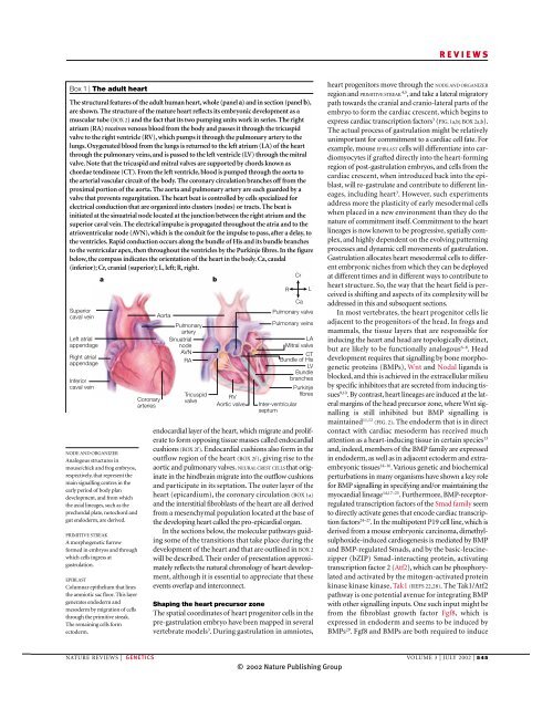
REVIEWSBox 2 | Heart developmentAMNIOTEA reptile, bird or mammal, inwhich a membrane, called theamnion, separates the conceptusfrom its environment.This box depicts the main transitions that occur in early heart development in AMNIOTES (on the basis of the events in mousedevelopment; see main text for more details). The whole embryo or isolated heart is shown on the left, whereas on the right,a representative section (transverse in panels b and d; longitudinal in panels f and h) illustrates the main internal features.All views are ventral. Staging in days of embryonic development (E) is based on mouse development. The myocardium andits progenitors are indicated in red. The cardiac progenitors are first recognizable as a crescent-shaped epithelium (thecardiac crescent) at the cranial and cranio-lateral parts of the embryo (panels a and b). The progenitor population extendscranially and laterally almost to the junction between the embryonic and extra-embryonic regions of the embryo (red arrowin panel b). Next, heart progenitors move ventrally to form the linear heart tube, which is composed of an endothelial liningthat is shrouded by a myocardial epithelium (panels c and d). Note that the inflow region of the linear heart tube is locatedcaudally, and its outflow region is located cranially. The myocardium remains attached to the ventral foregut through thedorsal mesocardium in continuity with the dorsal pericardial mesoderm. The linear heart tube undergoes a complexprogression termed cardiac looping, in which the tubular heart adopts a spiral shape with its outer surface sweepingrightwards (panels e and f ). During looping, the inflow portion of the heart, including the common atrium, is forceddorsally and cranially so that it is now above the developing ventricles. The internal relief of the heart at this stage has becomecomplex (panel f ). Endocardial cushions (EC), the precursors of the tricuspid and mitral valves (BOX 1), are forming in theatrioventricular (AV) canal. Endocardial cushions also form in the outflow tract and these are the precursors of theaorticopulmonary septum,which divides the outflowtract into the aorta andpulmonary artery. Thesecushions also give rise to theaortic and pulmonaryvalves. Other features of thisdevelopmental stage are theformation of trabeculae (T),the spongiform layer ofmyocytes along the innersurface of the ventriclesand the inter-ventricularseptum. During theremodelling phase of heartdevelopment (panels g andh), division of the heartRchambers by septation iscompleted, and distinct leftand right ventricles (LV andRV, respectively) and leftand right atria (LA and RA,respectively) are evident.This is achieved by furtherspiralling of the heart tubesuch that the outflow regionbecomes wedged betweenthe developing ventricles onRthe ventral side (panel g),and the inflow region spansthe ventricles dorsally(panel h). The chambersand vessels are now alignedas in the adult heart andbecome fully integrated.The muscular inter-atrialand inter-ventricular septaefuse with the non-muscularatrioventricular septum,which is derived from theRendocardial cushions of theatrioventricular canal,therefore completing theseparation of the chambers.Ca, caudal (inferior); Cr,cranial (superior); L, left; R, right.CrCracCaCreCaCrgCaCardiac cresent E7.75CaLinear heart tube E8.25LLooping heart E10.5LRemodelling heart E12.5LbdForegutDorsalmesocardiumfInter-ventricularseptumhECPrimitive foregutendodermRARVTLAECLVTNeuralepitheliumHead mesodermIntra-embryoniccoelomHeartprogenitorsDorsal pericardialmesodermMyocardiumEndocardiumOutflow tractCommon atriumAV canalForming rightventricleForming leftventricleInter-atrialseptumAV septumInter-ventricularseptum546 | JULY 2002 | VOLUME 3 www.nature.com/reviews/genetics© 2002 Nature Publishing Group