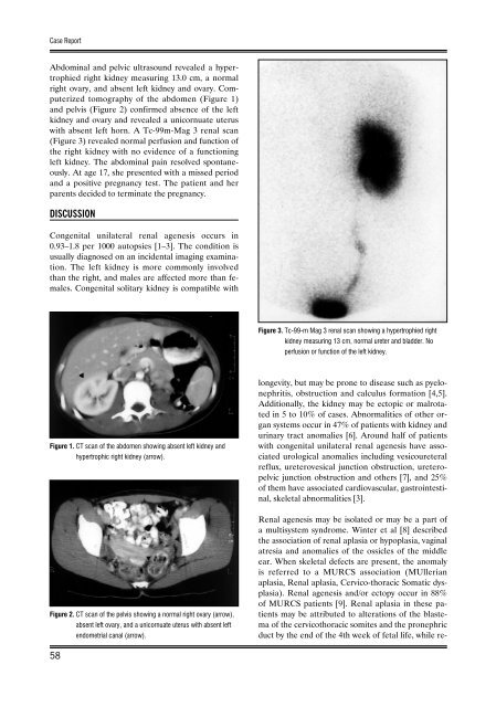
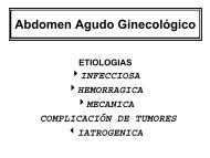
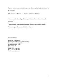
Case ReportAbdominal <strong>and</strong> pelvic ultrasound revealed a hypertrophiedright kidney measuring 13.0 cm, a normalright ovary, <strong>and</strong> absent left kidney <strong>and</strong> ovary. Computerizedtomography <strong>of</strong> the abdomen (Figure 1)<strong>and</strong> pelvis (Figure 2) confirmed absence <strong>of</strong> the leftkidney <strong>and</strong> ovary <strong>and</strong> revealed a unicornuate uteruswith absent left horn. A Tc-99m-Mag 3 <strong>renal</strong> scan(Figure 3) revealed normal perfusion <strong>and</strong> function <strong>of</strong>the right kidney with no evidence <strong>of</strong> a functioningleft kidney. The abdominal pain resolved spontaneously.At age 17, she presented with a missed period<strong>and</strong> a positive pregnancy test. The patient <strong>and</strong> herparents decided to terminate the pregnancy.DISCUSSIONCon<strong>genital</strong> <strong>unilateral</strong> <strong>renal</strong> <strong>agenesis</strong> occurs in0.93–1.8 per 1000 autopsies [1–3]. The condition isusually diagnosed on an incidental imaging examination.The left kidney is more commonly involvedthan the right, <strong>and</strong> males are affected more than females.Con<strong>genital</strong> solitary kidney is compatible withFigure 3. Tc-99-m Mag 3 <strong>renal</strong> scan showing a hypertrophied rightkidney measuring 13 cm, normal ureter <strong>and</strong> bladder. Noperfusion or function <strong>of</strong> the left kidney.Figure 1. CT scan <strong>of</strong> the abdomen showing absent left kidney <strong>and</strong>hypertrophic right kidney (arrow).Figure 2. CT scan <strong>of</strong> the pelvis showing a normal right ovary (arrow),absent left ovary, <strong>and</strong> a unicornuate uterus with absent leftendometrial canal (arrow).longevity, but may be prone to disease such as pyelonephritis,obstruction <strong>and</strong> calculus formation [4,5].Additionally, the kidney may be ectopic or malrotatedin 5 to 10% <strong>of</strong> cases. Abnormalities <strong>of</strong> other organsystems occur in 47% <strong>of</strong> patients with kidney <strong>and</strong>urinary tract <strong>anomalies</strong> [6]. Around half <strong>of</strong> patientswith con<strong>genital</strong> <strong>unilateral</strong> <strong>renal</strong> <strong>agenesis</strong> have associatedurological <strong>anomalies</strong> including vesicoureteralreflux, ureterovesical junction obstruction, ureteropelvicjunction obstruction <strong>and</strong> others [7], <strong>and</strong> 25%<strong>of</strong> them have associated cardiovascular, gastrointestinal,skeletal abnormalities [3].Renal <strong>agenesis</strong> may be isolated or may be a part <strong>of</strong>a multisystem syndrome. Winter et al [8] describedthe association <strong>of</strong> <strong>renal</strong> aplasia or hypoplasia, vaginalatresia <strong>and</strong> <strong>anomalies</strong> <strong>of</strong> the ossicles <strong>of</strong> the middleear. When skeletal defects are present, the anomalyis referred to a MURCS association (MUllerianaplasia, Renal aplasia, Cervico-thoracic Somatic dysplasia).Renal <strong>agenesis</strong> <strong>and</strong>/or ectopy occur in 88%<strong>of</strong> MURCS patients [9]. Renal aplasia in these patientsmay be attributed to alterations <strong>of</strong> the blastema<strong>of</strong> the cervicothoracic somites <strong>and</strong> the pronephricduct by the end <strong>of</strong> the 4th week <strong>of</strong> fetal life, while re-58
Barakat AJ – <strong>Association</strong> <strong>of</strong> <strong>unilateral</strong> <strong>renal</strong> <strong>agenesis</strong> <strong>and</strong> <strong>genital</strong>…nal ectopy, to a partial pronephric duct induction.The combination <strong>of</strong> absence <strong>of</strong> the vagina, abnormaluterus, <strong>renal</strong> <strong>and</strong> skeletal <strong>anomalies</strong> is known as theMayer-Rokitanski-Kuster-Hauser syndrome [10].Unilateral <strong>renal</strong> <strong>agenesis</strong> was also reported in patientswith Familial Kallmann syndrome, an X-linkedsyndrome <strong>of</strong> anosomic, hypogonadotropic hypogonadism[11].The patient in this report had <strong>unilateral</strong> <strong>renal</strong> <strong>agenesis</strong>associated with ipsilateral ovarian <strong>agenesis</strong> <strong>and</strong>unicornuate uterus with absent uterine horn on thesame side. Seventy to 89% <strong>of</strong> patients with <strong>unilateral</strong><strong>renal</strong> <strong>agenesis</strong> may have associated <strong>genital</strong> <strong>anomalies</strong>[12,13]. Renal <strong>anomalies</strong> are described in 40% <strong>of</strong> cases<strong>of</strong> mullerian aplasia [10], <strong>and</strong> 40% <strong>of</strong> women withunicornuate uterus [14]. Woolf <strong>and</strong> Allen [15] reported4 cases <strong>of</strong> unicornuate uterus <strong>of</strong> which 3 lackeda kidney on the same side <strong>of</strong> the mullarian <strong>agenesis</strong>,the fourth case had a pelvic kidney. Three out <strong>of</strong> 22females with <strong>unilateral</strong> <strong>renal</strong> <strong>agenesis</strong> described byAshley <strong>and</strong> Most<strong>of</strong>i [16] also had complete absence<strong>of</strong> one ovary, the fallopian tube on the same side <strong>and</strong>a hemiuterus. Rolen et al [17] found 67% <strong>of</strong> patientswith unicornuate uterus to have ipsilateral <strong>renal</strong> <strong>agenesis</strong>,<strong>and</strong> 13% <strong>of</strong> them have pelvic kidney. Li et al[18] reported <strong>renal</strong> <strong>agenesis</strong> in 30% <strong>of</strong> women withmullerian duct <strong>anomalies</strong>, <strong>and</strong> 80% <strong>of</strong> women withuterus didelphys. Unilateral <strong>renal</strong> <strong>agenesis</strong> has beendescribed also in association with a rudimentary uterinehorn, fallopian tube <strong>and</strong> ovary in an inguinalhernia sac [19].Genital <strong>anomalies</strong> occur in 37–60% <strong>of</strong> females <strong>and</strong>12% <strong>of</strong> males with con<strong>genital</strong> <strong>unilateral</strong> <strong>renal</strong> <strong>agenesis</strong>[3]. Abnormalities in females include <strong>agenesis</strong>,duplication, rudimentary, unicornuate or bicornuateuterus, uterus didelphys (double uterus, double cervix<strong>and</strong> double vagina), double or absent vagina, absentor hypoplastic ovary, absent fallopian tube, persistentGartner’s duct cyst, <strong>and</strong> abnormal external<strong>genital</strong>ia [3,13,18]. A double uterus with <strong>unilateral</strong>lyobstructed hemivagina is also a rare association [20].Fertility might not be impaired in the presence <strong>of</strong>mullerian <strong>agenesis</strong>; however, premature labor, spontaneousabortion <strong>and</strong> breech presentation occur morefrequently [21]. Abnormalities in males includecryptorchidism, seminal vesicle cyst, hypoplastic vas,<strong>unilateral</strong> prostatic <strong>agenesis</strong>, cystic testicular dysplasia,<strong>and</strong> hypospadias [3].The development <strong>of</strong> the urinary tract is a sequential<strong>and</strong> integrated process <strong>of</strong> the primitive <strong>renal</strong> elements.Abnormalities <strong>of</strong> this system result from defectsoccurring during embyogenesis between 15 <strong>and</strong>94 days <strong>of</strong> fetal life. Interaction between environmentalfactors such as maternal illness <strong>and</strong> exposureto toxic agents, as well as genetic factors around thisperiod result in malformations <strong>of</strong> this system [22].Abnormalities <strong>of</strong> the mullarian system, ovaries <strong>and</strong>kidney have the same embryologic defect since thewolffian <strong>and</strong> mullarian ducts develop in close anatomicalrelationship [16]. A defect in the entire region<strong>of</strong> the uro<strong>genital</strong> ridge formed from the gonad <strong>and</strong>mesonephros could account for this failure in multipleorgan development. In 1941, Gruenwald [23] demonstratedthat resection <strong>of</strong> the Wollfian duct resultedin absent kidney <strong>and</strong> fallopian tube <strong>and</strong> unicornuateuterus.Unilateral <strong>renal</strong> <strong>agenesis</strong> may be an expression <strong>of</strong>a single dominant gene [24]. The association <strong>of</strong> mullerian<strong>agenesis</strong> <strong>and</strong> <strong>renal</strong> <strong>agenesis</strong> could be an autosomaldominant disorder [25–27]. Buchta et al [26]described many generations <strong>of</strong> two families with hereditary<strong>renal</strong> adysplasia with or without mullerian<strong>anomalies</strong>. They suggested dominant inheritance.Wiersma et al [13] reported a mother <strong>and</strong> daughterwith double uterus, two cervices, a partial vaginalseptum, <strong>unilateral</strong> hematocolpos <strong>and</strong> ipsilateral <strong>renal</strong><strong>agenesis</strong>. Knudsen et al [28] described a 38-year-oldman with <strong>unilateral</strong> <strong>renal</strong> <strong>agenesis</strong> <strong>and</strong> ipsilateral seminalvesicle cyst. His sister had an embriologicallyanalogous malformation consisting <strong>of</strong> Gartner ductcyst, bicornuate uterus <strong>and</strong> <strong>renal</strong> <strong>agenesis</strong>. Schimke<strong>and</strong> King [25] observed a family with three-generationtransmission with <strong>renal</strong> <strong>agenesis</strong>/dysgenesis <strong>and</strong>uterine anomaly. They referred to it as ‘hereditaryuro<strong>genital</strong> adysplasia’, <strong>and</strong> suggested autosomal dominantinheritance with decreased penetrance <strong>and</strong>variable expressivity. The authors suggested that thedevelopmental defects <strong>of</strong> the mesonephric <strong>and</strong> paramesonephricducts might have a common genetic basis.Doray reported <strong>renal</strong> adysplasia in three generations[29]. McGillivray et al [30] suggested searchingsegment 5q11.2-q13.3 for the gene responsible forhereditary <strong>renal</strong> adysplasia. McCallum et al [31] postulatedthat <strong>unilateral</strong> <strong>renal</strong> <strong>agenesis</strong> <strong>and</strong> con<strong>genital</strong>bilateral absence <strong>of</strong> the vas deferens might havea non-cystic fibrosis mutation-mediated genetic basisthat leads to abnormal development <strong>of</strong> the entiremesonephric duct before seven weeks <strong>of</strong> gestation.CONCLUSIONSPrenatal diagnosis <strong>of</strong> the solitary kidney <strong>and</strong> other<strong>renal</strong> abnormalities by ultrasound is possible as earlyas 12 to 16 weeks <strong>of</strong> gestation [32]. Early detection <strong>of</strong>a con<strong>genital</strong> solitary kidney by routine prenatal ultrasoundor by incidental imaging studies should alert59