Association of unilateral renal agenesis and genital anomalies
Association of unilateral renal agenesis and genital anomalies
Association of unilateral renal agenesis and genital anomalies

You also want an ePaper? Increase the reach of your titles
YUMPU automatically turns print PDFs into web optimized ePapers that Google loves.
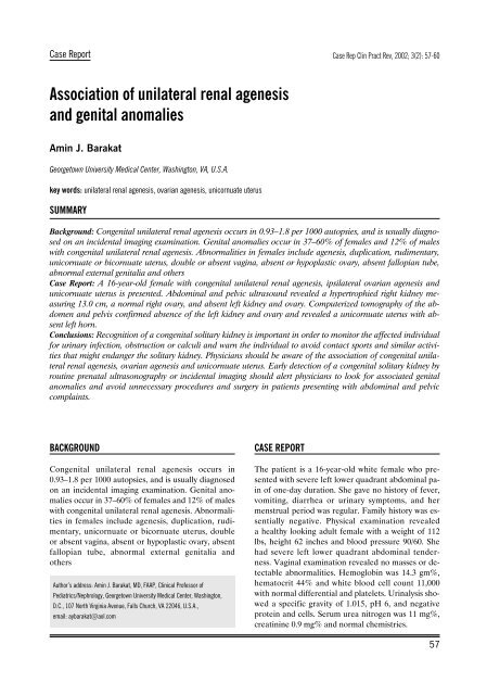
Case ReportCase Rep Clin Pract Rev, 2002; 3(2): 57-60<strong>Association</strong> <strong>of</strong> <strong>unilateral</strong> <strong>renal</strong> <strong>agenesis</strong><strong>and</strong> <strong>genital</strong> <strong>anomalies</strong>Amin J. BarakatGeorgetown University Medical Center, Washington, VA, U.S.A.key words: <strong>unilateral</strong> <strong>renal</strong> <strong>agenesis</strong>, ovarian <strong>agenesis</strong>, unicornuate uterusSUMMARYBackground: Con<strong>genital</strong> <strong>unilateral</strong> <strong>renal</strong> <strong>agenesis</strong> occurs in 0.93–1.8 per 1000 autopsies, <strong>and</strong> is usually diagnosedon an incidental imaging examination. Genital <strong>anomalies</strong> occur in 37–60% <strong>of</strong> females <strong>and</strong> 12% <strong>of</strong> maleswith con<strong>genital</strong> <strong>unilateral</strong> <strong>renal</strong> <strong>agenesis</strong>. Abnormalities in females include <strong>agenesis</strong>, duplication, rudimentary,unicornuate or bicornuate uterus, double or absent vagina, absent or hypoplastic ovary, absent fallopian tube,abnormal external <strong>genital</strong>ia <strong>and</strong> othersCase Report: A 16-year-old female with con<strong>genital</strong> <strong>unilateral</strong> <strong>renal</strong> <strong>agenesis</strong>, ipsilateral ovarian <strong>agenesis</strong> <strong>and</strong>unicornuate uterus is presented. Abdominal <strong>and</strong> pelvic ultrasound revealed a hypertrophied right kidney measuring13.0 cm, a normal right ovary, <strong>and</strong> absent left kidney <strong>and</strong> ovary. Computerized tomography <strong>of</strong> the abdomen<strong>and</strong> pelvis confirmed absence <strong>of</strong> the left kidney <strong>and</strong> ovary <strong>and</strong> revealed a unicornuate uterus with absentleft horn.Conclusions: Recognition <strong>of</strong> a con<strong>genital</strong> solitary kidney is important in order to monitor the affected individualfor urinary infection, obstruction or calculi <strong>and</strong> warn the individual to avoid contact sports <strong>and</strong> similar activitiesthat might endanger the solitary kidney. Physicians should be aware <strong>of</strong> the association <strong>of</strong> con<strong>genital</strong> <strong>unilateral</strong><strong>renal</strong> <strong>agenesis</strong>, ovarian <strong>agenesis</strong> <strong>and</strong> unicornuate uterus. Early detection <strong>of</strong> a con<strong>genital</strong> solitary kidney byroutine prenatal ultrasonography or incidental imaging should alert physicians to look for associated <strong>genital</strong><strong>anomalies</strong> <strong>and</strong> avoid unnecessary procedures <strong>and</strong> surgery in patients presenting with abdominal <strong>and</strong> pelviccomplaints.BACKGROUNDCon<strong>genital</strong> <strong>unilateral</strong> <strong>renal</strong> <strong>agenesis</strong> occurs in0.93–1.8 per 1000 autopsies, <strong>and</strong> is usually diagnosedon an incidental imaging examination. Genital <strong>anomalies</strong>occur in 37–60% <strong>of</strong> females <strong>and</strong> 12% <strong>of</strong> maleswith con<strong>genital</strong> <strong>unilateral</strong> <strong>renal</strong> <strong>agenesis</strong>. Abnormalitiesin females include <strong>agenesis</strong>, duplication, rudimentary,unicornuate or bicornuate uterus, doubleor absent vagina, absent or hypoplastic ovary, absentfallopian tube, abnormal external <strong>genital</strong>ia <strong>and</strong>othersAuthor’s address: Amin J. Barakat, MD, FAAP, Clinical Pr<strong>of</strong>essor <strong>of</strong>Pediatrics/Nephrology, Georgetown University Medical Center, Washington,D.C., 107 North Virginia Avenue, Falls Church, VA 22046, U.S.A.,email: aybarakat@aol.comCASE REPORTThe patient is a 16-year-old white female who presentedwith severe left lower quadrant abdominal pain<strong>of</strong> one-day duration. She gave no history <strong>of</strong> fever,vomiting, diarrhea or urinary symptoms, <strong>and</strong> hermenstrual period was regular. Family history was essentiallynegative. Physical examination revealeda healthy looking adult female with a weight <strong>of</strong> 112lbs, height 62 inches <strong>and</strong> blood pressure 90/60. Shehad severe left lower quadrant abdominal tenderness.Vaginal examination revealed no masses or detectableabnormalities. Hemoglobin was 14.3 gm%,hematocrit 44% <strong>and</strong> white blood cell count 11,000with normal differential <strong>and</strong> platelets. Urinalysis showeda specific gravity <strong>of</strong> 1.015, pH 6, <strong>and</strong> negativeprotein <strong>and</strong> cells. Serum urea nitrogen was 11 mg%,creatinine 0.9 mg% <strong>and</strong> normal chemistries.57
Case ReportAbdominal <strong>and</strong> pelvic ultrasound revealed a hypertrophiedright kidney measuring 13.0 cm, a normalright ovary, <strong>and</strong> absent left kidney <strong>and</strong> ovary. Computerizedtomography <strong>of</strong> the abdomen (Figure 1)<strong>and</strong> pelvis (Figure 2) confirmed absence <strong>of</strong> the leftkidney <strong>and</strong> ovary <strong>and</strong> revealed a unicornuate uteruswith absent left horn. A Tc-99m-Mag 3 <strong>renal</strong> scan(Figure 3) revealed normal perfusion <strong>and</strong> function <strong>of</strong>the right kidney with no evidence <strong>of</strong> a functioningleft kidney. The abdominal pain resolved spontaneously.At age 17, she presented with a missed period<strong>and</strong> a positive pregnancy test. The patient <strong>and</strong> herparents decided to terminate the pregnancy.DISCUSSIONCon<strong>genital</strong> <strong>unilateral</strong> <strong>renal</strong> <strong>agenesis</strong> occurs in0.93–1.8 per 1000 autopsies [1–3]. The condition isusually diagnosed on an incidental imaging examination.The left kidney is more commonly involvedthan the right, <strong>and</strong> males are affected more than females.Con<strong>genital</strong> solitary kidney is compatible withFigure 3. Tc-99-m Mag 3 <strong>renal</strong> scan showing a hypertrophied rightkidney measuring 13 cm, normal ureter <strong>and</strong> bladder. Noperfusion or function <strong>of</strong> the left kidney.Figure 1. CT scan <strong>of</strong> the abdomen showing absent left kidney <strong>and</strong>hypertrophic right kidney (arrow).Figure 2. CT scan <strong>of</strong> the pelvis showing a normal right ovary (arrow),absent left ovary, <strong>and</strong> a unicornuate uterus with absent leftendometrial canal (arrow).longevity, but may be prone to disease such as pyelonephritis,obstruction <strong>and</strong> calculus formation [4,5].Additionally, the kidney may be ectopic or malrotatedin 5 to 10% <strong>of</strong> cases. Abnormalities <strong>of</strong> other organsystems occur in 47% <strong>of</strong> patients with kidney <strong>and</strong>urinary tract <strong>anomalies</strong> [6]. Around half <strong>of</strong> patientswith con<strong>genital</strong> <strong>unilateral</strong> <strong>renal</strong> <strong>agenesis</strong> have associatedurological <strong>anomalies</strong> including vesicoureteralreflux, ureterovesical junction obstruction, ureteropelvicjunction obstruction <strong>and</strong> others [7], <strong>and</strong> 25%<strong>of</strong> them have associated cardiovascular, gastrointestinal,skeletal abnormalities [3].Renal <strong>agenesis</strong> may be isolated or may be a part <strong>of</strong>a multisystem syndrome. Winter et al [8] describedthe association <strong>of</strong> <strong>renal</strong> aplasia or hypoplasia, vaginalatresia <strong>and</strong> <strong>anomalies</strong> <strong>of</strong> the ossicles <strong>of</strong> the middleear. When skeletal defects are present, the anomalyis referred to a MURCS association (MUllerianaplasia, Renal aplasia, Cervico-thoracic Somatic dysplasia).Renal <strong>agenesis</strong> <strong>and</strong>/or ectopy occur in 88%<strong>of</strong> MURCS patients [9]. Renal aplasia in these patientsmay be attributed to alterations <strong>of</strong> the blastema<strong>of</strong> the cervicothoracic somites <strong>and</strong> the pronephricduct by the end <strong>of</strong> the 4th week <strong>of</strong> fetal life, while re-58
Barakat AJ – <strong>Association</strong> <strong>of</strong> <strong>unilateral</strong> <strong>renal</strong> <strong>agenesis</strong> <strong>and</strong> <strong>genital</strong>…nal ectopy, to a partial pronephric duct induction.The combination <strong>of</strong> absence <strong>of</strong> the vagina, abnormaluterus, <strong>renal</strong> <strong>and</strong> skeletal <strong>anomalies</strong> is known as theMayer-Rokitanski-Kuster-Hauser syndrome [10].Unilateral <strong>renal</strong> <strong>agenesis</strong> was also reported in patientswith Familial Kallmann syndrome, an X-linkedsyndrome <strong>of</strong> anosomic, hypogonadotropic hypogonadism[11].The patient in this report had <strong>unilateral</strong> <strong>renal</strong> <strong>agenesis</strong>associated with ipsilateral ovarian <strong>agenesis</strong> <strong>and</strong>unicornuate uterus with absent uterine horn on thesame side. Seventy to 89% <strong>of</strong> patients with <strong>unilateral</strong><strong>renal</strong> <strong>agenesis</strong> may have associated <strong>genital</strong> <strong>anomalies</strong>[12,13]. Renal <strong>anomalies</strong> are described in 40% <strong>of</strong> cases<strong>of</strong> mullerian aplasia [10], <strong>and</strong> 40% <strong>of</strong> women withunicornuate uterus [14]. Woolf <strong>and</strong> Allen [15] reported4 cases <strong>of</strong> unicornuate uterus <strong>of</strong> which 3 lackeda kidney on the same side <strong>of</strong> the mullarian <strong>agenesis</strong>,the fourth case had a pelvic kidney. Three out <strong>of</strong> 22females with <strong>unilateral</strong> <strong>renal</strong> <strong>agenesis</strong> described byAshley <strong>and</strong> Most<strong>of</strong>i [16] also had complete absence<strong>of</strong> one ovary, the fallopian tube on the same side <strong>and</strong>a hemiuterus. Rolen et al [17] found 67% <strong>of</strong> patientswith unicornuate uterus to have ipsilateral <strong>renal</strong> <strong>agenesis</strong>,<strong>and</strong> 13% <strong>of</strong> them have pelvic kidney. Li et al[18] reported <strong>renal</strong> <strong>agenesis</strong> in 30% <strong>of</strong> women withmullerian duct <strong>anomalies</strong>, <strong>and</strong> 80% <strong>of</strong> women withuterus didelphys. Unilateral <strong>renal</strong> <strong>agenesis</strong> has beendescribed also in association with a rudimentary uterinehorn, fallopian tube <strong>and</strong> ovary in an inguinalhernia sac [19].Genital <strong>anomalies</strong> occur in 37–60% <strong>of</strong> females <strong>and</strong>12% <strong>of</strong> males with con<strong>genital</strong> <strong>unilateral</strong> <strong>renal</strong> <strong>agenesis</strong>[3]. Abnormalities in females include <strong>agenesis</strong>,duplication, rudimentary, unicornuate or bicornuateuterus, uterus didelphys (double uterus, double cervix<strong>and</strong> double vagina), double or absent vagina, absentor hypoplastic ovary, absent fallopian tube, persistentGartner’s duct cyst, <strong>and</strong> abnormal external<strong>genital</strong>ia [3,13,18]. A double uterus with <strong>unilateral</strong>lyobstructed hemivagina is also a rare association [20].Fertility might not be impaired in the presence <strong>of</strong>mullerian <strong>agenesis</strong>; however, premature labor, spontaneousabortion <strong>and</strong> breech presentation occur morefrequently [21]. Abnormalities in males includecryptorchidism, seminal vesicle cyst, hypoplastic vas,<strong>unilateral</strong> prostatic <strong>agenesis</strong>, cystic testicular dysplasia,<strong>and</strong> hypospadias [3].The development <strong>of</strong> the urinary tract is a sequential<strong>and</strong> integrated process <strong>of</strong> the primitive <strong>renal</strong> elements.Abnormalities <strong>of</strong> this system result from defectsoccurring during embyogenesis between 15 <strong>and</strong>94 days <strong>of</strong> fetal life. Interaction between environmentalfactors such as maternal illness <strong>and</strong> exposureto toxic agents, as well as genetic factors around thisperiod result in malformations <strong>of</strong> this system [22].Abnormalities <strong>of</strong> the mullarian system, ovaries <strong>and</strong>kidney have the same embryologic defect since thewolffian <strong>and</strong> mullarian ducts develop in close anatomicalrelationship [16]. A defect in the entire region<strong>of</strong> the uro<strong>genital</strong> ridge formed from the gonad <strong>and</strong>mesonephros could account for this failure in multipleorgan development. In 1941, Gruenwald [23] demonstratedthat resection <strong>of</strong> the Wollfian duct resultedin absent kidney <strong>and</strong> fallopian tube <strong>and</strong> unicornuateuterus.Unilateral <strong>renal</strong> <strong>agenesis</strong> may be an expression <strong>of</strong>a single dominant gene [24]. The association <strong>of</strong> mullerian<strong>agenesis</strong> <strong>and</strong> <strong>renal</strong> <strong>agenesis</strong> could be an autosomaldominant disorder [25–27]. Buchta et al [26]described many generations <strong>of</strong> two families with hereditary<strong>renal</strong> adysplasia with or without mullerian<strong>anomalies</strong>. They suggested dominant inheritance.Wiersma et al [13] reported a mother <strong>and</strong> daughterwith double uterus, two cervices, a partial vaginalseptum, <strong>unilateral</strong> hematocolpos <strong>and</strong> ipsilateral <strong>renal</strong><strong>agenesis</strong>. Knudsen et al [28] described a 38-year-oldman with <strong>unilateral</strong> <strong>renal</strong> <strong>agenesis</strong> <strong>and</strong> ipsilateral seminalvesicle cyst. His sister had an embriologicallyanalogous malformation consisting <strong>of</strong> Gartner ductcyst, bicornuate uterus <strong>and</strong> <strong>renal</strong> <strong>agenesis</strong>. Schimke<strong>and</strong> King [25] observed a family with three-generationtransmission with <strong>renal</strong> <strong>agenesis</strong>/dysgenesis <strong>and</strong>uterine anomaly. They referred to it as ‘hereditaryuro<strong>genital</strong> adysplasia’, <strong>and</strong> suggested autosomal dominantinheritance with decreased penetrance <strong>and</strong>variable expressivity. The authors suggested that thedevelopmental defects <strong>of</strong> the mesonephric <strong>and</strong> paramesonephricducts might have a common genetic basis.Doray reported <strong>renal</strong> adysplasia in three generations[29]. McGillivray et al [30] suggested searchingsegment 5q11.2-q13.3 for the gene responsible forhereditary <strong>renal</strong> adysplasia. McCallum et al [31] postulatedthat <strong>unilateral</strong> <strong>renal</strong> <strong>agenesis</strong> <strong>and</strong> con<strong>genital</strong>bilateral absence <strong>of</strong> the vas deferens might havea non-cystic fibrosis mutation-mediated genetic basisthat leads to abnormal development <strong>of</strong> the entiremesonephric duct before seven weeks <strong>of</strong> gestation.CONCLUSIONSPrenatal diagnosis <strong>of</strong> the solitary kidney <strong>and</strong> other<strong>renal</strong> abnormalities by ultrasound is possible as earlyas 12 to 16 weeks <strong>of</strong> gestation [32]. Early detection <strong>of</strong>a con<strong>genital</strong> solitary kidney by routine prenatal ultrasoundor by incidental imaging studies should alert59
Case Reportthe physician to look for associated <strong>genital</strong> <strong>anomalies</strong>.This is particularly important in young femalessince one <strong>of</strong> every three with <strong>renal</strong> <strong>agenesis</strong> will alsohave a significant anomaly <strong>of</strong> the uterus, ovary or vagina[3]. Such knowledge is useful in avoiding unnecessaryprocedures <strong>and</strong> surgery in patients presentingwith abdominal or pelvic complaints. Recognition<strong>of</strong> a con<strong>genital</strong> solitary kidney is also importantin order to monitor the affected individual for urinaryinfection, obstruction or calculi <strong>and</strong> warn the individualto avoid contact sports <strong>and</strong> similar activitiesthat might endanger the solitary kidney.REFERENCES:1. Doroshou LW, Abeshouse BS: Con<strong>genital</strong> <strong>unilateral</strong> solitary kidney:Report <strong>of</strong> 37 cases <strong>and</strong> a review <strong>of</strong> the literature. Urol Surv, 1961; 11:219-292. Barakat AJ, Drougas JG: Occurrence <strong>of</strong> con<strong>genital</strong> abnormalities <strong>of</strong>the kidney <strong>and</strong> urinary tract in 13, 775 autopsies. Urology, 1991; 38:347-503. Thompson DP, Lynn HB: Genital <strong>anomalies</strong> associated with solitarykidney. Mayo Clin Proc, 1966; 41: 538-484. Dees JE: Prognosis <strong>of</strong> the solitary kidney. J Urol, 1960; 83: 550-25. Emanuel B, Nachman R, Aronson N, Weis H: Con<strong>genital</strong> solitary kidney.A review <strong>of</strong> 74 cases. Am J Dis Child, 1974; 127: 17-96. Barakat AJ, Drougas JG, Barakat R: <strong>Association</strong> <strong>of</strong> con<strong>genital</strong> abnormalities<strong>of</strong> the kidney <strong>and</strong> urinary tract with those <strong>of</strong> other organ systemsin 13, 775 autopsies. Child Nephrol Urol, 1988: 269-727. Cascio S, Paran S, Puri P: Associated urological <strong>anomalies</strong> in childrenwith <strong>unilateral</strong> <strong>renal</strong> <strong>agenesis</strong>. J Urol, 1999; 162: 1081-38. Winter JSD, Kohn G, Mellman WJ, Wagner S: A familial syndrome <strong>of</strong><strong>renal</strong>, <strong>genital</strong> <strong>and</strong> middle ear <strong>anomalies</strong>. J Pediatr, 1968; 72: 88-939. Duncan PA, Shapiro LR, Stangel JJ et al: The MURCS association:Mullerian duct aplasia, <strong>renal</strong> aplasia, <strong>and</strong> cervicothoracic somite dysplasia.J Pediatr, 1979; 95: 399-40210. Griffin JE, Edwards C, Madden JD et al: Con<strong>genital</strong> absence <strong>of</strong> thevagina: The Mayer- Rokitansky-Kuster-Hauser syndrome. Ann InternMed, 1976; 85: 224-3611. Wegenke JD, Vehling DT, Wear JB et al: Familial Kallmann syndromewith <strong>unilateral</strong> <strong>renal</strong> aplasia. Clin Genet, 1975; 7: 368-8112. Collins DC: Con<strong>genital</strong> <strong>unilateral</strong> <strong>renal</strong> <strong>agenesis</strong>. Ann Surg, 1932; 96:715-2613. Wiersma AF, Peterson LF, Justema EJ: Uterine <strong>anomalies</strong> associatedwith <strong>unilateral</strong> <strong>renal</strong> <strong>agenesis</strong>. Obstet Gynec, 1976; 47: 654-714. Fedele L, Bianchi S, Agnoli B et al: Urinary tract <strong>anomalies</strong> associatedwith unicornuate uterus. J Urol, 1996; 155: 847-815. Woolf RE, Allen WB: Concomittent malformation, the frequent simultaneousoccurrence <strong>of</strong> con<strong>genital</strong> malformations <strong>of</strong> the reproductive<strong>and</strong> urinary tracts. J Obstet Gynec, 1963; 2: 256-6516. Ashley DJ, Most<strong>of</strong>i FK: Renal <strong>agenesis</strong> <strong>and</strong> dysgenesis. J Urol, 1960;83: 211-3017. Rolen AC, Choquette AJ, Semmens JP: Rudementary uterine horn:Obstetric <strong>and</strong> gynecologic implications. Obstet Gynecol, 1966; 27:806-1318. Li S, Qayyum A, Coakley FV, Hricak H: <strong>Association</strong> <strong>of</strong> <strong>renal</strong> <strong>agenesis</strong><strong>and</strong> mullerian duct <strong>anomalies</strong>. J Comput Assist Tomogr, 2000; 24:829-3419. Keating JP, Yu MH, Grunewald B: Hernia uterus inguinale associatedwith <strong>unilateral</strong> <strong>renal</strong> <strong>agenesis</strong>. Aust NZ J Surg, 1995; 65: 688-9020. Phupong V, Pruksananonda K, Taneeppanichskul S et al: Double uteruswith <strong>unilateral</strong>ly obstructed hemivagina <strong>and</strong> ipsilateral <strong>renal</strong> <strong>agenesis</strong>:a variety presentation <strong>and</strong> a 10-year review <strong>of</strong> the literature.J Med Assoc Thai, 2000; 83: 569-7421. Bradley B, Gleicher N: Gr<strong>and</strong> multiparity associated with <strong>unilateral</strong> <strong>renal</strong>,ovarian <strong>and</strong> Mullerian <strong>agenesis</strong>. Mt Sinai J Med, 1980; 47: 418-2222. Temple JK, Shapira E: Genetic determinants <strong>of</strong> <strong>renal</strong> disease in neonates.Clin Perinatol, 1981; 8: 361-7323. Gruenwald P: The relation <strong>of</strong> the growing Mullerian duct <strong>and</strong> theWolffian duct <strong>and</strong> its importance for the genesis <strong>of</strong> malformations.Anat Rec, 1941; 81: 1-1924. Fitch N: Heterogeneity <strong>of</strong> bilateral <strong>renal</strong> <strong>agenesis</strong>. Canad Med Assoc J,1977; 116: 381-225. Schimke, RN, King CR: Hereditary uro<strong>genital</strong> adysplasia. Clin Genet,1980; 18: 417-2026. Buchta RM, Visekul C, Gilbert EF et al: Familial bilateral <strong>renal</strong> <strong>agenesis</strong><strong>and</strong> hereditary <strong>renal</strong> adysplasia. Z Kinderheilk, 1973; 115: 111-2927. Biedel CW, Pagon RA, Zapata JO: Mullerian <strong>anomalies</strong> <strong>and</strong> <strong>renal</strong><strong>agenesis</strong>: Autosomal dominant uro<strong>genital</strong> adysplasia. J Pediatr, 1984;104: 861-428. Knudsen JB, Brun B, Emus HC: Familial <strong>renal</strong> <strong>agenesis</strong> <strong>and</strong> uro<strong>genital</strong>malformations: seminal vesicle cyst <strong>and</strong> vaginal cyst with bicornuateuterus in siblings. Sc<strong>and</strong> J Urol Nephrol, 1979; 13: 109-1229. Doray B, Gasser B, Reinartz I, Stoll C: Hereditary <strong>renal</strong> adysplasia ina three generations family. Genet Counsel, 1999; 10: 251-730. McGillivray BC, Bassett AS, Langlois S et al: Familial 5q11. 2-q13. 3segmental duplication cosegragating with multiple <strong>anomalies</strong>, includingschizophrenia. Am J Med Genet, 1990; 35: 10-331. McCallum T, Milunsky J, Munarriz R et al: Unilateral <strong>renal</strong> <strong>agenesis</strong>associatedwith con<strong>genital</strong> bilateral absence <strong>of</strong> the vas deferens: phenotypicfindings <strong>and</strong> genetic considerations. Hum Reprod, 2001; 16:282-832. Barakat AY, Awazu M, Fleischer AC: Antenatal diagnosis <strong>of</strong> <strong>renal</strong> abnormalities:A review <strong>of</strong> the state <strong>of</strong> the art. Journal <strong>of</strong> the SouthernMedical <strong>Association</strong>, 1988; 82: 229-3460