Imaging of Glenohumeral Instability - NationalRad
Imaging of Glenohumeral Instability - NationalRad
Imaging of Glenohumeral Instability - NationalRad

Create successful ePaper yourself
Turn your PDF publications into a flip-book with our unique Google optimized e-Paper software.
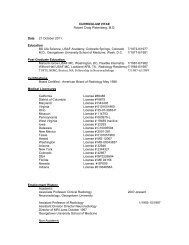
<strong>Imaging</strong> <strong>of</strong> glenohumeral instability 163many as 95% <strong>of</strong> all dislocations occur in the anterior directionand are not associated with a specific sport. In theyounger patient (age 35 years) the most common lesionassociated with an anterior subluxation event is a Bankartlesion (injury <strong>of</strong> the anterior-inferior labrum) or one <strong>of</strong> theBankart variants discussed below. 15 In the older patient population(age 35 years) the rotator cuff becomes the “weaklink” rather than the labrum and patients in this older agegroup experiencing a first-time dislocation are more likely tosustain a tear or avulsion <strong>of</strong> the rotator cuff tendon or possiblyan avulsion fracture <strong>of</strong> the greater tuberosity. 16In the patient presenting with an anteriorly dislocated humeralhead, the arm is <strong>of</strong>ten fixed in slight abduction andexternal rotation. The humeral head may be palpated in ananteromedial position, beneath the coracoid process. Followingreduction, an untreated Bankart lesion will lead to recurrentinstability rates that approach 80%-90%. The patient<strong>of</strong>ten complains <strong>of</strong> pain and a feeling <strong>of</strong> apprehension duringabduction and external rotation <strong>of</strong> the upper arm. Associatedinjuries include an impaction fracture <strong>of</strong> the posterosuperiorhumeral head (Hill-Sachs lesion), osseous or cartilaginousinjury <strong>of</strong> the anterior-inferior glenoid rim, or possibly astretching-type injury (neuropraxia) <strong>of</strong> the axillary nerve. Inthe patient with recurrent subluxation or continued signs <strong>of</strong>apprehension, the treatment <strong>of</strong> choice is surgical repair <strong>of</strong> thespecific lesion <strong>of</strong> instability.Radiographic FindingsAssociated With Anterior <strong>Instability</strong>An anterior dislocation <strong>of</strong> the humeral head is easily detectedon conventional radiographs. The AP radiograph will demonstrateinferior and medial displacement <strong>of</strong> the humeralhead into the subcoracoid location and an anterior dislocationis <strong>of</strong>ten referred to as a subcoracoid dislocation (Fig. 4A).The axillary and Scapular Y views will also demonstrate anteriordislocation, but are rarely necessary to establish thediagnosis. Once the humeral head is reduced, signs <strong>of</strong> previousdislocation include flattening <strong>of</strong> the posterolateral aspect<strong>of</strong> the humeral head (Hill-Sachs lesion) or a fracture <strong>of</strong> theanterior-inferior glenoid rim (osseus Bankart lesion) (Fig. 4Band C). The Hill-Sachs lesion is best depicted on the Strykernotch view or on an AP radiograph with the humeral head ininternal rotation. 17 The osseous Bankart lesion is best depictedon the West Point view (modified axillary view). 18 Inolder patients, an avulsion injury <strong>of</strong> the greater tuberositymay indicate a previous anterior dislocation.Figure 3 Abduction external rotation (ABER) imaging. (A) Normal patientpositioning for ABER imaging. The patient is supine, hand placed behindhead with arm in maximum abduction external rotation. A dual-coil configurationused. (B) A coronal scout is obtained and the ABER images areprescribed <strong>of</strong>f the scout images along the long axis <strong>of</strong> the humeral shaft. (C)ABER image shows the taut anterior band <strong>of</strong> the inferior glenohumeralligament (short arrows) and the normal-appearing anterior inferior labrum(long arrow). (Color version <strong>of</strong> figure is available online.)Computed Tomography FindingsAssociated With Anterior <strong>Instability</strong>Computed tomography (CT) imaging is helpful in detectingand delineating the extent <strong>of</strong> osseous involvement. In particular,sagittal reconstruction images through the glenoid fossawill demonstrate the size and number <strong>of</strong> fragments <strong>of</strong> anosseous Bankart lesion. The size <strong>of</strong> the osseous fragmentshould be reported along with the number <strong>of</strong> osseous fragmentsand the extent <strong>of</strong> displacement <strong>of</strong> fragments. In addition,an estimation regarding the percentage <strong>of</strong> the face <strong>of</strong> the
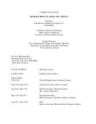
164 T.G. Sanders, M. Zlatkin, and J. Montgomeryglenoid that is involved should be reported (using the sagittalimaging plane). Extensive bony deficiency <strong>of</strong> the anteriorinferiorglenoid rim has been referred to as the inverted pearappearance (Fig. 5), and bone graft material may be requiredas a part <strong>of</strong> the reconstruction if the bony deficiency is greaterthan 30% <strong>of</strong> the face <strong>of</strong> the glenoid. 19MR FindingsAssociated With Anterior <strong>Instability</strong>Conventional MR imaging and direct MR arthrography areboth excellent techniques for detecting the extent <strong>of</strong> s<strong>of</strong>ttissueinjury after an anterior subluxation event. Direct MRarthrography has the added benefit <strong>of</strong> creating joint distention,which may increase the conspicuity <strong>of</strong> a subtle or nondisplacedlabral lesion, which will fill with high signal intensitygadolinium. The presence <strong>of</strong> gadolinium in the joint alsoallows the use <strong>of</strong> T1-weighted imaging to evaluate the labrum,which is a higher signal-to-noise image than the T2images typically required to detect labral pathology whenusing conventional MR imaging. The main disadvantage <strong>of</strong>direct MR arthrography is that it is minimally invasive andrequires the presence <strong>of</strong> a physician to perform the intraarticularinjection. The use <strong>of</strong> direct MR arthrography may beespecially beneficial in the evaluation <strong>of</strong> subtle lesions thatcan occur in the overhead athlete.Numerous Bankart variants have been described, eachdemonstrating a slightly different MR appearance with somevariations requiring a slightly different surgical repair technique.Therefore, knowledge <strong>of</strong> the various lesions and anFigure 4 Radiographic findings associated with anterior dislocation.AP radiograph (A) shows anterior dislocation <strong>of</strong> the humeralhead (long arrow) relative to the glenoid fossa (short arrow).The humeral head is positioned medial and inferior inposition. West-Point view (B) shows a small osseous Bankartlesion. True AP view <strong>of</strong> the glenohumeral joint (C) shows a smallosseous Bankart lesion (short arrow) and a large Hill-Sachs defect(long arrow).Figure 5 Osseous defect <strong>of</strong> the glenoid rim CT imaging. Sagittal CTreconstruction through the glenoid fossa demonstrates a large osseousdefect (arrows), indicating a Bankart lesion after anterior dislocation.The inverted pear appearance <strong>of</strong> the glenoid fossa indicatessignificant bone loss likely requiring bone graft to regain postoperativestability <strong>of</strong> the glenohumeral joint.
<strong>Imaging</strong> <strong>of</strong> glenohumeral instability 165accurate description <strong>of</strong> the instability lesion can be beneficialin obtaining optimal surgical results.Fibrous Bankart LesionA tear <strong>of</strong> the anterior-inferior glenoid labrum. This is themost common lesion after an anterior subluxation event. 15The labral fragment is <strong>of</strong>ten displaced and stripped awayfrom the adjacent glenoid. The axial imaging plane is theprimary plane for detecting Bankart lesion and MR imagingmay demonstrate an irregular fluid or contrast collection extendinginto the substance <strong>of</strong> or deep to the labrum with orwithout abnormal morphology <strong>of</strong> the labrum. 20,21 The adjacentmedial scapular periosteum is <strong>of</strong>ten stripped from theadjacent bone or possibly completely disrupted, resulting ina displaced labral fragment (Fig. 6). The location and extent<strong>of</strong> a labral tear should be described using either the quadrants<strong>of</strong> the glenoid as reference points or using the numbers <strong>of</strong> theface <strong>of</strong> the clock as reference points. The coronal imagingplane may also demonstrate fluid or contrast extending intothe substance <strong>of</strong> the anterior-inferior labrum and this hasbeen described as the double axillary pouch sign (Fig. 7).Perthes LesionA nondisplaced tear <strong>of</strong> the anterior-inferior glenoid labrum. 22The medial scapular periosteum remains intact in this Bankartvariant holding the labrum in near anatomic position(Fig. 8A). A chronic Perthes lesion may resynovialize or scarback down in place making this lesion difficult to detect onconventional MR imaging. This is the 1 lesion in which directMR arthrography with stress ABER imaging may be particularlyuseful in detecting an otherwise occult lesion <strong>of</strong> instability14 (Fig. 8B).Figure 7 Double axillary pouch sign, anterior labral tear in the coronalimaging plane. Coronal MR arthrographic image (A) shows thenormal-appearing anterior-inferior labrum (short arrow). CoronalMR arthrographic image (B) shows the double axillary pouch sign, acontrast collection (long arrow) located between the glenoid rimand torn anterior labrum (short arrow).Figure 6 Bankart lesion, MR imaging. Axial T2-weighted imagethrough the glenohumeral joint reveals a tear (long arrow) <strong>of</strong> theanterior inferior labrum (short arrow). Intermediate signal is seenextending completely beneath the labrum indicating a tear andcomplete detachment <strong>of</strong> the labrum.Anterior LabroligamentousPeriosteal Sleeve Avulsion LesionAnterior labroligamentous periosteal sleeve avulsion injury(medialized Bankart lesion) (Fig. 9) is a variant <strong>of</strong> the Bankartlesion in which the medial scapular periosteum remains intactand pulls the torn labral fragment in a medial direction. 23These lesions are easily detected in the acute setting, but maybe more difficult to detect in the chronic setting when themedialized labral fragment has scarred down to the adjacent
166 T.G. Sanders, M. Zlatkin, and J. Montgomeryimages, while the sagittal imaging plane is usually best suitedfor demonstrating the size and extent <strong>of</strong> the lesion (Fig. 10).Marrow edema <strong>of</strong> the glenoid as seen on MR imaging is usuallya very minor component <strong>of</strong> the abnormality. CT imaging,especially in the sagittal imaging plane can be helpful indepicting the extent <strong>of</strong> osseous abnormality and in detectingthe number <strong>of</strong> fracture fragments. The face <strong>of</strong> the glenoid asviewed in the sagittal imaging plane has been described ashaving a pear-shaped appearance. Loss <strong>of</strong> significant bonestock along the anterior-inferior margin has been describedas the inverted pear appearance <strong>of</strong> the glenoid (Fig. 5). This isimportant to describe as a bony deficiency <strong>of</strong> more than 30%<strong>of</strong> the face <strong>of</strong> the glenoid will likely require a bone graftprocedure to regain normal stability in the postoperative setting.19Glenolabral Articular Disruption LesionGlenolabral articular disruption 24 lesion most <strong>of</strong>ten resultsfrom an impaction <strong>of</strong> the humeral head against the face <strong>of</strong> theglenoid, resulting in a nondisplaced tear <strong>of</strong> the anterior-inferiorlabrum with an associated chondral injury <strong>of</strong> the inferiorglenoid (Fig. 11). 25 Often there is no history <strong>of</strong> a frank dislocation,and the patient typically presents with shoulder painbut only minimal signs <strong>of</strong> instability on physical examination.Surgical treatment includes labral repair or debridementcoupled with debridement <strong>of</strong> the chondral lesion.Humeral Avulsion <strong>of</strong> the<strong>Glenohumeral</strong> Ligament LesionHumeral avulsion <strong>of</strong> the glenohumeral ligament 26,27 lesionresults from an anterior dislocation or subluxation event andresults in a tear or avulsion <strong>of</strong> the anterior band <strong>of</strong> the inferiorFigure 8 Perthes lesion (nondisplaced labral tear). Axial (A) andABER (B) MR arthrographic images demonstrate a collection <strong>of</strong> contrast(long arrow), extending partially beneath the anterior inferiorlabrum (short arrow). The medial scapular periosteum (arrowheads) remains intact holding the labrum in near anatomic position.glenoid. In the chronic setting, these lesions may requireadditional surgical debridement <strong>of</strong> the fibrosis and scar tissuebefore completing the Bankart repair.Osseous Bankart LesionFracture <strong>of</strong> the anterior-inferior glenoid rim that accompaniesa tear <strong>of</strong> the glenoid labrum. These lesions may be difficultto detect on both radiographs and MR imaging. On MRimaging, the cortex <strong>of</strong> the anterior glenoid margin should bethoroughly evaluated in all 3 imaging planes. Disruption orirregularity <strong>of</strong> the cortex may be seen on axial and coronalFigure 9 ALPSA lesion (anterior labroligamentous periosteal sleeveavulsion), medialized Bankart. Axial MR arthrographic image demonstratesthe torn anterior-inferior labrum (long arrow) pulled in amedial direction by an intact medial scapular periosteum (shortarrow).
<strong>Imaging</strong> <strong>of</strong> glenohumeral instability 167band <strong>of</strong> the inferior glenohumeral ligament can be seen at theexpected humeral attachment site. In the acute setting, therewill be adjacent s<strong>of</strong>t-tissue edema and when performing directMR arthrography, contrast will <strong>of</strong>ten be seen leakingthrough the capsular defect.Batter’s ShoulderThis term has been used to describe a subluxation event <strong>of</strong>the glenohumeral joint that occurs when a baseball batter“swings and a misses” a pitch. When a powerful batter swingsa bat and misses the ball, tremendous forces are transmittedacross the glenohumeral joint and can result in a subluxationevent <strong>of</strong> the leading shoulder, (usually the nondominant ornonthrowing shoulder for a baseball player, unless the playeris a switch-hitter). We have seen numerous cases in our practicewith this exact mechanism <strong>of</strong> injury leading to a first-timesubluxation event resulting in immediate onset <strong>of</strong> disablingshoulder pain. We have seen both anterior and posteriorsubluxation events occurring with this exact mechanism <strong>of</strong>injury and in each case resulting in either a Bankart or “reverse”Bankart variant, <strong>of</strong>ten with an associated Hill-Sachs or“reverse” Hill-Sachs lesion, typically resulting in disablingpain and instability <strong>of</strong> the involved glenohumeral joint (Figs.13 and 14). Surgical repair is usually required for the patientto return to a preinjury level <strong>of</strong> play. This mechanism <strong>of</strong>injury has been reported in several pr<strong>of</strong>essional baseball playersand is <strong>of</strong>ten a season-ending injury.Figure 10 Osseous Bankart lesion. Axial T2-weighted image (A) andcoronal T1-weighted image (B) demonstrate a minimally displacedosseous Bankart lesion (short arrow). The axial image shows an area<strong>of</strong> cortical step-<strong>of</strong>f (long arrow), while the coronal image demonstratesa low signal intensity line indicating the fracture through theglenoid.glenohumeral ligament from the level <strong>of</strong> its humeral attachment(Fig. 12). There is no age predilection for this lesion.This lesion is difficult to detect using the standard arthroscopyportals, and it is therefore very important to alert thesurgeon to the possibility <strong>of</strong> this lesion preoperatively so thepotential lesion can be adequately evaluated at the time <strong>of</strong>surgery. A missed HAGL lesion has been reported as a commoncause for a failed shoulder reconstruction after anteriordislocation. On MR imaging, a disruption <strong>of</strong> the anteriorFigure 11 GLAD lesion (glenolabral articular disruption). Axial T2-weighted MR image with fat saturation demonstrates a nondisplacedtear <strong>of</strong> the anterior-inferior glenoid labrum with a smalladjacent full-thickness chondral defect involving the anterior-inferiorglenoid articular surface (arrow).
168 T.G. Sanders, M. Zlatkin, and J. Montgomerywith a misdiagnosis rate that approaches 50%. In the properclinical setting, a high index <strong>of</strong> suspicion is paramount inestablishing the correct diagnosis.Classically, posterior dislocations are associated with seizureor electrical shock; however, posterior instability withoutfrank dislocation has also been shown to be associatedwith numerous sporting activities. The typical patient withposterior instability is male, with age 20-30 years who participatein an overhead activity, such as a throwing, weight-Figure 12 HAGL lesion (humeral avulsion <strong>of</strong> the glenohumeral ligament).Coronal MR arthrographic image shows a complete avulsion<strong>of</strong> the anterior band <strong>of</strong> the inferior glenohumeral ligament (longarrow) from the humeral neck. Contrast (short arrow) is seen extendingthrough the capsular defect into the adjacent s<strong>of</strong>t tissues.Lesions <strong>of</strong> <strong>Instability</strong> in the OlderPatient With a First Time DislocationWhile the Bankart variants are the most common lesionsoccurring in young patients after a first-time dislocation,first-time dislocators aged more than 35 years rarely presentwith a Bankart lesion. In the older patient population, therotator cuff becomes the “weak link” and patients usuallypresent with either a tear or avulsion <strong>of</strong> the supraspinatustendon, avulsion <strong>of</strong> the subscapularis tendon, or an avulsionfracture <strong>of</strong> the greater tuberosity. 16 Tears <strong>of</strong> the supraspinatusor subscapularis tendons are treated surgically, while a nondisplacedavulsion fracture <strong>of</strong> the greater tuberosity is usuallytreated nonsurgically. Radiographs usually depict avulsionfractures <strong>of</strong> the greater tuberosity, but these lesions may occasionallybe occult radiographically. In these cases, MR imagingwill differentiate a surgical rotator cuff lesion from anonsurgical occult nondisplaced fracture <strong>of</strong> the greater tuberosity.Even in the setting <strong>of</strong> an avulsion fracture, MR imagingwill sometimes demonstrate a concurrent rotator cuff lesionthat will benefit from surgical repair.Posterior <strong>Instability</strong>Posterior glenohumeral subluxation events occur less commonlythan anterior subluxation events, accounting for approximately3% <strong>of</strong> all glenohumeral dislocations. 28 Posteriorsubluxation <strong>of</strong> the humeral head most <strong>of</strong>ten occurs because<strong>of</strong> a posteriorly directed force with the arm flexed, adducted,and maximally internally rotated. Posterior dislocations arecommonly missed at the time <strong>of</strong> initial clinical presentationFigure 13 Batter’s shoulder. Axial (A) and coronal (B) MR arthrographicimages <strong>of</strong> a pr<strong>of</strong>essional short stop who experienced acuteonset <strong>of</strong> pain <strong>of</strong> his loading shoulder after a swing and a miss at apitch. Follow-up MR arthrography revealed a large osseous Bankartlesion (arrows), indicating an anterior subluxation event.
<strong>Imaging</strong> <strong>of</strong> glenohumeral instability 169A patient with an acute posterior shoulder dislocation presentswith pain and has a tendency to hold the shoulder ininternal rotation and adduction. On physical examination theremay be loss <strong>of</strong> the normal rounded contour <strong>of</strong> the anteriorshoulder and a slight posterior bulge. With posterior glenohumeralinstability there can be voluntary posterior subluxation <strong>of</strong>the shoulder and pain when positioning the arm in the vulnerableposition <strong>of</strong> flexion, internal rotation, and adduction.Figure 14 Batter’s shoulder. (A) A 26-year-old baseball player heard apop and experienced immediate onset <strong>of</strong> pain when he swung at andmissed a pitch. T2-weighted axial image shows a reverse Bankart lesion(tear posterior labrum) (short arrow) and a small reverse Hills-Sachslesion (long arrow) with subcortical marrow edema, indicating a posteriorsubluxation event. (B) An 18-year-old baseball player heard a popand experienced immediate onset <strong>of</strong> pain after a swing and a miss at apitch. Axial T2-weighted MR image shows a reverse Bankart lesion(short arrow), and a reverse Hill-Sachs contusion (long arrow) indicatinga posterior subluxation event. The anterior-inferior labrum wasnormal. Arrowheads indicate the normal MGHL.lifting, gymnastics, swimming, or racquet sports. Posteriorinstability can also be seen in individuals who participate inrepetitive contact sports, such as football linemen who experiencerecurring trauma with the arm in the vulnerable position<strong>of</strong> flexion, adduction, and internal rotation. 29Radiographic FindingsAssociated With Posterior <strong>Instability</strong>While the presence <strong>of</strong> an anterior dislocation is usually quiteobvious on standard radiographic views <strong>of</strong> the shoulder, radiographicfindings in the patient with posterior dislocationcan be much more subtle and easily overlooked on AP radiographs,<strong>of</strong>ten delaying the diagnosis <strong>of</strong> posterior dislocation.Standard radiographic projections <strong>of</strong> the glenohumeral jointafter acute trauma should include the AP view, and a lateralview, such as the axillary or scapular Y view, to adequatelyevaluate for evidence <strong>of</strong> a posterior dislocation. 2During posterior dislocation, the humeral head most <strong>of</strong>tendislocates straight posteriorly, which can make diagnosis onan AP radiograph <strong>of</strong> the shoulder rather difficult. There areseveral subtle radiographic findings, which when present onthe AP radiograph should raise concern for a possible posteriordislocation. The first clue is that the humeral head willappear to be in the same position on both the internal andexternal rotation views <strong>of</strong> the shoulder. This occurs becausethe humeral head is locked in internal rotation at the time <strong>of</strong>posterior dislocation and the patient is unable to externallyrotate the humeral head. Another subtle clue may be theslight lateral positioning <strong>of</strong> the humeral head relative to theglenoid resulting in a slight gap on the AP radiograph referredto as the “rim” or “empty notch” sign (widening <strong>of</strong> the glenohumeraljoint 6 mm). Alternatively, the humeral head maybe slightly medially positioned resulting in an overlap <strong>of</strong> themedial cortex <strong>of</strong> the humeral head and the glenoid fossa.Finally, a posterior dislocation can result in an impaction <strong>of</strong>the anterior aspect <strong>of</strong> the humeral head against the posteriorglenoid rim, resulting in a “reverse” Hill-Sachs impactionfracture. On the AP radiograph, this will appear as a scleroticvertical line paralleling the medial humeral head cortex referredto as the “trough line” sign (Fig. 15A). All <strong>of</strong> these APradiographic signs are subtle and unreliable; therefore, it iscrucial to obtain a lateral radiograph if posterior subluxationis suspected clinically or on the basis <strong>of</strong> radiographic findingson the AP view. 2 Options include an axillary view orscapular Y view and posterior dislocation is obvious onthese views as the humeral head will be sitting posterior tothe glenoid (Fig. 15B).CT Findings AssociatedWith Posterior <strong>Instability</strong>CT imaging can be very helpful in defining the extent <strong>of</strong>osseous involvement after a posterior subluxation event. Flattening<strong>of</strong> the anterior medial aspect <strong>of</strong> the humeral headindicates a reverse Hill-Sachs impaction type fracture, while a
170 T.G. Sanders, M. Zlatkin, and J. Montgomeryfracture along the posterior margin <strong>of</strong> the glenoid rim denotesa reverse osseous Bankart lesion. CT can also be helpfulin the acute or subacute setting demonstrating a persistentlocked posterior dislocation or partial subluxation. 2MR Findings AssociatedWith Posterior <strong>Instability</strong>MR imaging will demonstrate the reverse Hill-Sachs and reverseBankart lesions similar to CT imaging, but in addition,can detect the presence or absence <strong>of</strong> subcortical marrowedema, which will help in determining the acuity <strong>of</strong> the lesion.MR imaging will also depict associated s<strong>of</strong>t-tissue injuriesafter posterior dislocation (Fig. 15C). 30Reverse Bankart LesionPosterior dislocation is <strong>of</strong>ten associated with a tear <strong>of</strong> theposterior glenoid labrum (Fig. 14). The tear is <strong>of</strong>ten nondisplacedand seen on MR imaging as high signal fluid or contrastextending into the substance or deep to the posteriorlabrum. A labral fragment may also be detached or displaced.A “reverse” osseous Bankart and or “reverse” Hill-Sachs lesionmay also be present (Fig. 15C).Reverse HAGLPosterior dislocation can also result in a tear <strong>of</strong> the posteriorband <strong>of</strong> the inferior glenohumeral ligament referred to as the“reverse HAGL” lesion. The presence <strong>of</strong> a small osseous avulsionfracture associated with the reverse HAGL lesion is referredto as the “reverse bony HAGL” (Fig. 16). ReverseHAGL lesions have also been described secondary to repetitivemicrotrauma in athletes without a frank posterior dislocation.30,31 Injuries <strong>of</strong> the teres minor muscle or tendon canoccasionally accompany a reverse HAGL lesion. S<strong>of</strong>t-tissueedema may be seen within the substance <strong>of</strong> the muscle indicatinga grade I muscle strain or contusion, or in severetrauma, a partial or full-thickness avulsion <strong>of</strong> the teres minortendon may occur. The presence <strong>of</strong> an isolated teres minormuscle injury should prompt a thorough search for the presence<strong>of</strong> a reverse HAGL lesion.Figure 15 Locked posterior dislocation. Thirty-seven years-oldfell <strong>of</strong>f son’s dirt bike and landed on left arm. AP view <strong>of</strong> theshoulder (A) obtained in the Emergency Department demonstratesa sclerotic line (arrows) paralleling the medial cortex <strong>of</strong>the humeral head, representing the trough sign, the radiographicsign indicating a reverse Hill-Sachs lesion. Scapular Y view (B)demonstrates posterior subluxation <strong>of</strong> the humeral head (longarrow) relative to the center <strong>of</strong> the glenoid fossa (short arrow).Axial T2-weighted MR image (C) obtained after initial radiographs,shows a posterior locked dislocation <strong>of</strong> the humeral headwith a reverse Hill-Sachs lesion (arrow).Bennett Lesion<strong>Glenohumeral</strong> instability is a common entity in the overheadthrowing athlete. These athletes are predisposed to chronictraction injuries <strong>of</strong> the posterior band <strong>of</strong> the inferior glenohumeralligament that occur during the deceleration phase <strong>of</strong>throwing. The Bennett lesion is an extra-articular posteriorcapsular avulsion injury that may be associated with a posteriorlabral tear, posterior undersurface rotator cuff tear, andposterior subluxation <strong>of</strong> the humeral head. Calcification occurswithin the posterior capsule at the site <strong>of</strong> avulsion injury.On MR imaging, the Bennett lesion appears as an area <strong>of</strong> lowsignal abnormality due to the mineralization within the capsuleat the site <strong>of</strong> avulsion possibly with associated thickening<strong>of</strong> the capsule or pericapsular edema (Fig. 17). CT and radiographywill show a crescentlike area <strong>of</strong> abnormal mineralizationalong the posterior margin <strong>of</strong> the osseous glenoid atthe level <strong>of</strong> the posterior capsular attachment. Patients with a
<strong>Imaging</strong> <strong>of</strong> glenohumeral instability 171<strong>of</strong> throwing. The motion <strong>of</strong> throwing has been broken downinto separate components which include1. Wind-up phase2. Early cocking phase3. Late cocking phase4. Acceleration phase5. Deceleration phase6. Follow-through phaseCurrent understanding <strong>of</strong> the biomechanics <strong>of</strong> throwing suggeststhat the majority <strong>of</strong> injuries occur either during the latecocking phase with the arm positioned in maximum abductionand external rotation or because <strong>of</strong> the tremendousforces that are generated across the glenohumeral joint duringthe acceleration and deceleration phases <strong>of</strong> throwing. 34,35The term “dead arm” has been coined to describe a pathologiccondition in which a throwing athlete is unable to continue tothrow with the same accuracy or control because <strong>of</strong> pain orbecause <strong>of</strong> a subjective unease in the shoulder. Our understanding<strong>of</strong> the pathophysiology <strong>of</strong> the dead arm in throwingathletes has evolved considerably in the past 30 years andwhile debate and research continues on this topic in theorthopedic community, it is clear that optimal treatment <strong>of</strong>the various lesions associated with throwing is predicatedupon not only an accurate identification <strong>of</strong> the lesion but alsoupon a thorough understanding <strong>of</strong> the pathophysiology leadingto these injuries. 34,35 Three theories now dominate theorthopedic community with regard to instability associatedwith pain in the throwing athlete. These include (1) extrinsicimpingement-instability overlap, (2) intrinsic impingement,and (3) GIRD.Figure 16 Reverse HAGL lesion. Coronal (A) and axial (B) MR arthrographicimages through the shoulder demonstrate a completeavulsion (long arrows) <strong>of</strong> the posterior band <strong>of</strong> the inferior humeralligament from the humeral neck. Contrast (short arrows) is seenextending through the capsular defect into the adjacent s<strong>of</strong>t tissuesalong the posterior aspect <strong>of</strong> the glenohumeral joint.Bennett lesion experience pain during the late cocking phaseand acceleration phase <strong>of</strong> throwing. 32,33Lesions <strong>of</strong> <strong>Instability</strong>Associated With ThrowingThe biomechanics <strong>of</strong> throwing have been studies extensivelyin an attempt to understand and prevent the disabling lesionsthat are <strong>of</strong>ten associated with the repetitive overhead activityFigure 17 Bennett lesion. Axial T2-weighted MR image in an 18-year-old pitcher with shoulder pain demonstrates a thick bandlikearea <strong>of</strong> low signal abnormality (short arrow) indicating an area <strong>of</strong>ossification, associated with an extra-articular avulsion <strong>of</strong> the posteriorcapsule. Pericapsular edema (long arrows) is also noted.
172 T.G. Sanders, M. Zlatkin, and J. MontgomeryExtrinsic Impingement-<strong>Instability</strong>Overlap (Secondary Impingement)In 1990, Jobe first put forth the theory that repetitive throwingor overhead activity results in stretching <strong>of</strong> the anteriorcapsule and the anterior support structures <strong>of</strong> the glenohumeraljoint resulting in subtle anterosuperior migration <strong>of</strong> thehumeral head during the late-cocking and early-accelerationphases <strong>of</strong> throwing. 36 This migration <strong>of</strong> the humeral headresults in extrinsic impingement <strong>of</strong> the rotator cuff against theundersurface <strong>of</strong> the osseous outlet. Extrinsic impingementinstabilityoverlap syndrome is thought to differ from thestandard theory <strong>of</strong> extrinsic impingement as the impingementresults from humeral head migration secondary to instability<strong>of</strong> the glenohumeral joint rather than resulting froman anatomic variation or a pathologic lesion <strong>of</strong> the osseousoutlet (secondary impingement). Radiographs are usuallyunremarkable. MR imaging may demonstrate cuff tendinosisinvolving either the supraspinatus or the infraspinatus tendonand if a rotator cuff tear is present, it is usually small andpartial-thickness in nature involving the articular side <strong>of</strong> thetendon. MR arthrography may be more sensitive in detectinga small partial-thickness rotator cuff tear in the young throwingathlete resulting from secondary impingement. Extrinsicimpingement-instability overlap is still widely accepted as asource <strong>of</strong> pain in the throwing athlete and is usually treatedwith anterior capsular tightening with a reported 50% returnrate to preinjury level <strong>of</strong> performance after surgery. 34Internal ImpingementJobe and Walsh later put forth the theory <strong>of</strong> internal impingementto explain the dead arm syndrome in certain throwingathletes. This theory differs slightly from the extrinsic impingement-instabilityoverlap theory described above. Withintrinsic impingement, the primary cause <strong>of</strong> the lesion isagain thought to be anterior capsular stretching resultingfrom repetitive microtrauma to these structures during thelate-cocking and early-acceleration phases <strong>of</strong> throwing. Ininternal impingement, however, the lesions rather than resultingfrom an extrinsic impingement, occur because <strong>of</strong> theimpingement <strong>of</strong> the undersurface <strong>of</strong> the posterior cuff betweenthe posterior aspect <strong>of</strong> the greater tuberosity and theposterosuperior labrum. 34 This theory <strong>of</strong> internal impingementput forth by Jobe and Walsh further supported the idea<strong>of</strong> anterior capsular stretching as the primary lesion in throwingathletes. However, the anatomic lesions differed slightlywith the lesions <strong>of</strong> internal impingement primarily seen astendinosis and partial thickness articular-sided tears <strong>of</strong> theposterior cuff combined with posterior superior labral tears.Radiographs again are usually normal but subtle nonspecificfindings <strong>of</strong> sclerosis and subchondral cysts <strong>of</strong> the posterioraspect <strong>of</strong> the greater tuberosity may be seen on AP radiographsand the Grashey view may demonstrate subtle inferiorsubluxation <strong>of</strong> the humeral head associated with anteriorcapsular laxity. MR imaging typically demonstrates somecombination <strong>of</strong> 4 abnormalities (Fig. 18). These include (1)tendinosis and or partial thickness-articular sided tearing <strong>of</strong>the posterior cuff, (2) abnormal signal or a frank tear <strong>of</strong> theFigure 18 Internal impingement. A 24-year-old pr<strong>of</strong>essional baseballpitcher demonstrates changes in internal impingement. Coronal (A)and axial (B) MR arthrographic images demonstrate fraying, irregularityand a tear <strong>of</strong> the posterior superior labrum (short arrows),tendinosis and partial thickness articular sided tearing <strong>of</strong> the infraspinatustendon (long arrows), and subcortical cystic changesand marrow edema <strong>of</strong> the greater tuberosity (arrowheads).posterosuperior labrum, (3) marrow edema and or subcorticalcystic change within the posterior aspect <strong>of</strong> the humeralhead, and finally (4) internal impingement seen on ABERimaging. The presence <strong>of</strong> abnormalities <strong>of</strong> the posterior cuffcombined with an abnormality <strong>of</strong> the superior labrum in thesymptomatic overhead athlete appears to be most sensitivewith regard to the diagnosis <strong>of</strong> internal impingement. 37-39The presence <strong>of</strong> subcortical marrow changes in the greater
<strong>Imaging</strong> <strong>of</strong> glenohumeral instability 173tuberosity is most variable, while the presence <strong>of</strong> internalimpingement <strong>of</strong> the posterior cuff between the greater tuberosityand the posterosuperior labrum on ABER imaging is theleast specific MR finding with regard to internal impingement.These lesions <strong>of</strong> the posterior cuff and superior labrumcan be quite subtle and the use <strong>of</strong> direct MR arthrography canimprove detection <strong>of</strong> these lesions in the young overheadathlete with symptoms <strong>of</strong> internal impingement.The theory <strong>of</strong> internal impingement put forth by Jobe andWalsh further supported the idea that the primary lesion <strong>of</strong>the “dead arm” is that <strong>of</strong> anterior capsular stretching injuryresulting from the repetitive microtrauma <strong>of</strong> throwing. Treatment<strong>of</strong> internal impingement is again anterior capsular reconstructionor tightening with as many as 50% <strong>of</strong> overheadathletes returning to preinjury level <strong>of</strong> pitching after surgery.<strong>Glenohumeral</strong> InternalRotation Deficit DisorderGIRD is the most recent theory put forth by Dr Burkhart inthe early 1990s regarding the pathophysiology <strong>of</strong> the “deadarm” syndrome and still remains controversial in some corners<strong>of</strong> the orthopedic community. 34,35 This theory suggeststhat the primary lesion in many overhead athletes is not astretching <strong>of</strong> the anterior capsule but rather scarring andfibrosis <strong>of</strong> the posterior capsule or tightening <strong>of</strong> the posteriorcuff musculature secondary to repetitive overhead activity.This new theory suggests that the primary lesion is one <strong>of</strong>posterior capsular tightness rather than anterior capsular laxitythat lead to the various lesions <strong>of</strong> the labrum and rotatorcuff in the “dead arm” syndrome. It is theorized that posteriorcapsular scarring or posterior muscle tightness pulls the humeralhead in a posterior direction, shifting the humeral head“point <strong>of</strong> contact” on the face <strong>of</strong> the glenoid in a posterosuperiordirection during the late-cocking phase <strong>of</strong> throwing. This resultsin a loss <strong>of</strong> internal rotation <strong>of</strong> the glenohumeral joint to less than180° when compared with the nonthrowing arm.In GIRD, it is theorized that the posterior capsular scarringor posterior muscle tightening results from repetitive traumato the posterior shoulder during the late cocking phase <strong>of</strong>throwing. This results in a cascade <strong>of</strong> injuries, beginning witha posterosuperior shift <strong>of</strong> the “point <strong>of</strong> contact” <strong>of</strong> the humeralhead on the glenoid face during the late cocking phase<strong>of</strong> throwing. Loss <strong>of</strong> internal rotation <strong>of</strong> the glenohumeraljoint <strong>of</strong> the throwing arm ensues and as a result, during thelate cocking phase <strong>of</strong> throwing, abnormal twisting rotationalforces are applied to the anchor <strong>of</strong> the long <strong>of</strong> the bicepstendon and a peel back injury occurs to the posterior superiorlabrum during the acceleration phase <strong>of</strong> throwing resulting ina tear <strong>of</strong> the posterosuperior labrum. These changes in theposterior capsule and posterosuperior labrum are <strong>of</strong>ten seenin conjunction with changes <strong>of</strong> the posterior cuff resultingfrom internal impingement.Radiographs are usually normal and there are still onlyspotty reports in the literature regarding MR findings associatedwith GIRD (Fig. 19). The most commonly reported findingsinclude a focal area <strong>of</strong> thickening or fibrosis <strong>of</strong> the posteriorcapsule immediately adjacent to the glenoid attachment. Also,Figure 19 GIRD (glenohumeral internal rotation deficit disorder). An18-year-old baseball pitcher with clinical findings <strong>of</strong> glenohumeralinternal rotation deficit disorder. Axial proton-density image (A)demonstrates marked thickening (arrows) <strong>of</strong> the posterior capsule.(B) Coronal T2-weighted image also shows signs <strong>of</strong> internal impingementwith tendinosis <strong>of</strong> the infraspinatus tendon (long arrows)and signal alteration within the posterosuperior labrum (shortarrow).the changes in internal impingement including abnormalities <strong>of</strong>the posterior cuff and greater tuberosity are seen. Finally, a peelback or avulsion-type lesion <strong>of</strong> the posterior superior labrummay be seen. 40Preventive treatment for GIRD includes posterior capsule/muscle stretching exercises after each pitching outing, whichhas been shown to decrease the incidence <strong>of</strong> GIRD in the highly
174 T.G. Sanders, M. Zlatkin, and J. MontgomeryMiscellaneousLesions <strong>of</strong> <strong>Instability</strong>Paralabral CystsParalabral cysts <strong>of</strong> the shoulder have been described as analogousto parameniscal cysts <strong>of</strong> the knee and most <strong>of</strong>ten occurin conjunction with a labral tear and demonstrate a highassociation with glenohumeral joint instability. They typi-Figure 20 Paralabral cyst with compression <strong>of</strong> the suprascapularnerve. (A) Axial T2-weighted image demonstrates a tear (short arrow)<strong>of</strong> the posterior labrum with an associated paralabral cyst (longarrow) dissecting into the spinoglenoid notch posteriorly. SagittalT2-weighted image (B) shows the paralabral cyst (long arrow) dissectinginto the spinoglenoid notch posteriorly. There is diffuseedema (short arrows) isolated to the infraspinatus muscle indicatingneurogenic edema associated with a compression <strong>of</strong> the suprascapularnerve at the level <strong>of</strong> the spinoglenoid notch.competitive overhead athlete. Once a labral lesion occurs, treatmentincludes superior labrum anterior posterior repair <strong>of</strong> thepeel back lesion rather than debridement and rotator cuff debridementor repair as needed. Early results suggest a 70%-80%return to preinjury level <strong>of</strong> activity. 34,35Figure 21 Axillary nerve neuropraxy. Coronal T2-weighted imagewithout fat saturation (A) and coronal STIR (B) images in a patientafter anterior dislocation <strong>of</strong> the shoulder who complains <strong>of</strong> pain andshoulder weakness. There is diffuse edema present throughout theteres minor (short arrows) and deltoid (long arrows) muscles, indicatingneurogenic edema associated with a stretching injury <strong>of</strong> theaxillary nerve.
<strong>Imaging</strong> <strong>of</strong> glenohumeral instability 175Axillary Nerve NeuropraxyThe axillary nerve is the shortest branch <strong>of</strong> the brachialplexus and traverses the quadrilateral space adjacent to theposterior humeral circumflex artery. Anterior dislocation <strong>of</strong>the glenohumeral joint can result in a stretching type injury<strong>of</strong> the axillary nerve, leading to denervation <strong>of</strong> the teres minormuscle and less commonly the deltoid muscle. Loss <strong>of</strong> deltoidmuscle function may mimic rotator cuff pathology onphysical examination. MR imaging can be very helpful indetecting signs that suggest a previous stretching-type injury<strong>of</strong> the nerve (Fig. 21). In the acute setting, as in compressivetype lesions <strong>of</strong> the nerve, the denervated muscle will demonstratediffuse edema, while in the chronic setting, fatty infiltrationand atrophy <strong>of</strong> the muscle will occur. 43Figure 22 Recurrent Bankart lesion after labral repair. Axial MR arthrographicimage through the shoulder demonstrating contrast(long arrow) extending deep to the anterior-inferior labrum indicatinga recurrent tear after previous labral repair. Short arrow indicatesa suture anchor. Arrowheads indicate a recurrent tear <strong>of</strong> theanterior scapular periosteum.cally occur because <strong>of</strong> extrusion <strong>of</strong> fluid through a tear <strong>of</strong> thelabrum. They can be small and focal or quite large dissectinginto various spaces adjacent to the joint. They are best detectedon T2-weighted images with fat saturation, this beingone <strong>of</strong> the main reasons that most shoulder protocols includeat least 1 T2-weighted sequence, even with shoulder MRarthrography. They may communicate with the joint space,but only rarely fill with contrast during direct MR arthrography.MR imaging usually depicts an oval or multiseptated fluidfilled mass on T1- and T2-weighted images. The lesion maydissect into the suprascapular notch or into the spinoglenoidnotch resulting in compression <strong>of</strong> the suprascapular nerve(Fig. 20). Rarely, a paralabral cyst may dissect into the quadrilateralspace, resulting in compression <strong>of</strong> the axillary nerve.Nerve compression can result in denervation <strong>of</strong> the musclesenervated by the affected nerve. In the acute setting the musclewill demonstrate diffuse edema, at this point the muscleand nerve damage is still considered reversible. In thechronic setting muscle edema is replaced by fatty atrophy/fatty infiltration <strong>of</strong> the involved muscle. At this point thenerve and muscle damage is considered irreversible. Compression<strong>of</strong> the suprascapular nerve at the level <strong>of</strong> the suprascapularnotch results in denervation <strong>of</strong> both the supraspinatusand infraspinatus muscles, while compression <strong>of</strong> thenerve at the level <strong>of</strong> the spinoglenoid notch results in isolateddenervation <strong>of</strong> the infraspinatus muscle, as the branch to thesupraspinatus muscle has already been given <strong>of</strong>f. Compression<strong>of</strong> the axillary nerve generally results in denervation <strong>of</strong>the teres minor and sometimes the deltoid muscle. 41,42Surgical Treatment for<strong>Glenohumeral</strong> <strong>Instability</strong>Several procedures have been developed using indirect surgicaltechniques to correct glenohumeral instability. Theseprocedures rather than repair the torn labrum, attempt toprevent recurrent instability through indirect means. Some <strong>of</strong>these procedures include a staple capsulorrhaphy (DuToitand Roit repair), subscapularis manipulation (Putti Platt andMagnuson Stack procedure) and repositioning <strong>of</strong> the coracoidprocess (Bristow procedure). With advances in arthroscopictechniques, these indirect repair procedures are onlyrarely performed today and are mainly <strong>of</strong> historical interest,although occasionally a patient will present for imaging whohas undergone one <strong>of</strong> these procedures years ago.Figure 23 Artifact associated with hardware from prior osseous Bankartrepair. Axial MR arthrographic image demonstrates markedartifact (arrow) obscuring much <strong>of</strong> the anatomy <strong>of</strong> the anterior inferiorglenohumeral joint. This artifact is associated with a screwthat was used to repair an osseous Bankart lesion.
176 T.G. Sanders, M. Zlatkin, and J. Montgomerywith a screw, may result in severe artifact obscuring visualization<strong>of</strong> the labrum on subsequent MR imaging (Fig. 23). Inthese cases CT arthrography may better delineate the appearance<strong>of</strong> the postoperative labrum in suspected cases <strong>of</strong> reinjury.Figure 24 CT imaging appearance after a Laterjet procedure. AxialCT image through the glenohumeral joint after a Laterjet procedure(bone graft procedure to repair an osseous Bankart lesion). A largescrew (long arrow) holds the bone graft (short arrow) in place. Thesize <strong>of</strong> the screw shows why MR imaging <strong>of</strong>ten demonstratesmarked susceptibility artifact after repairs <strong>of</strong> an osseous Bankartlesion.Direct repair techniques usually consist <strong>of</strong> debridementand or suturing <strong>of</strong> the torn labrum using suture anchorsplaced within the glenoid to reattach the torn labral fragment.Osseous Bankart lesions may be repaired using a screw toreaffix the bone fragment or may require use <strong>of</strong> allograftmaterial to correct a bony deficit <strong>of</strong> the osseous glenoid. Anengaging Hill-Sachs lesion may also require the use <strong>of</strong> bonegraft material to repair a large Hill-Sachs defect.Following labral repair, suture anchor artifact is <strong>of</strong>ten seenwithin the osseous glenoid adjacent to the site <strong>of</strong> labral repair.In some cases, this artifact may be severe enough to partiallyobscure the repaired labrum, but in most cases, the artifact isminimal and does not prevent adequate evaluation <strong>of</strong> thelabrum. The normal postoperative labrum may appearblunted or irregular in appearance. MR findings suggesting arecurrent tear include a detached or displaced labral fragmentor fluid undermining the labrum (Fig. 22). Recurrenttears may occur in an area <strong>of</strong> previous repair or may extendbeyond the site <strong>of</strong> repair to involve adjacent labral tissue thatwas normal at the time surgery. The use <strong>of</strong> direct MR arthrographymay help in the evaluation <strong>of</strong> the postoperative labrum.44,45Surgical repair <strong>of</strong> an osseous Bankart lesion is usually performedif the original bony defect involves more than 30% <strong>of</strong>the face <strong>of</strong> the glenoid. 46 If there is a single large bone fragment,a screw may be adequate to repair the fractured glenoid(Fig. 23). If the bone fragment is comminuted, this may requiredebridement and placement <strong>of</strong> graft material (Laterjetprocedure) (Fig. 24). Repair <strong>of</strong> an osseous Bankart lesionFigure 25 Hardware artifact after arthroscopic Bankart repair. AxialMR arthrographic image (A) <strong>of</strong> a patient with continued pain afterarthroscopic repair <strong>of</strong> a Bankart lesion demonstrates artifact associatedwith a prior Bankart repair. An area <strong>of</strong> prominent susceptibilityartifact (arrow) is suspicious for a proud suture anchor. The patientwas taken to the CT scanner immediately after the MR examination.Axial CT image (B) through the shoulder demonstrates a proudsuture anchor (arrow) as the source <strong>of</strong> continued pain.
<strong>Imaging</strong> <strong>of</strong> glenohumeral instability 177MR <strong>Imaging</strong> Appearance<strong>of</strong> Postoperative ComplicationsPostoperative complications after shoulder reconstructionfor instability can include displaced or loosening <strong>of</strong> hardwareor suture anchor devices (Figs. 25 and 26). MR arthrographymay be helpful in detecting the intra-articular location <strong>of</strong>displaced hardware, some <strong>of</strong> which is nonradiopaque, andtherefore occult on radiographs. 44 Osteolysis or osteomyelitiscan also occur adjacent to hardware resulting in loosening orfluid signal adjacent to the hardware or screws. Postoperativesynovitis may result in a large joint effusion and maymimic postoperative infection on MR imaging, which willpresent with a large joint effusion, synovial thickening,and enhancement with administration <strong>of</strong> intravenous gadolinium(Fig. 27).Acute chondrolysis <strong>of</strong> the glenohumeral joint refers to arapid onset <strong>of</strong> widespread chondrocyte death occurring overa short interval <strong>of</strong> time. This is a devastating complicationthat has been reported with increasing frequency after arthroscopicreconstruction <strong>of</strong> the glenohumeral joint in youngindividuals for shoulder instability. The patient typically presentswith rapid onset <strong>of</strong> shoulder discomfort and a painfuldecreased range <strong>of</strong> motion, which usually occurs within thefirst 6-12 months after shoulder reconstruction. Treatment issupportive with the patient <strong>of</strong>ten eventually requiring jointarthroplasty for severe glenohumeral osteoarthritis. The exactetiology remains unclear, but there is evidence to suggestthat acute chondrolysis may be related to the use <strong>of</strong> thermalprobes or Marcaine pumps at the time <strong>of</strong> surgery, and mostsurgeons now avoid the use <strong>of</strong> these ancillary techniques inan attempt to avoid this devastating complication. Other suggestedetiologies include an unknown infectious agent orimmune response to bioabsorbable material.Preoperative imaging usually demonstrates normal symmetricjoint space on radiographs and normal articular cartilageon MR imaging (Fig. 28A). At the time <strong>of</strong> onset <strong>of</strong> symptomsin the postoperative patient, radiographs usually depictsevere glenohumeral joint space narrowing and minimal subchondralsclerosis without other signs <strong>of</strong> osteoarthritis (Fig.Figure 26 Hardware complication after shoulder surgery. CoronalT2-weighted image shows a displaced tack (arrow) within the axillarypouch as a source <strong>of</strong> continued shoulder pain after surgery.Figure 27 Osteomyelitis and septic arthritis after rotator cuff repairand superior labral debridement. Coronal T1-weighted (A) and T2-weighted (B) images demonstrate marrow edema (short arrows)surrounding a suture anchor in the humeral head and a small complexjoint effusion (long arrow) in this patient who developed osteomyelitisand septic arthritis after a rotator cuff repair and superiorlabral debridement.
178 T.G. Sanders, M. Zlatkin, and J. Montgomery28B). In the acute stages <strong>of</strong> chondrolysis <strong>of</strong> the glenohumeraljoint, MR imaging shows diffuse chondral loss on both sides<strong>of</strong> the glenohumeral joint. Minimal subchondral marrow signalchanges are noted. There is usually a paucity <strong>of</strong> jointeffusion (Fig. 28C), unlike in a postoperative septic jointwhich usually presents with a large complex joint effusion.Even in suspected cases <strong>of</strong> acute chondrolysis, septic arthritisshould be considered in the differential diagnosis with appropriateculture <strong>of</strong> joint fluid. In most cases <strong>of</strong> acute chondrolysis,no infectious agent is ever identified. 47Conclusions<strong>Glenohumeral</strong> instability is a very complex and challengingtopic both for the clinician and the imager. Numerous subtlelesions can be detected on radiographs, CT, and MR imagingand a thorough understanding <strong>of</strong> not only the anatomy <strong>of</strong> theglenohumeral joint but the pathophysiology <strong>of</strong> these injuriesis essential to adequately diagnose and treat the various lesions<strong>of</strong> instability.Figure 28 Acute chondrolysis <strong>of</strong> the glenohumeral joint after Bankartrepair. Preoperative AP radiograph <strong>of</strong> the shoulder (A) demonstratesa normal glenohumeral joint space (arrows). AP radiograph<strong>of</strong> the shoulder approximately 6 months after surgery (B)demonstrates marked narrowing the glenohumeral joint space(arrows) in this patient who began experiencing progressive onset<strong>of</strong> pain and decreased range <strong>of</strong> motion. Axial T2-weightedimage (C) shows complete loss <strong>of</strong> the normal articular cartilageon both sides <strong>of</strong> the glenohumeral joint (long arrows) with subchondralmarrow edema (short arrow). Note the paucity <strong>of</strong> jointfluid which can be a useful MR imaging sign to help differentiateacute chondrolysis from a postoperative septic arthritis.References1. Kroener K, Lind T, Jensen J: The epidemiology <strong>of</strong> shoulder dislocations.Arch Orthop Trauma Surg 108:288-290, 19892. Sanders TG, Jersey SL: Conventional radiograph <strong>of</strong> the shoulder. SeminRoentgenol 40:207-222, 20053. Merrill V: Shoulder girdle, in Ballinger PW (ed): Merrill’s Atlas <strong>of</strong> RadiographicPositions and Radiographic Procedures, Vol I (ed 6). StLouis, MO, Mosby, pp 101-150, 19864. Loredo R, Longo C, Salonen D, et al: Glenoid labrum: MR imaging withhistologic correlation. Radiology 196:33-41, 19955. Bencardino JT, Beltran J: MR imaging <strong>of</strong> the glenohumeral ligaments.Radiol Clin North Am 44:489-502, 20066. Tirman PF, Steinbach LS, Feller JF, et al: Humeral avulsion <strong>of</strong> theanterior shoulder stabilizing structures after anterior shoulder dislocation:demonstration by MRI and MR arthrography. Skeletal Radiol 25:743-748, 19967. Wolf E, Chang J, Dickson K: Humeral avulsion <strong>of</strong> the glenohumeralligaments as a cause <strong>of</strong> anterior shoulder instability. Arthroscopy 11:600-607, 19958. Kaplan PA, Bryans KC, Davick JP, et al: MR imaging <strong>of</strong> the normalshoulder: variants and pitfalls. Radiology 184:519-524, 19929. Legan JM, Burkhard TK, G<strong>of</strong>f WB II, et al: Tears <strong>of</strong> the glenoid labrum:MR imaging <strong>of</strong> 88 arthroscopically confirmed cases. Radiology 179:241-246, 199110. Yeh L, Kwak S, Kim Y, et al: Anterior labroligamentous structures <strong>of</strong> theglenohumeral joint: correlation <strong>of</strong> MR arthrography and anatomic dissectionin cadavers. AJR Am J Roentgenol 171:1229-1236, 199511. Tirman PFJ, Feller JF, Palmer WE, et al: The Buford complex—a variation<strong>of</strong> normal shoulder anatomy: MR arthroscopic imaging features.AJR Am J Roentgenol 166:869-873, 199612. Williams MM, Snyder SJ, Buford D: The Buford complex—the cordlikemiddle glenohumeral ligament and absent anterosuperior labrum complex:a normal anatomic capsulolabral variant. Arthroscopy 10:241-247, 199413. Cvitanic O, Tirman PJF, Feller JF, et al: Using abduction and externalrotation <strong>of</strong> the shoulder to increase sensitivity <strong>of</strong> MR arthrography inrevealing tears <strong>of</strong> the glenoid labrum. AJR Am J Roentgenol 169:837-844, 199714. Wischer TK, Bredella MA, Genant HK, et al: Perthes lesion (a variant <strong>of</strong>the Bankart lesion): MR imaging and MR arthrographic findings withsurgical correlation. AJR Am J Roentgenol 178:233-237, 200215. Bankart A: The pathology and treatment <strong>of</strong> recurrent dislocation <strong>of</strong> theshoulder joint. Br J Surg 26:23-29, 1938
<strong>Imaging</strong> <strong>of</strong> glenohumeral instability 17916. Neviaser RJ, Neviaser TJ, Neviaser JS: Concurrent rupture <strong>of</strong> the rotatorcuff and anterior dislocation <strong>of</strong> the shoulder in the older patient. J BoneJoint Surg 70A:1308-1311, 198817. Hall RH, Issac F, Booth CR: Dislocations <strong>of</strong> the shoulder with specialreferences to accompanying small fractures. J Bone Joint Surg 41A:489-494, 195918. Rokous JR, Feagin JA, Abbott HG: Modified axillary roentgenogram. Auseful adjunct in the diagnosis <strong>of</strong> recurrent instability <strong>of</strong> the shoulder.Clin Orthop 82:84-86, 197219. Chuang TY, Adams CR, Burkhart SS: Use <strong>of</strong> preoperative three-dimensionalcomputed tomography to quantify glenoid bone loss in shoulderinstability. Arthroscopy 24:376-378, 200820. Gusman PB, Potter HG, Schatz JA, et al: Labral injuries: accuracy <strong>of</strong>detection with unenhanced MR imaging <strong>of</strong> the shoulder. Radiology200:519-524, 199621. Palmer W, Brown J, Rosenthal D: Labroligamentous complex <strong>of</strong> theshoulder: evaluation with MR arthrography. Radiology 190:645-651,199422. Perthes G: Ueber operationen bei habitueller schulterluxationen. DeutschZtschr Chir 85:199-227, 190623. Neviaser T: The anterior labroligamentous periosteal sleeve avulsionlesion: a cause <strong>of</strong> instability <strong>of</strong> the shoulder. Arthroscopy 9:17-21, 199324. Neviaser T: The GLAD lesion another cause <strong>of</strong> anterior shoulder pain.Arthroscopy 9:22-23, 199325. Sanders TG, Tirman PF, Linares R, et al: The glenolabral articulardisruption lesion: MR arthroscopy with arthroscopic correlation. AJRAm J Roentgenol 172:171-175, 199926. Tirman PF, Steinbach LS, Feller JF, et al: Humeral avulsion <strong>of</strong> theanterior shoulder stabilizing structures after anterior shoulder dislocation:demonstration by MR and MR arthrography. Skeletal Radiol 25:743-748, 199627. Wolf E, Cheng J, Dickson K: Humeral avulsion <strong>of</strong> glenohumeral ligamentsas a cause <strong>of</strong> anterior shoulder instability. Arthroscopy 11:600-607, 199528. Pollock RG, Bigliani LU: Recurrent posterior shoulder instability: diagnosisand treatment. Clin Orthop 291:85-96, 199329. Tibone JE, Bradley JP: The treatment <strong>of</strong> posterior subluxation in athletes.Clin Orthop 291:124-137, 199330. Shah N, Tung GA: <strong>Imaging</strong> signs <strong>of</strong> posterior instability. AJR Am JRoentgenol 192:730-735, 200331. Chhabra A, Diduch DR, Anderson M: Arthroscopic repair <strong>of</strong> a posteriorhumeral avulsion <strong>of</strong> the inferior glenohumeral ligament (HAGL). Arthroscopy20:73 -76, 2004 (suppl 2)32. Nakagawa S, Yoneda M, Hayashida N, et al: Posterior shoulder pain inthrowing athletes with a Bennett lesion: factors that influence throwingpain. J Shoulder Elbow Surg 15:72-77, 200633. De Maeseneer M, Jaovisidha JA, Jacobson JA, et al: The Bennett lesion <strong>of</strong>the shoulder. J Comput Assist Tomogr 22:31-34, 199834. Burkhart SS, Morgan CD, Kibler WB: The disabled shoulder: spectrum<strong>of</strong> pathology. Part I: pathoanatomy and biomechanics. Arthroscopy19:404-420, 200335. Burkhart SS, Morgan CD, Kibler WB: The disabled shoulder: spectrum<strong>of</strong> pathology. Part II: evaluation and treatment <strong>of</strong> SLAP lesions inthrowers. Arthroscopy 20:531-539, 200336. Jobe FW, Kvitne RS, Giangarra CE: Shoulder pain in the overhandthrowing athlete. The relationship <strong>of</strong> anterior instability and rotatorcuff impingement. Orthop Rev 18:963-975, 198937. Grainger AJ: Internal impingement syndromes <strong>of</strong> the shoulder. SeminMusculoskelet Radiol 12:127-135, 200838. Giaroli EL, Major NM, Higgins LD: MRI <strong>of</strong> internal impingement <strong>of</strong> theshoulder. AJR Am J Roentgenol 185:925-929, 200539. Kaplan LD, McMahon PJ, Towers J, et al: Internal impingement: findingson magnetic resonance imaging and arthroscopic evaluation. Arthroscopy20:701-704, 200440. Tuite MJ, Petersen BD, Wise SM, et al: Arthrography <strong>of</strong> the posteriorlabrocapsular complex in overhead throwers with pathologic internalimpingement and internal rotation deficit. Skeletal Radiol 36:495-502,200741. Sanders TG, Tirman PFJ: Paralabral cyst: an unusual etiology for quadrilateralspace syndrome. Arthroscopy 15:632-637, 199942. Tung GA, Entzian D, Stern JB, et al: MR imaging and MR arthrography<strong>of</strong> paraglenoid labral cysts. AJR Am J Roentgenol 174:1707-1715, 200043. Wilson L, Sundaram M, Piraino DW, et al: Isolated teres minor atrophy:manifestation <strong>of</strong> quadrilateral space syndrome or traction injury to theaxillary nerve? Orthopedics 29:447-450, 200644. McMenamin D, Koulouris G, Morrison WB: <strong>Imaging</strong> <strong>of</strong> the shoulderafter surgery. Eur J Radiol 68:106-119, 200845. Yao JC, Lee YS, Tae SK, et al: Magnetic resonance imaging appearance<strong>of</strong> a repaired capsulolabral complex after arthroscopic Bankart repair.Am J Sports Med 36:2310-2316, 200846. Lo IK, Parten PM, Burkart SS: The inverted pear glenoid: an indicator <strong>of</strong>significant glenoid bone loss. Arthroscopy 20:169-174, 200447. Sanders TG, Zlatkin MB, Paruchuri NB, et al: Chondrolysis <strong>of</strong> theglenohumeral joint after arthroscopy: findings on radiography and lowfield-strengthMRI. AJR Am J Roentgenol 188:1094-1098, 2007