- Page 1 and 2:
http://researchspace.auckland.ac.nz
- Page 3 and 4:
E vte*tJ(!rsHl.q"*$tBgHfrrsgJ{.ji .
- Page 5 and 6:
o P. taxifoliao P. ferruginea_ 5-.
- Page 7 and 8:
FFig.3.1
- Page 9 and 10:
Fig. 3.2
- Page 11 and 12: [..#Hll,';',i. IFig. 3.3
- Page 13 and 14: Fig. 3.4
- Page 15 and 16: .'TA,{-t,I-t-_--lr:Tfii: i, 1,..1;1
- Page 17 and 18: Fig. 3.6
- Page 20 and 21: Figure 3.8: Sporangial wall of the
- Page 22 and 23: Figure 3.9: (Prumnopitys taxifolial
- Page 24 and 25: Figure 3.10: Development of the mic
- Page 26 and 27: Figure 3.1 1: (Prumnopitys taxifoli
- Page 28 and 29: Figure 3.12: Germination of the mic
- Page 30 and 31: Figure 3.13: The exine, intine and
- Page 32 and 33: Figure 3.14: Cellular and free-nucl
- Page 34 and 35: Figure 3.15: Germinated pollen grai
- Page 36 and 37: Figure 3.16: Development of the mal
- Page 38 and 39: Figure 3.17: Pollen tube and body c
- Page 40 and 41: Figure 3.18: The body cell nucleus
- Page 42 and 43: Figure 3.19: Targetting of archegon
- Page 44 and 45: Figure 3.20: The male gametes in Pr
- Page 46 and 47: Figure 4.1: Morphology and developm
- Page 48 and 49: Figure 42 : Earl development of tlt
- Page 50 and 51: Figure 4.3 : Initiation of fertile
- Page 52 and 53: Figure 4.4 : The ovular complex at
- Page 54 and 55: Figure 4.5: (Prumnopitys taxifolial
- Page 56 and 57: Figure 4.6:The nucellus cone before
- Page 58 and 59: Figure 4.7: Post-pollination develo
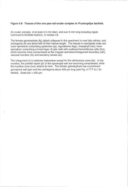
- Page 60 and 61: Figure 4.8: Transverse sections of
- Page 64 and 65: Figure 4.10: (Prumnopitys taxifolia
- Page 66 and 67: Figure 4.11: (Prumnopitys taxifolia
- Page 68 and 69: Figure 4.12: Development of the meg
- Page 70 and 71: Figure 4.13: Megaspore tetrads in P
- Page 72 and 73: Figure 4.14: Development of the fem
- Page 74 and 75: Figure 4.15: (Prumnopitys taxifolia
- Page 76 and 77: Figure 4.16: Occasionally two or ev
- Page 78 and 79: Figure 4.17: ln Prumnopitys taxifol
- Page 80 and 81: Figure 4.18: Features of the centra
- Page 82 and 83: Figure 4.19: Characteristics of the
- Page 84 and 85: Figure 4.20: The archegonium reache
- Page 86 and 87: Figure 4.21: The developing central
- Page 88 and 89: Figure 4.22: (Prumnopitys taxifolia
- Page 90 and 91: Figure 4.23: (Prumnopitys taxifolia
- Page 92 and 93: Figure 4.24: Stages in the maturati
- Page 94 and 95: Figure 4.25: Volumes of thirty-thre
- Page 96 and 97: Figure 4.26: ln Prumnopitys taxifol
- Page 98 and 99: Figure 4.27: (Prumnopitys taxifolia
- Page 100 and 101: Figure 4.28: Up to four archegonia
- Page 102 and 103: Figure 4.29: The cell wall of the m
- Page 104 and 105: Figure 4.30: Some characteristics o
- Page 106 and 107: Figure 4.31: Another example of an
- Page 108 and 109: Figure 4.32: ln Prumnopitys taxifol
- Page 110 and 111: Figure 4.33: Electron micrographs o
- Page 112 and 113:
Figure 4.34: Electron-micrographs o
- Page 114 and 115:
Figure 5.1: lntact and penetrated n
- Page 116 and 117:
Figure 5.2 Material from the pollen
- Page 118 and 119:
Figure 5.3: ln Prumnopitys taxifoli
- Page 120 and 121:
Figure 5.4: ln Prumnopitys taxifoli
- Page 122 and 123:
Figure 5.5 After fertilization, the
- Page 124 and 125:
Figure 5.6: Unfertilized archegonia
- Page 126 and 127:
Figure 5.7: Changes in unfertilized
- Page 128 and 129:
Figure 5.8: An unusual archegonium
- Page 130 and 131:
Figure 5.9: ln Prumnopitys taxifoli
- Page 132 and 133:
Figure 5.10: ln Prumnopitys taxifol
- Page 134 and 135:
Figure 5.11: ln Prumnopitys taxifol
- Page 136 and 137:
Figure 5.12: ln Prumnopitys taxifol
- Page 138 and 139:
Figure 5.13: Outside the newly cell
- Page 140 and 141:
Figure 5.14: ln Prumnopitys taxifol
- Page 142 and 143:
Figure 5.15: ln Prumnopitys taxitol
- Page 144 and 145:
Figure 5.16: Five-celled and eight-
- Page 146 and 147:
Figure 5.17: Some proembryos can ha
- Page 148 and 149:
Figure 5.18 : ln Prumnopitys taxifo
- Page 150 and 151:
Figure 5.19: ln Prumnopitys taxitol
- Page 152 and 153:
Figure 5.20: The middle of the susp
- Page 154 and 155:
Figure 5.21 : ln Prumnopitys taxifo
- Page 156 and 157:
Figure 5.22: ln Prumnopitys taxifol
- Page 158 and 159:
Figure 5.23: ln Prumnopitys taxifol
- Page 160 and 161:
Figure 5.24: Within the female game
- Page 162 and 163:
Figure 5.25: Transverse sections of
- Page 164 and 165:
Figure 5.26: One embryo was observe
- Page 166 and 167:
Figure 5.27: The dehiscence zone in
- Page 168 and 169:
Figure 5.28: Transition between hyp
- Page 170 and 171:
Figure 5.29: In the germinating emb
- Page 172 and 173:
Figure 5.30 : Further tissue develo
- Page 174 and 175:
Figure 5.31: In the germinating emb