Download PDF - Springer
Download PDF - Springer
Download PDF - Springer
You also want an ePaper? Increase the reach of your titles
YUMPU automatically turns print PDFs into web optimized ePapers that Google loves.
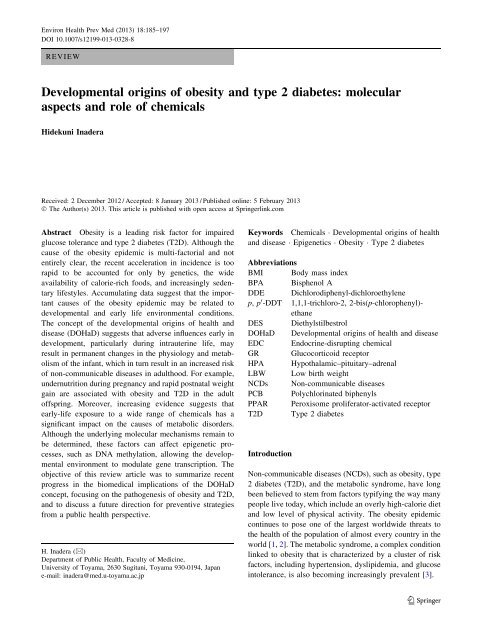
Environ Health Prev Med (2013) 18:185–197DOI 10.1007/s12199-013-0328-8REVIEWDevelopmental origins of obesity and type 2 diabetes: molecularaspects and role of chemicalsHidekuni InaderaReceived: 2 December 2012 / Accepted: 8 January 2013 / Published online: 5 February 2013Ó The Author(s) 2013. This article is published with open access at <strong>Springer</strong>link.comAbstract Obesity is a leading risk factor for impairedglucose tolerance and type 2 diabetes (T2D). Although thecause of the obesity epidemic is multi-factorial and notentirely clear, the recent acceleration in incidence is toorapid to be accounted for only by genetics, the wideavailability of calorie-rich foods, and increasingly sedentarylifestyles. Accumulating data suggest that the importantcauses of the obesity epidemic may be related todevelopmental and early life environmental conditions.The concept of the developmental origins of health anddisease (DOHaD) suggests that adverse influences early indevelopment, particularly during intrauterine life, mayresult in permanent changes in the physiology and metabolismof the infant, which in turn result in an increased riskof non-communicable diseases in adulthood. For example,undernutrition during pregnancy and rapid postnatal weightgain are associated with obesity and T2D in the adultoffspring. Moreover, increasing evidence suggests thatearly-life exposure to a wide range of chemicals has asignificant impact on the causes of metabolic disorders.Although the underlying molecular mechanisms remain tobe determined, these factors can affect epigenetic processes,such as DNA methylation, allowing the developmentalenvironment to modulate gene transcription. Theobjective of this review article was to summarize recentprogress in the biomedical implications of the DOHaDconcept, focusing on the pathogenesis of obesity and T2D,and to discuss a future direction for preventive strategiesfrom a public health perspective.H. Inadera (&)Department of Public Health, Faculty of Medicine,University of Toyama, 2630 Sugitani, Toyama 930-0194, Japane-mail: inadera@med.u-toyama.ac.jpKeywords Chemicals Developmental origins of healthand disease Epigenetics Obesity Type 2 diabetesAbbreviationsBMI Body mass indexBPA Bisphenol ADDE Dichlorodiphenyl-dichloroethylenep, p 0 -DDT 1,1,1-trichloro-2, 2-bis(p-chlorophenyl)-ethaneDES DiethylstilbestrolDOHaD Developmental origins of health and diseaseEDC Endocrine-disrupting chemicalGR Glucocorticoid receptorHPA Hypothalamic–pituitary–adrenalLBW Low birth weightNCDs Non-communicable diseasesPCB Polychlorinated biphenylsPPAR Peroxisome proliferator-activated receptorT2D Type 2 diabetesIntroductionNon-communicable diseases (NCDs), such as obesity, type2 diabetes (T2D), and the metabolic syndrome, have longbeen believed to stem from factors typifying the way manypeople live today, which include an overly high-calorie dietand low level of physical activity. The obesity epidemiccontinues to pose one of the largest worldwide threats tothe health of the population of almost every country in theworld [1, 2]. The metabolic syndrome, a complex conditionlinked to obesity that is characterized by a cluster of riskfactors, including hypertension, dyslipidemia, and glucoseintolerance, is also becoming increasingly prevalent [3].123
Environ Health Prev Med (2013) 18:185–197 187Table 1Studies of the Dutch hunger winter familiesSubjects Major findings ReferencesObesity and type 2 diabetesAge 19 years Obesity Ravelli et al. [6]Age 50 years 2-h glucose levels were elevated after a glucose load Ravelli et al. [28]Increase in body mass index and waist circumference in women Ravelli et al. [26]Age 58 years 2-h glucose levels were elevated after a glucose load de Rooij SR et al.[127]Impaired insulin secretion after a glucose loadde Rooij SR et al.[128]Glucose intolerance was differed by PPAR-gamma 2 genotypede Rooij SR et al.[129]Increased weight and fat deposition in women Stein et al. [130]No association between prenatal famine and metabolic syndrome de Rooij et al. [131]Lipid profilesAge 50 years Increased ratio of low-density to high-density lipoproteins Roseboom et al. [132]Age 59 years Increased total cholesterol and triglyceride levels in women Lumey et al. [133]Blood pressureAge 50 years No differences were found in systolic or diastolic pressure Roseboom et al. [134]No association with adult blood pressure Roseboom et al. [135]Age 59 years Moderate increase in systolic but not diastolic blood pressure Stein et al. [136]Greater blood pressure increase during stress Painter et al. [137]Atherosclerosis and mortalityAge 50 yearsIncreased prevalence of coronary heart disease in those exposed to the famine in early Roseboom et al. [138]gestationHigher incidence of coronary artery disease Painter et al. [139]Age 57 years No effect on adult mortality Painter et al. [140]Reduced carotid artery intima media thickness Painter et al. [141]Age between 18 and64 yearsMiscellaneousHigher overall adult mortality risk in womenvan Abeelen et al.[142]Age 24–48 years Increased risk for schizophrenia Susser et al. [143]Age 50 years Decreased factor VII concentrations Roseboom et al. [144]Increased prevalence of obstructive airways disease Lopuhaa et al. [145]Age 58 years No difference in cortisol concentrations after dexamethasone suppression test de Rooij et al. [146]PPAR Peroxisome proliferator-activated receptorMechanisms of the developmental originsof metabolic diseasesAlthough our understanding of the molecular mechanismsunderlying the effects of fetal undernutrition and LBW onthe development of NCDs later in life is far from complete,possible mechanisms have been proposed. Generally,organisms possess an evolved ability to respond to externalsignals by adjusting their phenotype during development tomatch their environment. Thus, poor nutrition of thepregnant mother may signal to the fetus that nutrients arescarce in the postnatal environment. Therefore, a fetus thatis exposed to signals it interprets as reflecting nutrientdeficiency or maternal stress will adapt its metabolictrajectory to suit an environment of limited energy availability.When the postnatal environment then fails to matchthe experienced prenatal environment, maladaptationoccurs, resulting in onset of obesity and T2D [38]. Indeed,it has been shown that those who were most likely todevelop T2D in adult life had LBW and underwent rapidpostnatal weight gain [39]. In humans, the combination ofLBW and rapid childhood growth has been associated withlater insulin resistance [38, 39]. Recent animal studies haveconfirmed that it is mainly the discrepancy between thepre- and postnatal environments that affects adult onsetdiseases, rather than gestational undernutrition itself [40,41]. The process whereby a stimulus or insult during asensitive or critical period has irreversible long-term123
188 Environ Health Prev Med (2013) 18:185–197effects on development is often referred to as ‘metabolicprogramming’ [42].The stress response may be involved in the pathogenesisof DOHaD via the regulation of the hypothalamic–pituitary–adrenal (HPA) axis, which has potent effects onmetabolism and vasculature [43, 44]. Changes in the HPAaxis have been proposed as a mechanism behind theepidemiological link between LBW and later increasedblood pressure [45]. Offspring of rat dams given dexamethasoneduring pregnancy have reduced birth weightand increased blood pressure and glucose intolerance inadulthood [46, 47]. Intrauterine stress is associated withinsulin resistance and accompanying dysregulation of theHPA axis, with chronically excessive adrenal glucocorticoidsecretion and increased stress responses [48].Mechanistic analysis has shown that intrauterine glucocorticoidexposure leads to reduced numbers of glucocorticoidreceptors in the hypothalamus, resulting inimpaired negative feedback and hence long-term upregulationof the HPA axis after birth [49]. This, in turn, maycontribute to increased blood pressure and glucose intolerance.In humans, exposure to antenatal betamethasonecaused signs of insulin resistance in adult offspring at30 years of age [50]. Thus, increased maternal corticosteroidlevels as a result of stress induced by reducednutrient availability induce hypertension in the offspring[51]. A recent study reported that maternal iron restriction,independent of maternal macronutrient or caloricintake, also works as a fetal stressor that programs metabolicand circulatory functions in the offspring [52].Animal studies are used to clarify the underlyingmechanisms of clinical observations in more detail. Fetalnutrition is a key regulator of fetal growth and thus anobvious candidate as an influence on programming [53]. Infact, rats living in a postnatal obesogenic environment havevery different physiological responses depending on whetherthey were born to undernourished or well-fed mothers,with the former becoming more obese, more hypertensive,and more hyperphagic than the latter [54]. Imbalance ofprotein and carbohydrate intake during pregnancy has beenassociated with reduced birth weight and increased bloodpressure in the offspring [55]. Many studies have shownthat prenatal protein restriction results in LBW and programshypertension in experimental animals [56–58].Embryos collected from mothers fed a low-protein diethave reduced cell numbers [59]. In particular, a strongcorrelation has been found between nephron number andbirth weight [60], and a reduction in nephron number isassociated with increased blood pressure [61]. Exposure ofrats to a low-protein diet in utero also decreases b-cellproliferation, islet size, and islet vascularization [62].Subsequent accelerated growth leads to an excessive metabolicdemand on this limited cell mass.Transcriptome-wide analysis using the AffymetrixMouse 430A_2.0 array (Affymetrix, Santa Clara, CA)showed that feeding mice a protein-restricted diet betweengestational days 10.5 and 17.5 altered the expression of 235genes in the placenta [63]. In the adult male offspring ofdams fed a protein-restricted diet, 311 genes in the liverdiffered significantly from those in the offspring of controldams, as determined by the Agilent 014879 whole ratgenome array (Agilent Technologies, Santa Clara, CA)[64]. Thus, an altered maternal diet during pregnancyinduces persistent changes in the transcriptome. Whateverthe mechanisms, these events may occur in humans ifundernutrition in utero is followed by an abundant postnataldiet, such in small babies born into a food-richWestern society.Role of epigeneticsThe induction of altered phenotypes during development inresponse to environmental stimuli involves epigeneticchanges. Epigenetic regulation has been defined as ‘‘heritablechanges in gene function that occur without changesin the nucleotide sequence’’ [65]. Epigenetic factorsinclude DNA methylations, histone modifications, andmicroRNAs. Epigenetic changes, in particular in DNAmethylation, provide a ‘memory’ of developmental plasticresponses to the early environment and are central to thegeneration of phenotypes and their stability throughout thelife course. Methylation at the 5 0 position of cytosine inDNA within a CpG dinucleotide (the p denotes the interveningphosphate group) is a common modification inmammalian genomes and constitutes a stable epigeneticmark that is transmitted through DNA replication and celldivision [66]. CpG dinucleotides are not randomly distributedthroughout the genome but are clustered at the 5 0ends of genes/promoters in regions known as CpG islands.Hypermethylation of these CpG islands is generally associatedwith transcriptional repression, while hypomethylationof CpG islands is generally associated withtranscriptional activation.Data from animal models suggest that epigenetic processesare an important link between the early life environmentand altered metabolism and body composition inthe adult offspring [67, 68]. A growing body of literaturehas reported a role for epigenetic factors in the complexinterplay between genes and the environment [4, 69–71].Epigenetic changes may explain how an altered maternaldiet during pregnancy, such as a protein restricted diet,induces persistent changes in the transcriptome.Environmental developmental influences, such as thematernal diet or chemical exposure, can affect the offspringphenotype via epigenetic effects [72, 73]. A diet that is poor123
Environ Health Prev Med (2013) 18:185–197 189or enriched in methyl donors and cofactors of DNA methylation,especially during fetal growth and development,may influence the epigenotype. For example, a methyl-richmaternal diet during gestation was found to alter the bodycomposition of the offspring in agouti mice, which wasaccompanied by epigenetic changes in metabolic controlgenes [74]. Thus, environmentally induced epigeneticmodifications alter gene expression and may increase theoffspring’s susceptibility for later disease [75, 76].It has been shown in rodent models that unbalancedmaternal diets during pregnancy induce changes in DNAmethylation and covalent histone modifications in the 5 0regulatory regions of specific non-imprinted genes,affecting the offspring’s later body composition and metabolicphenotype [77, 78]. For example, the offspring of ratdams fed a protein-restricted diet had lower levels of CpGmethylation and greater expression of the peroxisomeproliferators-activated receptor (PPAR) a and glucocorticoidreceptor (GR) genes in the liver than control animals[77]. Changes in the epigenetic regulation of these genesmay result in alterations in the activity of pathways controlledby their target genes, such as phosphoenolpyruvatecarboxykinase and acyl-CoA oxidase, which subsequentlyaffects lipid and carbohydrate metabolism. Hypomethylationof the hepatic PPARa and GR promoters has beenreported in both F1 and F2 offspring of F0 rats fed a protein-restricteddiet during pregnancy without furthernutritional challenge to the F1 generation, indicating thatchanges in the epigenome that occur during developmentmay be passed on to subsequent generations [68].Intrauterine growth restriction has been associated withprogressive epigenetic silencing of Pdx1, a pancreatic andduodenal homeobox 1 transcription factor critical for b-celldevelopment, resulting in impaired b-cell function andT2D in the adult offspring in rats [79]. Lack of methylationof the retrotransposon with a methylation-sensitive promoterwas associated with subsequent offspring obesity inmice [80]. In contrast, supplementation of the diets ofpregnant animals with methyl donors, such as folic acid,vitamin B12, choline, or betaine, increased DNA methylationof specific genes in the offspring [74, 81–83].Overall, these results show that dietary modifications caninduce altered phenotypes through epigenetic changes inspecific genes and that these changes in phenotype can bemodulated by nutritional interventions during pregnancy.As yet there are limited published human data linkingmaternal nutrition to epigenetic changes in the offspring.Adults who were exposed to famine in utero showedaltered DNA methylation in the promoters of severalimprinted and non-imprinted genes in white blood cells.Two studies on the Dutch hunger winter families reportedthe effects of prenatal undernutrition on promoter methylation[84, 85]. Individuals with periconceptual exposure tofamine had reduced DNA methylation of the imprintedinsulin-like growth factor-2 (IGF2) gene, a key factor inhuman growth and development, as compared with theirunexposed, same-sex siblings at 59 years of age [84]. Inaddition, individuals from Dutch hunger winter familiesalso had lower DNA methylation of the imprinted INS-IGF2 (INSIGF) gene, but increased DNA methylation ofthe guanine nucleotide-binding protein (GNASAS), maternallyexpressed 3 (MEG3), interleukin-10 (IL10), ATPbindingcassette A1 (ABCA1), and leptin (LEP) genes inparallel with impaired glucose tolerance compared withtheir unexposed same-sex siblings [85]. A recent studyreported that higher methylation of retinoid X receptor-achr9 was associated with lower maternal carbohydrateintake in early pregnancy, which is also associated withhigher neonatal adiposity [53]. Thus, it is clear that fetalstress can affect the methylation status of several subsets ofgenes, supporting the hypothesis that associations betweenearly developmental conditions and health outcomes laterin life may be mediated by changes in the epigeneticinformation. However, the differences in methylation statusof these genes between the exposed and unexposedindividuals were relatively small, although statisticallysignificant. Further studies with clear end points are neededto determine whether measurement of epigenetic marks inearly life can be used as biomarkers to identify individualswho have experienced environmental perturbations indevelopment and thus who are more likely to developobesity and metabolic disease in later adulthood. It iscrucial to identify such epigenetic marks that are predictiveof a later phenotype so that they can be used as relevantbiomarkers for disease prevention.Role of chemicalsBecause the obesity epidemic coincided with the rapidincrease in the use of industrial chemicals, the hypothesisthat exposure to chemicals, combined with genetic predispositionand consumption of a high-calorie diet, may bea major contributor to the obesity epidemic was proposednearly 10 years ago [86, 87]. Since then, a number ofstudies have specifically addressed the effects of environmentalchemical exposure on weight gain. Chemicals thataffect human fetal development are generally calledendocrine-disrupting chemicals (EDCs). EDCs are compoundsthat act upon the body’s hormonal systems andinclude industrial contaminants, plastics, pesticides, andother compounds [88]. EDC contamination is a globalproblem. Bisphenol A (BPA), the prototypical EDC, isproduced in large quantities for use in the production ofpolycarbonate and epoxy resins. However, when used infood and drink containers, it can ‘leach’ into the contents,123
190 Environ Health Prev Med (2013) 18:185–197resulting in ingestion of BPA with food and drink [89]. Asa result, human exposure to BPA is widespread, and BPAhas been detected in urine in more than 90 % of all humansamples tested [89, 90]. Some EDCs are highly resistant todegradation and remain persistent in the environment. Forexample, persistent organic pollutants, which includepolychlorinated dibenzo-p-dioxins, polychlorinated dibenzofurans,and polychlorinated biphenyls (PCBs) canaccumulate in the human body, especially in adipose tissuebecause of their lipophilic nature. Although the productionof PCBs was banned by the USA in the 1970s, PCBsremain ubiquitous contaminants in the human populationeven today because of their stability [91]. Many studieshave been conducted to study the link between chemicalexposure and metabolic disorders. The findings of studieson the effects of environmental chemicals on obesity andT2D are summarized in Table 2.Epidemiological studies on maternal smoking haveindicated that the adjusted odds ratio for obesity is between1.5- and 2.0-fold greater if children were exposed during,but not before or after, pregnancy [92–94], suggesting thatthere are critical periods during embryogenesis when theembryo is the most sensitive to exposure to xenobiotics.These studies also indicated that chemical toxicants, suchas nicotine, can contribute to the etiology of later obesity.In humans, there is epidemiological evidence for theassociation between developmental exposure to chemicalsand metabolic disorders later in life [95]. It has beendemonstrated that chemicals cross the placenta and directlyaffect the fetus [96]. Maternal exposure to chemical substancesduring pregnancy has been associated with anincreased body mass index (BMI) in the offspring [97–99].Several studies have indicated that organochlorine exposuremay be associated with the development of T2D [97,98, 100–103]. For example, the results of a Spanish cohortstudy showed that prenatal exposure to the organochlorinehexachlorobenzene was associated with increased BMI atage 6 years [97]. Prenatal exposure to dichlorodiphenyldichloroethylene(DDE) was significantly associated withincreased weight and BMI in adult female offspring [98].DDE in cord blood was associated with increased BMI inyoung children, and this effect was exacerbated bymaternal smoking [103]. A recent prospective studyreported that in utero exposure to perfluorooctanoate waspositively associated with risk of overweight at age 20years in female but not in male offspring [104]. Collectively,these results support the hypothesis that the fetus isvulnerable to exposure to environmental chemicals,resulting in an increased risk of excessive body weight gainand susceptibility to T2D later in life. Moreover, genderand the postnatal environment in combination with prenatalchemical exposure can modify the onset, as well as theprogression and outcome of the disease.In experimental cell cultures and animal studies, avariety of chemicals have been shown to act as adipogenesisor obesity-inducing agents. A number of chemicals areknown to promote obesity by increasing the number ofadipocytes or the storage of fat into existing adipocytes.The organochlorine compound 1, 1, 1-trichloro-2, 2-bis(p-chlorophenyl)-ethane (p, p 0 -DDT) induces a concentration-dependentincrease in in vitro 3T3-L1 adipocyte differentiation[105]. In mouse studies, treatment with thesynthetic estrogen diethylstilbestrol (DES) on days 1–5 ofneonatal life at 0.001 mg/day increased body weight andthe percentage of body fat [106]. Treatment of pregnantmice with xenoestrogen BPA resulted in increased bodyweight in the female offspring on postnatal day 22 comparedwith unexposed controls [107]. Recent studies haveindicated that perinatal exposure to approximately 70 lg/kg/day BPA via drinking water alters early adipogenesisand increases body weight in rodent models [108, 109].Mice perinatally exposed to DES or the phytoestrogengenistein increased weight after puberty [106, 110, 111].Wright et al. [112] reported that exposure to octylphenol,another xenoestrogen, during fetal and postnatal life infemale lambs led to increased weight at puberty. Duringdevelopment, estrogen induces an increase in adipocytenumbers and affects adipocyte function [113]. Overall,these results show that exposure to organochlorine compoundsand/or xenoestrogens during sensitive windows ofdevelopment may have obesogenic effects, and may lead topermanent changes in the metabolic pathways that regulatebody weight.Other chemicals that can work as potential obesogensare the organotins. Organotins represent a class of persistentorganic pollutants that may reach harmful levels inexposed populations [114]. In culture systems, organotinsinduce adipocyte differentiation [115, 116]. Tributyltin isalso known to induce adipogenesis in vivo. Mice treatedprenatally with tributyltin are born with more stored fatthan controls [117]. Multipotent stromal cells harvestedfrom white adipose tissue at 8 weeks of age in mice prenatallyexposed to tributyltin had high numbers of preadipocytesand cells preprogrammed to prefer the adipogenicfate, an effect that will likely lead to an increase in adiposemass over time [118]. Organotins work via direct activationof the PPARc–retinoid X receptor (RXR) heterodimer[116]. Activation of the PPARc–RXR heterodimer hasbeen found to favor lipid biosynthesis and storage, andinduces adipocyte differentiation. Interestingly, a recentreport indicated that tributyltin can work as a xenoestrogenvia estrogen receptors in vivo [119]. Among commonlyused phthalates as plasticizers, monoethyl-hexyl-phthalatecan work as an activator for PPARc [120].The mechanisms of action of these chemicals are diverseand probably involve epigenetic molecular changes,123
Environ Health Prev Med (2013) 18:185–197 191Table 2Studies on environmental chemicals on obesity and type 2 diabetesSubjects Chemicals a Major findings ReferencesHuman studiesAdults in southern Taiwan Arsenic High prevalence of T2D Lai et al. [147]Air Force veterans TCDD Increased prevalence of T2D Henriksen et al. [148]Air Force veterans TCDD Serum level was correlated with incidence of Longnecker et al. [149]T2DPubertal boys DDE Increased body weight Gladen et al. [150]Adults POPs Correlated with the prevalence of T2D Lee et al. [100]Adult women PCB Increased incidence of T2D Vasiliu et al. [102]Mexican Americans p, p 0 -DDT Increased prevalence of T2D Cox et al. [151]Adult native Americans HCB Serum level was positively correlated with Codru et al. [152]incidence of T2DU.S. population Organochlorine pesticides Positively associated with metabolic syndrome Lee et al. [153]Children aged 6 years HCB Increased BMI (body mass index) and body Smink et al. [97]weightYucheng poisoning women PCBs Increased prevalence of T2D Wang et al. [95]Residents in Cd-contaminated area Cd Correlated with diabetic nephropathy Hanswell-Elkins et al. [154]Air Force veterans TCDD Increased prevalence of T2D Michalek et al. [155]Adult female offspring DDE Increased weight and BMI Karmaus et al. [98]Women aged 50–59 years p, p 0 -DDE Increased prevalence of T2D Rignell-Hydbom et al. [156]Workers PFOS Increased prevalence of T2D Lundin et al. [157]Children aged 3 years DDE and PCBs Intrauterine exposure was associated with BMI Verhulst et al. [103]Great Lakes sport fish consumers DDE Increased incidence of T2D Turyk et al. [158]Women in southern Spain Nonylphenol Positively associated with BMI Lopez-Espinosa et al. [159]Adults of eastern Slovakia PCBs Increased prevalence of T2D Ukropec et al. [160]Koreans Organochlorine pesticides Increased prevalence of T2D Son et al. [161]Adults PCBs Increased prevalence of T2D Lee et al. [162]Koreans Heptachlor epoxide Positively associated with metabolic syndrome Park et al. [163]Children at 14 months DDE Elevated BMI Mendez et al. [164]Adults aged 18–74 BPA Higher exposure was associated with general Carwile et al. [165]and central obesityWomen aged 20 years PFOA Maternal PFOA concentrations were positively Halldorsson et al. [104]associated with BMIAdult populations of Catalonia PCBs and HCBs Positively associated with diabetes andGasull et al. [166]prediabetesAnimal studiesRats Cd Neonatal exposure increased diabetic prevalence Merali et al. [167]Female mice BPA Perinatal exposure led to obesity Howdeshell et al. [107]Female rats BPA Perinatal exposure led to obesity Rubin et al. [168]Rats BPA Perinatal exposure led to impaired glucose Wei et al. [109]toleranceFemale ewe lambs Octylphenols Gestational exposure led to obesity Wright et al. [112]Mice DES Perinatal exposure led to obesity Newbold et al. [106]Mice TBT In utero exposure led to obesity Grun et al. [117]Female mice BPA Perinatal and postnatal exposure led to obesity Miyawaki et al. [169]Female mice PFOS Work as developmental obesogen Hines et al. [170]Female rats BPA Gestational exposure led to obesity Somm et al. [108]Mice TBT In utero exposure led to multipotent stem cells tobecome adipocytesKirchner et al. [118]TD2 Type 2 diabetes, BMI body mass indexa TCDD 2, 3, 7, 8-Tetrachlorodibenzo-p-dioxin, DDE dichlorodiphenyl-dichloroethylene, POPs persistent organic pollutants, PCB polychlorinatedbiphenyl, p, p 0 -DDT dichlorodiphenyltrichloroethane, HCB hexachlorobenzene, Cd cadmium, PFOS perfluorooctanesulfonic acid, BPA bisphenol A,PFOA perfluorooctanoate, DES diethylstilbestrol, TBT tributyltin, PFOS perfluorooctanoic acid123
192 Environ Health Prev Med (2013) 18:185–197including DNA methylation and histone modifications[121]. Environmental compounds may induce the establishmentof specific epigenetic patterns during key developmentalperiods that influence phenotypic variation,which in some cases lead to disease states [122, 123].Indeed, maternal exposure to BPA decreased DNA methylationin the retrotransposon upstream of the agouti genein mice and shifted coat color distribution in the offspringby stably altering the epigenome [74]. Whatever themechanism, complex events, including exposure to obesogenicor diabetogenic chemicals during development, maybe contributing to the obesity and T2D epidemics. Clarifyingthe mechanisms involved in weight homeostasis is anovel target in the study of abnormal programming inducedby environmental chemicals, which should be re-named‘metabolic disrupting chemicals’ [124].Future perspectivesWe are in the midst of a global epidemic of obesity withsubstantial negative health and socio-economic consequences.There is now growing evidence that developmentalinfluences have lifelong effects on metabolicfunction (Fig. 1). Perturbations of the developmental milieucan have a profound impact on the onset and incidence ofobesity and T2D. Phenotypic outcomes with long-termconsequences involve the interplay between environmental,developmental, and genetic influences. The pathogenesis ofobesity and T2D resides in a mixture of genetic and environmentalfactors. It is therefore probable that not onlymaternal nutrition and stress, but also maternal size, parity,and maternal age can affect the offspring phenotype and/orepigenetic states. Moreover, possible interplay among prenatalexposure and the postnatal environmental factors,such as nutrition, stress, chemical exposure, and aging, canaffect disease outcome. Further studies are merited toclarify the interactions that mediate metabolic outcomes.Transient environmental conditions during human gestationcan be recorded as persistent changes in the epigenome.Epigenetic changes, in particular those in DNAmethylation, are central to the generation of novel phenotypesand their stability throughout the life course. In thefuture, epigenetic research may have substantial benefitsfor public health because specific components of the epigeneticstate at birth may predict later obesity and T2D.Researchers need to identify such effective epigeneticbiomarkers which can be measured at early stage of life.These epigenetic marks may be important biomarkers,which not only indicate responses to prenatal challenges,but also predict later risk for obesity and T2D. Currentstudies are limited to analyzing specific candidate loci, andthe complete epigenome has yet to be explored. Newtechnologies, such as methods for rapid sequencing fordifferentially methylated regions of the genome, need to bedeveloped before large-scale epidemiological studies canbe conducted. It is likely that comprehensive studies of theepigenome will be helpful to shed light on gene–environmentinteractions in the pathogenesis of obesity and T2D.Accumulating data indicate that low doses of environmentalchemicals have adverse effects on human health[125]. These days, individuals are exposed to a mixture ofEDCs. Indeed, dozens of environmental chemicals aredetected in human tissues and fluids [126]. However, verylittle is known about how these chemicals act in combination.So far, almost all epidemiological studies havelooked for the associations between metabolic disordersand the exposure to a single EDC at a single time, ratherthan to the whole mixture of toxicants to which human areexposed. These mixtures are likely to have unexpected andunpredictable effects. Further prospective studies withclear end points are required to determine whetherMalnutritionStressAdult-onsetObesityEpigenetic changesDNA methylation etc.DiabetesDyslipidemiaMetabolic syndromeChemicalsSteroidsFig. 1Influences during critical fetal periods may cause the adult onset of non-communicable diseases, such as obesity and type 2 diabetes123
Environ Health Prev Med (2013) 18:185–197 193exposure to mixtures of EDCs in early life can influencethe development of obesity and T2D.NCDs are preventable, but new initiatives are needed toinstitute prevention. If the risk of many common diseasesof adulthood in our communities is largely determinedbefore birth, adult lifestyle interventions will only reducethe risk transiently or to a small degree because they occurtoo late. Thus, from a public health perspective, it isimportant to determine whether interventions after theneonatal period can reverse the adverse effects of unbalancedprenatal nutrition. If the effects of adult lifestyleinterventions are limited, maximum effect will be gainedfrom timely interventions in early life. Improved nutritionwill not only benefit the present population but may alsoreduce disease in future generations. If so, more careshould be given to the consumption of a healthy diet duringpregnancy and improved fetal nutrient availability, whichmay lead to a more normal birth weight and early lifegrowth, thereby reducing the risk for programmed metabolicdisease. Clear evidence and good communication ofthis evidence may lead to policy responses that will openthe possibility of nutritional or pharmacological interventionsto combat the rapid rise in obesity and T2D.Acknowledgments The author thanks two anonymous reviewerswhose comments and suggestions greatly improved this review.Conflict of interestNone.Open Access This article is distributed under the terms of theCreative Commons Attribution License which permits any use, distribution,and reproduction in any medium, provided the originalauthor(s) and the source are credited.References1. Zimmet P, Alberti KG, Shaw J. Global and societal implicationsof the diabetes epidemic. Nature. 2001;414:782–7.2. Finucane MM, Stevens GA, Cowan MJ, Danaei G, Lin JK,Paciorek CJ, et al. National, regional, and global trends in bodymassindex since 1980: systematic analysis of health examinationsurveys and epidemiological studies with 960 country-yearsand 9.1 million participants. Lancet. 2011;377:557–67.3. Alexander CM, Landsman PB, Teutsch SM, Haffner SM.NCEP-defined metabolic syndrome, diabetes, and prevalence ofcoronary heart disease among NHANES III participants age50 years and older. Diabetes. 2003;52:1210–4.4. Gluckman PD, Hanson MA, Cooper C, Thornburg KL. Effect ofin utero and early-life conditions on adult health and disease.N Engl J Med. 2008;359:61–73.5. Godfrey KM, Barker DJ. Fetal programming and adult health.Public Health Nutr. 2001;4:611–24.6. Ravelli GP, Stein ZA, Susser MW. Obesity in young men afterfamine exposure in utero and early infancy. N Engl J Med.1976;295:349–53.7. Hofman PL, Regan F, Jackson WE, Jefferies C, Knight DB,Robinson EM, et al. Premature birth and later insulin resistance.N Engl J Med. 2004;351:2179–86.8. Gluckman PD, Hanson MA, editors. Developmental origins ofhealth and disease. Cambridge: Cambridge University Press;2006.9. Huxley R, Neil A, Collins R. Unravelling the fetal originshypothesis: is there really an inverse association betweenbirthweight and subsequent blood pressure? Lancet. 2002;360:659–65.10. Newsome CA, Shiell AW, Fall CH, Phillips DI, Shier R, LawCM. Is birth weight related to later glucose and insulin metabolism?—asystematic review. Diabet Med. 2003;20:339–48.11. Huxley R, Owen CG, Whincup PH, Cook DG, Colman S,Collins R. Birth weight and subsequent cholesterol levels:exploration of the ‘‘fetal origins’’ hypothesis. JAMA. 2004;292:2755–64.12. Gluckman PD, Hanson MA. Living with the past: evolution,development, and patterns of disease. Science. 2004;305:1733–6.13. Barker DJ, Osmond C. Infant mortality, childhood nutrition, andischemic heart disease in England and Wales. Lancet. 1986;1:1077–81.14. Barker DJ, Winter PD, Osmond C, Margetts B, Simmonds SJ.Weight in infancy and death from ischemic heart disease.Lancet. 1989;2:577–80.15. Hales CN, Barker DJ. Type 2 (non-insulin-dependent) diabetesmellitus: the thrifty phenotype hypothesis. Diabetologia.1992;35:595–601.16. Barker DJ, Gluckman PD, Godfrey KM, Harding JE, Owens JA,Robinson JS. Fetal nutrition and cardiovascular disease in adultlife. Lancet. 1993;341:938–41.17. Barker DJ, Hales CN, Fall CH, Osmond C, Phipps K, Clark PM.Type 2 (non-insulin-dependent) diabetes mellitus, hypertensionand hyperlipidaemia (syndrome X): relation to reduced fetalgrowth. Diabetologia. 1993;36:62–7.18. Phillips DI, Barker DJ, Hales CN, Hirst S, Osmond C. Thinnessat birth and insulin resistance in adult life. Diabetologia.1994;37:150–4.19. Frankel S, Elwood P, Sweetnam P, Yarnell J, Smith GD.Birthweight, body-mass index in middle age, and incident coronaryheart disease. Lancet. 1996;348:1478–80.20. Stein CE, Fall CH, Kumaran K, Osmond C, Cox V, Barker DJ.Fetal growth and coronary heart disease in south India. Lancet.1996;48:1269–73.21. Benyshek DC, Martin JF, Johnston CS. A reconsideration of theorigins of the type 2 diabetes epidemic among Native Americansand the implications for intervention policy. Med Anthropol.2001;20:25–64.22. Kajantie E, Osmond C, Barker DJ, Forsen T, Phillips DI, ErikssonJG. Size at birth as a predictor of mortality in adulthood: afollow-up of 350000 person-years. Int J Epidemiol.2005;34:655–63.23. Leon DA, Lithell HO, Vagero D, Koupilova I, Mohsen R,Berglund L, et al. Reduced fetal growth rate and increased riskof death from ischemic heart disease: cohort study of 15000Swedish men and women born 1915–29. Br Med J.1998;317:241–5.24. Slomko H, Heo HJ, Einstein FH. Epigenetics of obesity anddiabetes in humans. Endocrinology. 2012;153:1025–30.25. Lumey LH, Stein AD, Kahn HS, van der Pal-de Bruin KM,Blauw GJ, Zybert PA, et al. Cohort profile: the Dutch HungerWinter families study. Int J Epidemiol. 2007;36:1196–204.26. Ravelli AC, van der Meulen JH, Osmond C, Barker DJ, BlekerOP. Obesity at the age of 50 y in men and women exposed tofamine prenatally. Am J Clin Nutr. 1999;70:811–6.27. Roseboom TJ, van der Meulen JH, Ravelli AC, Osmond C,Barker DJ, Bleker OP. Perceived health of adults after prenatalexposure the Dutch famine. Paediatr Perinat Epidemiol.2003;17:391–7.123
194 Environ Health Prev Med (2013) 18:185–19728. Ravelli AC, van der Meulen JH, Michels RP, Osmond C, BarkerDJ, Hales CN, et al. Glucose tolerance in adults after prenatalexposure to famine. Lancet. 1998;351:173–7.29. Painter RC, Roseboom TJ, Bleker OP. Prenatal exposure to theDutch famine and disease in later life: an overview. ReprodToxicol. 2005;20:345–52.30. Phillips DI. Insulin resistance as a programmed response to fetalundernutrition. Diabetologia. 1996;39:1119–22.31. Curhan GC, Willett WC, Rimm EB, Spiegelman D, AscherioAL, Stampfer MJ. Birth weight and adult hypertension, diabetesmellitus, and obesity in US men. Circulation. 1996;94:3246–50.32. Nobili V, Alisi A, Panera N, Agostoni C. Low birth weight andcatch-up-growth associated with metabolic syndrome: a ten yearsystematic review. Pediatr Endocrinol Rev. 2008;6:241–7.33. Dufour S, Petersen KF. Disassociation of liver and muscleinsulin resistance from ectopic lipid accumulation in low-birthweightindividuals. J Clin Endocrinol Metab. 2011;96:3873–80.34. Barouki R, Gluckman PD, Grandjean P, Hanson M, Heindel JJ.Developmental origins of non-communicable disease: implicationsfor research and public health. Environ Health. 2012;11:42.35. Wang Y, Wang X, Kong Y, Zhang JH, Zeng Q. The GreatChinese Famine leads to shorter and overweight females inChongqing Chinese population after 50 years. Obesity.2010;18:588–92.36. Hult M, Tornhammar P, Ueda P, Chima C, Bonamy AK,Ozumba B, et al. Hypertension, diabetes and overweight:looming legacies of the Biafran famine. PLoS One. 2010;5:e13582.37. Stein AD, Lumey LH. The relationship between maternal andoffspring birth weights after maternal prenatal famine exposure:the Dutch famine Birth Cohort Study. Hum Biol. 2000;72:641–54.38. Eriksson JG, Forsen TJ, Osmond C, Barker DJ. Pathways ofinfant and childhood growth that lead to type 2 diabetes. DiabetesCare. 2003;26:3006–10.39. Eriksson JG, Osmond C, Kajantie E, Forsen TJ, Barker DJ.Patterns of growth among children who later develop type 2diabetes or its risk factors. Diabetologia. 2006;49:2853–8.40. Dai Y, Thamotharan S, Garg M, Shin BC, Devakar SU.Superimposition of postnatal calorie restriction protects theaging male intrauterine growth-restricted offspring from metabolicmaladaptations. Endocrinology. 2012;153:4216–26.41. Garg M, Thamotharan M, Dai Y, Thamotharan S, Shin BC,Stout D, et al. Early postnatal caloric restriction protects adultmale intrauterine growth-restricted offspring from obesity.Diabetes. 2012;61:1391–8.42. Pinney SE, Simmons RA. Metabolic programming, epigenetics,and gestational diabetes mellitus. Curr Diab Rep. 2012;12:67–74.43. Barker DJ, Osmond C, Simmonds SJ, Wield GA. The relation ofsmall head circumference and thinness at birth to death fromcardiovascular disease in adult life. Br Med J. 1993;306:422–6.44. Phillips DI. Programming of the stress response: a fundamentalmechanism underlying the long-term effects of the fetal environment?J Intern Med. 2007;261:453–60.45. Benediktsson R, Lindsay RS, Noble J, Seckl JR, Edwards CR.Glucocorticoid exposure in utero: new model for adult hypertension.Lancet. 1993;341:339–41.46. Levitt NS, Lindsay RS, Holmes MC, Seckl JR. Dexamethasonein the last week of pregnancy attenuates hippocampal glucocorticoidreceptor gene expression and elevates blood pressurein the adult offspring in the rat. Neuroendocrinology.1996;64:412–8.47. Nyirenda MJ, Lindsay RS, Kenyon CJ, Burchell A, Seckl JR.Glucocorticoid exposure in late gestation permanently programsrat hepatic phosphoenolpyruvate carboxykinase andglucocorticoid receptor expression and causes glucose intolerancein adult offspring. J Clin Invest. 1998;101:2174–81.48. Buhl ES, Neschen S, Yonemitsu S, Rossbacher J, Zhang D,Morino K, et al. Increased hypothalamic–pituitary–adrenal axisactivity and hepatic insulin resistance in low-birth weight rats.Am J Physiol Endocrinol Metab. 2007;293:E1451–8.49. Gluckman PD, Hanson MA, Beedle AS. Early life events andtheir consequences for later disease: a life history and evolutionaryperspective. Am J Hum Biol. 2007;19:1–19.50. Dalziel SR, Walker NK, Parag V, Mantell C, Rea HH, RodgersA, et al. Cardiovascular risk factors after antenatal exposure tobetamethasone: 30-year follow-up of a randomised controlledtrial. Lancet. 2005;365:1856–62.51. Langley-Evans SC. Hypertension induced by foetal exposure toa maternal low-protein diet, in the rat, is prevented by pharmacologicalblockade of maternal glucocorticoid synthesis.J Hypertens. 1997;15:537–44.52. Bourque SL, Komolova M, McCabe K, Adams MA, Nakatsu K.Perinatal iron deficiency combined with a high-fat diet causesobesity and cardiovascular dysregulation. Endocrinology.2012;153:1174–82.53. Godfrey KM, Sheppard A, Gluckman PD, Lillycrop KA, BurdgeGC, McLean C, et al. Epigenetic gene promoter methylation atbirth is associated with child’s later adiposity. Diabetes.2011;60:1528–34.54. Vickers MH, Breier BH, Cutfield WS, Hofman PL, GluckmanPD. Fetal origins of hyperphagia, obesity, and hypertension andpostnatal amplification by hypercaloric nutrition. Am J PhysiolEndocrinol Metab. 2000;279:E83–7.55. Shiell AW, Campbell-Brown M, Haselden S, Robinson S,Godfrey KM, Barker DJ. High-meat, low-carbohydrate diet inpregnancy: relation to adult blood pressure in the offspring.Hypertension. 2001;38:1282–8.56. Langley-Evans SC, Phillips GJ, Jackson AA. In utero exposureto maternal low protein diets induces hypertension in weanlingrats, independently of maternal blood pressure changes. ClinNutr. 1994;13:319–24.57. Maloney CA, Gosby AK, Phuyal JL, Denyer GS, Bryson JM,Caterson ID. Site-specific changes in the expression of fat-partitioninggenes in weanling rats exposed to a low-protein diet inutero. Obes Res. 2003;11:461–8.58. Langley-Evans SC. Critical differences between two low proteindiet protocols in the programming of hypertension in the rat. IntJ Food Sci Nutr. 2005;51:11–7.59. Kwong WY, Wild AE, Roberts P, Willis AC, Fleming TP.Maternal undernutrition during the preimplantation period of ratdevelopment causes blastocyst abnormalities and programmingof postnatal hypertension. Development. 2000;127:4195–202.60. McMillen IC, Robinson JS. Developmental origins of the metabolicsyndrome: prediction, plasticity, and programming.Physiol Rev. 2005;85:571–633.61. Alexander BT. Fetal programming of hypertension. Am JPhysiol Regul Integr Comp Physiol. 2006;290:R1–10.62. Snoeck A, Remacle C, Reusens B, Hoet JJ. Effect of a lowprotein diet during pregnancy on the fetal rat endocrine pancreas.Biol Neonate. 1990;57:107–18.63. Gheorghe CP, Goyal R, Holweger JD, Longo LD. Placentalgene expression responses to maternal protein restriction in themouse. Placenta. 2009;30:411–7.64. Lillycrop KA, Rodford J, Garratt ES, Slater-Jefferies JL, GodfreyKM, Gluckman PD, et al. Maternal protein restriction withor without folic acid supplementation during pregnancy altersthe hepatic transcriptome in adult male rats. Br J Nutr.2010;103:1711–9.65. Jones PA, Takai D. The role of DNA methylation in mammalianepigenetics. Science. 2001;293:1068–70.123
Environ Health Prev Med (2013) 18:185–197 19566. Bird A. DNA methylation patterns and epigenetic memory.Genes Dev. 2002;16:6–21.67. Drake AJ, Walker BR, Seckl JR. Intergenerational consequencesof fetal programming by in utero exposure to glucocorticoids inrats. Am J Physiol Regul Integr Comp Physiol. 2005;288:R34–8.68. Burdge GC, Slater-Jefferies J, Torrens C, Phillips ES, HansonMA, Lillycrop KA. Dietary protein restriction of pregnant rats inthe F0 generation induces altered methylation of hepatic genepromoters in the adult male offspring in the F1 and F2 generations.Br J Nutr. 2007;97:435–9.69. Burdge GC, Lillycrop KA. Nutrition, epigenetics, and developmentalplasticity: implications for understanding human disease.Annu Rev Nutr. 2010;30:315–39.70. Ooki S. Life course genetic epidemiologic study based on longitudinaltwin-family data: a new perspective (in Japanese).Nihon Eiseigaku Zasshi. 2011;66:31–8.71. Drong AW, Lindgren CM, McCarthy MI. The genetic andepigenetic basis of type 2 diabetes and obesity. Clin PharmacolTher. 2012;92:707–15.72. Waterland RA, Jirtle RL. Early nutrition, epigenetic changes attransposons and imprinted genes, and enhanced susceptibility toadult chronic diseases. Nutrition. 2004;20:63–8.73. Godfrey KM, Lillycrop KA, Burdge GC, Gluckman PD, HansonMA. Epigenetic mechanisms and the mismatch concept of thedevelopmental origins of health and disease. Pediatr Res.2007;61:5R–10R.74. Dolinoy DC, Huang D, Jirtle RL. Maternal nutrient supplementationcounteracts bisphenol A-induced DNA hypomethylationin early development. Proc Natl Acad Sci USA.2007;104:13056–61.75. Ling C, Groop L. Epigenetics: a molecular link between environmentalfactors and type 2 diabetes. Diabetes. 2009;58:2718–25.76. Heindel JJ, vom Saal FS. Role of nutrition and environmentalendocrine disrupting chemicals during the perinatal period onthe aetiology of obesity. Mol Cell Endocrinol. 2009;304:90–6.77. Lillycrop KA, Phillips ES, Jackson AA, Hanson MA, BurdgeGC. Dietary protein restriction of pregnant rats induces and folicacid supplementation prevents epigenetic modification ofhepatic gene expression in the offspring. J Nutr. 2005;135:1382–6.78. Bogdarina I, Welham S, King PJ, Burns SP, Clark AJ. Epigeneticmodification of the renin-angiotensin system in the fetalprogramming of hypertension. Circ Res. 2007;100:520–6.79. Park JH, Stoffers DA, Nicholls RD, Simmons RA. Developmentof type 2 diabetes following intrauterine growth retardation inrats is associated with progressive epigenetic silencing Pdx 1.J Clin Invest. 2008;118:2316–24.80. Waterland RA, Jirtle RL. Transposable elements: targets forearly nutritional effects on epigenetic gene regulation. Mol CellBiol. 2003;23:5293–300.81. Sinclair KD, Allegrucci C, Singh R, Gardner DS, Sebastian S,Bispham J, et al. DNA methylation, insulin resistance, and bloodpressure in offspring determined by maternal periconceptional Bvitamin and methionine status. Proc Natl Acad Sci USA.2007;104:19351–6.82. Lillycrop KA, Phillips ES, Torrens C, Hanson MA, Jackson AA,Burdge GC. Feeding pregnant rats a protein-restricted diet persistentlyalters the methylation of specific cytosines in thehepatic PPAR alpha promoter of the offspring. Br J Nutr.2008;100:278–82.83. Burdge GC, Lillycrop KA, Jackson AA, Gluckman PD, HansonMA. The nature of the growth pattern and of the metabolicresponse to fasting in the rat are dependent upon the dietaryprotein and folic acid intakes of their pregnant dams and postweaningfat consumption. Br J Nutr. 2008;99:540–9.84. Heijmans BT, Tobi EW, Stein AD, Putter H, Blauw GJ, SusserES, et al. Persistent epigenetic differences associated with prenatalexposure to famine in humans. Proc Natl Acad Sci USA.2008;105:17046–9.85. Tobi EW, Lumey LH, Talens RP, Kremer D, Putter H, SteinAD, et al. DNA methylation differences after exposure to prenatalfamine are common and timing- and sex-specific. HumMol Genet. 2009;18:4046–53.86. Baillie-Hamilton PF. Chemical toxins: a hypothesis to explainthe global obesity epidemic. J Altern Complement Med.2002;8:185–92.87. Heindel JJ. Endocrine disruptors and the obesity epidemic.Toxicol Sci. 2003;76:247–9.88. Diamanti-Kandarakis E, Bourguignon JP, Giudice LC, HauserR, Prins GS, Soto AM, et al. Endocrine-disrupting chemicals: anEndocrine Society scientific statement. Endocr Rev.2009;30:293–342.89. Calafat AM, Ye X, Wong LY, Reidy JA, Needham LL. Exposureof the U.S. population to bisphenol A and 4-tertiaryoctylphenol:2003–2004. Environ Health Perspect. 2008;116:39–44.90. Vandenberg LN, Hauser R, Marcus M, Olea N, Welshons WV.Human exposure to bisphenol A. Reprod Toxicol. 2007;24:139–77.91. Mullerova D, Kopecky J. White adipose tissue: storage andeffector site for environmental pollutants. Physiol Res. 2007;56:375–81.92. Toschke AM, Koletzko B, Slikker W Jr, Hermann M, von KriesR. Childhood obesity is associated with maternal smoking inpregnancy. Eur J Pediatr. 2002;161:445–8.93. Power C, Jefferis BJ. Fetal environment and subsequent obesity:a study of maternal smoking. Int J Epidemiol. 2002;31:413–9.94. Al Mamun A, Lawlor DA, Alati R, O’Callaghan MJ, WilliamsGM, Najman JM. Does maternal smoking during pregnancyhave a direct effect on future offspring obesity? Evidence from aprospective birth cohort study. Am J Epidemiol. 2006;164:317–25.95. Wang SL, Tsai PC, Yang CY, Leon Guo Y. Increased risk ofdiabetes and polychlorinated biphenyls and dioxins: a 24-yearfollow-up study of the Yucheng cohort. Diabetes Care.2008;31:1574–9.96. Park JS, Bergman A, Linderholm L, Athanasiadou M, Kocan A,Petrik J, et al. Placental transfer of polychlorinated biphenyls,their hydroxylated metabolites and pentachlorophenol in pregnantwomen from eastern Slovakia. Chemosphere.2008;70:1676–84.97. Smink A, Ribas-Fito N, Garcia R, Torrent M, Mendez MA,Grimalt JO, et al. Exposure to hexachlorobenzene during pregnancyincreases the risk of overweight in children aged 6 years.Acta Paediatr. 2008;97:1465–9.98. Karmaus W, Osuch JR, Eneli I, Mudd LM, Zhang J, Mikucki D,et al. Maternal level of dichlorodiphenyl-dichloroethylene(DDE) may increase weight and body mass index in adultfemale offspring. Occup Environ Med. 2009;66:143–9.99. Janesick A, Blumberg B. Endocrine disrupting chemicals andthe developmental programming of adipogenesis and obesity.Birth Defects Res C Embryo Today. 2011;93:34–50.100. Lee DH, Lee IK, Song K, Steffes M, Toscano W, Baker BA,et al. A strong dose-response relation between serum concentrationof persistent organic pollutants and diabetes: results fromthe National Health and Examination Survey 1999–2002. DiabetesCare. 2006;29:1638–44.101. Porta M. Persistent organic pollutants and the burden of diabetes.Lancet. 2006;368:558–9.102. Vasiliu O, Cameron L, Gardiner J, Deguire P, Karmaus W.Polybrominated biphenyls, polychlorinated biphenyls, body123
196 Environ Health Prev Med (2013) 18:185–197weight, and incidence of adult-onset diabetes mellitus. Epidemiology.2006;17:352–9.103. Verhulst SL, Nelen V, Hond ED, Deguire P, Karmaus W.Intrauterine exposure to environmental pollutants and body massindex during the first 3 years of life. Environ Health Perspect.2009;117:122–6.104. Halldorsson TI, Rytter D, Haug LS, Bech BH, Danielsen I,Becher G, et al. Prenatal exposure to perfluorooctanoate and riskof overweight at 20 years of age: a prospective cohort study.Environ Health Perspect. 2012;120:668–73.105. Moreno-Aliaga MJ, Matsumura F. Effects of 1,1,1-trichloro-2,2-bis(p-chlorophenyl)-ethane (p, p 0 -DDT) on 3T3-L1 and 3T3F442A adipocyte differentiation. Biochem Pharmacol. 2002;63:997–1007.106. Newbold RR, Padilla-Banks E, Snyder RJ, Jefferson WN.Developmental exposure to estrogenic compounds and obesity.Birth Defects Res A Clin Mol Teratol. 2005;73:478–80.107. Howdeshell KL, Hotchkiss AK, Thayer KA, Vandenberg JG,vom Saal FS. Exposure to bisphenol A advances puberty. Nature.1999;401:763–4.108. Somm E, Schwitzgebel VM, Toulotte A, Cederroth CR,Combescure C, Nef S, et al. Perinatal exposure to bisphenol aalters early adipogenesis in the rat. Environ Health Perspect.2009;117:1549–55.109. Wei J, Lin Y, Li Y, Ying C, Chen J, Song L, et al. Perinatalexposure to bisphenol A at reference dose predisposes offspringto metabolic syndrome in adult rats on a high-fat diet. Endocrinology.2011;152:3049–61.110. Newbold RR, Padilla-Banks E, Jefferson WN. Perinatal exposureto environmental estrogens and the development of obesity.Mol Nutr Food Res. 2007;51:912–7.111. Newbold RR, Padilla-Banks E, Jefferson WN. Environmentalestrogens and obesity. Mol Cell Endocrinol. 2009;304:84–9.112. Wright C, Evans AC, Evans NP, Duffy P, Fox J, Boland MP,et al. Effect of maternal exposure to the environmental estrogen,octylphenol, during fetal and/or postnatal life on onset of puberty,endocrine status, and ovarian follicular dynamics in ewelambs. Biol Reprod. 2002;67:1734–40.113. Cook PS, Naaz A. Role of estrogens in adipocyte developmentand function. Exp Biol Med. 2004;229:1127–35.114. Rantakokko P, Turunen A, Verkasalo PK, Kiviranta H, MannistoS, Vartiainen T. Blood levels of organotin compounds andtheir relation to fish consumption in Finland. Sci Total Environ.2008;399:90–5.115. Inadera H, Shimomura A. Environmental chemical tributyltinaugments adipocyte differentiation. Toxicol Lett. 2005;74:226–34.116. Kanayama T, Kobayashi N, Mamiya T, Nakanishi T, NishikawaJ. Organotin compounds promote adipocyte differentiation asagonists of the peroxisome proliferators-activated receptorgamma/retinoid X receptor pathway. Mol Pharmacol.2005;67:766–74.117. Grun F, Watanabe H, Zamanian Z, Maeda L, Arima K, CubachaR, et al. Endocrine-disrupting organotin compounds are potentinducers of adipogenesis in vertebrates. Mol Endocrinol.2006;20:2141–55.118. Kirchner S, Kieu T, Chow C, Casey S, Blumberg B. Prenatalexposure to the environmental obesogen tributyltin predisposesmultipotent stem cells to become adipocytes. Mol Endocrinol.2010;24:526–39.119. Penza M, Jeremic M, Marrazzo E, Maggi A, Ciana P, Rando G,et al. The environmental chemical tributyltin chloride (TBT)shows both estrogenic and adipogenic activities in mice whichmight depend on the exposure dose. Toxicol Appl Pharmacol.2011;255:65–75.120. Feige JN, Gelman L, Rossi D, Zoete V, Metivier R, Tudor C,et al. The endocrine disruptor monoethyl-hexyl-phthalate is aselective peroxisome proliferator-activated receptor gammamodulator that promotes adipogenesis. J Biol Chem.2007;282:19152–66.121. Anway MD, Cupp AS, Uzumcu M, Skinner MK. Epigenetictransgenerational actions of endocrine disruptors and male fertility.Science. 2005;308:1466–9.122. Jirtle RL, Skinner MK. Environmental epigenomics and diseasesusceptibility. Nat Rev Genet. 2007;8:253–62.123. Skinner MK, Anway MD, Savenkova MI, Gore AC, Crews D.Transgenerational epigenetic programming of the brain transcriptomeand anxiety behavior. PLoS One. 2008;3:e3745.124. Casals-Casas C, Desvergne B. Endocrine disruptors: fromendocrine to metabolic disruption. Annu Rev Physiol.2011;73:135–62.125. Vandenberg LN, Colborn T, Hayes TB, Heindel JJ, Jacobs DRJr, Lee DH, et al. Hormones and endocrine-disrupting chemicals:low-dose effects and nonmonotonic dose responses. EndocrRev. 2012;33:378–455.126. Woodruff TJ, Zota AR, Schwartz JM. Environmental chemicalsin pregnant women in the US: NHANES 2003–2004. EnvironHealth Perspect. 2011;119:878–85.127. de Rooij SR, Painter RC, Roseboom TJ, Phillips DI, Osmond C,Barker DJ, et al. Glucose tolerance at age 58 and the decline ofglucose tolerance in comparison with age 50 in people prenatallyexposed to the Dutch famine. Diabetologia. 2006;49:637–43.128. de Rooij SR, Painter RC, Phillips DI, Osmond C, Michels RP,Godsland IF, et al. Impaired insulin secretion after prenatalexposure to the Dutch famine. Diabetes Care. 2006;29:1897–901.129. de Rooij SR, Painter RC, Phillips DI, Osmond C, Tanck NW,Defesche JC, et al. The effects of the Pro12Ala polymorphism ofthe peroxisome proliferators-activated receptor-gamma2 geneon glucose/insulin metabolism interact with prenatal exposure tofamine. Diabetes Care. 2006;29:1052–7.130. Stein AD, Kahn HS, Rundle A, Zybert PA, van der Pal-de BruinK, Lumey LH. Anthropometric measures in middle age afterexposure to famine during gestation: evidence from the DutchFamine. Am J Clin Nutr. 2007;85:869–76.131. de Rooij SR, Painter RC, Holleman F, Bossuyt PM, RoseboomTJ. The metabolic syndrome in adults prenatally exposed to theDutch famine. Am J Clin Nutr. 2007;86:1219–24.132. Roseboom TJ, van der Meulen JH, Osmond C, Barker DJ, RavelliAC, Bleker OP. Plasma lipid profiles in adults after prenatalexposure to the Dutch famine. Am J Clin Nutr.2000;72:1101–6.133. Lumey LH, Stein AD, Kahn HS, Romijn JA. Lipid profiles inmiddle-aged men and women after famine exposure duringgestation: the Dutch Hunger Winter Families Study. Am J ClinNutr. 2009;89:1737–43.134. Roseboom TJ, van der Meulen JH, Ravelli AC, van MontfransGA, Osmond C, Barker DJ, et al. Blood pressure in adults afterprenatal exposure to famine. J Hypertens. 1999;17:325–30.135. Roseboom TJ, van der Meulen JH, van Montfrans GA, RavelliAC, Osmond C, Barker DJ, et al. Maternal nutrition duringgestation and blood pressure in later life. J Hypertens.2001;19:29–34.136. Stein AD, Zybert PA, van der Pal-de Bruin K, Lumey LH.Exposure to famine during gestation, size at birth, and bloodpressure at age 59 years: evidence from the Dutch Famine. Eur JEpidemiol. 2006;21:759–65.137. Painter RC, de Rooij SR, Bossuyt PM, Phillips DI, Osmond C,Barker DJ, et al. Blood pressure response to psychological123
Environ Health Prev Med (2013) 18:185–197 197stressors in adults after prenatal exposure to the Dutch famine.J Hypertens. 2006;24:1771–8.138. Roseboom TJ, van der Meulen JH, Osmond C, Barker DJ, RavelliAC, Schroeder-Tanka JM, et al. Coronary heart diseaseafter prenatal exposure to the Dutch famine, 1944–45. Heart.2000;84:595–8.139. Painter RC, de Rooij SR, Bossuyt PM, Simmers TA, Osmond C,Barker DJ, et al. Early onset of coronary artery disease afterprenatal exposure to the Dutch famine. Am J Clin Nutr.2006;84:322–7.140. Painter RC, Roseboom TJ, Bossuyt PM, Osmond C, Barker DJ,Bleker OP. Adult mortality at age 57 after prenatal exposure tothe Dutch famine. Eur J Epidemiol. 2005;20:673–6.141. Painter RC, de Rooij SR, Hutten BA, Bossuyt PM, de Groot E,Osmond C, et al. Reduced intima media thickness in adults afterprenatal exposure to the Dutch famine. Atherosclerosis.2007;193:421–7.142. van Abeelen AF, Veenendaal MV, Painter RC, de Rooij SR,Dijkgraaf MG, Bossuyt PM, et al. Survival effects of prenatalfamine exposure. Am J Clin Nutr. 2012;95:179–83.143. Susser E, Neugebauer R, Hoek HW, Brown AS, Lin S, LabovitzD, et al. Schizophrenia after prenatal famine. Further evidence.Arch Gen Psychiatry. 1996;53:25–31.144. Roseboom TJ, van der Meulen JH, Ravelli AC, Osmond C,Barker DJ, Bleker OP, et al. Plasma fibrinogen and factor VIIconcentrations in adults after prenatal exposure to famine. Br JHaematol. 2000;111:112–7.145. Lopuhaa CE, Roseboom TJ, Osmond C, Barker DJ, Ravelli AC,Bleker OP, et al. Atopy, lung function, and obstructive airwaysdisease after prenatal exposure to famine. Thorax. 2000;55:555–61.146. de Rooij SR, Painter RC, Phillips DI, Osmond C, Michels RP,Bossuyt PM, et al. Hypothalamic–pituitary–adrenal axis activityin adults who were prenatally exposed to the Dutch famine. EurJ Endocrinol. 2006;155:153–60.147. Lai MS, Hsueh YM, Chen CJ, Shyu MP, Chen SY, Kuo TL,et al. Ingested inorganic arsenic and prevalence of diabetesmellitus. Am J Epidemiol. 1994;139:484–92.148. Henriksen GL, Ketchum NS, Michalek JE, Swaby JA. Serumdioxin and diabetes mellitus in veterans of Operation RanchHand. Epidemiology. 1997;8:252–8.149. Longnecker MP, Michalek JE. Serum dioxin level in relation todiabetes mellitus among Air Force veterans with backgroundlevels of exposure. Epidemiology. 2000;11:44–8.150. Gladen BC, Ragan NB, Rogan WJ. Pubertal growth anddevelopment and prenatal and lactational exposure to polychlorinatedbiphenyls and dichlorodiphenyl dichloroethene.J Pediatr. 2000;136:490–6.151. Cox S, Niskar AS, Narayan KM, Marcus M. Prevalence of selfreporteddiabetes and exposure to organochlorine pesticidesamong Mexican Americans: Hispanic health and nutritionexamination survey, 1982–1984. Environ Health Perspect.2007;115:1747–52.152. Codru N, Schymura MJ, Negoita S, Akwesasne Task Force onEnvironment, Rei R, Carpenter DO. Diabetes in relation toserum levels of polychlorinated biphenyls and chlorinated pesticidesin adult Native Americans. Environ Health Perspect.2007;115:1442–7.153. Lee DH, Lee IK, Porta M, Steffes M, Jacobs DR Jr. Relationshipbetween serum concentrations of persistent organic pollutantsand the prevalence of metabolic syndrome among non-diabeticadults: results from the National Health and Nutrition ExaminationSurvey 1999–2002. Diabetologia. 2007;50:1841–51.154. Hanswell-Elkins M, Satarug S, O’Rourke P, Moore M, Ng J,McGrath V, et al. Striking association between urinary cadmiumlevel and albuminuria among Torres Strait Islander people withdiabetes. Environ Res. 2008;106:379–83.155. Michalek JE, Pavuk M. Diabetes and cancer in veterans ofOperation Ranch Hand after adjustment for calendar period,days of spraying, and time spent in Southeast Asia. J OccupEnviron Med. 2008;50:330–40.156. Rignell-Hydbom A, Lidfeldt J, Kiviranta H, Rantakokko P,Samsioe G, Agardh CD, et al. Exposure to p, p 0 -DDE: a riskfactor for type 2 diabetes. PLoS One. 2009;4:e7503.157. Lundin JI, Alexander BH, Olsen GW, Church TR. Ammoniumperfluorooctanoate production and occupational mortality. Epidemiology.2009;20:921–8.158. Turyk M, Anderson H, Knobeloch L, Imm P, Persky V. Organochlorineexposure and incidence of diabetes in a cohort ofGreat Lakes sport fish consumers. Environ Health Perspect.2009;117:1076–82.159. Lopez-Espinosa MJ, Freire C, Arrebola JP, Navea N, Taoufiki J,Fernandez MF, et al. Nonylphenol and octylphenol in adiposetissue of women in Southern Spain. Chemosphere. 2009;76:847–52.160. Ukropec J, Radikova Z, Huckova M, Koska J, Kocan A, SebokovaE, et al. High prevalence of prediabetes and diabetes in apopulation exposed to high levels of an organochlorine cocktail.Diabetologia. 2010;53:899–906.161. Son HK, Kim SA, Kang JH, Chang YS, Park SK, Lee SK, et al.Strong associations between low-dose organochlorine pesticidesand type 2 diabetes in Korea. Environ Int. 2010;36:410–4.162. Lee DH, Steffes MW, Sjodin A, Jones RS, Needham LL, JacobsDR Jr. Low dose of some persistent organic pollutants predictstype 2 diabetes: a nested case-control study. Environ HealthPerspect. 2010;118:1235–42.163. Park SK, Son HK, Lee SK, Kang JH, Chang YS, Jacobs DR,et al. Relationship between serum concentrations of organochlorinepesticides and metabolic syndrome among non-diabeticadults. J Prev Med Public Health. 2010;43:1–8.164. Mendez MA, Garcia-Esteban R, Guxens M, Vrijheid M, KogevinasM, Goni F, et al. Prenatal organochlorine compoundexposure, rapid weight gain, and overweight in infancy. EnvironHealth Perspect. 2011;119:272–8.165. Carwile JL, Michels KB. Urinary bisphenol A and obesity:NHANES 2003–2006. Environ Res. 2011;111:825–30.166. Gasull M, Pumarega J, Tellez-Plaza M, Castell C, Tresserras R,Lee DH, et al. Blood concentrations of persistent organic pollutantsand prediabetes and diabetes in the general population ofCatalonia. Environ Sci Technol. 2012;46:7799–810.167. Merali Z, Singhal RL. Diabetogenic effects of chronic oralcadmium administration to neonatal rats. Br J Pharmacol.1980;69:151–7.168. Rubin BS, Murray MK, Damassa DA, King JC, Soto AM.Perinatal exposure to low doses of bisphenol A affects bodyweight, patterns of estrous cyclicity, and plasma LH levels.Environ Health Perspect. 2001;109:657–80.169. Miyawaki J, Sakayama K, Kato H, Yamamoto H, Masuno H.Perinatal and postnatal exposure to bisphenol a increases adiposetissue mass and serum cholesterol level in mice. J AtherosclerThromb. 2007;14:245–52.170. Hines EP, White SS, Stanko JP, Gibbs-Flournoy EA, Lau C,Fenton SE. Phenotypic dichotomy following developmentalexposure to perfluorooctanoic acid (PFOA) in female CD-1mice: low doses induce elevated serum leptin and insulin, andoverweight in mid-life. Mol Cell Endocrinol. 2009;304:97–105.123


