Biovalve Safe - Vygon (UK)
Biovalve Safe - Vygon (UK)
Biovalve Safe - Vygon (UK)

Create successful ePaper yourself
Turn your PDF publications into a flip-book with our unique Google optimized e-Paper software.
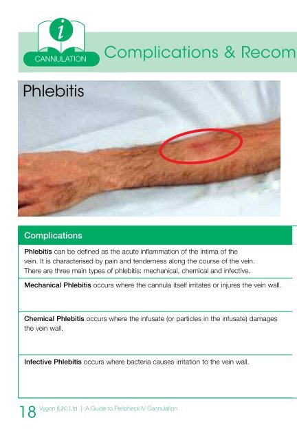
CANNULATIONComplications & Recom mendationsPhlebitisVisual Infusion Phlebitis (VIP) ScoreIV site appears healthyOne of the following is evident:l Slight pain near I.V. site l Slight redness near I.V. siteTwo of the following are evident:l Pain at I.V. site l Swelling l ErythemaAll of the following are evidentl Pain along the path of the cannula l Erythemal SwellingAll of the following are evident & extensivel Pain along the path of the cannula l Erythemal Swelling l Palpable venous cordAll of the following are evident & extensivel Pain along the path of the cannula l Erythemal Swelling l Palpable venous cord l Pyrexia012345No signs of phlebitisl Observe cannulaPossible first signs of phlebitisl Observe cannulaEarly stage of phlebitisl Resite cannulaMedium stage of phlebitisl Resite cannula l Consider treatmentAdvanced stage of phlebitis(or start of thrombophlebitis)l Resite cannula l Consider treatmentAdvanced stage of Thrombophlebitisl Resite cannula l Initiate treatmentOxford Radcliffe Trust Infection Control Services. Updated from A Jackson. 1997 OM141067ComplicationsPhlebitis can be defined as the acute inflammation of the intima of thevein. It is characterised by pain and tenderness along the course of the vein.There are three main types of phlebitis: mechanical, chemical and infective.Mechanical Phlebitis occurs where the cannula itself irritates or injures the vein wall.Chemical Phlebitis occurs where the infusate (or particles in the infusate) damagesthe vein wall.Infective Phlebitis occurs where bacteria causes irritation to the vein wall.18<strong>Vygon</strong> (<strong>UK</strong>) Ltd | A Guide to Peripheral IV CannulationRecommendationsTo prevent phlebitis - use aseptic insertion techniques, choose the smallest gaugecannula possible for the prescribed treatment, secure the cannula properly toprevent movement. It is important to do regular checks for the signs of phlebitis.Use the smallest gauge cannula neccessary for prescribed therapy in order tominimise catheter and vein wall contact. Stabilise the cannula with a transparent,semi-permeable dressing and assess the cannula site regularly.This can be avoided by ensuring the infusate is filtered, does not exceed a finalosmolarity of 500mmol/l, pH between 5 and 9, dextrose concentrations of >10%.Selecting the smallest gauge cannula and the largest vein possible will allow agreater volume of blood to flow around the cannula tip, thus diluting the infusate.The principles of asepsis, including handwashing, minimal touch technique and thecleansing of access points prior to use are essential. This must also include the useof a sterile dressing to cover the cannula insertion site. Cannula changes of at least72-96 hours are recommended.<strong>Vygon</strong> (<strong>UK</strong>) Ltd | A Guide to Peripheral IV Cannulation19