Pigeonpea and Chickpea Insect Identification Handbook - Agropedia
Pigeonpea and Chickpea Insect Identification Handbook - Agropedia
Pigeonpea and Chickpea Insect Identification Handbook - Agropedia

Create successful ePaper yourself
Turn your PDF publications into a flip-book with our unique Google optimized e-Paper software.
P o d Borers<br />
Helicoverpa (Heliothis) armigera ( H u b n e r )<br />
Helicoverpa zea ( B o d d i e )<br />
Helicoverpa punctigera ( W a l l e n g r e n )<br />
Heliothis virescens (F.)<br />
( L e p i d o p t e r a : N o c t u i d a e )<br />
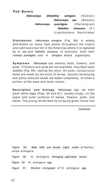
D i s t r i b u t i o n . Helicoverpa armigera (Fig. 58) is widely<br />
d i s t r i b u t e d on many host plants t h r o u g h o u t the t r o p i c s<br />
<strong>and</strong> s u b t r o p i c s but not in the A m e r i c a s w h e r e it is replaced<br />
by H. zea <strong>and</strong> Heliothis virescens. In Australia, both Helicoverpa<br />
punctigera <strong>and</strong> H. armigera attack p i g e o n p e a .<br />
S y m p t o m s . Helicoverpa s p p d e s t r o y buds, flowers, <strong>and</strong><br />
pods. If f l o w e r s <strong>and</strong> p o d s are not available, t h e y feed u p o n<br />
leaflets (Fig. 59), leaving the veins. On p o d s , c o n s p i c u o u s<br />
holes are m a d e by the entry of larvae. Usually d e v e l o p i n g<br />
<strong>and</strong> partly m a t u r e d seeds are eaten c o m p l e t e l y . At times a<br />
p o r t i o n of the seed <strong>and</strong> testa remain.<br />
D e s c r i p t i o n a n d b i o l o g y . Helicoverpa spp lay their<br />
small w h i t e eggs (Figs. 60 <strong>and</strong> 61), usually singly, on the<br />
upper <strong>and</strong> o u t e r surfaces of leaves, f l o w e r s , pods, <strong>and</strong><br />
stems. T h e y o u n g larvae feed by scraping green tissue <strong>and</strong><br />
Continued<br />
Figure 58. Male (left) <strong>and</strong> female (right) moths of H e l i c o -<br />
verpa armigera.<br />
Figure 59. H. armigera damaging pigeonpea leaves.<br />
Figure 60. H. a r m i g e r a egg.<br />
Figure 61. Electron micrograph of H. a r m i g e r a egg.<br />
62