A Physical Shepp-Logan Head Phantom for MRI - Waikato Digital ...
A Physical Shepp-Logan Head Phantom for MRI - Waikato Digital ...
A Physical Shepp-Logan Head Phantom for MRI - Waikato Digital ...

Create successful ePaper yourself
Turn your PDF publications into a flip-book with our unique Google optimized e-Paper software.
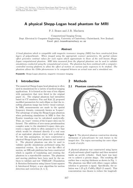
P. J. Bones, J. R. Maclaren, ‘A <strong>Physical</strong> <strong>Shepp</strong>-<strong>Logan</strong> <strong>Head</strong> <strong>Phantom</strong> <strong>for</strong> <strong>MRI</strong>’, Proceedingsof Image and Vision Computing New Zealand 2007, pp. 317–320, Hamilton,New Zealand, December 2007.A physical <strong>Shepp</strong>-<strong>Logan</strong> head phantom <strong>for</strong> <strong>MRI</strong>P. J. Bones and J. R. MaclarenComputational Imaging Group,Dept. Electrical & Computer Engineering, University of Canterbury, Christchurch, New Zealand.Email: phil.bones@canterbury.ac.nzAbstractA head phantom which is compatible with magnetic resonance imaging (<strong>MRI</strong>) has been constructed fromlayers of polycarbonate. When imaged using the appropriate scanner parameters, the partial volumeeffect generates contrast values <strong>for</strong> each region which approximate to those of the well known <strong>Shepp</strong>-<strong>Logan</strong> computational phantom. <strong>MRI</strong> data measured from the physical phantom can be used to validatesimulations conducted using the computational version. The phantom has been combined with a computercontrolledmoving plat<strong>for</strong>m to allow the effect of motion on various pulse sequences to be studied. Thephantom allows the Gibbs phenomenon to be compared between an actual scan and a simulated one.Keywords: <strong>Shepp</strong>-<strong>Logan</strong> phantom, magnetic resonance imaging1 IntroductionThe numerical <strong>Shepp</strong>-<strong>Logan</strong> head phantom is oftenused in simulations <strong>for</strong> a variety of medical imagingapplications. It is <strong>for</strong>med as the sum of ten ellipseswith parameters that were listed in the originalpaper [1]. The original phantom had intensitiesbased on CT numbers; Pan and Kak [2] proposedmodified parameters <strong>for</strong> each ellipse so that the resultingphantom image has better visual contrast.In <strong>MRI</strong>, measurements are made in the spatialfrequency domain, commonly known as ‘k-space’.The advantage of using the <strong>Shepp</strong>-<strong>Logan</strong> phantomwhen per<strong>for</strong>ming simulations in <strong>MRI</strong> is that theFourier trans<strong>for</strong>m can be calculated analytically.Thus, an ‘exact’ version of the k-space data can be<strong>for</strong>med. The addition of zero-mean pseudorandomcomplex noise to the simulated k-space data generatesa signal which is often assumed to be thatwhich would be obtained directly if a real scanof an equivalent physical object were per<strong>for</strong>med.To test this assumption, we have constructed aphysical <strong>Shepp</strong>-<strong>Logan</strong> head phantom and scannedit. The physical phantom can also be used tovalidate specific simulations per<strong>for</strong>med using thenumerical version. In order to test the effect ofmotion, an MR-safe plat<strong>for</strong>m has been constructedwhich is capable of translating and rotating thephantom in a reproducible manner. The phantomand moving plat<strong>for</strong>m have been used by the authorsto validate and test ‘TRELLIS’ (Translation& Rotation Estimation using Linear Least-squares& Interleaved Strips), a new algorithm <strong>for</strong> motiondetection and correction [3, 4, 5].2 Methods2.1 <strong>Phantom</strong> construction(a)Figure 1: The physical phantom construction showingthicknesses of polycarbonate <strong>for</strong> each feature in theimaged slice. The region surrounding the outer ellipseis made of acrylic.A physical phantom was constructed using layersof polycarbonate glued together and then mountedinside an acrylic cylinder. Each layer was lasercut to achieve a precise shape. The resulting 3Dsurface has a varying thickness as shown in Fig. 1.The phantom was filled with dilute copper sulfatesolution to generate a signal. Specifically, the solutionwas 1.25 g/L CuSO 4 .5H 2 O (5 mmol). Thisconcentration was chosen to reduce the longitudi-317
nal relaxation time constant (T 1 ) and the transversalrelaxation time constant (T 2 ) to levels consistentwith achieving a realistic contrast in a reasonableacquisition interval. The layers of polycarbonatecombined occupy less than half the depth of thecylinder. The remainder of the cylinder containsfurther circular disks featuring imageable cutoutshapes (a kiwi on one disk, a fernleaf and “NZ” onthe other). Sufficient spare depth occurs to allowa uni<strong>for</strong>m circular disk containing fluid only to beimaged as well.The copper sulfate solution produces a strong signalwhile the polycarbonate and acrylic producezero signal. The range of intensities required toproduce a realistic <strong>Shepp</strong>-<strong>Logan</strong> phantom are generatedthrough the partial volume effect. To achievethis, the phantom must be imaged using a slicethickness of 10 mm and all the polycarbonate layersmust be contained within one slice.2.2 <strong>Phantom</strong> motionIn order to test our new TRELLIS algorithm <strong>for</strong>motion detection and correction [3, 4], we havebuilt and successfully tested a moving plat<strong>for</strong>m<strong>for</strong> the phantom [5]. The plat<strong>for</strong>m is shown inFig. 2. It is constructed entirely from non-metallicmaterials so as not to affect the magnetic fieldhomogeneity in the scanner or pose a safety hazard.Two large plastic syringes act as pneumatic rams:one translates the plat<strong>for</strong>m while the other rotatesthe plat<strong>for</strong>m. A programmable logic controller locatedoutside the scanner room is used to couplecompressed air and vacuum to the syringes andthereby to generate a repeatable motion sequence.256 resolution, echo train length = 3), both whilestationary and while moving in a pre-programmedsequence. Images were reconstructed directly fromthe raw data without explicit filtering of k-space.3 ResultsA comparison of images of the numerical <strong>Shepp</strong>-<strong>Logan</strong> phantom and our physical version (fixed inposition) is shown in Fig. 3. The SNR used was35 dB, chosen to match the SNR in the imageof the physical phantom. Likewise, the numericalphantom was rotated by 2.2 ◦ to match the physicalphantom. It is evident, from both the imagespaceand k-space magnitudes, that reconstructedimages from the two phantoms are very similar. Animperfection can be seen in Fig. 3(b) at the baseof the physical phantom: this is caused by a smallbubble in the copper sulfate solution. Motion effectsobtained using the phantom with the movingplat<strong>for</strong>m (not shown here) also match simulationswell.(a)(b)(c)(d)Figure 2: The physical phantom (the cylinder) ismounted on a pneumatically controlled plat<strong>for</strong>m (entirelyMR compatible with no metallic components) sothat studies of the effects of bulk material motion canbe assessed.2.3 ImagingThe phantom was imaged on a 1.5 T GE scannerusing a fast spin echo sequence (FSE-XL, 256 ×Figure 3: Comparison of images of the numerical andthe physical phantom: (a) and (c), the numericalphantom in image-space and its computed k-spacemagnitude (log scale), respectively; (b) and (d), thephysical phantom reconstructed in image-space and itsmeasured k-space magnitude (log scale), respectively.The resolution in both cases was 256 × 256.However, one interesting difference is the reducedamount of Gibbs ringing in the physical phantomreconstruction compared to the numerical phantomreconstruction. This discrepancy is apparentin both the phase-encode and frequency-encode directions.A cross-section shows this more clearlythan the images themselves. One such cross-sectionis given in Fig. 4. This is taken horizontally through318