Fundamentals of neuroscience: cells, axons, and synapses
Fundamentals of neuroscience: cells, axons, and synapses
Fundamentals of neuroscience: cells, axons, and synapses

Create successful ePaper yourself
Turn your PDF publications into a flip-book with our unique Google optimized e-Paper software.
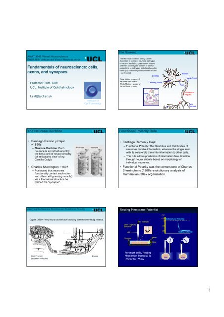
ANAT 3045 Visual NeuroscienceBIOS 3001 Advanced Visual Neuroscience<strong>Fundamentals</strong> <strong>of</strong> <strong>neuroscience</strong>: <strong>cells</strong>,<strong>axons</strong>, <strong>and</strong> <strong>synapses</strong>Pr<strong>of</strong>essor Tom SaltUCL Institute <strong>of</strong> Ophthalmologyt.salt@ucl.ac.ukInstitute <strong>of</strong>OphthalmologyThe NeuroneThe Nervous system’s wiring can bedescribed in terms <strong>of</strong> neuronal cell typesin each <strong>of</strong> its distinct gray matter regions<strong>and</strong> their stereotyped pattern <strong>of</strong> axonalprojections to cell types both locally <strong>and</strong> inother gray matter regions (or other tissues– eg muscle)Grey Matter – areas <strong>of</strong>neuronal cell bodiesWhite Matter – areas <strong>of</strong>nerve fibres (<strong>axons</strong>)VesicleDendritesCell Body (Soma)AxonSynapticCleftAxonNucleusMyelin SheathDirection <strong>of</strong>ImpulseAxonTerminalsThe Neurone DoctrineFunctional Polarity Rule• Santiago Ramon y Cajal~1890s– Neurone Doctrine: Eachneurone is an individual entity,the basic unit <strong>of</strong> neural circuitry(cf ‘reticularist view’ <strong>of</strong> egCamillo Golgi)• Charles Sherrington ~1897– Postulated that neuronesfunctionally contact each other<strong>and</strong> other cell types (eg muscle)via a theoretical structure hetermed the “synapse”.ReticularNeurone• Santiago Ramon y Cajal– Functional Polarity: The Dendrites <strong>and</strong> Cell bodies <strong>of</strong>neurones receive information, whereas the single axonwith its collaterals transmits information to other <strong>cells</strong>.– This rule allows prediction <strong>of</strong> information flow directionthrough neural circuits based on morphology <strong>of</strong>individual neurones.• Functional Polarity was the cornerstone <strong>of</strong> CharlesSherrington’s (1906) revolutionary analysis <strong>of</strong>mammalian reflex organisation.Applying the Neurone Doctrine <strong>and</strong> Functional PolarityResting Membrane PotentialmVCajal’s (1909-1911) neural architecture drawing based on the Golgi method.Glass CapillaryElectrodeTo Voltmeter+200-40Extracellular PotentialKCl-80Resting PotentialChamberSaline0- +0- +CellsVVOptic Tectum(superior colliculus)RetinaFor most <strong>cells</strong>, RestingMembrane Potential is-55mV to -75mVCellCell1
Distribution <strong>of</strong> Major Ions Across theMembrane <strong>of</strong> the Squid Giant AxonDistribution <strong>of</strong> Major Ions Across theMembrane <strong>of</strong> the Frog MuscleIon Cytoplasm ExtracellularK + 40020Cl - 52 560Na + 50 440Ion Cytoplasm ExtracellularK + 124 2.3Cl - 1.5 78Na + 10 109(mM)(mM)(mM)(mM)Resting potential ca. -60mVResting potential ca. -100mVWhat Forces Govern the Movements <strong>of</strong> Ions?1. Concentration Gradients. i.e. diffusionfrom high to low concentration areas.2. Electric Charge Separation. i.e. ionstend to move towards regions <strong>of</strong> oppositeelectric charge.3. Cell Membrane. i.e. ion movement isrestricted by the physical barrier imposedby the cell membrane.Selectively Permeable MembraneIn OutA -A -K +K +A -A - K + K +K +K + A -K + K +A -A -A - K +K +A - K +A - K +K + A -K + A -K +K + A -A - A -A -Selectively Permeable MembraneSelectively Permeable MembraneIn OutIn OutA -A -K + K +A - A -K +KK ++ K +A - A - K + A - A -K +K +K +KK ++K + AĀ- KA -+K +A -A- A - K +A -A -K +A - K +K +K +A -K +KA - +A - K +K +K K + A -K ++A -K + A - A -KK + A - + A -A - K + K +A -A -A - A - A -- - - + + +- - - - + + + +2
Nernst EquationRTE K =__ZFWhereln____ [K + ] o[K + ] iE K = K + Equilibrium Pot’l.R = Gas ConstantT = Absolute TemperatureZ = Valence <strong>of</strong> K +F = Faraday Constant[K + ] o,i =K + concentrationsSubstituting,E K = 26mVNerve___ 20ln400 = -75mVRelationship <strong>of</strong> Membrane Potential (V m ) to [K + ] o0V m25mV5075100Nernst1 10 [K + ] 100o mM1. How can concentration gradients forNa + , K + , <strong>and</strong> Cl - all be maintainedacross the cell membrane?2. How do these gradients interact todetermine the resting membranepotential?IonDistribution <strong>of</strong> Major Ions Across theMembrane <strong>of</strong> the Squid Giant AxonCytoplasmExtracellularFluidNernstPotentialPassive <strong>and</strong> Active Movement <strong>of</strong> Ions through the MembraneMembraneIntracellularK + K +E K - V m = -10mVExtracellularK + 400 20-75Cl - 52 560 -60Na + 50 440 +55(mM) (mM) (mV)MetabolicEnergy(ATP)Na +Na + -K +PumpK +Na +Resting Pot’l (V m ) = -75mVE Na - V m = 155mVResting potential ca. -60mVGOLDMAN EQUATIONV m =RT __FlnIf P K>> P Na<strong>and</strong> P Cl,_______________________P K[K + ] o+ P Na[Na + ] o + P Cl[Cl - ] iP K[K + ] + iP Na[Na + ] i+ P Cl[Cl - ] oRT __V m ~FTherefore,The greater the permeability <strong>and</strong> concentration <strong>of</strong>an ion, the greater will be its contribution to V m .ln_____ P K[K + ] oP K[K + ] iwhere P = Permeability Membrane potential (Vm(Vm) ) is determined primarily by K+ <strong>and</strong>Na+. Membrane potential will be closest to the Nernst(Equilibrium) Potential <strong>of</strong> the ion with the greatestconcentrations <strong>and</strong> membrane permeability. At Rest, Membrane Potential is close to the potassiumequilibrium potential (EK+) because the membrane is mostpermeable to K+. At Rest, as EK+ is slightly more negative than Vm, , there is asteady K+ efflux, balanced by a steady Na+ influx. Thesetwo passive fluxes are balanced by active pumping <strong>of</strong> Na+<strong>and</strong> K+ in the opposite directions. Note: this is a steadystate, not an equilibrium. Under most physiological conditions the bulk concentrations<strong>of</strong> Na+, K+ <strong>and</strong> Cl- inside <strong>and</strong> outside <strong>of</strong> the cell remainconstant.3
Electrotonic Potential SpreadACTION POTENTIALTHRESHOLDCurrentPotential (V)CurrentElectrotonicPotential+20mV0Overshoot-60mVUpstroke0 100 time (ms)-20Threshold-40UpstrokeRepolarisationCurrent-60-80Resting Pot’lThreshold-80Current Pulses2.5 mm0 ms 50 ms 5Voltage-dependent Na + ChannelsSodium Channel StructureDEPOLARISATION“Positive Feedback”or “Regeneration”Na + EntersCellVoltagedependentNa +Channels OPENNote: Voltage-dependent Na + ChannelsINACTIVATEThe Refractory PeriodPERMEABILITY CHANGES DURING THE ACTION POTENTIAL+20+20 V m 0V m0-20Na + K +-20-40mV-60-800 ms 5300MembranePermeabilityorConductancemS/cm 2-40mV-60-80Threshold0Absolute Relative5 msREFRACTORY PERIOD4
PROPAGATION OF THE ACTION POTENTIAL - 1Unmyelinated nerve membranePROPAGATION OF THE ACTION POTENTIAL - 1Unmyelinated nerve membraneExtracellular+ + + + + + + + + + + + + + + + + + + + + + + + + + + + + + + + + + + + +- - - - - - - - - - - - - - - - - - - - - - - - - - - - - - - - - - - -IntracellularNa +Extracellular+ + - - + + + + + + + + + + + + + + + + + + + + + + + + + + + + + + +- + + - - - - - - - - - - - - - - - - - - - - - - - - - - - - -IntracellularNote• No significant change in ionic concentrations during the action potential.• No change in Na + Pump activity during the action potential.PROPAGATION OF THE ACTION POTENTIAL - 1Unmyelinated nerve membranePROPAGATION OF THE ACTION POTENTIAL - 2Unmyelinated nerve membraneNa +Extracellular+ + - - + + + + + + + + + + + + + + + + + + + + + + + + + + + + + + +- + + - - - - - - - - - - - - - - - - - - - - - - - - - - - - -Local Circuit CurrentsIntracellularNa +Extracellular+ + + + + + - - + + + + + + + + + + + + + + + + + + + + + + + + + +- - - - - - + + - - - - - - - - - - - - - - - - - - - - - - - -K +IntracellularPROPAGATION OF THE ACTION POTENTIAL - 3Unmyelinated nerve membranePROPAGATION OF THE ACTION POTENTIAL - 3Unmyelinated nerve membraneNa +Extracellular+ + + + + + + + + + + + + - - + + + + + + + + + + + + + + + + + + + + +Na +Extracellular+ + + + + + + + + + + + + + + + + - - + + + + + + + + + + + + + + + +- - - - - - - - - - - - + + - - - - - - - - - - - - - - - - - - - -- - - - - - - - - - - - - - - - + + - - - - - - - - - - - - - - -<strong>and</strong> so forthK + IntracellularK +IntracellularNote• An action potential is triggered in each portion <strong>of</strong> the membrane• The action potential can travel in all directions from the initial triggering point.• The action potential cannot immediately reinvade a portion <strong>of</strong> membrane,because <strong>of</strong> the refractory period.5
The Erlanger / Gasser Classification <strong>of</strong> Nerve FibresFibreTypeFunction (examples)Avg. Fibre Dia.(m)AAAABCThe Lloyd Hunt Classification <strong>of</strong> Nerve FibresGroup Function (examples)Avg. Fibre Dia.(m)IIIIIVPrimary Muscle Spindle AfferentsMotor to Skeletal MusclesCutaneous Touch <strong>and</strong> Pressure AfferentsMotor to Muscle SpindlesCutaneous Temperature <strong>and</strong> Pain AfferentsSympathetic PreganglionicCutaneous Pain AfferentsSympathetic PostganglionicPrimary Muscle Spindle AfferentsAfferents from Tendon OrgansCutaneous Mechano-receptorsDeep Pressure Sensors in MuscleUnmyelinated Pain Afferents1585< 331(unmyelinated)13931Avg. Cond. Vel.(m/s)100 (70-120)50 (30-70)20 (15-30)15 (12-30)7 (3-15)1 (0.5-2)Avg. Cond. Vel.(m/s)75 (70-120)55 (25-70)11 (10-25)1 (0.5-2)Myelinated Nerve Fibres• Conduction velocity increases with fibre diameter, but the greatest influence iswhether or not an axon is myelinated.• Myelin sheath consists <strong>of</strong> membranes <strong>of</strong> Schwann Cells wrapped around the<strong>axons</strong>. This wrapping is periodically interrupted at what are termed the Nodes <strong>of</strong>Ranvier.• The effect <strong>of</strong> this sheath is to increase the resistance across the cell membranefrom the inside <strong>of</strong> the axon to the extracellular space, <strong>and</strong> to concentrate chargedistribution at the Nodes <strong>of</strong> Ranvier.Action Potential Propagation in Myelinated AxonsAction Potential Propagation in Myelinated Axons+ ++ +Myelinated nervemembrane+ +ExtracellularNa ++ +Myelinated nervemembrane+ +Extracellular- -- -- -- -- -IntracellularIntracellularIncreased chargedistribution at theNodes <strong>of</strong> RanvierAction Potential Propagation in Myelinated AxonsAction Potential Propagation in Myelinated AxonsNa ++ +Myelinated nervemembrane+ +ExtracellularMyelinated nervemembraneNa ++ +Extracellular- -Local Circuit Currents- -IntracellularK +- -Intracellularlocal circuit current flow is from one Node <strong>of</strong> Ranvier to the next6
Action Potential Propagation in Myelinated AxonsVoltage ClampTwo-electrode voltage clamp <strong>of</strong> squid axon, after Hodgkin & HuxleyExtracellular+ +- -IntracellularMyelinated nervemembraneNa +K +<strong>and</strong> so forth• Effectively the action potential is propagated from one node <strong>of</strong> ranvierto the next. This is referred to as Saltatory Conduction.• The net result <strong>of</strong> this is that an action potential can travel morequickly by jumping along a myelinated axon rather than by beingconventionally propagated along an umyelinated axon.The voltage-clamp technique keeps the voltage across themembrane constant so that the amplitude <strong>and</strong> time course<strong>of</strong> <strong>of</strong> ionic currents can be measuredVoltage Clamp Reveals Ionic CurrentsVoltage Clamp recordings from CellsTwo electrode voltage clamp(large <strong>cells</strong> eg oocytes)MeasureVoltage-70mVInjectCurrentSingle electrodevoltage clamp(neurones) • Measure voltage• is it what we want ?• Pass current to adjustvoltageMembranecurrents-70mVTEA - TetraEthylAmmonium – blocks K + currentsTTX - TetrodoToXin – blocks Na + currentsWhole Cell (Patch) recordingsExtracellular Recording methodswholecellMembranecurrentsGlycine 50uM100pAMicroelectrodeSingle neurone (unit) spikes500uVFlash1 sGlycine 50uMneuroneField Potential Recordingisolatedpatch5pA1 sFlash7
SynapticCleftActionPotentialCHEMICAL SYNAPSESAxonTerminalPostsynaptic Cell! Action Potential Propagated tothe Nerve Terminal.! Arrival <strong>of</strong> an Action Potentialleads to release <strong>of</strong> a chemical(i.e. the Neurotransmitter)from the terminal.! Neurotransmitter diffusses to thePostsynaptic Membrane.! Binding <strong>of</strong> Transmitter moleculeto Receptors causes apostsynaptic effect (e.g.Membrane Depolarisation).Process <strong>of</strong> Chemical Neurotransmission Synthesis <strong>of</strong> neurotransmitter in the presynapticneurone Storage <strong>of</strong> the neurotransmitter <strong>and</strong>/or itsprecursor in the presynaptic nerve terminal Release <strong>of</strong> the neurotransmitter into the synapticcleft Binding <strong>and</strong> recognition <strong>of</strong> the neurotransmitterby target receptors Termination <strong>of</strong> action <strong>of</strong> the released transmitterLife Cycle <strong>of</strong> Synaptic VesiclesTYPES OF NEUROTRANSMITTERAminesAcetycholineNoradrenalineDopamineSerotonin (5HT)PeptidesEnkephalinsSubstance PSomatostatinCholecystokininExcit. Amino AcidsL-Glutamic AcidL-Aspartic AcidL-Homocysteic AcidPurinesAdenosineATPInhibitory Amino AcidsGlycineGABAFree RadicalsNitric Oxide (NO)LipidsCannabinoidsVanillinoidsThe “vesicle hypothesis”8