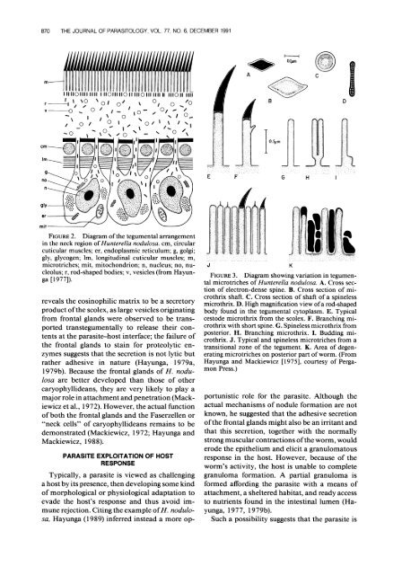
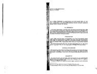
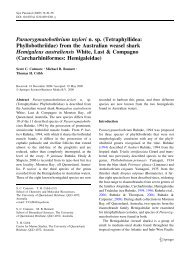
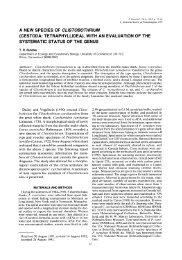
870 THE JOURNAL OF PARASITOLOGY, VOL. 77, NO. 6, DECEMBER 1991Iul#lIIIHHII rlul1u1!111lt!1lllf!( A,t.A^Q1mI>71111101111 11i1I110111110111110111-11111111011 111-100 -,'0 -~IO.5jmEFGH. ..IFIGURE 2. Diagram <strong>of</strong> the tegumental arrangementin the neck region <strong>of</strong> Hunterella nodulosa. cm, circularcuticular muscles; er, endoplasmic reticulum; g, golgi;gly, glycogen; lm, longitudinal cuticular muscles; m,microtriches; mit, mitochondrion; n, nucleus; no, nucleolus;r, rod-shaped bodies; v, vesicles (from Hayunga[1977]).reveals the eosinophilic matrix to be a secretoryproduct <strong>of</strong> the scolex, as large vesicles originatingfrom frontal glands were observed to be transportedtranstegumentally to release their contentsat the parasite-host interface; the failure <strong>of</strong>the frontal glands to stain for proteolytic enzymessuggests that the secretion is not lytic butrather adhesive in nature (Hayunga, 1979a,1979b). Because the frontal glands <strong>of</strong> H. nodulosaare better developed than those <strong>of</strong> othercaryophyllideans, they are very likely to play amajor role in attachment and penetration (Mackiewiczet al., 1972). However, the actual function<strong>of</strong> both the frontal glands and the Faserzellen or"neck cells" <strong>of</strong> caryophyllideans remains to bedemonstrated (Mackiewicz, 1972; Hayunga andMackiewicz, 1988).PARASITE EXPLOITATION OF HOSTRESPONSETypically, a parasite is viewed as challenginga host by its presence, then developing some kind<strong>of</strong> morphological or physiological adaptation toevade the host's response and thus avoid immunerejection. Citing the example <strong>of</strong> H. nodulosa,Hayunga (1989) inferred instead a more op-JFIGURE 3. Diagram showing variation in tegumentalmicrotriches <strong>of</strong> Hunterella nodulosa. A. Cross section<strong>of</strong> electron-dense spine. B. Cross section <strong>of</strong> microthrixshaft. C. Cross section <strong>of</strong> shaft <strong>of</strong> a spinelessmicrothrix. D. High magnification view <strong>of</strong> a rod-shapedbody found in the tegumental cytoplasm. E. Typicalcestode microthrix from the scolex. F. Branching microthrixwith short spine. G. Spineless microthrix fromposterior. H. Branching microthrix. I. Budding microthrix.J. Typical and spineless microtriches from atransitional zone <strong>of</strong> the tegument. K. Area <strong>of</strong> degeneratingmicrotriches on posterior part <strong>of</strong> worm. (FromHayunga and Mackiewicz [1975], courtesy <strong>of</strong> PergamonPress.)portunistic role for the parasite. Although theactual mechanisms <strong>of</strong> nodule formation are notknown, he suggested that the adhesive secretion<strong>of</strong> the frontal glands might also be an irritant andthat this secretion, together with the normallystrong muscular contractions <strong>of</strong> the worm, woulderode the epithelium and elicit a granulomatousresponse in the host. However, because <strong>of</strong> theworm's activity, the host is unable to completegranuloma formation. A partial granuloma isformed affording the parasite with a means <strong>of</strong>attachment, a sheltered habitat, and ready accessto nutrients found in the intestinal lumen (Hayunga,1977, 1979b).Such a possibility suggests that the parasite isK
HAYUNGA-MORPHOLOGICAL ADAPTATIONS OF INTESTINAL HELMINTHS 871capable <strong>of</strong> far more than passive evasion <strong>of</strong> hostdefenses and that, conceivably, parasites mayevolve strategies that actively exploit the host'sresponse for their own benefit (Hayunga, 1989).Careful examination <strong>of</strong> other parasites may revealthat strategies <strong>of</strong> exploitation are far morecommon than have previously been thought(Damian, 1987).COST OF SPECIALIZATIONSpecialization in terms <strong>of</strong> morphological adaptationhas resulted in highly efficient means <strong>of</strong>attachment for some intestinal helminths thatpresumably have given these species a competitiveedge. By adapting so well to their preferredsites, cestodes appear to be specialists with regardto space. To a somewhat lesser degree, trematodesand acanthocephalans also may be regardedas specialists. Nematodes seem less limitedin site selection by their anatomy and thus appearto be generalists in this regard, although theirhabitat selection may be restricted by physiologicalfactors. For nematodes it would appear thatniche stratification is more a question <strong>of</strong> whatthey eat rather than where they eat.Although the specialist may be highly efficient,the cost <strong>of</strong> specialization is dependence on onetype <strong>of</strong> host, as clearly demonstrated by studies<strong>of</strong> host specificity. One need only ask the question,"Whatever happened to the parasites <strong>of</strong> thedinosaurs?" to envision the risks <strong>of</strong> a strategy <strong>of</strong>specialization. But, if anything, helminths haveshown themselves to be highly resilient and innovative,as shown by the ability <strong>of</strong> polystomatidmonogeneans to inhabit both the gills <strong>of</strong> tadpolesand the urinary bladders <strong>of</strong> adult frogs, or by thestrategy <strong>of</strong> neotenic development when a desiredhost is no longer available. The morphologicaladaptations described here represent only oneexample <strong>of</strong> many highly effective strategies forsurvival.ACKNOWLEDGMENTSI am grateful to Robin M. Overstreet and JohnC. Holmes, both for inviting me to speak at thissymposium and for keeping me on the programdespite the turmoil <strong>of</strong> world events this year. Iparticularly acknowledge the influence <strong>of</strong> JohnMackiewicz, who introduced me to the field <strong>of</strong>parasitology and to the fascinating Caryophyllidea.Many <strong>of</strong> the ideas that I discussed werelearned in his classes, and the original researchthat I described was done in his laboratory. Thespecimen shown in Figure 1 was prepared by Dr.Mackiewicz. Observations on competition andsite selection by caryophyllideans were made incollaboration with Anthony J. Grey, a fellowgraduate student, now at the New York StateDepartment <strong>of</strong> Health in Albany. Finally, I givecredit to the pioneering efforts <strong>of</strong> prominent parasitologistswhom I have quoted extensively inthis paper.LITERATURE CITEDACKERT, J. E., S. A. EDGAR, AND L. P. FRICK. 1939.Goblet cells and age resistance <strong>of</strong> animals to parasitism.Transactions <strong>of</strong> the American MicroscopicalSociety 58: 81-89.BEFUS, D., AND J. BIENENSTOCK. 1982. Factors involvedin symbiosis and host resistance at the mucosa-parasiteinterface. Progress in Allergy 31: 76-177.BEGUIN, F. 1966. Etude au microscope electroniquede la cuticule et de ses structures associ6es chezquelques cestodes. Essai d'histologie comparee.Zeitschrift fuir Zellforschung 72: 30-46.BERGER, J., AND D. F. METTRICK. 1971. Microtrichialpolymorphism among hymenolepid tapeworms asseen by scanning electron microscopy. Transactions<strong>of</strong> the American Microscopical Society 90:393-403.BRATEN, T. 1968. The fine structure <strong>of</strong> the tegument<strong>of</strong> Diphyllobothrium latum (L.)-A comparison <strong>of</strong>the plerocercoid and adult stages. Zeitschrift furParasitenkunde 30: 104-112.COLWELL, R. K., AND E. R. FUENTES. 1975. Experimentalstudies <strong>of</strong> the niche. Annual Review <strong>of</strong>Ecology and Systematics 6: 281-310.CROMPTON, D. W. T. 1973. The sites occupied bysome parasitic helminths in the alimentary tract<strong>of</strong> vertebrates. Biological Reviews 48: 27-83.1975. Relationships between Acanthocephalaand their hosts. Symposia <strong>of</strong> the Society forExperimental Biology 29: 467-504.DAMIAN, R. T. 1987. The exploitation <strong>of</strong> host immuneresponses by parasites. Journal <strong>of</strong> Parasitology73: 3-13.DOGIEL, V. A. 1958. Ecology <strong>of</strong> the parasites <strong>of</strong> freshwaterfishes. In Parasitology <strong>of</strong> fishes, V. A. Dogiel,G. K. Petrushka, and Y. I. Polyanski (eds.). LeningradUniversity Press. [English translation by Z.Kabata, Oliver & Boyd, Edinburgh, xi + 384 p.,1961.]ERASMUS, D. A. 1969. Studies on the host-parasiteinterface <strong>of</strong> strigeoid trematodes. V. Regional differentiation<strong>of</strong> the adhesive organ <strong>of</strong> Apatemongracilis minor Yamaguti, 1933. Parasitology 59:245-256.1977. The host-parasite interface <strong>of</strong> trematodes.Advances in Parasitology 15: 202-242., AND C. OHMAN. 1963. The structure andfunction <strong>of</strong> the adhesive organ in strigeid trematodes.Annals <strong>of</strong> the New York Academy <strong>of</strong> Sciences113: 7-35.GREY, A. J., AND E. G. HAYUNGA. 1980. Evidencefor alternative site selection by Glaridacris laruei(Cestoidea: Caryophyllidea) as a result <strong>of</strong> inter-