PEARL OYSTER FISHERIES MARINE BIOLOGY OF CEYLON,
PEARL OYSTER FISHERIES MARINE BIOLOGY OF CEYLON,
PEARL OYSTER FISHERIES MARINE BIOLOGY OF CEYLON,

Create successful ePaper yourself
Turn your PDF publications into a flip-book with our unique Google optimized e-Paper software.
REPORTTO THE GOVERNMENT <strong>OF</strong> <strong>CEYLON</strong>ONTHE<strong>PEARL</strong> <strong>OYSTER</strong> <strong>FISHERIES</strong><strong>OF</strong> THEGULF <strong>OF</strong> MANAAR,BYW. A. HERDMAN, D.Sc, F.R.S., P.L.S.,Professor of Natural History in the University of Liverpool.WITH SUPPLEMENTARY REPORTSUPON THE<strong>MARINE</strong> <strong>BIOLOGY</strong> <strong>OF</strong> <strong>CEYLON</strong>,BY OTHER NATURALISTS.Part II.PUBLISHED AT THE REQUEST <strong>OF</strong> THECOLONIALGOVERNMENTBYTHE ROYAL SOCIETY.LONDON1904.
[77]THE PARASITES <strong>OF</strong> THE <strong>PEARL</strong> <strong>OYSTER</strong>.BYARTHUR E. SHIPLEY, M.A., F.R.S.,FELLOW AND TUTOR <strong>OF</strong> CHRIST'S COLLEGE, CAMBRIDGE,ANDJAMES HORNELL, F.L.S., .<strong>MARINE</strong> BIOLOGIST TO THE <strong>CEYLON</strong> GOVERNMENT AND INSPECTOR <strong>OF</strong> <strong>PEARL</strong> BANKS.[With FOUR PLATES.]INTRODUCTION.The history of the formation of pearls in Europeanmussels around the Cercaria ofTrematodes is recorded in the papers of Garner (1871), Giard (1897), Dubois (1901)and others, and it has recently been re-told in more detail by Dr. H. LysterJameson.* The main part of Dr. Jameson's own observation was directed to theLeiicit/nidcndrium (Distomum) somaterice (Levinsen) of the eider-duck (Somateriamottissima, Linn.) and of a scoter (Oedemia nigra, L.), the sporocyst of which hestates is found in Tapes decussatus, Gmel., and in the cockle, Cardium edule, L.,whilst the tailless Cercaria infests the mussel, Mytilus edulis, and iswhich the small lustreless pearls of that mollusc are formed.the centre roundIt has been pointed outsince that two points are still left in an unsettled condition by Jameson's paper,viz.(1) the exact mode of origin of the epithelial sac around the parasite whichsecretes the pearl,and (2) the supposed transference of the parasite from the cockleto the mussel.With regard to the history of the relationship of internal parasites to the pearlsformed in the Ceylon pearl oyster, Margaritifera vulgaris, Schum. not a trueoyster but a member of the AviculibvE, and so allied to the mussel, Mytilus eduliswe quote two short paragraphs from Professor Herdman's Introduction to the firstvolume of this Report :" To Dr. Kelaart (1857 to 1859) belongs the honour of havingformation of pearls in the Ceylon oyster with the presenceparasites.first connected theof vermeanIt is true that Filippi, seven years before, in 1852, showed thatthe Trematode Distomum duplicatum was the cause of pearl formation in thefresh-water mussel Anodonta, and Kitchenmeister (1856), Mobitts (1857),* 'P. Zool. Soc.,' London, 1902, p. 110.
78 <strong>CEYLON</strong> <strong>PEARL</strong> <strong>OYSTER</strong> REPORT.and others extended the discovery to other pearl-producing oysters, and toother parasites; but it ispossible that Kelaaet knew nothingof thesepapers, and that he made his discovery in regard to the Ceylon oysters quiteindependently. He (and the Swiss Zoologist, Humbert, who was with him'at a pearl fishery) found in addition to the Filaria and Circaria, three otherparasitical worms infesting the viscera and other parts of the pearl oyster.We both agree that these worms play an important part in the formation ofpearls and it may ; yet be found p>ossible to infect oysters in other beds withthese worms, and thus increase the quantity of these gems.' Thus we haveKelaart, in 1859, definitely stating the possibility,in the case of the Ceylonpearl oyster, of infecting other beds with the larvae of the pearl-producingPlatyhelminthian parasites in order to increase the quantity of pearls."Thurston, in 1894, confirmed Kelaart's observation, finding in the tissues andalso in the alimentary canal of the Ceylon oyster, ' larva of some Platyhelminthian(fat worm).' He figures ('Madras Mus. Bull.,' I., Plate II., fig. 1)a section showing two of the parasites encysted between the alimentary canaland generative tubes. Here the matter rested so far as the Ceylon pearloysterwas concerned."Kelaart's work has been tillrecently somewhat neglected, and we therefore givein a note* the whole paragraph which deals with parasites from his last Report.!The large number of oysters dissected during the fifteen months of Dr. Herdman'sand Mr. Hornell's expedition enable us now to furnish what is probably a fairlyexhaustive list of the Entozoa of the pearl oyster, seven in number. They compriseone Cestode (Tetrarhynchus unionifactor, n.sp.), three Trematodes (Muttuamargaritiferaz, n. sp., Musalia herdmani, n. sp., Aspidogaster margaritiferm, n. sp.)and three Nematodes, Ascaris meleagrinie, n. sp.,Cheiracanthus uncinatus, and anunidentified species of Oxyuris.* " I have not in this paper detailed some very interesting discoveries made since my last Report, onthe Anatomy and Physiology of the Pearl Oyster, believing that they are better fitted for a treatise onthe subject than to be embodied in a Report to the Ceylon Government, which must necessarily bewritten in popular form. However, as this Report may, like the precedingones, fall into the hands ofscientific men, I shall merely mention here that Monsieur Humbert, a Swiss zoologist, has, bymicroscopic observations at the last pearl fishery,his owncorroborated all I have stated about the ovaria orgenital glands and their contents ;and that he has discovered, in addition to the Filaria and Circaria,three other parasitical worms infesting the viscera and other partsof the pearl oyster.We both agreethat these worms play an important part in the formation of pearls ;and itmay yet be found possibleto infect oysters in other beds with these worms, and thus increase the quantity of these gems. Thenucleus of an American pearl, drawn by Mobius, is nearly of the same form as the Circaria found in thepearl oysters of Ceylon. It will be curious to ascertain if the oystersin the Tinnevelly banks have thesame species of worms as those found in the oysters on the banks at Arripo."t Reprinted in the 'Proceedings of the Madras Government Revenue Department,' 17th February,1864.
THE PARASITES <strong>OF</strong> THE <strong>PEARL</strong> <strong>OYSTER</strong>. 79The ectoparasites of the pearl oyster consist principally of shell-boring animalswhich tunnel more or less extensively into the nacreous portion of the valves. Ofthese, the only one whose depredationsare of economic importanceis the boringsponge Cliona niaiyart/ij'ertr,Dknmy. The other two, the Polychgete wormLeucodore sp.,and the Lamellibranch mollusc Lithodomus sp. (date-shell), are never sonumerous as to constitute a serious danger to the oyster population of the pearl1 i.inks.I. CESTODES <strong>OF</strong> THE <strong>PEARL</strong> <strong>OYSTER</strong>.I. Cestode Larv.e in the Pearl Oyster, Margaritifera vulgaris, Schtjm.The cestode larvse of the pearl oyster pass through several stagesin the tissues ofthe host, that is if they escape being entombed within the centre of a pearl.Economically these unpleasant little creatures are of supreme importance to theCeylon pearl fishery, as their presence in the oyster causes the formation of the finestquality of pearl and those with the highest lustre.These larvae first attracted attention during the second cruise'" of the " LadyHavelock," on March 6th, 1902, and the following clays,when numbers of the earlyglobular stage were dissected out from the livers of oysters dredged from the WestCheval Paar.Subsequently, during the investigation carried out at the GalleBiological Laboratory, a second and more advanced stage of a Tetrarhynchus larvawas found in the same animal. Details of the morphology and histology werethen worked out, and the relationship which the larvre bear to pearl formation wasinvestigated.It is usual for a Tetrarhynchus to enter its first host as an embryoenclosed in an' ;egg-shell. As VaullegeardI says, L'oeuf doit etre avale" par un animal marin ;ne se developpe que si cet etre lui fournit un milieu convenable, mais les Tetrarhynquesparaissent peu tenir a une espece ;" the drawings made by one of us(Plate I., fig. 1) of a free-swimming larva taken in the plankton at the north end ofthe Muttuvaratu Paar seems to point to the fact that the embryo of the pearlformingparasites may leave the egg-shell before entering their first host, and thismust be so if the organisms in question are truly the young of the Tetrarhynclnis ;this point, however, requires corroboration.The earlier and more globular-shaped of the two stages met with in the pearloyster is by far the more numerous, as may naturally be expected. It accountsfor all save one per cent, of the total cases found. The liver, the gills (Plate I.,figs. 3 and 4), and the connective tissue of the mantle (Plate I., fig. 2) are theorgans chiefly favoured ;the muscles are practically free, while the gonads yieldilcomparatively few. * See " Narrative," this Report, Part I., p. 70.t ' Mem. Soc. Normandie,' XIX., 1897 to 1899, p. 353.
80 <strong>CEYLON</strong> <strong>PEARL</strong> <strong>OYSTER</strong> REPORT.In the liver and in the base of the gills they usually reach the greatest size ;in the gill-filaments, where they occasionally abound, they seldom attain other thanminute dimensions. Each isenveloped in a tough, elastic, and fibrous capsule ofspherical form, derived from the adjacent connective-tissue cells (Plate L, figs. 5, 6,and 8).The capsules of those found in the liver and gill-bases are specially thickand strong, and slightly opalescent. In extreme cases the fibrous capsule has athickness equal to fullyhalf the diameter of the enclosed parasite (Plate I., fig.5 a,and fig.8 a and b).The minute larvae encapsuled in the gill-filaments and these are easy of observationbecause of the transparency of the capsule membrane (Plate I., figs.5 and 8)may be seen occasionally to rotate slowly within their prison. The bodyis subglobularand remains so, even after considerable increase in size. The oldest larvaehowever tend gradually to assume an elongated cylindrical form, with a pointedprotrusible head, a denticulated collar, and a long oval body, foreshadowing thechange to the rarer second or hooded larval phase (Plate I., figs. 12 and 13).The larva in this stage closely resembles one of those figured in a paper byProfessor Giard on " L'Origine Parasitaire des Perles."* It was observed byMonsieur Setjrat, who not unnaturally mistook it for Amphistoma.In the globular stage, whether minute or comparatively large, the structure differsbut in details. Thus the body consists of two regions, an anterior, the smaller, and alarge bladder-like posterior division. The former measures approximately but onethirdof the total diameter. Viewed en face, this region has the appearance of abroad convexly-annular sucker, with a wide central orifice, wherefrom protrudesslightly the rounded summit of a low eminence (Plate I., fig. 9).The saccate hinder portion of the body is thin-walled and filled with granularcontents wherein rounded refractile corpuscles lie scattered abundantly, especiallyinthe peripheral layer (Plate I., tigs. 12 and 13).Under slight pressure, as first seen,it exhibited a striking resemblance to a tiny Trematode, or itmightbe mistaken fora large Gregarine.When freed from its investing membranes, the larva progresses slowly by means ofthe alternate elongation and contraction of the body, the low central anterioreminence assisting by protrusion and retraction. The anterior regionis clear andslightly tinged with yellow ; the hinder is colourless and granular.The cephalic" sucker"is in reality a head-sheath, the low median eminencewithin being a proboscis-like head rudiment, the scolex-rudiment (Plate I., fig. 6).When the latter is retracted the normal condition when at rest within the cystcapsuleit lies sunk within the sheath, separated from it by a deep and narrowencircling groove. Under the influence of slight pressure, the head is seen (Plate I.,fig.11) to be shot out in the form of a blunt cone, the head groove being temporarilyobliterated. The muscular walls of this cephalic apparatus, sheath as well as head,* 'C. R. Soc. Biol.,' LV., 1903, p. 1222.
THE PARASITES <strong>OF</strong> THE <strong>PEARL</strong> <strong>OYSTER</strong>. 81are highly developed, and consist of two series, one running longitudinally, the otherin a circular manner.The cuticle of the head and its groove is smooth that of the sheath is ornamented;with a multitude of closely set minute shagreendike points, resembling in shapedenticles from the skin of Scyllium;the central point having a tiny basal projectionon either side (Plate I., figs.10 and 11, Plate II., fig. 18).The hinder portion of this larva is the morphological equivalent of the " bladder "of the Cysticercus stage of Tarda, differing however in that the contents, in lieu ofbeing fluid, are composed of a loose connective-tissue parenchyma,in which areilistributed the rounded retractile bodies already referred to. These effervesce upon theapplication of acid, and by this test, taken together with the similarity in appearance,they are shown to be calcareous corpuscles identical with those so widely met withthe Cestoda.amongEnsheathing the body parenchyma are two muscular layers of similar arrangementto those of the head, but of extremely feeble development. These in turn areoverlaid by a cuticle of peculiar appearance. In the parenchyma ramifythe tubulesof the vascular system.The connective-tissue framework of the cortical region of the isparenchyma ratherdenser than it is in the centre. In the former, accumulating at the periphery,lie themajority of the calcareous corpuscles.Of these, while some are nearly spherical, thegreater number are slightly reniform, or else obscurely compound, double, or evenroughly trifoliate, shallow depressions marking some out into two or even threedistinct areas (Plate II., figs.1G and 17).With pressure, they can be broken intoirregular sharp-edged fragments, just as a glass ball shatters under a blow.The traces of a distinct and fine capsule around many of the corpuscles, which canbe detected in the living tissue, is probably the remains of the cells in which theconcretions arise (Plate II., fig. 17).A closely woven network of anastomosing tubules of exceeding delicacy, suffusedwith the palest of pink tints, is also to be seen in the cortical parenchyma. Suchrepresents a generalised and rudimentary excretory system which becomes specializedin succeeding stages (Plate I., fig. 1:2).In the peripheral region just under the cuticle are occasionally to be made outlarge clear-walled cells filled with ' colourless transparent globules ; probably theseare the " bladder-cells" which ooze through the cuticular tubules under pressure.When a living larva is examined microscopically, the body behind the denticulatedhinder portion of the head-sheath shows a distinct appearance of being clothed ina densely ciliated envelope. So distinct is the appearance, that time after timereferences to the " ciliated surface " of these larva? appear in the earlier notes madeon the steamer "Lady Havelock." Never however were these cilia seen in motion,and this suspicious circumstance led to a more minute scrutiny, which revealed thatthe appearanceis due to an optical illusion. In reality the apparently ciliated layerM
82 <strong>CEYLON</strong> <strong>PEARL</strong> <strong>OYSTER</strong> REPORT.is an extremely delicate semi-mucilaginous external cuticular layer the epicuticleaswe may perhapsterm itderiving its deceptive appearance ofciliation from numerousclosely-set vertical tulmli (Plate I., fig. 15). Underlyingthis is the thin truecuticle, perforated with pores corresponding in position and continuous with thetubules of the epicuticle. In consistence it is elastic, firm, and strong.When one of these larvae is being examined alive under pressure, after some time,as activity and vitality decrease, the epicuticle begins to disintegrate, delicatebladder-like cells filled with clear, colourless, oil-like globules ooze throughthe tubulesfrom the sub-cuticular tissues, the mucilaginous epicuticle disappears concurrently,apparently dissolving, so that after the lapse of a few minutes the larva shows butthe merest traces of this investment. The body appears then to be boundedexternally by the firm and very thin true cuticle, a multitude of free thin-walled sacsfull of oil-globules clustering cloud-like around the larva (Plate I., fig. 15).In the elongated Balanoglossus-like older individuals belongingto this first larvalstage, the head rudiment becomes longer and more proboscis-like ; its Imlk increasesmore rapidly than its sheath, which tends to assume a collar-like form (Plate I.,figs.12 and 13).At this stage, movements become more active, and the larva,facilitated by its more vermean form, crawls restlessly about when freed from itscapsule.Histologically,the tissues show no further differentiation.The larvae we have described correspond so closely with the figures of those found byMonsieur Seurat given in Professor Giard's article mentioned above, that we thinkthere is little doubt that they are at least generically the same. Professor Giardidentifies these as belonging to the family Monobothria of van Beneden's OrderPseudophvllidea. If Professor Giard's identification be correct, then our larvae alsobelong to this family.We have, however, found a larval stage of an undoubtedTetrarhynchus living in the pearl oyster, and it is not impossible, thoughimprobable, that this stageit seemsis but a later one of such of the larvae described above asescape entombment in a pearl.This second stage, or larval Tetrarhynchus, shows a greatadvance on the larvadescribed above. In lengthit measures from 4"5 millims. to 5"5 millims., and has asagittate outline. The body, covered with a sculptured cuticle, is sub-cylindrical,decreasing slightly in diameter at the extreme posterior extremity (Plate II., fig. 19).The anterior end is sub-conical, furnished laterally with two hood-like lappets,which are laid back ear-like, one on either side, when the creature isquiescent.They function as organs both of progression and of adhesion. Two muscular cupshapeddepressions occur in each, constituting two pairs of simple suckers (Plate II.,fig. 22).When at rest, these suckers are not apparent;but as the lappets sweepforwards, either as swimming or locomotor organs, or as organs of prehension andattachment, the pits or suckers become conspicuous and comparatively deep. Thelappets are extremely mobile, changing form continuously.The short caudal region, slightly less in diameter than the rest of the body,
THE PAEASITES <strong>OF</strong> THE <strong>PEARL</strong> <strong>OYSTER</strong>. 83terminates bluntly and is armed over the posterior moiety with stiff cilia-like hairs(Plate N.. fig. 20).The internal structure of the body is obscured by the massing of innumerablecalcareous corpusclesin the cortical region. No details of the excretory systemcould be made out, save the presenceof the main longitudinal trunks, and ofa well developed terminal contractile vesicle openingto the exterior at theposterior extremity (Plate 11., figs. 1!) and 20), and a few loops in the head (Plate II.,fig. 21).Conspicuous within tin- second quarter ot the body lie the four great proboscissacs.Each proboscis passesforwards to its point of emergence between the cephalicsuckers (Plate II., fig. 22).ne of their hooks is shown in Plate II., fig.2:3.The cuticle possessesa certain surface ornamentation, consisting of tiny mammillationsor tubercles, irregularly disposed (Plate II., fig. 20), except upon the caudalportion, where the markings assume a meandering Greek pattern of graceful andintricate curves. The latter are possibly the expression of a post-mortem shrinkage,they were not observed in all cases, and are exaggerated in our figure.II. Cestodbs in the File or Trigger Fishes.In looking for the second host of the itpearl oyster parasites, was natural toexamine the species of Batistes, the Trigger or File fishes. It has been asserted, andalso contradicted, that these fishes feed largely on pearl oysters and other molluscs.We ha \e. however, confirmed the truth of the statement, having on many occasionsfound pearl oyster shells in the stomachs of Balistes taken on the banks. Thepresence in the body of both B. mitis and B. stellatus of numerous Tetrarhynchidcysts led to the hope that a further stage in the life history of the parasite uponwhich jewellers are so greatly dependent had been discovered. A more minuteexamination however, renders the connexion between the parasites of the pearloyster and those of the file fish a doubtful one.We have found so far in the Balistes two Tetrarhynchi belonging in our opinion totwo distinct species,and these we have named Tetrarhynchus balistidis and T. pinncerespectively.The latter is ensheathed in a large bladder-like vesicle, four or fiverimes the length of the scolex. and perhaps twice as broad, the whole somewhatresembling a Lima-bean. The other form, T. balistidis, is also ensheathed in a vesicle,but of somewhat different form ;it has, for instance, little space between the inner andouter wall, and is simply a double membrane closely appliedto the head of thescolex. It does not arise from the extreme end of the scolex, and so envelop thewhole scolex, but it arises a littlewaybehind the head and is folded forward as awoman might turn a short cape over her head. The result is that in this secondform the scolex projects behind the vesicle (Plate II., fig. 24), whilst in the firstthe vesicle projects behind the scolex (Plate II., figs.31 and 32).M 2
84 <strong>CEYLON</strong> <strong>PEARL</strong> <strong>OYSTER</strong> REPORT.Macerated specimens and sectionsare very different in shape,show that the teeth of these two Tetrarlnjnelusize and number. In T. halistidis the teeth are few innumber, slightly curved or hooked, with a well pronounced broad base (Plate II.,figs.33 and 35).Seen in optical section, there are but two teeth visible in atransverse row, and probably each transverse ringof teeth consists of but four tosix of these structures (Plate II., fig. 34).The teeth of T. pinna' are, on the otherhand, exceptionally numerous. They arise in two hemicircles, and the centreof each hemicircle lies posterior to the free ends. The rightalternate, as is seen in Plate II., fig.oblique N^ shape in which they are arrangedand left hemicircles37. The teeth are so numerous that theis not evident when the teeth arethemselves examined. Then only a forest of fine blades is seen like the clashingswords of an army acclaiming an Emperor, but if the lens be focussed on to thebases from which the teeth originate the orderly arrangement, as of soldiers drawnup in rank, becomes apparent. The teeth of this form differ from the stout teethof T. halistidis not only in their number, but in their size and shape. They aresmaller and far more delicate in outline, and are shaped like a Malay kriss(Plate II.,fig. 36).At the proximal end is a small haft or handle, which probably representsthe portion embedded in the flesh.T. halistidis occurs encysted in the liver, and beneath the peritoneum of thespotted file or trigger fishes (Balistes stelhdiis and B. mitis).Its lengthis about12 millims. to 13 millims., and its breadth 1*5 millims. to 2 millims. It consists of arounded triangular head and an elongated, melon-seed-shaped body. The head isenveloped in a protective sheath like an amnion but remaining open at a median pore(Plate II., fig. 24).The hooked introverts and their muscular sheathes are allconfined to the head, and are envelopedin the sheath. Traces of the bothria orsuckers are visible. The bodyisquite free from the sheath and in no partsurrounded by it. Both body and head are crowded with calcareous corpuscles.What we take to be a later stage of T. halistidis also occurs beneath the peritoneumof Balistes mitis and B. stellatus. This stage is represented by the squarish headof a Tetrarhynchus with the introverts protrudingand the bothria well marked(Plate II., fig. 25). This form has thrown off itsbody aud its amnion, in fact thewhole vesicle has disappeared. It shows distinct signs of 4 bothria, such as exist inTetrarhynchus bisulcatus, Linton. The introverts and their muscular sacs lie oneon either side of the level of the bothria. The small papilla or protuberance atthe posterior end indicates the beginning of the body or string of proglottides otthe adult.We have thus in Balistes mitis and B. stellatus in allthe speciesT.halistidis.probability two stages otT. pinna? lies in more definite cysts formed by the pathological growth of thetissues of its host, and from which they can be easily shelled. The cysts areelongated oval or sausage-shaped, whitish or brownish-yellow in colour, and some
'ID niillinis. to 15 millims. inTHE PARASITES <strong>OF</strong> THE <strong>PEARL</strong> <strong>OYSTER</strong>. 85length (Plate IT, figs. 28, 2D, and 30). The larva withthe cyslis covered bya thin cuticle, beneath which lie some poorly developed circularand longitudinal muscular fibres (Plate 11.. fig. 38).The body of the vesicle isveryfluid, consisting of a highly vacuolated parenchyma.In the parenchymatous cellsnumerous oil dropsand calcareous bodies lie.Finally we have a very small Tetrarhynchus found amongst the spiralvalves of theintestine of a sting-ray, Tceniura melanospilos, Blkk., kindly identified for us byMr. G. A. BOULENGEE of the British Museum (Plate IV.. figs. 67, 70, 71, and 72).In the stomach of this fish, known locally as the " Pulli-thirikkai, two entire andquite unmutilated Batistes were found. Dr. von Linstow has kindly furnished uswith a diagnosis of this Tetrarhynchus, which he has named T. mini urns, and this wesubjoin in the systematic part dealing with the Cestoda (p. 89).Amongst the specimens of Tetrarhynchus minimus found in the Tasniura was asingle example of Polypocephalus, a genus established by Braun* for some specimenshe described from the stomach of Rhinobatis granulatus,
THE PARASITES <strong>OF</strong> THE PEAEL <strong>OYSTER</strong>. 87'None of these features are reproduced in either of the Tetrarhynchi encysted in thetissues of the Batistes. The more advanced Larvse from the pearl oyster have arrivedat a later stage in development than the larvae found in the Balistes, and, unlikethem, seem to belong to that group of the Tetrarhynchidae which form no vesicle.Tin' teeth on the introvert differ in all respects from those of the Tetrarhynchi of theBalistes and from another Tetrarhynehus sp.found in Trygon walga. Those of thetwo species from the Balistes differ in nearly everydetail. It would be rash to makeadogmatic statement as to the future fate of the Tetrarhynehus of the pearl oyster.The trigger or file fishes (Balistes),known to the Tamil fishermen as the Kilathi,have by our own observation been proved guilty of feeding largely on pearl oysters.Fragments of pearl oysters are frequently found in their stomachs, aud altogetherthey seem to be the most likely host for the further development of the oysterparasite.We do not, however, think that it is at present clear that they are thesecond or final host.Like the pearl oysters, the infested file fishes are by no means confined to theoyster banks in the Gulf of Manaar. Our Ceylon Expedition took them at Trincomaleeand again at Galle ;at the latter place one was taken so infested with youngTetrarhynchi that the mass of their cysts equalled in hulk the whole of the stomachand intestine. A second pointis that the form with the large vesicle, T. pinnce, wasalso found in the tissues of a large Pinna sp., a mollusc more widely distributedaround Ceylon than Margaritifera vulgaris.The nature of the teeth and their arrangement in T. minimus argues against anyrelationship between the pearl oyster parasite aud the small Tetrarhynehus found inTceniura melanospilos, and this want of relationshipis corroborated by its veryminute size. At present the final host of the pearl-forming Cestode does not seem tobe Tceniura melanospilos. Still the adult Tetrarhynchi live almost exclusivelyin thealimentarycanal of Elasmobranchs.Other members of this order which were foundin the neighbourhood of the pearl fisheries were Pteroplatea micrura, Bl. and Schist.,and Tru
88 <strong>CEYLON</strong> <strong>PEARL</strong> <strong>OYSTER</strong> REPORT.from Trygon centrura. Diesing* has also described T. {Kit.) rubromaculatus fromTrygon pastinaca, and T. {Hh.) heteromerus from Trygon brucco, but the Trygonsfrom the seas around Ceylon have yet to lie investigated for their Cestode parasites.None of these Tetrarhynchi hitherto described from Trygons sufficiently resemble tbeforms described in this Memoir to warrant any claim to relationship.Systematic Description.The followingis an attempt to draw up the systematiccharacters of the threeTetrarhynchids described above, but it must not be forgotten that these charactersare taken from larval forms. Although the later stages of the larva? obviouslyapproximate to the adult condition, the characterisation of a species from a larvalform isalways open to objection.Cestodes from Pearl Oyster.1. Tetrarhynchus unionifactor, n. sp., Plate II., figs.19 and 20.Well advanced larva, about 6"5 millims. to 7 millims. in length.Head with twolappets at the bottom of each two bothria. Introverts with teeth spirally arranged,teeth shaped as in figure, uniform in shape and size. Surface of body covered withlow warts except the hindermost end, where the last thirtieth of the total bodylength was ornamented with coiling markings, perhaps post-mortem wrinkling. Theposterior half of this regionhears stiff cilia, The sacs of the introverts stretchthroughout the anterior half of the bodv. There is no trace of vesicle.Habitat :Inthe tissues of the pearl oyster. Margaritifera vulgaris, Schum,the Gulf of Manaar, Ceylon.fromCestodes from File or Trigger Fish.The three Cestodes hitherto recorded from the genus Batistes, and they are allfrom Batistes capriscus, L., are :(i.) Rhynchobothrium paleaceum, Rue, which in a larval state is found in Mullusbarbatus, L., and is a synonym of Tetrabothriorhynchus migratorius, Dies.1''According to von Linstow's Compendium der Helminthologie ' this speciesis figured in Oisson's " Entozoa, iakttagna hos Scandinaviska hafsfiskar."| Inthe explanation to the Plate, however, the Cestode issimply called a Tetrarhynchuswithout specific name.* 'S.-B. Ak. Wien,' XLVIII.t 'Diesing 'Systenia Helminthum,' p. 573, and 'Rev. d. Cephal. Param.,'p. 294.\ 'Lund. Univ. Ais-skr.,' 1886 (IV.), Plate II., figs. 35, 36, 37.'S.-B. Ak. Wien,' XLVIII.,
THE PARASITES <strong>OF</strong> THE <strong>PEARL</strong> <strong>OYSTER</strong>. 80(ii.) Tetinrhynchus balistes-caprisci, Wag.* Two figures are givenof a Tetrarhyncliusfrom />. capriscus, but there is no diagnosis of the species,figures are hardly sufficientfor purposes of precise determination.and the(iii.)Cestoscolex capriscus, Patjona, is mentioned in a list given in ' Res Ligusticae,'II." Vermi Parasiti in Animali della Liguria,"t but no description and no figuresare given.From the above brief resume of the literature of the subjectit seems impossibleto identity the specimensat our disposal with any of those mentioned by previouswriters. We have therefore made a fresh start and described the two species fromBatistes as new.2.Tetrarhynclius balistidis, n. sp., Plate II., fig. 24.Well advanced metacestoid larva, still retaining the body, 12 millims. to 13 millims.in length.Head triangular, enveloped by a closely wrapping vesicle which leavesthe body free'. Bady crowded with calcareous corpuscles. Teeth of introvertfew, only 4 or 6 in a transverse row, strongly hooked. Introvert sheathes confinedto the head ssnd not entering the body, which, it seems, is after a certain time thrownoff (cf. fig. 25) with the vesicle. Apparently 4 lappets.Habitat : The metacestoid stage orcysticercoid occurs in the sub-peritonealtissues of Batistes stcllatus and B. mitis from Ceylon.3.Tetrarhynclius pinn2 ; n. sp.,Plate II., figs.31 and 32 and 38.The advanced larva or metacestoid is enclosed in a large vesicle, which not onlycovers the head, hut the entire body, and is much larger than the body,1 millim. to15 millims. long. The teeth on the introvert are very numerous and arrangedinoblique lines. Each tooth is slender, very slightly hooked, and isshaped like aMalav kriss. The proboscis sheaths extend nearly to the posterior end of thescolex. Two lappets.Habitat : The metacestoid larva lives in cystsin the tissues around the alimentarycanal of Batistes stellalus and B. mitis, the younger larva? probably in a Pinna sp.from (Jeylon.Cestodes from Sting-Ray.4.Tetrarhynclius minimus, n. sp., vox Linstow. Plate IV.,Jfigs. 67, 70, 71, 72.Length 37 millims., the lastproglottis measures 16 millims. in length and0"39 millim. in breadth. The body consists of about G proglottides. The scolex orhead bears on its anterior third 4 roundish projections directed backwards; these*1'Acta Ac. German, XXIV. Suppl. 1854, p. 77, Plate XIV., %-s. 179, 180.t 'Ann. Mas. Geneva,' Ser. II., IV., 18S6, p. 486.\Plates III. and IV. have, by mi take, been lettered 3 and 4 in Arabic,N
90 <strong>CEYLON</strong> <strong>PEARL</strong> <strong>OYSTER</strong> REPORT.are the proboscis sheathes from which the proboscidesarc protruded (Plate IV.,figs. 71 and 72).The projections bear very minute, closely packed hooks, from theirapices the proboscides protrude, and these bear larger hooks at wider intervals.There is a regular gradation in the size of the proboscis hooks which is shown inPlate IV., fig. 67. The part of the proboscis which is retracted is arranged in awavy fashion. The reproductive pore is lateral on the posteriorthird of eachproglottis, but for the most part, only immature proglottides were present. The ovaare thin-shelled, spherical, with a diameter of 0'0039 millim. This is the smallestof all species of Tetrarhynclms.Habitat: The folds of the spiral valve of the intestine of Tceniura melanospilos,Blkr,, taken off Ceylon, at Trincomalee.The very peculiar and regular arrangement of the teeth, and the gradation intheir size and shapeis a remarkable feature in the Tetrarhynchus from theTceniura. These features seem to separateit off both from the Tetrarhynchi of thepearl oyster and from those of the file fish, and to bring us to the conclusion thatthese forms which seemed at first as if they might be stages in one life-history arereally independent species, and that our knowledge of the life-cvcle of the parasitewhich causes the formation of pearls is still incomplete.II.TREMATODES <strong>OF</strong> THE <strong>PEARL</strong> <strong>OYSTER</strong>.Three species of Trematodes inhabit the pearl oyster,all of them in an immaturecondition.They all appear to be new speciesand we have called them MuttuamargaritiferoB, Musalia herclmani, and Aspidogaster margaritiferce respectively.1. Muttua marguritiferae, n. gen. and n. sp. Plate III., figs. 53, 54, 55, 56 and 57.Minute, 0'9 millim. to 0'75 millim. inlength. Lanceolate shape, slightlynarrower anteriorly, both ends bluntly rounded. Cuticle covered with minutepointed scales which extend over posterior end. Suckers almost equal in size, theposterior lies behind the middle of the body. No prepharynx. Pharynx medium insize, no oesophagus, the links of the alimentary canal extend to the vesicle of theexcretory system, i.e., almost to the posterior end of the body. Excretory vesicleLarge, transversely placed and somewhat basin shaped, the coils of the excretorytubules are very marked on each side of the pharynx. Genital pore to the rightof the posterior sucker. The vagina is plicated, the penis is large.The cercariastage lias two black eye-spots, one on each side of the pharynx. The charactersof the species are those of the genus.Habitat: The cercaria stage inhabits the pearl oyster, Margaritifera vulgaris,ScHUM. It is usually found in the gills, and is most frequently met with in thoseoysters which live on a rocky substratum, such as those of the Muttuvaratu Paar.Of the three Trematodes associated with the pearl oyster,this is the only onefound in any abundance; the other two species are so rare that no more than some
THE PARASITES <strong>OF</strong> THE <strong>PEARL</strong> <strong>OYSTER</strong>. 9]half-dozen individuals have been met with during the course of the presentinvestigation.The local distribution of M. margaritifercB is noteworthy. Its Cercariae swarm inthe stunted pearl oysters of the Muttuvaratu Paar ;but in those from the EasternChevaJ they are very rare.Rocky ground predominatesin the former locality, sandin the latter. As a consequence the mollusean, annelidan and fish constituents ofthe fauna show considerable divergence, and this, in turn, influences the numbers ofthe two unknown animals which lodge respectively the sporocyst and the adult formofthis Trematode, and thus produces the erratic local distribution noted.These Cercarian larvse are usually found in the gills of the pearl oyster; there theyoccur frequently in considerable numbers. In a fragment of gill-lamella from aMuttuvaratu individual, made up of L2 filaments, as manyas 6 of these Trematodeswere found. To a much smaller degree theyinfest the mantle. In the other organsthey .seldom appear.Their abundance is strangely variable even in oysters from the same "paar. Inthose from the Muttuvaratu, the majoritycontain from 20 to 40 each; occasionalindividuals are, however, found infested by an extremely limited number, aud in afew instances there is a total absence of the parasite. On the other baud, in thelarge well-grown oysters from the Eastern Cheval, during the past eighteen months itwas exceptional to find even a singleone.To give an account of the life-history of the Trematode and to discover the hostsof the other stages in its life-cycle,it will be needful to anchor on the MuttuvaratuPaar for a sufficiently long period to permitof an exhaustive examination of theprincipal organisms that live there in association with the oysters. A lengthyvisitto this particular paaris also required in order to determine whether this Trematodehas any importance as a pearl-inducing factor, although, so far as present evidencegoes, it is strongly against such a presumption every one of the dozen cyst-pearlsobtained from this bank which have been decalcified has yielded a Cestode larva asnucleus.The stage of Muttua margaritifercB, which is met with in the pearl oyster, is aquiescent Cercarian form of an advanced character, the alimentary canal and thecopulatory organs being fully developed. What appear to be testes are also present,but no tract; ol ovary was detected.The larva lies coiled or, rather, doubled upon itself (Plate III., fig. 54) within a thinmembranous adventitious capsule. It frequently changes position, rotating slowly\\ ithin its prison.When liberated by the rupture of the investing sac, the bodygracefullyis seen to belanceolate in outline when viewed from either the dorsal or the ventralaspect. Anteriorly it is somewhat truncate, posteriorly it narrows rapidly, ending ina blunt rounded angle (Plate III., fig. 55). In lengthit isremarkably uniform,0"75 millim. to 0'9 millim. coveringall the individuals measured.N 2
92 <strong>CEYLON</strong> <strong>PEARL</strong> OYSTEE REPORT.The two suckers are equal in diameter, that of each equal to one-third of the width otthe body at its widest part.The posterior or ventral sucker (v.s.) is placed just behindthe middle point in the longitudinal axis, its anterior margin approximately coincidingwith this point.The muscular structure of the suckers presents no unusual feature.Transverse rows of minute spines, closely and regularly disposed, beset the cuticle.The points are directed backwards, and as a rule they alternate in position withthose of the row in front. The rows encircle the body, and, being arranged withperfect regularity, they impart a distinct appearance of annulation. There are about150 or more of these encircling rows (Plate III., fig. 57, c.s.).Two black eye-spots are conspicuous, even when the larva is viewed within itscapsule (Plate III., figs.54 and 55, e).One lies on either side, close to the junctionof the oral sucker with the muscular pharynx. The diameter of each eye is equalto the width of two of the transverse rows of cuticular spines.A wide aperture in the centre of the oral sucker opens into its capaciousfunnel-shaped cavity, a chamber continually varying in size. The mouth lies at thebase of this funnel, whence a short buccal passage leads directly into the muscularpharynx (ph.), elliptical in optical section. There is practically no oesophagus, andthe two long tubular digestive creca (d.c.)arise close to the pharynx. Each of these,as it passes backwards, curves outwards till itapproaches close to the lateral margin,thereafter pursuing a nearly straight posterior course. The crcca terminate at apoint close to the anterior bolder of the excretory vesicle (c.r.). They are neverdistended with food material, as happens in the case ol the succeeding species; littleis to be seen save a number of rather large clear globules. All the organs are,indeed, remarkably clear and free from the massing of opaque granules so frequentinmany Cercariaj and which is so marked a feature of the species next to bedescribed.The excretory system consists of two tubular lateral trunks extending the wholelength of the body. Anteriorly, in the pharyngeal region, they are much convoluted;posteriorly they empty into a capacious median vesicle which communicateswith the exterior by means of a narrow funnel-shaped pore at the hinder end. Theexcretory vesicle varies considerably in form ; sometimes, as when the larva is lyingwithin its capsule, it is broadly ovate, the narrow end directed posteriorly (Plate III.,fig. 54, c.r.) ; at other times, when the worm iscrawling about after liberation, theA^esicle shortens and widens, and appears as a broad transverse chamber, roughlytriangular in outline, the base directed forwards (Plate III., fig. 55, c.v.).Theepithelial cells lining its interior are very conspicuous they consist of very large;cells, markedly convex on the free surface.Two paired glands, which appear to be the male gonads, are present. They aresituated laterally, one on either flank of the ventral sucker. Each is an elongatedsac, broader behind than in front, and full of denselv packed cells containingnumerous clear globuledike bodies.No ovary can be traced.
THE PARASITES OE THE <strong>PEARL</strong> <strong>OYSTER</strong>. !)3Both of the copulatory organs are present and well developed. They open sideby side to the immediate rightof the ventral sucker at about the level of itscentre. That of the male consists of a great cylindrical penis-sheath (p.sh.) lying,in great part, posterior to the ventral sucker. It contains a well-defined seminalvesicle (s.v.)at the posterior end together with the rudiments of the penis itself.The distal portion of the sheath is somewhat narrowed to form a distinct neck.The female organ, the vagina, lies to the right, alongside and parallel with thepenis-sheath. The walls are thrown into a number of circular folds or pleats widegrooves, concave in section, alternating with sharp-angled encompassing projectingfolds that giveit much the appearance of a broadly spindle-shaped Chinese lantern,No trace of uterus can be made out.The encysted cercarise of this Trematode resemble those figured and described byJameson in some features, but differ from them in other particulars, which indeedexclude it from the sub-family Brachycoelinaj to which Leucithodendrium belongs,e.g., the two branches of the alimentary canal extend far beyond the ventral sucker,and indeed reach almost to the binder end of the body.It is difficult to establish a nsw genus upon a form which is not yet adult, butafter going through the twelve sub-families into which Looss* has split up theDistomidae we cannot bring our specimens into line with any of them. Some of theircharacters appear in one sub-family and some in another, but the totality ofcharacters does not appearin any one of them.Owing to the immaturity, several of thechief features of the adult anatomy, such as the disposition of the uterus, could notbe made out.The increasing difficulty of coining names hitherto unoccupied has induced us tofall back on Tamil, and we suggest the name Muttv.a (Muttu means a pearl)for thegenus. This will recall the particular paar (Muttuvaratu) where it occurs in thegreatest abundance.5. Kusalia herdmani, n. gen. and n. sp. Plate III., fig. 51, and Plate IV.,figs. 58, 59, and 65.Skin smooth, without denticles.Pharynxrather smaller than oral sucker. Ventralsucker very large and protrusible;its diameter, as compared with that of the oral, isas 7 : 3.(Esophagus very short, a median backward pouch projects between theorigin of the two limbs of the alimentary canal. The latter are long and reach backto the end of the body. The reproductive openingslie between the ventral suckerand the oral, but nearer the latter. The testes lie behind the ventral sucker and areinclined at an angle one to another, the ovarylies behind them. Excretory bladdersmall and triangular.Habitat : The larval encysted stageis found in Margaritifera vulgaris, Schum.,encapsuled in the muscles, the mantle, and the foot.* 'Zool, Jahrb. Syst.,' XII., 1899, p. 522,
94 <strong>CEYLON</strong> PEAEL <strong>OYSTER</strong> REPORT.This species is exceedingly rare in the pearl oyster.Four individuals only havebeen found, two from oysters hailing from the PeriyaPaar Kerrai, and a like numberfrom the Muttuvaratu. Both of the former were found in the muscular pallialregion in front of the base of the foot (Plate IV., fig. 65) in separate oysters. Of theothers, one layin the floor of the visceral mass, the other in the posterior ventralregion of the mantle.As in Muttua iiiiirijiiritifcrce,the stage met with bad the outward form and therudiments at least of all the organs of the adult individual. The specimens foundwere, however, not sexually mature, and being in an encysted condition must beconsidered as a Cercaria. Their length when in a normal non-contracted conditionis |-of an inch (3 millims.). They are of an elongated narrow lanceolate shape, witha ratio of length to breadth of about C> to I(Plate IV., figs.58 and 59).The cuticle isperfectly smooth, without denticles or ornamentation of anydescription.The slickers are of greatly disproportionate size, the ventral rather more than twicethe diameter of the oral (ratio of 7 :3).The former (v.s.)is rendered further conspicuousby being pedunculate, rising boss-like from justbehind the centre of thebody. The peduncle is rather longer than half the diameter of the sucker (Plate IV.,fig. 58). It has a large degree of mobility.When in situ, and also when freed from" its capsule, to the naked eyethe wormappears of a pale pink tinge ; under a low power of the microscope this is resolvedinto a dark yellow coloration confined to the oesophagus and wide digestive csecaof the alimentary canal. The only other colour present, when viewed by transmittedlight,isthe black of the narrow excretory trunks.The mouth is situated at the bottom of the funnel-shaped cavity of the oralsucker (Plate IV., fig. 59, o.s.). It opens almost immediatelyinto the short muscularpharynx (/>/>.).A rather wide aperture admits in turn to a peculiar saccate, subglobular(esophagus (a 7 .)which gives off laterallyand dorsally a branch on eitherside. These pass outwards, at right angles,for a short distance, then, turningposteriorly, they are continued as very wide blind sacs, the digestive caeca (d.c.) asfar as the anterior bolder of the excretory vesicle (c.v.).A great mass of yellowgranules distends both oesophagus and digestive caeca, imparting a characteristic deepyellow hue to the digestive system. By reason of their great bulk these organsoccupy the major portion of the body of this worm a condition contrasting notablywith the transparent and practically empty state of the alimentary canal in Muttuamargariiifei ce.The main trunks of the excretory system (ex.tr.) are two narrow tubes, black byreflected light, coursing backwards in sinuous manner from the pharyngeal region, oneon eitherside.Posteriorly they empty into the slender pyriform excretory vesicle(e.r. ), transparent and contractile. The products of excretion pass to the exterior by aterminal excretory pore (ex.pX
THE PAEASITES <strong>OF</strong> THE <strong>PEARL</strong> OYSTEE. 95liisprobable tliat this species is protogynous, for while the testes are .-is yetempty, the ovary is densely packed with granular tissue. In the present restingcondition the former organs appear as two paired ovoid sacs, clear, and with nodistinguishable contents. They lie immediately posterior to the ventral sucker.Behind the left testis lies the globular ovary, opaque with the mass of its crowdedgranular contents.The copulatory organslie near the anterior end of the body. They open side byside just behind the oesophagealdilatation. The penis-sheath (p.sh.) has a nearlymedian position; the vaginalies a little to the left. The former iscylindrical,showing the rudiment of a seminal vesicle The(s.v.). vagina is slightly curved andappears to have glandular walls. The uterus (ut.) is barely distinguishable in pressurepreparations as a long, transparent coiled tube running from the base of the vaginabackwards to connect with the ovary. Vasa deferentia are not to be made out.In many respects this larval form conforms to the characters of Looss' suit-familyPhilophthalminje.* It is a larval form, so that the difference in size is immaterial.More importantis the fact that the testes in the new species are, roughly speaking,on a level, not one behind the other, and that they are before, and not behind theovary, as they are in the Philophthalminse. The members of this family live, accordingto Looss, " An geschiitztenStellen der Korperoberflache bei Vogeln."Until we have succeeded in tracing the life-history of this form, it would be unwiseto dogmatize as to its systematic position.We have, however, little doubt that it isa new genus;and we have named it after Musali, the district of which the pearlfishery coast is part.The Adigar of Musali is the native official responsiblefor alldetails when a fishery camp is being organized. The present holder of the office,Mr. V. Vraspillai, typifies everything that is best in the headman system in voguein ( !eylon.3. Aspidogaster margaritiferae, n. sp. Plate IV., figs. GO, 61, G2, 66, 68, 69.Length in immature specimens G millims., colour brown ochre dorsally, but the foothas a beautiful rose-red hue. Four rows of alveoli or suckers.on the foot arranged;alternately the number of alveoli is not precisely known, itprobably increases withage, but there are something like 20 in the outer rows and 18 in the two medianrows. A number of " tube-feet" project from the area between the outer rows andthe middle rows and between the two central rows. There are none on the outer sideof the outer rows.Habitat. Pericardial cavity of Margaritifera vulgaris, Schum. Taken at the
96 <strong>CEYLON</strong> <strong>PEARL</strong> <strong>OYSTER</strong> REPORT.areas of the Cheval Paar. They were pinkish red to the naked eye,and of comparativelylarge si/.e, quite ] inch (6 millims.)when extended. From its distinctivehabitat, we may appropriately apply the name margaritiferce to this species.The bodyiscomposed of two distinct regions, an anterior neck-like portion, slenderand cylindrical, bearing oral sucker and mouth at the freeextremity, and a posteriorstout region which spreads laterally, on the ventral aspect, into a broad, oblong,pedal disc, armed with rows of numerous sucker-pits and short, digitate tube-feet(Plate 111., figs. 49 and 50, and Plate IV., figs.Gl and 66).The dorsum isminutelywrinkled or annulated transversely (Plate IV., fig. 62).The oral suctorial apparatus (o.s.) is not of the definite rosette form typical ofDistomum; itappears as a transverse slit bounded by thin mobile lips.The lipsdivaricate when about to make adhesion, as in the manner characteristic of an ordinarylipped sucker.Careful examination duringlife showed the pedal disc(p.d.)to possess awonderfully complex structure. The surface is excavated into numerous cup-shapedhemispherical pits, associated with which are numbers of small tube feet of remarkablecharacteristics. Both series are arranged with perfect regularity.The shallow pitsor suckers (s.p.)are disposed in four longitudinal rows, the individual pits of one rowalternating with those of the adjoining, an economy of space which permits theaccommodation of the largest possible number of pits (Plate IV., figs.60 and 61).The mouth of each sucker-pit is simple, bordered by a membranous edge containingmuscular elements. When the animal is detached from its hold, the apertures arefrequently seen to close by an approximation of the muscular margin. In most casesthe edges meet in a tri-radiate manner; in others the lips close upon a single slit, theaxis of this being at right angle-! to the adjacent margin of the pedal disc (Plate IV.,fig. 62).The tube-feet project from the angles between the sucker-pits, forming thereforethree double ranks of feet disposed in zig-zag pattern (Plate III., fig. 52, Plate IV.,figs. 61, 60, and 68).There are none along the margin of the pedal disc. They arehollow, thin-walled, and tubular, capable of great extension and of complete retractionby inversion, in manner similar to the eversion and retraction of the proboscides ofTetrarhynchus. They are hollow erectile organs of the simplest structure, possessingthe power of extension in an extraordinary degree.When fully extended, theyassume the form of slender cylinders tapering very gradually to an acuminate apex(Plate 111., fig. 52, Plate IV, fig. 68). Partially retracted, they exhibit a closelyannulated or wrinkled appearance, reminding one of the annulation of an earthworm,the anterior extremity of which they greatlyresemble. When drawn in more fully,they show as low truncate pillars or stumps.The alimentary canal is median and unbranched, ending blindly near the posteriorend of the body. The mouth, situated at the base of the oral sucker-slit, leads intoa short narrow buccal canal opening into the strongly muscular pharynx (j'li-) oblong
THE PARASITES <strong>OF</strong> THE <strong>PEARL</strong> <strong>OYSTER</strong>. 97inoptical section. Immediately behind this is a thin-walled vesicle representingthe oesophagus.From this issues the long unbranched, thick-walled digestivecaecum (d.c).The excretory systemis more highly specialized than in the two Distomids alreadydescribed. As in them, it consists of a lateral trunk system opening behind into acontractile vesicle.In place, however, of arising in the pharyngeal region and passingbackwards direct to the contractile vesicle, each trunk is doubled and consists of aproximaland a distal section. Theproximal,which receives numerous branch feedersin its course, arises in the posterior portion of the body, close to the termination ofthe digestive caecum. Thence it runs forwards to the anterior end of the pharynx,where it loops and turns upon itself, passing backwards over nearly the same courseas it came. As itgoes it coils around the primary or proximal portion. Bothdivisions are richly ciliated, a current is observable passing forwards in the cavity ofthe proximal limb, towards the hinder end in the other, or the distal limb. Another(third) tube or band is very faintly visible running longitudinally. Possiblysexual duct.it is aThe only specimen which could be spared to the knife was immature. There is amedian aperture just between the pharynxpenis is well-marked, the testis is single and so is the ovary,and the anterior end of the foot. Theboth lie in a mass ofparenchyma which is separated above from the mass in which the alimentary canallies, and below from the foot by two sheets of muscles. No vitellaria, uterus, orL-VTJREr's canal were distinguishable.A pale smoky yellow tint suffuses the entire body, saving in the tissues lyingdorsal to the tube-feet, where a warm brick-red tint is distinctive. The tube-feetappear to be colourless.When one of these Aspidogastersis extracted alive from the pericardium,itexhibits an active and restless disposition, crawling freely about in a watch-glass. Itshabit when thus isolated is to attach itself firmly by the suckers of the pedal disc andto wave the long neck-like anterior region from side to side, upwards and downwards,after the manner of a leech scenting or searching for prey, swaying to the extremeright, back almost to the posterior end of the body, then swiftly swinginground itrepeats the search upon the left side, then forwards and above (Plate IV., figs.61and 69).The manner of progression resembles that of a leech.Makingadhesion over thewhole of its ventral sucking disc, the mouth rises from the surface to which it has tillnow adhered ;the neck stretches forwards, lengthening to the utmost, it curvesdownwards, the lips part,and the oral sucker makes a fresh adhesion (Plate IV.,figs. 69).The posterior portion of the isbody next drawn forwards, the anteriorsuckers of the disc remaining fixed the while ; then, where the posterior regioniswell shortened, the whole of the ventral disc is freed and drawn along to the pointwhere the oral sucker is fixed, when the disc re-attaches first at the anterior end andO
98 <strong>CEYLON</strong> <strong>PEARL</strong> <strong>OYSTER</strong> REPORT.then posteriorly. Thus the animal moves one step forwards and isbroughtback tothe attitude ithad at the bep-inninsr.In Bronn's Trematoda, Braun enumerates four species of Aspidogaster, viz.,A. conchicola, v. Baer, A. limacoides, Diesing, A. insignis, Lediz, and A. macdonaldi,Mont. In his recent revision of the family Aspidobothridre, Nickerson*places the third of these, A. insignis, in the genus Cotylaspis, thus reducing thenumber of species of Aspidogaster to three. Of these three our isspecies mostclearly allied to A. macdonaldi, inasmuch as these two species, and these two speciesalone, ai'eprovided with the remarkable "tentacles" or "tube-feet" which projectbetween the suckers of the foot. It however differs from this species in the following:particularsMacdonald's (i.)specimens were tallowy in colour, while ours are of aninchochreous brown with a deep, rose-pink foot ;(ii.) Macdonald's specimens were -to xo mcn m length, ours are inch Macdonald long mentions " caeca" in the;(iii.)intestine of A. macdonaldi, ours have a simple alimentary canal, possibly Macdonaldhas made a mistake in this respect;(iv.)Macdonald records some 180 tentaclesand some 120 alveoli or suckers, the numbers in our specimens are fewer ; (v.) Macdonaldfound his specimens " creeping about in the respiratory siphon of a largeMelo, or melon-shell, in Shark Bay, Western Australia," our specimens occurred inthe pericardial chamber of Margaritifera vulgaris, Schum., on the Cheval Paar,Ceylon.At the end of his paper Nickerson raises the questionas to whether Macdonald'sspecies does not deserve generic rank. If it does it carries our speciesThe chief genericwith it.character would be the possession of the tentacles or " tube-feet."At present, and until more specimens have been investigated and until we knowmore of the life-history,it seems wise to regard these forms as well-marked speciesof Aspndogaster.III.NEMATODES <strong>OF</strong> THE <strong>PEARL</strong> <strong>OYSTER</strong>, AND <strong>OF</strong> THEFILE FISH.The only previous record of a Nematode from the pearl oyster that we have beenable to find is in a listby Dr. L. Obley of the Nematodes in the British Museum, twhere the name appears of Ascaris melcagrina, Kollar, from the pearl oyster.Vincenz Kollar wrote almost exclusively on insects, and we were unable to traceany reference to this Nematode in such of his writings as we have been able toinspect.We therefore applied to Mr. C. D. Sherborn for help. Together withProfessor F. Jeffrey Bell, he very kindly made an inspection of Orley's MS.,* 'Zool. Jahrb. Syst.,' XV., 1901 to 1902, p. 597, Nickerson makes no mention of A. valid, Stors,described in a Memoir I have not seen, from the (esophagus and stomach of Thalassochelys caretta(M. Stossich, 'Appunti di Elmintologia ').t Ann. Nat. ' Hist,,' IX., oth Series, 1*82, p. 310.
THE PARASITES <strong>OF</strong> THE <strong>PEARL</strong> <strong>OYSTER</strong>. 91)where the name Kollae occurs, but when this was compared with W. Baied'scopy of Gray's Catalogue, a note in Baied's handwriting was found containing thewords Ascaris meleagrina, Kelaart. There seems little doubt that Oeeey miscopiedKOLLAR lor Kela \i:t. We have not, however, hern able to find any diagnosis in thereportsof KELAART, or any figure of this animal which would enable us to recognizethe species,and so it seems that the name is a nomen nudum.Three species of Nematoda, representing as many genera, Oxyuris, Ascaris, andCheiracautliiis, were found in the pearl oysters on this expedition.The Oxyuris was met with hut twice, and both specimens were unfortunatelylost.They measured barely inch in length. They were found in the intestine of thepearl oyster. The other two species were found encysted in the tissues of the pearloyster.These were kindly examined for us by Dr. von Ltnstow, who identifies oneas new. The specimens which reached England, and which were submitted to him,were both larvae.Dr. von Ltnstow has been good enough to give us the followingdescriptions :Ascaris nieleagrinae, n. sp.Plate III., figs.42 and 4:!.The greatest lengthis 29 millims., the breadth is 0'55 millim. On the anteriorend there are 3 lips,of these the dorsal one is round, with 2 large papillae directedforward ;on the anterior edge a row of small teeth or projections occur, and betweenthe 3 chief lips lie 3 secondary and much less prominent lips.The cuticle is regularlyringed. The oesophagusis g-L- of the total length, and the conically pointed tailisjyg of the same. A pair of anal glands occurs in the end of the alimentarycanal.Habitat :Thelarva in the tissues of the pearl oyster Margaritifera vulgaris,Schum., the adult in the intestine of the filefishes Balistes mitis and B. stellatus.This Ascarid is usually found within the gonad of the pearl oyster, less frequentlywithin the tissues of the mantle, in all cases in an encysted condition. It appeared,in life, transparent and slightly yellowish to the naked eye.Under the microscope,the intestine is seen to be a brownish -yellow tint. The surface of the body isdistinctly and closely annulated.In the pearl oyster, itsprimary host, this Nematode does not attain sexualmaturity, remaining encysted and immature so- long as this host lives. The maturestage is reached only after the pearl oyster, happening to be devoured by one of theoyster-eating species of file fishes usuallyBalistes mitis or B. stellatus suffersdigestion within the stomach of the fish.Being thus set free, the Ascarid finds itsway into the intestine and attains there eventually a notably larger size than wheninits primary host, the pearl oyster.Statistics and Details. Out of 24 pearl oysters,3 to 3^ years old, from the PeriyaPaar Kerrai, dissected on 7th November, 1902, as manyas 10 contained 1 each of thisAscarid. The cysts were lodged chieflyin the gonad in the immediate vicinity ofO 2
'100 <strong>CEYLON</strong> <strong>PEARL</strong> <strong>OYSTER</strong> REPORT.the stomach, most to one or other side, a few above the roof of this organ. Oneindividual was found beneath the mouth in the substance of the labial palps.Out of a further lot of 1G dissected on 7th November, 1902, and coming fromNorth-east Cheval Paar, 5 contained this species of Nematode. As manyas 5 ofthese worms were found in one individual ;3 were within the gonad, a fourth wascontained in a large cystin the palpar region of the mantle close to the pallialline.In a second pearl oyster,2 Nematodes were found in the gonad. A third andfourth each had one encysted in the same region, while in a fifth theNematode cystwas in the mantle.On 14th November, 1902, 4.. pearl oysters from South-west Cheval yielded but 3individuals harbouring this parasite, found in each case in the gonad. In anotherlot, one individual had one of these Ascarids in a cystin the antero-ventral regionof the mantle.On 5th November, none were found in 30 oysters from South-east Cheval, and onGth November, none in 1 5 oysters from the same.On 11th November, two out of 20 dissected oysters from North-west Cheval yieldedeach 1 Nematode from the gonad.On 12 November, 20 oysters from West Cheval gave but one, which contained thisparasite.It was encysted within the wall of the stomach.On 15th and lGth November, none were found in Gl pearl oysters from SoutheastCheval.On the 18th and 20th November, none were yielded by the dissection of 55individuals from the Dutch Modragamand Muttuvaratu Paars.On 3rd and 4th February, 1903, 5 individuals out of 83 dissected oysters fromSouth-east Cheval showed 1 Nematode each in the gonad.On 8th and 13th February, 1903, 3 oysters only were infected out of a lot of 120dissected from North-east Cheval, in each case the cyst was within the gonad.Of 15 oystersdissected on 11th February, 1903, from Periya Paar Kerrai, 1onlyyielded an Ascarid, found in the usual position.On 12th February, 1903, 32 oysters from the North Cheval contained no traceof this worm.Of a total of 534 pearl oysters dissected speciallyin search for Nematodes, 30individuals yielded specimens of this species.The cysts were usuallyof distinct pyriform outline, flattened laterally. Occasionallythe form was irregular.Cheiracanthus vmcinatus, Molin. Plate III., figs. 41, 44, 45, 4G, 47, and 48.Echinocephalus uncinatus, Molin,Denk. Ak. Wien,' XIX., 1861, 2nd Abth., p. 311.Tbia Nematode was also in a larval state. The specimens averaged 12 8 millims.in lengthand 0'43 millim. in breadth, The cuticle is swollen. On the thickened
THE PARASITES <strong>OF</strong> THE <strong>PEARL</strong> <strong>OYSTER</strong>. 101head end are (i rows of about 50 hooks about 0031 niillim. long. The head-lappetsare rounded behind and abut on one another in the lateral line. There are 6 lipson the head. The oesophagusis of4-5the entire length, a id the conieally pointed'(ail end is:> 4 of the total length. Four tubes lie beneath the anterior end of theoesophagus; these are 2 - 05 millinis. in length, and, as is characteristic for the genusCheir acanthus, these are shorter than the (esophagus and show externally a layerof spiral muscles.The larvas were found encysted in the adductor muscle.The adult of these forms is in allprobability the Cheiracanthus uncinatus,described and figured by Molix under the name Echinocephaliis uncinatus. It isfound in the alimentary canal of Trygon pastinaca and of Trygon brucco.This second Nematode infesting the pearl oysteris a robust species, readilydistinguishable at sight from the Ascarid, by its comparative shortness and the subglobularenlargement of the cephalic extremity, which is armed with 6 concentricrings of backwardly-directed spines (Plate III, figs. 41, 44, 45). This globularcephalic inflation is characteristic, but at times it appears in a deflated condition, asshown in Plate III., fig. 45, when the form becomes that of a truncated cone.To the naked eye it appears when lying in its cyst of a faint pinkish tint, undermagnification the alimentary canal is seen to be of a dirty pale-yellowhue. Thecolourless transparent tissue lining the body wall has a distinct areolar appearance,due to the presence of large saccate cells. The bodyis smooth, with no trace ofannulation.This Nematode favours exclusively as its habitat the substance of the adductormuscle, lying coiled up therein in an ovoid cyst. Plate III., fig. 47, shows some of theusual regionsin the muscle where it is found. Its occurrence is strictly limited tothis particular organ, but occasionallyit would appear to become freed from itsenvelope and to leave the muscle, as instances occurred of this species being entombedin the nacreous lining of the shell. In several cases the coverino- film of nacreobscured scarcely anything of theouter form of the worm's body, the globular headand curved and pointed tail being especially conspicuous.Later stages have been found by one of us in the trigger fishes Balistes mitisand B. xtellatus, both pearl oyster-eating species, as proved by the presence of theshell fragments in the stomach.This spinous-headed Nematode is found both in the alimentary canal and in thevisceral cavityof Balistes. In the latter case, where it is much the more frequentlyfound, it burrows in the peritoneal membranes and adjacent connective-tissue.Judging from this habitat, it would appear to use its spine-armed cephalic extremityas a burrowino; organ.It is most probable that the adult also lives in some species of Trygonwhich areknown to feed on file fish, and also on pearl oysters.Statistics and Details. In all the cases noted below the Nematode was found inthe adductor muscle one Nematode in each case (Plate III., fig. 47).
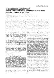
THE PARASITES <strong>OF</strong> THE <strong>PEARL</strong> <strong>OYSTER</strong>. 103EXPLANATION <strong>OF</strong> THE PLATES.PLATE I.Fig.1. Planarian-like free-living organism, possibly the larva of a species of Cestode, taken in theplankton on Muttuvaratu Paar, 19th November, 1902. It is most probably of the samespecies as the encysted larvae found so abundantly in the pearl oysters of the paar named.Actual length when elongated, 0-37 millim. The rudiment of what may be the proboscis isalready apparent, a-h, show various in specimens various attitudes; ex., calcareouscorpuscles; cut., the thick mucilaginous cuticular layer; Pr., possible rudiment of a proboscis.2. Three fragments a, b,and c of the distal or marginal region of the mantle of a pearl oyster,showing Cestode cysts scattered in the intermuscular connective-tissue. Natural size.:Mg.Pall., pallial margin Mg.vel., velar margin; T.cy., Cestode cysts.3. A Cestode cyst (cy.)in an interlamellar junction at the base of a branchia. x 3 diameters.4.Fragment of a branchia of a pearl oyster showing encysted larval Cestode x 12 diameters.(cy.).5. a. Nucleus of a "fine" pearl from the posteriorear region of a Cheval Paar oyster. Theproboscis sheath and the central pit within which the proboscis is retracted are clearly shown./.-. A " fine " pearl, showing distinct resemblance in outer form with the characteristicappearance of a Cestode larva (see fig. 5, i), viewed anteriorly ; c, the same viewed laterally ;d and e,natural size of the same. i. Outline of a young Cestode larva for comparison withthe preceding. /. An elongated pearl, also having resemblance in outline to a Tetrarhynchidlarva. h. Natural size of same. /. A lenticular pearl having an equatorial band of brownprismatic pearl-substance.6. Section through an encysted larva of about the same shape as that represented in fig.i. Therostrum or proboscis (a)is still retracted in the body.7. a and b, two of the later larval stagesof the Cestode met with, in encysted condition, in thetissues of the pearl oyster. Compare with fig.13. x 80 diameters.8. A. A partially calcified Cestode cyst from the muscular pallial region of a 3-year old oysterfrom the Muttuvaratu Paar. The larva was dead and slightly changed in outline partialdisintegration having taken place. In colour it was dirty yellow. In the outer layers (b) ofthe cyst capsule a deposit of lime salts had begun. B. Another cyst.b. The outer layers ofcyst c. Nucleus, obtained by decalcification, of a fine pearl from the muscular pallial region;of a pearl oyster from the Cheval Paar. C. Nucleus of another " fine " pearl from the ventralpallial region of a pearl oyster from the same locality as before ;a few calcareous corpusclesif.seen within the larva as solution brought the character of the nucleus into view. D. Theinnermost of the membraneous laminaj left after solution of the corresponding pearl coats ;c,a dead Cestode larva forming the actual nucleus; a ball-shapedCestode larva is seen. (Weight 195 milligrs.)calcification within the9.Very young Cestode larva extracted from a thick-walled cyst in the mantle of a pearl oyster.x 50 diameters. (Note. The outer layer is very faintly seen under a low power, and has anappearance closely resembling ciliation. The distal limit is not so strongly marked as shownhere, see fig. 10.) c.c, calcareous corpuscles; Pr., proboscis; Pr.sk, proboscis sheath... 10. Young Cestode larva extracted from a cyst within the 'mantle of a pearl oyster.the minute denticulation of the proboscis collar.x 25 diameters.Itshows.,11. The same, seen under slight pressure, whereby the proboscis, Pr., has been evaginated forcibly.Pr.sh., proboscis sheath.x 25 diameters.
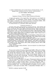
104 <strong>CEYLON</strong> <strong>PEARL</strong> <strong>OYSTER</strong> REPORT.Fig. 12. Another larva of the same stage, seen under slighter pressure, and higher magnification,x 40diameters. The vertically striated mucilaginous cuticle is clearly shown, together with a networkof anastomosing vessels. Cut., cuticle ;Ex. p., excretory pore.,, 13. Shows the beginning of the change from a spherical to an elongated form of body. Taken froma cyst from the viscero-pedal mass of the pearl oyster.,,14. Portion of same larva seen in optical section at region of proboscis collar, showing a fragment ofthe anastomosing network of vessels and the large bladder-like cells full of clear globules, lyingbeneath the dermal layer (semi-diagrammatic). The calcareous corpuscles are omitted for thesake of clearness. Den., denticles ; Par., sub-dermal bladder-like cells ; Pr.sh., proboscis sheath ;/"'*-.,anastomosing vessels.,,15. a. Optical section of body- wall of a Cestode larva from the pearl oyster, showingthe normalappearance./'. Final stage, when, under continual pressure, the mucilaginous cuticular layerhas disintegrated, its place being taken by one or two layers of the bladder-like cells, whichthus come to invest the body of the larva, c. Tho same as a under slight pressure ;aninvasion of the delicate cuticular layer by bladder-like cells full of clear globules is in progress,PLATE II.Fig. 1C. Forms of the calcareous corpuscles found in the larvae of Tetrarhynchus unionif actor.17. The same, enclosed in the cells that secrete them.,, 18. Form of denticles which ornament the proboscis collar of the early larva of TetrarhynchusUMonifactor.,, 19. The oldest larval stage of Tetrarhynchus unionifactor met with in the tissues of tho pearl oyster.Arm. cil., armature of stiff cilia around excretory pore; cs.,one of the two pairs of cephalicsuckers; Ex.v., external orifice of excretory system ; Pr., protractile proboscides; Tr.ex., lateraltrunks of excretory system.,,20. Surface view of the posterior extremity of the same individual on an enlarged scale.a. Shows the warted pattern of the whole surface of the body, except at the extremeposterior extremity, where, as shown (b), the surface marking becomes sinuous ;other lettersas in fig.19.21. A loop of the excretory network in the cephalic region of the same individual.22. Anterior extremity of an individual of the same stage as that depicted in fig.19 seen undergreater magnification. Pr.sh., sheath of proboscis ; Eet.m., retractor muscle of proboscis.,,23. One of the hooklets from a proboscis.24. A larval form of Tetrarhynchusbalistidis from the liver of a Batistes, sp.x 16 diameters.C.c, calcareous corpuscles; Tr.ex., excretory trunks. The natural size is shown by the smallfigure to the right.25. Sub-spherical larva of Tetrarhynchus balistidis found in oval and in rounded cystsbeneath theperitoneum of Batistes, sp. ; Pr.pro., proboscis protruded.,, 26. A slightly older example of the same.27. Natural size of a cyst of T. balistidis from abdominal cavity of a Batistes, sp.28. Cyst from peritoneum of Batistes, nat. size, containing T. pinnas.,, 29. The larval T. pinnce freed from cyst membrane. Nat. size.Figs. 30, 31, 32. The same, under higher magnification. C.c, calcareous corpuscles; Cy. mem., cystmembrane; Pr.ret., proboscides retracted ; Sc, scolex.Fig. 33. Teeth of T. balistidis, isolated by maceration and highly magnified.,,34. Teeth of T. balistidis in situ on the introvert.,, 35. More teeth of T, balistidis for comparison with fig. 36,
106 <strong>CEYLON</strong> <strong>PEARL</strong> <strong>OYSTER</strong> REPORT.Fig. 61. The same seen from the ventral aspect while crawling over a glass plate.The dotted outlinesa and b show the leech-like mobility of the anterior region of the body in attitudes assumedfrequently when the worm is restless, f.f., tube feet. The other letters as in fig. 60.62. View of the posterior extremity of the same, from above, showing the sucker-pits (s.p.) closedby the approximation of their lips.The fine transverse wrinkling characteristic of the dorsumof the trunk is also shown in the figure.63. Ciliated ectoparasite from gillsof pearl oyster with paired trilobed eyes.61. Another characteristic appearance of the same. A contractile vacuole is seen posteriorly and twotriradiate eye-spots anteriorly.65. Diagram of pearl oyster to show at a the position of the cyst enclosing MvsaUa hcrdmani, in thetissues of the pearl oyster. x .66. A portion of the pedal disc seen when an Aspidogasier margaritiferce is detached from its hold andturned uponits back. The edges of the disc then recurve inwards to varying degrees.67. Highly magnified view of the end of a proboscis of Tetrwrhynchus minimus, showing the varioussized hooks and their arrangement.68. Tube feet of Aspidogaster margaritiferce in various states of extension and retraction.69. a, b, c, successive attitudes assumed by .-/. margaritiferce during progression, d, another view ofthe same when about to change position.70. Head of Tetrwrhynchus minimus.71. Optical section of the same.72. Tetrarhynchus minimus, showing proglottides.Note. Owing to an error in the lithographer's office, Plates III. and IV. have beenprinted as 3 and 4, in Arabic numerals.
I'iARL<strong>CEYLON</strong> <strong>PEARL</strong> <strong>OYSTER</strong> REPORT PLATE1^^Vbd/viiHj Pall7'-.'/.J .,4/p*\ 12q'RASITES I<strong>OYSTER</strong>
. -<strong>CEYLON</strong> <strong>PEARL</strong> <strong>OYSTER</strong> REPORT PLATE 11Ex. o.! TER.
\<strong>CEYLON</strong> PEARI. <strong>OYSTER</strong> REJ PLATE 3.414643::,,1435156.ph52^..53.54.55.ex. p.J-H.iA.E.S.del.PARASITES <strong>OF</strong>-A.RL<strong>OYSTER</strong>.
.<strong>CEYLON</strong> <strong>PEARL</strong> <strong>OYSTER</strong> REPORT PLATE1ex.t-.s.p.62.J.H.tAi S.del.Wilson, Cambridge.