ApoTome - Carl Zeiss International
ApoTome - Carl Zeiss International
ApoTome - Carl Zeiss International

Create successful ePaper yourself
Turn your PDF publications into a flip-book with our unique Google optimized e-Paper software.
16<br />
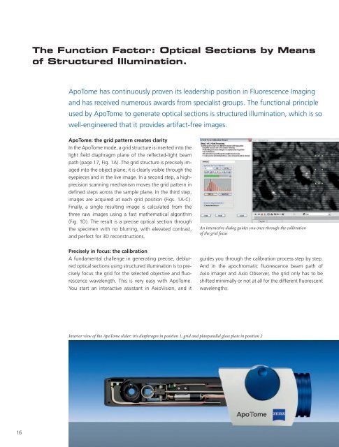
The Function Factor: Optical Sections by Means<br />
of Structured Illumination.<br />
<strong>ApoTome</strong> has continuously proven its leadership position in Fluorescence Imaging<br />
and has received numerous awards from specialist groups. The functional principle<br />
used by <strong>ApoTome</strong> to generate optical sections is structured illumination, which is so<br />
well-engineered that it provides artifact-free images.<br />
<strong>ApoTome</strong>: the grid pattern creates clarity<br />
In the <strong>ApoTome</strong> mode, a grid structure is inserted into the<br />
light field diaphragm plane of the reflected-light beam<br />
path (page 17, Fig. 1A). The grid structure is precisely imaged<br />
into the object plane; it is clearly visible through the<br />
eyepieces and in the live image. In a second step, a highprecision<br />
scanning mechanism moves the grid pattern in<br />
defined steps across the sample plane. In the third step,<br />
images are acquired at each grid position (Figs. 1A-C).<br />
Finally, a single resulting image is calculated from the<br />
three raw images using a fast mathematical algorithm<br />
(Fig. 1D). The result is a precise optical section through<br />
the specimen with no blurring, with elevated contrast,<br />
and perfect for 3D reconstructions.<br />
Precisely in focus: the calibration<br />
A fundamental challenge in generating precise, deblurred<br />
optical sections using structured illumination is to precisely<br />
focus the grid for the selected objective and fluorescence<br />
wavelength. This is very easy with <strong>ApoTome</strong>.<br />
You start an interactive assistant in AxioVision, and it<br />
An interactive dialog guides you once through the calibration<br />
of the grid focus<br />
guides you through the calibration process step by step.<br />
And in the apochromatic fluorescence beam path of<br />
Axio Imager and Axio Observer, the grid only has to be<br />
shifted minimally or not at all for the different fluorescent<br />
wavelengths.<br />
Interior view of the <strong>ApoTome</strong> slider: iris diaphragm in position 1, grid and planparallel glass plate in position 2