IVUS AND OCT
IVUS and OCT
IVUS and OCT
- No tags were found...

Create successful ePaper yourself
Turn your PDF publications into a flip-book with our unique Google optimized e-Paper software.
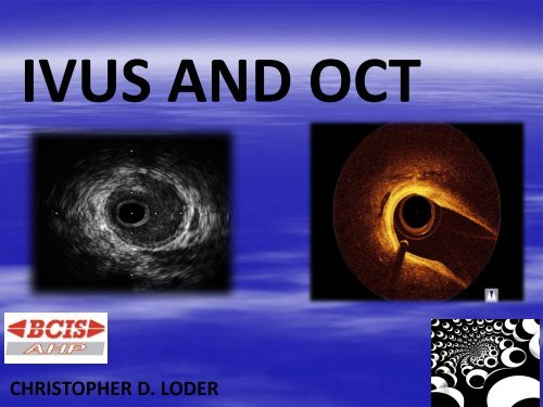
<strong>IVUS</strong> <strong>AND</strong> <strong>OCT</strong><br />
CHRISTOPHER D. LODER
• Ultrasound waves have a frequency<br />
of >20 000 cycles per second (i.e.<br />
above the audible range). For<br />
medical imaging we use frequencies<br />
in the range of millions of cycles per<br />
second (megahertz = MHz)
Frequency<br />
• Obstetric US 3-5MHz<br />
• Echo ~ 3.5MHz<br />
• TOE 5-7 MHz<br />
• Musculo-Skeletal US ~<br />
12MHz<br />
• <strong>IVUS</strong> 40MHz
What is a transducer?<br />
• Transducers are devices that converts one<br />
type of energy into another<br />
• <strong>IVUS</strong> transducers convert electrical energy<br />
into ultrasound energy and returning<br />
ultrasound energy back into electrical energy<br />
• This is then converted into a grey scale planar<br />
image
TWO TYPES<br />
• Mechanical<br />
Single element transducer<br />
rotated mechanically on<br />
a drive shaft to create a<br />
tomographic image<br />
• Solid State<br />
Cylindrical array of<br />
transducers firing<br />
sequentially in a 360 arc<br />
to produce a tomographic<br />
image
<strong>IVUS</strong> Transducers<br />
Mechanical Transducer – 40 MHz Atlantis Pro (BosSci)<br />
Solid-State Transducer – 25 MHz EagleEye (Volcano)
INDICATIONS<br />
• Left Main<br />
•Planning PCI<br />
• Instent restenosis (ISR)<br />
• Post stent analysis<br />
• Bifurcations<br />
•Ambiguous appearances
What is normal ?
Artery wall anatomy
Cross section of normal coronary artery<br />
Adventitia is composed of<br />
collagen that is highly reflected<br />
by ultrasound (appears white)<br />
Media is made of<br />
homogeneous smooth muscle<br />
cells and is not reflected by<br />
ultrasound (appears dark)<br />
Intima is dense and will<br />
appear as a “white” layer<br />
between the media and the<br />
blood speckles.<br />
14<br />
Confidential information of Boston Scientific Corporation. Do not copy or distribute.
• CSA Cross Sectional Area<br />
• EEM External Elastic Membrane<br />
• P&M Plaque and Media<br />
• NURD Non-Uniform Rotational Distortion<br />
• MaxLD Maximum Lumen Diameter<br />
• MLD Minimum Lumen Diameter<br />
• L Mode Longitudinal view (sagital)<br />
• PRD Proximal Reference Diameter<br />
• DRD Distal Reference Diameter
CSA Cross sectional area
L-Mode (Longitudinal)<br />
LLL MODE<br />
MODE
Wire shadow<br />
Visualization of the wire<br />
shadow in the lumen area.<br />
19<br />
Confidential information of Boston Scientific Corporation. Do not copy or distribute.
Normal / Diseased<br />
Normal<br />
Diseased<br />
20<br />
Confidential information of Boston Scientific Corporation. Do not copy or distribute.
Soft Plaque / Fibrotic Plaque<br />
Soft Plaque<br />
Not as bright as the<br />
adventitia (hypoechoic):<br />
“Soft” refers to<br />
echogenicity, due to<br />
high lipid content<br />
21<br />
Confidential information of Boston Scientific Corporation. Do not copy or distribute.
Calcium<br />
small<br />
22<br />
Confidential information of Boston Scientific Corporation. Do not copy or distribute.
Calcium<br />
medium<br />
23<br />
Confidential information of Boston Scientific Corporation. Do not copy or distribute.
Calcium NAPKIN RING
Stent Struts<br />
25<br />
Confidential information of Boston Scientific Corporation. Do not copy or distribute.
Stent…underdeployed<br />
26<br />
Confidential information of Boston Scientific Corporation. Do not copy or distribute.
Angiography versus <strong>IVUS</strong><br />
ANGIOGRAPHY<br />
<strong>IVUS</strong><br />
2 dimensional<br />
Planar<br />
Shadow of lumen<br />
Wall structures not imaged<br />
Intermittent snapshots or repeat<br />
contrast injections necessary<br />
QCA measurements prone to<br />
magnification errors<br />
360º view<br />
Tomographic and sagittal<br />
Visualisation of shape and location<br />
Visualisation of inner wall structures<br />
and morphology<br />
Continuous image<br />
Precise measurements
QCA =61%
LAD
Statistical recommendations
LMS ≤ 6mm²<br />
Ostial LAD<br />
Ostial<br />
LAD & LCX<br />
≤4mm²<br />
Left main<br />
Ostial<br />
LCX
72YO Male UA Diabetic, Hypertensive, CRF,<br />
Hypercholesterolaemia
<strong>OCT</strong><br />
OPTICAL COHERENCE TOMOGRAPHY
<strong>OCT</strong> USES LIGHT<br />
Light emitted by the<br />
optical fiber is reflected<br />
back by different types of<br />
tissue<br />
The system measures the<br />
time delay of the<br />
reflected light waves<br />
An <strong>OCT</strong> image is<br />
generated showing<br />
vessel anatomy and<br />
tissue microstructure
SHORT MONORAIL WIRE MOUNTED<br />
CATHETER
<strong>OCT</strong> PHYSICS<br />
<strong>OCT</strong> systems incorporate near-infrared light sources and optical<br />
components that operate in a wavelength band centred around<br />
1310nm. <strong>OCT</strong> measures the depth of reflections from tissue<br />
according to the round-trip propagation time of reflected energy. An<br />
interferometer is required to measure the backscattered light. The<br />
image is formed by the backscattering from the vessel wall or the<br />
time it takes for emitted light to travel between the target tissue<br />
and back to the lens, producing an “echo time delay” with a<br />
measurable signal intensity or magnitude.
As blood has a high amount of Iron in it (haemoglobin), light is unable to<br />
pass through it well.<br />
Because of this, it is imperative that the vessel is cleared of blood, and<br />
this is done by (traditionally) injecting contrast or 50/50.
WHATS NEXT?