Cytoline 1 and Cytoline 2 Macroporous microcarriers
Cytoline 1 and Cytoline 2 Macroporous microcarriers
Cytoline 1 and Cytoline 2 Macroporous microcarriers
- No tags were found...
You also want an ePaper? Increase the reach of your titles
YUMPU automatically turns print PDFs into web optimized ePapers that Google loves.
data file<br />
Cell culture<br />
<strong>Cytoline</strong> 1 <strong>and</strong> <strong>Cytoline</strong> 2<br />
<strong>Macroporous</strong> <strong>microcarriers</strong><br />
<strong>Cytoline</strong> <strong>microcarriers</strong> are designed for use in packed,<br />
fluidized bed reactors for the culture of CHO <strong>and</strong> suspension<br />
cells intended for the production of recombinant proteins for<br />
therapeutic use. <strong>Cytoline</strong> <strong>microcarriers</strong> are macroporous <strong>and</strong><br />
have a matrix based on polyethylene which is weighted with<br />
silica. The <strong>microcarriers</strong> yield high cell concentrations which<br />
can be sustained over long periods of time.<br />
• <strong>Macroporous</strong> <strong>microcarriers</strong><br />
• High density cell culture<br />
• Designed for fluidized bed cultures<br />
<strong>Macroporous</strong> <strong>microcarriers</strong><br />
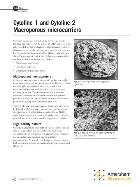
Cells gain easy access to the interior of <strong>Cytoline</strong> due to the<br />
macroporous structure of the <strong>microcarriers</strong> (Figure 1). Inside<br />
<strong>Cytoline</strong>, cells are protected from both physical <strong>and</strong><br />
environmental stresses, <strong>and</strong> are able to create their own<br />
micro-environment. This micro-environment increases<br />
metabolic communication between the cells <strong>and</strong> permits<br />
protein-free perfusion, which in turn decreases media costs,<br />
particularly in long-term continuous processes.<br />
Fig. 1. Scanning electron micrograph of<br />
<strong>Cytoline</strong> 1.<br />
The polyethylene base matrix causes the <strong>microcarriers</strong> to be<br />
hydrophobic while the silica content gives <strong>Cytoline</strong> a slight<br />
negative charge. <strong>Cytoline</strong> is further primed by an NaOH<br />
wash during preparation to optimize performance, the effects<br />
of which are particularly noticeable in protein-free media.<br />
High density culture<br />
<strong>Cytoline</strong> <strong>microcarriers</strong> offer both an external surface <strong>and</strong> an<br />
interior space which can be populated by anchoragedependent<br />
cells or cells grown in suspension. Cell retention<br />
during perfusion is improved due to metabolic<br />
communication. As a result, cell density per reactor volume is<br />
high in contrast to when conventional <strong>microcarriers</strong> are used<br />
(Figure 2).<br />
Fig. 2. Close-up reveals the tight clustering of<br />
CHO cells on <strong>Cytoline</strong> 1.<br />
df<br />
18-1060-65 AC, 2002-09 • p1
Cell culture<br />
Designed for fluidized bed culture<br />
<strong>Cytoline</strong> 1 is optimized for the culture of CHO cells in<br />
fluidized beds. CHO cells attach well to <strong>Cytoline</strong> 1 <strong>and</strong> final<br />
cell densities are very high. These high densities are possible<br />
because of the high sedimentation rate of <strong>Cytoline</strong> 1, 120 to<br />
220 cm/min. A high sedimentation rate enables the use of a<br />
high circulation rate which ensures an adequate supply of<br />
oxygen to the reactor in order to maintain the high cell<br />
concentration.<br />
<strong>Cytoline</strong> 2 is primarily intended for the culture of<br />
hybridomas (Figure 3) <strong>and</strong> other stress-sensitive cells in<br />
packed fluidized beds. (<strong>Cytoline</strong> 2 supports a lower density<br />
of cells than <strong>Cytoline</strong> 1.) The sedimentation rate for <strong>Cytoline</strong><br />
2 is 25 to 75 cm/min, which means that the circulation rate is<br />
lower <strong>and</strong> shear stress on the cells is less. The lower<br />
sedimentation rate also allows <strong>Cytoline</strong> 2 to be used in<br />
stirred cultures.<br />
Good adhesion during long periods<br />
of culture<br />
When culturing recombinant CHO cells using Cytodex <br />
<strong>microcarriers</strong>, the cells occasionally have a tendency to peel<br />
off the <strong>microcarriers</strong> after about 10 days in culture. When<br />
using <strong>Cytoline</strong> to culture CHO cells, there is no sign of cell<br />
detachment even after more than 60 days in culture.<br />
Characteristics of <strong>Cytoline</strong> <strong>microcarriers</strong><br />
• Steam sterilizable (in situ) 121 °C, 1 bar<br />
• Alkali <strong>and</strong> acid resistant<br />
• Lot-to-lot consistency<br />
• No material of biological origin is used<br />
• Suitable for immobilization <strong>and</strong> growth of adherent <strong>and</strong> nonadherent<br />
cell types<br />
• Suitable for bioreactor systems that are in use in industrial<br />
environments, such as stirred tanks, airlift culture systems <strong>and</strong><br />
fluidized beds<br />
• Increases culture surface area when used in routine laboratory<br />
culture techniques, such as roller, spinner culture or shaker<br />
flasks<br />
• Lens-shape advantageous for nutrient /oxygen supply diffusion<br />
• Storage in the dark gives optimal shelf-life<br />
Specifications of <strong>Cytoline</strong><br />
<strong>Cytoline</strong> 1 <strong>Cytoline</strong> 2<br />
Fig. 3. Human hybridoma cells cultured within<br />
<strong>Cytoline</strong> 2.<br />
Sedimentation velocity (cm/min) 120–220 25–75<br />
Length (mm) 1.7–2.5 1.7–2.5<br />
Thickness (mm) 0.4–1.1 0.4–1.1<br />
Density (g/cm 3 ) 1.32 1.03<br />
Pore size (µm) 10–400 10–400<br />
Surface area (m 2 /g) >0.3 >0.1<br />
df<br />
18-1060-65 AC, 2002-09 • p2
Cell culture<br />
Preparing <strong>Cytoline</strong><br />
– General washing procedure<br />
1 To the desired volume of <strong>Cytoline</strong> (1 or 2), add twice the<br />
amount of distilled water <strong>and</strong> autoclave the mixture for<br />
10 min at 121 °C (1 bar). The maximum temperature the<br />
<strong>microcarriers</strong> can withst<strong>and</strong> is 121 °C.<br />
2 Sieve the mixture to remove the water supernatant, then<br />
add twice the amount of fresh distilled water.<br />
3 Stir the microcarrier/water suspension for 10 min.<br />
4 Repeat 3 times steps 2 <strong>and</strong> 3.<br />
5 Add an equal volume of 0.1 M NaOH to the <strong>microcarriers</strong><br />
<strong>and</strong> incubate this mixture overnight.<br />
6 Wash 3 or 4 times with distilled water until all alkali is<br />
removed (check the pH of the wash water).<br />
7 Autoclave the <strong>microcarriers</strong> in distilled water or PBS at<br />
121 °C (1 bar) for 30 min.<br />
– Testing the microcarrier<br />
1 Transfer 2 ml of dry <strong>Cytoline</strong> to either 125 ml spinner flask<br />
or 100 ml shaker flask.<br />
2 Add 10 ml of distilled water <strong>and</strong> autoclave the mixture for<br />
10 min at 121 °C.<br />
3 Sieve the mixture to remove the water supernatant, then<br />
add 10 ml of distilled water.<br />
4 Stir the microcarrier/water suspension for 10 min.<br />
5 Repeat this procedure 3 times.<br />
6 Add 10 ml of 0.1 M NaOH to the <strong>microcarriers</strong> <strong>and</strong><br />
incubate this mixture overnight.<br />
7 Remove alkali using the same wash procedure as outlined<br />
above.<br />
8 The final wash should have a pH below 8.5 or close to that<br />
of the added distilled water.<br />
9 The <strong>microcarriers</strong> are now ready to be autoclaved.<br />
NOTE. Do not use temperatures higher than 121 °C (1 bar).<br />
Never autoclave the carriers without any liquid.<br />
Washing of the <strong>microcarriers</strong> inside the fluidized bed reactor<br />
is described in the User Manual for Cytopilot Mini<br />
(Code No. 18-1114-41).<br />
df<br />
18-1060-65 AC, 2002-09 • p3
Cell culture<br />
Ordering information<br />
Designation Pack size Code No.<br />
<strong>Cytoline</strong> 1 50 ml 17-1268-01<br />
<strong>Cytoline</strong> 1 500 ml 17-1268-02<br />
<strong>Cytoline</strong> 1 5 litres 17-1268-03<br />
<strong>Cytoline</strong> 2 50 ml 17-1269-01<br />
<strong>Cytoline</strong> 2 500 ml 17-1269-02<br />
<strong>Cytoline</strong> 2 5 litres 17-1269-03<br />
to order:<br />
Asia Pacific Tel: +852 2811 8693 Fax: +852 2811 5251 Australasia Tel: +61 2 9899 0999 Fax: +61 2 9899 7511 Austria Tel: 01 576 0616 20 Fax: 01 576 0616 27 Belgium Tel: 0800 73 888 Fax: 03 272 1637<br />
Canada Tel: 1 800 463 5800 Fax: 1 800 567 1008 Central, East, South East Europe Tel: +43 1 982 3826 Fax: +43 1 985 8327 Denmark Tel: 45 16 2400 Fax: 45 16 2424 Finl<strong>and</strong> & Baltics Tel: +358 (0)9 512 3940<br />
Fax: +358 (0)9 512 1710 France Tel: 0169 35 67 00 Fax: 0169 41 9677 Germany Tel: 0761 4903 401 Fax: 0761 4903 405 Italy Tel: 02 27322 1 Fax: 02 27302 212 Japan Tel: 81 3 5331 9336 Fax: 81 3 5331 9370 Latin<br />
America Tel: +55 11 3933 7300 Fax: +55 11 3933 7315 Middle East <strong>and</strong> Africa Tel: +30 (1) 96 00 687 Fax: +30 (1) 96 00 693 Netherl<strong>and</strong>s Tel: 0165 580 410 Fax: 0165 580 401 Norway Tel: 2318 5800 Fax: 2318 6800<br />
Portugal Tel: 21 417 7035 Fax: 21 417 3184 Russia & other C.I.S. & N.I.S. Tel: +7 (095) 232 0250,956 1137 Fax: +7 (095) 230 6377 South East Asia Tel: 60 3 8024 2080 Fax: 60 3 8024 2090 Spain Tel: 93 594 49 50<br />
Fax: 93 594 49 55 Sweden Tel: +46 18 612 19 00 Fax: +46 18 612 19 10 Switzerl<strong>and</strong> Tel: 01 802 81 50 Fax: 01 802 81 51 UK Tel: 0800 616 928 Fax: 0800 616 927 USA Tel: +1 800 526 3593 Fax: +1 877 295 8102<br />
<strong>Cytoline</strong> <strong>and</strong> Cytodex are trademarks of Amersham Biosciences Limited. Amersham <strong>and</strong> Amersham Biosciences are trademarks of Amersham plc. Cytopilot is a trademark of Vogelbusch GmbH. Amersham Biosciences<br />
AB Björkgatan 30, SE-751 84 Uppsala, Sweden. Amersham Biosciences UK Limited Amersham Place, Little Chalfont, Buckinghamshire HP7 9NA, Engl<strong>and</strong>. Amersham Biosciences Corporation 800 Centennial Avenue,<br />
PO Box 1327, Piscataway, NJ 08855 USA. Amersham Biosciences Europe GmbH Munzinger Strasse 9, D-79111 Freiburg, Germany. Amersham Biosciences KK Sanken Building, 3-25-1, Hyakunincho, Shinjuku-ku,<br />
Tokyo 169-0073, Japan. All goods <strong>and</strong> services are sold subject to the terms <strong>and</strong> conditions of sale of the company within the Amersham Biosciences group that supplies them. A copy of these terms <strong>and</strong> conditions<br />
is available on request. © Amersham Biosciences AB 2002 – All rights reserved.<br />
Produced by Wikströms, Sweden 1021091, 09.2002<br />
Printed matter. Licence 341 051<br />
df<br />
18-1060-65 AC, 2002-09 • p4


