Nerve Supply to the Heart / SA & AV Nodes / EKG
Nerve Supply to the Heart / SA & AV Nodes / EKG
Nerve Supply to the Heart / SA & AV Nodes / EKG
- No tags were found...

Create successful ePaper yourself
Turn your PDF publications into a flip-book with our unique Google optimized e-Paper software.
Chapter 19<br />
<strong>Nerve</strong> <strong>Supply</strong> <strong>to</strong> <strong>the</strong> <strong>Heart</strong> /<br />
<strong>SA</strong> & <strong>AV</strong> <strong>Nodes</strong> / <strong>EKG</strong>
<strong>Nerve</strong> <strong>Supply</strong> <strong>to</strong> <strong>Heart</strong> (1 of 3)<br />
Sinoatrial Node (<strong>SA</strong> node) and Atrioventricular Node<br />
(<strong>AV</strong> node) “leak sodium ions”<br />
do not maintain a “static” resting potential<br />
nodal cardiocytes <strong>to</strong> reach threshold first will<br />
depolarize and set <strong>the</strong> rate of depolarization for all<br />
<strong>the</strong> o<strong>the</strong>r myocardiocytes in <strong>the</strong> heart<br />
Sinoatrial Node = Pacemaker (leak sodium faster!)<br />
Atrioventricular Node = Secondary Pacemaker<br />
The rate of depolarization of <strong>the</strong>se nodes are<br />
“modified” by <strong>the</strong> au<strong>to</strong>nomic nervous system<br />
• Sympa<strong>the</strong>tic NS<br />
• Parasympa<strong>the</strong>tic NS
<strong>Nerve</strong> <strong>Supply</strong> <strong>to</strong> <strong>Heart</strong> – Sympa<strong>the</strong>tic <strong>Nerve</strong>s (2 of 3)<br />
• sympa<strong>the</strong>tic nerves<br />
– sympa<strong>the</strong>tic pathway <strong>to</strong> <strong>the</strong> heart originates in <strong>the</strong><br />
lower cervical <strong>to</strong> upper thoracic segments of <strong>the</strong><br />
spinal cord<br />
– continues <strong>to</strong> adjacent sympa<strong>the</strong>tic chain ganglia<br />
– some pass through cardiac plexus in mediastinum<br />
– continue as cardiac nerves <strong>to</strong> <strong>the</strong> heart<br />
– fibers terminate in<br />
• <strong>SA</strong> and <strong>AV</strong> nodes<br />
• in atrial and ventricular myocardium<br />
• coronary arteries (as well as <strong>the</strong> aorta, pulmonary trunk)<br />
– increase heart rate and contraction strength<br />
– dilates coronary arteries <strong>to</strong> increase myocardial blood<br />
flow
<strong>Nerve</strong> <strong>Supply</strong> <strong>to</strong> <strong>Heart</strong> (3 of 3)<br />
• parasympa<strong>the</strong>tic nerves<br />
– pathway begins with nuclei of <strong>the</strong> vagus nerves in<br />
<strong>the</strong> medulla oblongata<br />
– extend <strong>to</strong> cardiac plexus and continue <strong>to</strong> <strong>the</strong> heart by<br />
way of <strong>the</strong> cardiac nerves<br />
– fibers of right vagus nerve lead <strong>to</strong> <strong>the</strong> <strong>SA</strong> node<br />
– fibers of left vagus nerve lead <strong>to</strong> <strong>the</strong> <strong>AV</strong> node<br />
– little or no vagal stimulation of <strong>the</strong> myocardium<br />
• parasympa<strong>the</strong>tic stimulation reduces <strong>the</strong> heart rate<br />
– slows heart rate
Cardiac Rhythm<br />
• cycle of events in heart given special names<br />
– sys<strong>to</strong>le – atrial or ventricular contraction<br />
– dias<strong>to</strong>le – atrial or ventricular relaxation<br />
• sinus rhythm<br />
– normal heartbeat triggered by <strong>the</strong> <strong>SA</strong> node<br />
– set by <strong>SA</strong> node at 60 – 100 bpm<br />
– adult at rest is 70 <strong>to</strong> 80 bpm (vagal <strong>to</strong>ne)<br />
• ec<strong>to</strong>pic focus<br />
– caused by ano<strong>the</strong>r parts of heart that fires before <strong>SA</strong><br />
node discharges<br />
– caused by hypoxia, electrolyte imbalance, caffeine,<br />
nicotine, cocaine and o<strong>the</strong>r drugs<br />
– see next slide
Abnormal <strong>Heart</strong> Rhythms<br />
• spontaneous firing from some part of heart o<strong>the</strong>r than <strong>the</strong> <strong>SA</strong><br />
node<br />
– ec<strong>to</strong>pic foci<br />
• region of spontaneous firing<br />
• nodal rhythm – if <strong>SA</strong> node is damaged, heart rate is set by<br />
<strong>AV</strong> node, 40 <strong>to</strong> 50 bpm<br />
• intrinsic ventricular rhythm – if both <strong>SA</strong> and <strong>AV</strong> nodes are<br />
not functioning, rate set by o<strong>the</strong>r myocardiocytes at 20 <strong>to</strong> 40<br />
bpm<br />
– Arrhythmia<br />
– this requires artificial pacemaker <strong>to</strong> sustain life long term<br />
• any abnormal cardiac rhythm<br />
• failure of conduction system <strong>to</strong> transmit signals (heart block)<br />
• bundle branch block<br />
• <strong>to</strong>tal heart block (damage <strong>to</strong> <strong>AV</strong> node)
• atrial fibrillation<br />
– ec<strong>to</strong>pic foci in atria<br />
Cardiac Arrhythmias<br />
– atria beat 200 - 400 times per minute<br />
– May not be fatal<br />
• ventricular fibrillation<br />
– serious arrhythmia caused by electrical signals reaching different regions at<br />
widely different times<br />
– heart can’t pump blood and no coronary perfusion<br />
– kills quickly if not s<strong>to</strong>pped<br />
• defibrillation - strong electrical shock whose intent is <strong>to</strong> depolarize <strong>the</strong><br />
entire myocardium, s<strong>to</strong>p <strong>the</strong> fibrillation, and reset <strong>SA</strong> nodes <strong>to</strong> sinus<br />
rhythm<br />
• premature ventricular contractions (PVCs)<br />
– caused by stimulants, stress or lack of sleep
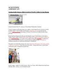
Pacemaker Physiology<br />
• <strong>SA</strong> node does not have a stable resting membrane potential<br />
– note <strong>the</strong>se cells are different than <strong>the</strong> typical myocyte!<br />
– starts at -60 mV and drifts upward from a slow inflow of Na +<br />
– gradual depolarization is called pacemaker potential<br />
– when reaches threshold of -40 mV<br />
• voltage-gated fast Ca 2+ and Na + channels open <strong>to</strong><br />
depolarize <strong>the</strong> cell (contraction)<br />
• faster depolarization occurs peaking at 0 mV<br />
• K + channels <strong>the</strong>n open and K + leaves <strong>the</strong> cell<br />
– causing repolarization (relaxation of cardiocyte)<br />
– once K + channels close, pacemaker potential starts over<br />
– each depolarization of <strong>the</strong> <strong>SA</strong> node sets off one heartbeat<br />
• at rest, fires every 0.8 seconds or 75 bpm<br />
• <strong>SA</strong> node is <strong>the</strong> system’s pacemaker
<strong>SA</strong> Node Potentials<br />
Copyright © The McGraw-Hill Companies, Inc. Permission required for reproduction or display.<br />
+10<br />
Membrane potential (mV)<br />
0<br />
–10<br />
–20<br />
–30<br />
–40<br />
–50<br />
–60<br />
–70<br />
Fast<br />
Ca 2+ –Na +<br />
inflow<br />
Slow Na +<br />
inflow<br />
Fast K +<br />
outflow<br />
Threshold<br />
Pacemaker<br />
potential<br />
Action<br />
potential<br />
0 .4 .8<br />
1.2 1.6<br />
Time (sec)
Impulse Conduction <strong>to</strong> Myocardium<br />
• signal from <strong>SA</strong> node stimulates two atria <strong>to</strong> contract almost<br />
simultaneously<br />
– reaches <strong>AV</strong> node in 50 msec<br />
• signal slows down through <strong>AV</strong> node<br />
– thin cardiocytes have fewer gap junctions<br />
– delays signal 100 msec which allows <strong>the</strong> ventricles <strong>to</strong> fill<br />
• signals travel very quickly through <strong>AV</strong> bundle and Purkinje<br />
fibers<br />
– entire ventricular myocardium depolarizes and contracts in<br />
near unison<br />
• papillary muscles contract an instant earlier than <strong>the</strong> rest,<br />
tightening slack in chordae tendineae<br />
• ventricular sys<strong>to</strong>le progresses up from <strong>the</strong> apex of <strong>the</strong> heart<br />
– spiral arrangement of cardiocytes twists ventricles slightly<br />
– like someone wringing out a <strong>to</strong>wel
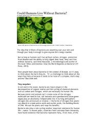
Cardiac Conduction System<br />
1<br />
<strong>SA</strong> node fires.<br />
Right atrium<br />
Sinoatrial node<br />
(pacemaker)<br />
Atrioventricular<br />
node<br />
Atrioventricular<br />
bundle<br />
2<br />
1<br />
3<br />
2<br />
4<br />
5<br />
Left<br />
atrium<br />
Purkinje<br />
fibers<br />
Bundle<br />
branches<br />
2<br />
3<br />
4<br />
5<br />
Excitation spreads through<br />
atrial myocardium.<br />
<strong>AV</strong> node fires.<br />
Excitation spreads down <strong>AV</strong><br />
bundle.<br />
Purkinje fibers distribute<br />
excitation through<br />
ventricular myocardium.<br />
Purkinje fibers
Electrical Behavior of Cardiocyte<br />
• cardiocytes have a stable resting potential of -90 mV<br />
• depolarize only when stimulated<br />
– depolarization phase (very brief)<br />
• stimulus opens voltage regulated Na + gates, (Na + rushes in)<br />
membrane depolarizes rapidly<br />
• action potential peaks at +30 mV<br />
• Na + gates close quickly<br />
– plateau phase lasts 200 <strong>to</strong> 250 msec, sustains<br />
contraction for expulsion of blood from heart<br />
• Ca 2+ channels are slow <strong>to</strong> close and SR is slow <strong>to</strong> remove<br />
Ca 2+ from <strong>the</strong> cy<strong>to</strong>sol<br />
– repolarization phase -Ca 2+ channels close, K +<br />
channels open, rapid diffusion of K + out of cell returns it<br />
<strong>to</strong> resting potential<br />
• has a long absolute refrac<strong>to</strong>ry period of 250 msec<br />
compared <strong>to</strong> 1 – 2 msec in skeletal muscle<br />
– prevents wave summation and tetanus which would s<strong>to</strong>p<br />
<strong>the</strong> pumping action of <strong>the</strong> heart
Action Potential of Myocardiocyte
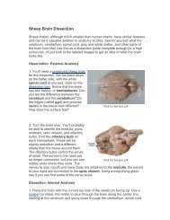
Electrocardiogram (ECG or <strong>EKG</strong>)<br />
• composite of all action potentials of nodal and<br />
myocardial cells<br />
• detected, amplified and recorded by electrodes on<br />
arms, legs and chest<br />
0.8 second<br />
R<br />
R<br />
+1<br />
Millivolts<br />
0<br />
P wave<br />
T wave<br />
PQ<br />
segment<br />
ST<br />
segment<br />
PR<br />
interval<br />
Q<br />
S<br />
QT<br />
interval<br />
QRS interval<br />
–1<br />
Atria<br />
contract<br />
Ventricles<br />
contract<br />
Atria<br />
contract<br />
Ventricles<br />
contract
ECG Deflections<br />
• P wave<br />
– <strong>SA</strong> node fires<br />
– atria depolarize<br />
0.8 second<br />
• PQ segment<br />
– atrial sys<strong>to</strong>le<br />
– atrial sys<strong>to</strong>le begins 100 msec after<br />
<strong>SA</strong> signal<br />
• QRS complex<br />
– ventricular depolarization<br />
– complex shape of spike due <strong>to</strong><br />
different thickness and shape of <strong>the</strong><br />
two ventricles<br />
Millivolts<br />
+1<br />
0<br />
P wave<br />
PR<br />
interval<br />
R<br />
Q<br />
S<br />
QT<br />
interval<br />
T wave<br />
PQ<br />
segment<br />
R<br />
ST<br />
segment<br />
QRS interval<br />
–1<br />
• ST segment<br />
– ventricular sys<strong>to</strong>le<br />
– plateau in myocardial action<br />
potential<br />
Atria<br />
contract<br />
Ventricles<br />
contract<br />
Atria<br />
contract<br />
Ventricles<br />
contract<br />
• T wave<br />
– ventricular repolarization and<br />
relaxation
Electrical Activity of Myocardium<br />
1) atrial depolarization<br />
begins<br />
2) atrial depolarization<br />
complete (atria<br />
contracted)<br />
Key<br />
Wave of<br />
depolarization<br />
Wave of<br />
repolarization<br />
Copyright © The McGraw-Hill Companies, Inc. Permission required for reproduction or display.<br />
P<br />
P<br />
R<br />
Q<br />
S<br />
1 Atria begin depolarizing.<br />
4 Ventricular depolarization complete.<br />
3) ventricles begin <strong>to</strong><br />
depolarize at apex; atria<br />
repolarize (atria relaxed)<br />
P<br />
P<br />
R<br />
Q<br />
S<br />
T<br />
4) ventricular depolarization<br />
complete (ventricles<br />
contracted)<br />
5) ventricles begin <strong>to</strong><br />
repolarize at apex<br />
2 Atrial depolarization complete.<br />
3<br />
Ventricular depolarization begins at apex<br />
and progresses superiorly as atria repolarize.<br />
P<br />
R<br />
Q<br />
5<br />
6<br />
Ventricular repolarization begins at apex<br />
and progresses superiorly.<br />
P<br />
R<br />
Q<br />
S<br />
Ventricular repolarization complete; heart<br />
is ready for <strong>the</strong> next cycle.<br />
T<br />
6) ventricular repolarization<br />
complete (ventricles<br />
relaxed)
Normal Electrocardiogram (ECG)<br />
Copyright © The McGraw-Hill Companies, Inc. Permission required for reproduction or display.<br />
0.8 second<br />
R<br />
R<br />
+1<br />
PQ<br />
segment<br />
ST<br />
segment<br />
Millivolts<br />
0<br />
P wave<br />
T wave<br />
PR<br />
interval<br />
Q<br />
S<br />
QT<br />
interval<br />
QRS interval<br />
–1<br />
Atria<br />
contract<br />
Ventricles<br />
contract<br />
Atria<br />
contract<br />
Ventricles<br />
contract
Diagnostic Value of ECG<br />
• abnormalities in conduction<br />
pathways<br />
• myocardial infarction<br />
• nodal damage<br />
• heart enlargement<br />
• electrolyte and hormone<br />
imbalances
ECGs: Normal and Abnormal<br />
Copyright © The McGraw-Hill Companies, Inc. Permission required for reproduction or display.<br />
• abnormalities in conduction<br />
pathways<br />
(a) Sinus rhythm (normal)<br />
• myocardial infarction<br />
• heart enlargement<br />
(b) Nodal rhythm—no <strong>SA</strong> node activity<br />
• electrolyte and hormone<br />
imbalances