marine microbial thiotrophic ectosymbioses - HYDRA-Institute
marine microbial thiotrophic ectosymbioses - HYDRA-Institute
marine microbial thiotrophic ectosymbioses - HYDRA-Institute

You also want an ePaper? Increase the reach of your titles
YUMPU automatically turns print PDFs into web optimized ePapers that Google loves.
2727_C04.fm Page 99 Wednesday, June 30, 2004 12:00 PM<br />
Marine Microbial Thiotrophic Ectosymbioses 99<br />
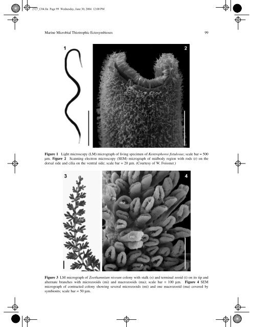
Figure 1 Light microscopy (LM) micrograph of living specimen of Kentrophoros fistulosus;<br />
scale bar = 500<br />
mm.<br />
Figure 2 Scanning electron microscopy (SEM) micrograph of midbody region with rods (r) on the<br />
dorsal side and cilia on the ventral side; scale bar = 20 mm. (Courtesy of W. Foissner.)<br />
Figure 3 LM micrograph of Zoothamnium niveum colony with stalk (s) and terminal zooid (t) on its tip and<br />
alternate branches with microzooids (mi) and macrozooids (ma); scale bar = 100 mm. Figure 4 SEM<br />
micrograph of contracted colony showing several microzooids (mi) and one macrozooid (ma) covered by<br />
symbionts; scale bar = 50 mm.