

In vivo delivery technique of nucleic acid compounds using ...
In vivo delivery technique of nucleic acid compounds using ...
In vivo delivery technique of nucleic acid compounds using ...

Create successful ePaper yourself
Turn your PDF publications into a flip-book with our unique Google optimized e-Paper software.
Takei and Kadomatsu: <strong>In</strong> <strong>vivo</strong> <strong>delivery</strong> <strong>technique</strong> <strong>of</strong> <strong>nucleic</strong> <strong>acid</strong> <strong>compounds</strong> <strong>using</strong> atelocollagen<br />
expression and then carried out treatment experiments<br />
<strong>using</strong> a subcutaneous tumor model in nude mice. We used<br />
atelocollagen, a biomaterial, for the <strong>delivery</strong> <strong>of</strong> antisense<br />
DNA into the tumor.<br />
II. Atelocollagen<br />
Gene introduction <strong>technique</strong>s aim to deliver gene<br />
expression vectors and antisense DNA to cells in the<br />
whole body or, specifically, to the targeted disease site at<br />
the optimal concentration and timing. For maximal<br />
therapeutic effects a biomaterial (biophilic substance) is<br />
most <strong>of</strong>ten used as a carrier (Ochiya et al, 2001). Recently,<br />
novel bio-introduction <strong>technique</strong>s <strong>using</strong> biomaterials<br />
combined with <strong>nucleic</strong> <strong>acid</strong> <strong>compounds</strong>, which are<br />
required for the conventional drug <strong>delivery</strong> system (DDS)<br />
and successful gene therapy, have been developed. The<br />
following biomaterials have been reported to be effective<br />
for gene introduction: polyethylene vinyl co-acetate<br />
(EVAc, Luo et al, 1999), poly(lactide-co-glycolide)<br />
(PLCG, Shea et al, 1999), gene activated matrix (GAM,<br />
Bonadio et al, 1999), poly[alpha-(4-aminobutyl)-Lglycolic<br />
<strong>acid</strong>] (PAGA, Lim et al, 2000), imidazolecontaining<br />
polymer (Pack et al, 2000), alginate<br />
microsphere (Mittal et al, 2001), chitosan (Roy et al,<br />
1999), gelatin (Truong-Le et al, 1999), PLA-DX-PEG<br />
(Saito et al, 2001) and atelocollagen (Ochiya et al, 1999).<br />
We here introduce a <strong>technique</strong> to deliver <strong>nucleic</strong> <strong>acid</strong><br />
<strong>compounds</strong> to tissue in <strong>vivo</strong> <strong>using</strong> atelocollagen. The<br />
collagen molecule forms a helix consisting <strong>of</strong> three<br />
polypeptide chains and exhibits a stick-like structure, 300<br />
nm in length and 1.5 nm in diameter. This molecule has<br />
telopeptides at both ends, which account for most <strong>of</strong> the<br />
antigenicity <strong>of</strong> collagen. It becomes atelocollagen and<br />
loses most <strong>of</strong> its antigenicity after removal <strong>of</strong> the<br />
telopeptides by pepsin digestion (Fujioka et al, 1995). The<br />
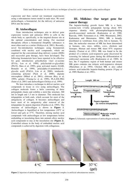
structure <strong>of</strong> atelocollagen is shown in Figure 1.<br />
Atelocollagen is liquidified at low temperature (10°C or<br />
lower) and gels at 37°C. Therefore, mixing <strong>nucleic</strong> <strong>acid</strong><br />
<strong>compounds</strong> with atelocollagen at low temperature before<br />
embedding or inoculating them into animals allow <strong>nucleic</strong><br />
<strong>acid</strong> <strong>compounds</strong> to stay at the inoculation site (Figure 2).<br />
Controlled release in the body <strong>of</strong> embedded <strong>nucleic</strong> <strong>acid</strong><br />
<strong>compounds</strong> is achieved by digestion with collagenase.<br />
258<br />
III. Midkine: Our target gene for<br />
cancer therapy<br />
The heparin-binding growth factor MK is a basic,<br />
cysteine-rich 13-kDa polypeptide having about 50%<br />
sequence identity with pleiotrophin/heparin-binding<br />
growth-associated molecule (Kadomatsu et al, 1988;<br />
Rauvala, 1989; Tomomura et al, 1990; Muramatsu, 2002;<br />
Kadomatsu and Muramatsu, 2004). MK is broadly<br />
distributed in vertebrates from zebra fish to humans. To<br />
date, the molecular structure <strong>of</strong> MK has been determined<br />
in humans, rats, mice, rabbits, cows, chickens and<br />
Xenopus. Human and mouse MK share 87% sequence<br />
identity (Tsutsui et al, 1991). MK was found to be the<br />
product <strong>of</strong> a retinoic <strong>acid</strong>-responsive gene discovered by<br />
screening for induced genes during the differentiation <strong>of</strong><br />
embryonal carcinoma cells (Kadomatsu et al, 1989). <strong>In</strong><br />
fact, the 5’-regulatory region <strong>of</strong> both human and mouse<br />
MK genes contains one retinoic <strong>acid</strong>-responsive element<br />
(Matsubara et al, 1994). Chicken MK is also called<br />
retinoic <strong>acid</strong>-inducible heparin-binding protein (Vigny et<br />
al, 1989; Raulais et al, 1991).<br />
Figure 1. Schematic representation <strong>of</strong> atelocollagen molecule.<br />
Figure 2. <strong>In</strong>jection <strong>of</strong> atelocollagen/<strong>nucleic</strong> <strong>acid</strong> compound complex in artificial subcutanerous tumors.


