

01. Gene therapy Boulikas.pdf - Gene therapy & Molecular Biology
01. Gene therapy Boulikas.pdf - Gene therapy & Molecular Biology
01. Gene therapy Boulikas.pdf - Gene therapy & Molecular Biology

Create successful ePaper yourself
Turn your PDF publications into a flip-book with our unique Google optimized e-Paper software.
<strong>Gene</strong> Ther Mol Biol Vol 1, 1-172. March, 1998.<br />
<strong>Gene</strong> Therapy and <strong>Molecular</strong> <strong>Biology</strong> Vol 1, page 1<br />
Status of gene <strong>therapy</strong> in 1997: molecular<br />
mechanisms, disease targets, and clinical<br />
applications<br />
Teni <strong>Boulikas</strong><br />
Institute of <strong>Molecular</strong> Medical Sciences, 460 Page Mill Road, Palo Alto, California 94306<br />
and Regulon Inc., 249 Matadero Avenue, Palo Alto, CA 94306<br />
__________________________________________________________________________________________________<br />
Correspondence: Teni <strong>Boulikas</strong>, Regulon Inc., 249 Matadero Avenue, Palo Alto, CA 94306, Tel (650) 813-9264, Fax: (650) 424-9594,<br />
E-mail: <strong>Boulikas</strong>@Worldnet.att.net<br />
Key words: gene <strong>therapy</strong>, gene transfer, clinical trials, cancer, immuno<strong>therapy</strong>, p53, adenovirus, retrovirus, adeno-associated virus,<br />
HIV-1, HSV-1, EBV, AIDS, tumor vaccines, IFN-γ, TNF-α, VEGF, retinoblastoma, purine nucleoside phosphorylase, HSV-tk, E1A,<br />
E1B, Cre, LoxP, recombination, HIV vectors, liposomes, fusogenic peptides, plasmovirus, transcription factor, TIL, IL-2, IL-3, IL-7, IL-<br />
12, GM-CSF, prostate cancer, p21, p16, apoptosis, Bcl-2, Bax, Bcl-xs, E2F, bystander effect, MDR1, IGF-I, antisense, triplex DNA,<br />
Parkinson’s disease, lysosomal storage disease, hemophilia, cystic fibrosis, CFTR, rheumatoid arthritis, hypertension, familial<br />
hypercholesterolemia, LDL, angiopoietin, restenosis, angiogenesis, TGF-β, arterial injury, atherosclerosis, ADA deficiency, obesity,<br />
leptin.<br />
Summary<br />
<strong>Gene</strong> <strong>therapy</strong> is a newly emerging field of biomedical research aimed at introducing therapeutically important<br />
genes into somatic cells of patients; a new and revolutionary era in molecular medicine has begun. Diseases already<br />
shown to be amenable to <strong>therapy</strong> with gene transfer in clinical trials include cancer (melanoma, breast, lymphoma,<br />
head & neck, ovarian, colon, prostate, brain, chronic myelogenous leukemia, non-small cell lung, lung<br />
adenocarcinoma, colorectal, neuroblastoma, glioma, glioblastoma, astrocytoma, and others), AIDS, cystic fibrosis,<br />
adenosine deaminase deficiency, cardiovascular diseases (restenosis, familial hypercholesterolemia, peripheral<br />
artery disease), Gaucher disease, α1-antitrypsin deficiency, rheumatoid arthritis and a few others. Human diseases<br />
expected to be the object of clinical trials include hemophilia A and B, Parkinson’s disease, ocular diseases,<br />
xeroderma pigmentosum, high blood pressure, obesity and many others. The establishment of novel animal models<br />
for human disease, the discovery of new genes, and improvements in successful gene delivery open bright new<br />
prospects for molecular medicine. A wide variety of delivery vehicles for genes have been tested including murine<br />
retroviruses, recombinant adenoviral vectors, adeno-associated virus, HSV, EBV, HIV vectors, and baculovirus.<br />
Nonviral gene delivery methods use cationic or neutral liposomes, direct injection of plasmid DNA, and polymers.<br />
Various strategies to enhance efficiency of gene transfer have been tested such as fusogenic peptides in combination<br />
with liposomes, or polymers, to enhance the release of plasmid DNA from endosomes. Recombinant retroviruses<br />
stably integrate into the DNA and require host DNA synthesis; adenoviruses can infect nondividing cells but cause<br />
immune reactions leading to the elimination of therapeutically transduced cells. Adeno-associated virus (AAV) is<br />
not pathogenic, does not elicit immune responses but new strategies are required to obtain high AAV titers for<br />
preclinical and clinical studies. Wild-type AAVs integrate into chromosome 19 whereas recombinant AAVs are<br />
deprived of site-specific integration and may also persist episomally; HSV vectors can infect nonreplicating cells<br />
such as neuron cells, have a high payload capacity for foreign DNA but inflict cytotoxic effects. It seems that each<br />
delivery system will be developed independently of the others and that each will prove its strengths for specific<br />
applications. At present, retroviruses are most commonly used in human clinical trials followed by adenoviruses,<br />
cationic liposomes and AAV. Polymer-encapsulated syngeneic or allogeneic cells implanted into a tissue of a patient<br />
can be used to secrete therapeutic proteins; the method is in trials for amyotrophic lateral sclerosis using the ciliary<br />
neurotrophic factor gene, and can be extended to Factor VIII and IX for hemophilia, interleukin genes, dopamine-<br />
1
<strong>Boulikas</strong>: An overview on gene <strong>therapy</strong><br />
secreting cells to treat Parkinson's disease, nerve growth factor for Alzheimer's disease and other diseases.<br />
Ingenious techniques under development with great future prospects for human gene <strong>therapy</strong>, include the Cre-<br />
LoxP recombinase system to rid of undesirable viral DNA sequences used for gene transfer, use of tissue-specific<br />
promoters to express a gene in a particular cell type or use of ligands, such as peptides selected from random<br />
peptide libraries, recognizing surface molecules to direct the gene vehicle to a particular cell type, designing p53<br />
“gene bombs” that explode into tumor cells, exploit the HIV-1 virus to engineer vectors for gene transfer, the<br />
combining of viruses with polymers or cationic lipids to improve gene transfer, the attachment of nuclear<br />
localization signal peptides to oligonucleotides to direct them to nuclei, and the invention of molecular switch<br />
systems allowing genes to be turned on or off at will.<br />
Although many human tumors are non- or weakly immunogenic, the immune system can be reinforced and<br />
instructed to eliminate cancer cells after transduction of patient’s cells ex vivo with the cytokine genes GM-CSF, IL-<br />
12, IL-2, IL-4, IL-7, IFN-γ, and TNF-α, followed by cell vaccination of the patient (e.g. intradermally) to potentiate<br />
T-lymphocyte-mediated antitumor effects (cancer immuno<strong>therapy</strong>). DNA vaccination with genes encoding tumor<br />
antigens and immuno<strong>therapy</strong> with synthetic tumor peptide vaccines are further developments in this exciting field.<br />
The genes used for cancer gene <strong>therapy</strong> in human clinical trials include a number of tumor suppressor genes (p53,<br />
RB, BRCA1, E1A), antisense oncogenes (antisense c-fos, c-myc, K-ras), and suicide genes (HSV-tk, in combination<br />
with ganciclovir, cytosine deaminase in combination with 5-fluorocytosine). Important in gene <strong>therapy</strong> are also the<br />
genes of bcl-2, MDR-1, p21, p16, bax, bcl-xs, E2F, IGF-I VEGF, angiostatin, CFTR, LDL-R, TGF-β, and leptin.<br />
Reports on human clinical trials using adenoviral and retroviral injections of the p53 gene have been very<br />
encouraging; future directions might go toward the use of genes involved in the control of tumor progression and<br />
metastasis. The molecular mechanisms of carcinogenesis have been largely elucidated and improvements in gene<br />
delivery methods are likely to lead to the final victory of the human race in the fight against cancer and other<br />
deadly diseases.<br />
Abbreviations:<br />
α1-AT, α1-antitrypsin<br />
5FC, 5-fluorocytosine<br />
5FU, 5-fluorouracil<br />
aa, amino acid<br />
AAV, adeno-associated virus<br />
Ad, adenovirus<br />
ADA, adenosine deaminase<br />
aFGF, acidic fibroblast growth factor<br />
AIDS, acquired immunodeficiency<br />
syndrome<br />
APCs, antigen-presenting cells<br />
bFGF, basic fibroblast growth factor<br />
bp, base pairs<br />
CAT, chloramphenicol acetyltransferase<br />
CD, cytosine deaminase<br />
CDKs, cyclin-dependent kinases<br />
CEA, carcinoembryonic antigen<br />
CF, cystic fibrosis<br />
CFTR, cystic fibrosis transmembrane<br />
regulator<br />
cfu, colony forming units<br />
CMV IE, cytomegalovirus immediate-early<br />
CMV, cytomegalovirus<br />
CNS, central nervous system<br />
CNTF, ciliary neurotrophic factor<br />
CTLs, cytotoxic T lymphocytes<br />
DBD, DNA-binding domain<br />
DSBs, double-strand DNA breaks<br />
EBV, Epstein-Barr virus<br />
EGF, epidermal growth factor<br />
EGFR, epidermal growth factor receptor<br />
FH, familial hypercholesterolemia<br />
GCV, ganciclovir<br />
GFP, green fluorescent protein<br />
GM-CSF, granulocyte-macrophage colony<br />
stimulating factor<br />
HIV-1, human immunodeficiency virus<br />
type 1<br />
HPV, human papillomavirus<br />
HSC, hematopoietic stem cells<br />
HSV, herpes simplex virus<br />
i.m., intramuscular<br />
i.p., intraperitoneal<br />
i.v., intravenous<br />
ICE, interleukin-1β converting enzyme<br />
IFN-γ, interferon-γ<br />
IGF-I, insulin-like growth factor I<br />
IGF-IR, insulin-like growth factor I<br />
receptor<br />
IL, interleukin<br />
IL-1β, interleukin-1β<br />
ITR, inverted terminal repeat<br />
LAK, lymphokine-activated killer cells<br />
LDL-R, low density lipoprotein receptor<br />
LTR, long terminal repeat<br />
mAb, monoclonal antibody<br />
MAR, matrix-attached region<br />
MeP-dR, 6-methylpurine-2’-deoxyriboside<br />
MHC, major histocompatibility complex<br />
MLV, murine leukemia virus<br />
MMTV, mouse mammary tumor virus<br />
Mo-MLV, Moloney murine leukemia virus<br />
MOI, multiplicity of infection<br />
MT, metallothionein<br />
Neo R , neomycin phosphotransferase<br />
NK, natural killer cells<br />
nt, nucleotides<br />
ODNs, oligodeoxynucleotides<br />
ORFs, open reading frames<br />
ORIs, origins of replication<br />
2<br />
PAI-1, plasminogen activator inhibitor-1<br />
PARP, poly(ADP-ribose) polymerase<br />
PBL, peripheral blood lymphocytes<br />
PCNA, proliferating cell nuclear antigen<br />
PDGF, platelet-derived growth factor<br />
PGF, placenta growth factor<br />
PKC, protein kinase C<br />
PMGT, particle-mediated gene transfer<br />
PNP, purine nucleoside phosphorylase<br />
PSA, prostate specific antigen<br />
PVR, proliferative vitreoretinopathy<br />
RA, rheumatoid arthritis<br />
RA-SF, rheumatoid arthritis synovial<br />
fibroblasts<br />
rAAV, recombinant adeno-associated virus<br />
RAC, recombinant advisory committee<br />
RB, retinoblastoma<br />
RLU, relative luciferase units<br />
RSV, Rous sarcoma virus<br />
s.c., subcutaneous<br />
SCID, severe combined immunodeficient<br />
SMC, smooth muscle cell<br />
TAD, transactivation domain<br />
TGF-β, transforming growth factor-β<br />
TIL, tumor-infiltrating lymphocyte<br />
TK, thymidine kinase<br />
TNF-α, tumor necrosis factor α<br />
tPA, tissue plasminogen activator<br />
uPA, urokinase plasminogen activator<br />
VEGF, vascular endothelial growth factor<br />
VSMC, vascular smooth muscle cell<br />
VSMC, vascular smooth muscle cells<br />
VSV, vesicular stomatitis virus<br />
wt, wild-type
I. Introduction<br />
Monumental progress in several fields including DNA<br />
replication, transcription factors and gene expression,<br />
repair, recombination, signal transduction, oncogenes and<br />
tumor suppressor genes, genome mapping and sequencing,<br />
and on the molecular basis of human disease are providing<br />
the foundation of a new era of biomedical research aimed<br />
at introducing therapeutically important genes into somatic<br />
cells of patients. The main targets of gene <strong>therapy</strong> are to<br />
repair or replace mutated genes, regulate gene expression<br />
and signal transduction, manipulate the immune system, or<br />
target malignant and other cells for destruction (reviewed<br />
by Anderson, 1992; Nowak, 1995; <strong>Boulikas</strong>, 1996a,b;<br />
Culver, 1996; Ross et al, 1996).<br />
Two main approaches have been pursued for gene<br />
transfer to somatic cells (i) direct gene delivery using<br />
murine retroviruses, adenoviruses, adeno-associated virus,<br />
HSV, EBV, liposomes, polymers, or direct plasmid<br />
injection (gene <strong>therapy</strong> in vivo); and (ii) ex vivo gene<br />
<strong>therapy</strong> involving removal of syngeneic cells from a<br />
specific organ or tumor of an individual, genetic<br />
correction of the defect in cell culture (ADA deficiency,<br />
LDL-R for FH) or transfer of a different gene (IL-2 to<br />
tumor infiltrating lymphocytes to potentiate the<br />
cytotoxicity to tumors, cytokine genes to tumor cells from<br />
a patient for cancer immuno<strong>therapy</strong>, multidrug resistance<br />
gene transfer to render bone marrow cells resistant to<br />
certain antineoplastic drugs), followed by reimplantation<br />
of the cells. The reimplanted cells produce the therapeutic<br />
protein.<br />
Several key factors or steps appear to be involved for<br />
the effective gene transfer to somatic cells in a patient or<br />
animal model: (i) the type of vehicle used for gene<br />
delivery (liposomes, adenoviruses, retroviruses, AAV,<br />
HSV, EBV, polymer, naked plasmid) which will<br />
determine not only the half-life in circulation, the<br />
biodistribution in tissues, and efficacy of delivery but also<br />
the route through the cell membrane and fate of the<br />
transgene in the nucleus; (ii) interaction of the genevehicle<br />
system with components in the serum or body<br />
fluids (plasma proteins, macrophages, immune response<br />
cells); (iii) targeting to the cell type, organ, or tumor, and<br />
binding to the cell surface; (iv) port and mode of entrance<br />
to the cell (poration through the cell membrane, receptormediated<br />
endocytosis), (v) release from cytoplasmic<br />
compartments (endosomes, lysosomes), (vi) transport<br />
across the nuclear envelope (nuclear import); (vii) type<br />
and potency of regulatory elements for driving the<br />
expression of the transferred gene in a particular cell type<br />
including DNA sequences that determine integration<br />
versus maintenance of a plasmid or recombinant<br />
virus/retrovirus as an extrachromosomal element; (viii)<br />
expression (transcription) of the transgene producing<br />
heterogeneous nuclear RNA (HnRNA) which is then (ix)<br />
spliced and processed in the nucleus to mature mRNA and<br />
<strong>Gene</strong> Therapy and <strong>Molecular</strong> <strong>Biology</strong> Vol 1, page 3<br />
3<br />
is (x) exported to the cytoplasm to be (xi) translated into<br />
protein. Additional steps may include posttranslational<br />
modification of the protein and addition of a signal peptide<br />
(at the gene level) for secretion.<br />
All steps can be experimentally manipulated and<br />
improvements in each one can enormously enhance the<br />
level of expression and therapeutic index of a gene <strong>therapy</strong><br />
approach. It has been proposed that the plasmid vector is<br />
unable to translocate to the nucleus unless complexed in<br />
the cytoplasm with nuclear proteins possessing nuclear<br />
localization signals (NLSs). NLSs are short karyophilic<br />
peptides on proteins destined to function in the nucleus<br />
used for binding to specific transporter molecules in the<br />
cytoplasm, mediating their passage through the pore<br />
complexes to the nucleus (see <strong>Boulikas</strong>, 1998, this<br />
volume). NLS are present on histones, transcription<br />
factors, nuclear enzymes, and a number of other nuclear<br />
proteins; nascent chains of DNA-binding polypeptides<br />
could bind to the supercoiled plasmid in the cytoplasm<br />
mediating its translocation to the nucleus.<br />
During delivery of foreign DNA in vivo vehicles may<br />
be attacked by macrophages, lymphocytes, or other<br />
components of the immune system and the vast majority<br />
will be cleared from blood, intracellular, or other body<br />
fluids before it is given the chance to reach the membrane<br />
of the cell target; the half-life of naked plasmids injected<br />
intravenously into animals is about 5 min (Lew et al,<br />
1995). Cationic lipids, other than being very toxic,<br />
mediate efficient gene delivery passing through biological<br />
membranes; those lipid-DNA complexes surviving the<br />
immediate neutralization by serum proteins in the blood<br />
can reach the lung, heart and other tissues after vein or<br />
artery injection with one heart beat and transform<br />
endothelial vascular cells (reviewed by <strong>Boulikas</strong>, 1996d).<br />
A variety of viral vectors have been developed to<br />
exploit the characteristic properties of each group to<br />
maintain persistence and viral gene expression in infected<br />
cells. Retroviral vectors and AAV integrate into target<br />
chromosomes and the transgene they carry can be<br />
inactivated from position effects from chromatin<br />
surroundings. Vectors with persistence/integration<br />
functions may not result in high levels of gene delivery in<br />
vivo.<br />
Adenoviruses and retroviruses which are of the most<br />
frequently used vehicles for gene transfer can<br />
accommodate up to 7kb of total foreign DNA into their<br />
genome because of packaging limitations. This precludes<br />
their use for the transfer of large genomic regions.<br />
Transfer of intact yeast artificial chromosome (YAC) into<br />
transgenic mice will enable the analysis of large genes or<br />
multigenic loci such as human β-globin locus (reviewed<br />
by Peterson et al, 1997).<br />
A small portion of plasmid molecules crossing the cell<br />
membrane will escape degradation from nucleases in the<br />
lysosomes and become released to the cytoplasm; even a
smaller portion of these molecules will enter nuclei;<br />
finally, after successfully reaching the nucleus, plasmids<br />
with therapeutic genes are usually degraded by nuclear<br />
enzymes and transgene expression is permanently lost<br />
after about 2-7 days from animal tissues following<br />
successful gene delivery. During the peak of transgene<br />
expression (usually 7-48 h from injection) the transgene<br />
transcript can follow the normal fate of other nuclear<br />
transcripts when proper polyadenylation signals are<br />
provided; its processed mRNA will be exported to the<br />
cytoplasm and translated into the therapeutic protein.<br />
The choice of the appropriate delivery system for<br />
successful somatic gene transfer demands understanding<br />
of the drawbacks and advantages of each delivery system,<br />
such as limitations in the total length of the DNA that can<br />
be introduced, including the cDNA of the therapeutically<br />
important gene and control elements. Understanding the<br />
pathophysiology of the disease and the cell targets can<br />
give clues on the way of introducing the gene (i.v., i.p.,<br />
intratumoral, s.c. injection) or direct the gene therapist to<br />
designing methods to target and secrete a therapeutic<br />
protein from a tissue which is not the normal site of<br />
production of a therapeutic protein. The type of control<br />
elements required for the anticipated tissue-specific<br />
expression of the construct, the presence of viral or other<br />
origins of replication as well as of the cDNA encoding the<br />
viral replication initiator protein for an episomal<br />
replication of the transgene, sequences that prompt<br />
integration and others that insulate the gene from the<br />
chromatin surroundings at the integration site, are also<br />
important for successful gene transfer.<br />
Cancer gene <strong>therapy</strong> and immuno<strong>therapy</strong> has been the<br />
first priority of human gene <strong>therapy</strong> protocols. New gene<br />
targets are being defined and new clinical protocols are<br />
being proposed and approved. Effective eradication of a<br />
great variety of tumors with drugs which inhibit<br />
angiogenesis has been extraordinarily successful on<br />
animal models and the method moves fast to clinical trials;<br />
transfer of anti-angiogenesis genes will be the next step. A<br />
number of anticancer genes are being tested in preclinical<br />
or clinical cancer trials including p53, RB, BRCA1, E1A,<br />
bcl-2, MDR-1, HER2, p21, p16, bax, bcl-xs, E2F,<br />
antisense IGF-I, antisense c-fos, antisense c-myc,<br />
antisense K-ras and the cytokine genes GM-CSF, IL-12,<br />
IL-2, IL-4, IL-7, IFN-γ, and TNF-α. A promising<br />
approach is transfer of the herpes simplex virus thymidine<br />
kinase (HSV-tk) gene (suicide gene) and systemic<br />
treatment with the prodrug ganciclovir which is converted<br />
by HSV-tk into a toxic drug killing dividing cells.<br />
Theoretically, expression of therapeutic genes<br />
preferentially in cancer cells could be achieved by<br />
regulatory elements from tumor-specific genes such as<br />
carcinoembryonic antigen.<br />
The first gene <strong>therapy</strong> products are expected to receive<br />
FDA approval by the year 2000; the market for gene<br />
<strong>Boulikas</strong>: An overview on gene <strong>therapy</strong><br />
4<br />
<strong>therapy</strong> products is expected to exceed $45 billion by<br />
2010.<br />
This article reviews the molecular mechanisms and<br />
recent developments for the gene <strong>therapy</strong> of cancer, HIV,<br />
ADA deficiency, Parkinson's disease, lysosomal storage<br />
disease, hemophilia A and B, α1-antitrypsin deficiency,<br />
cystic fibrosis, rheumatoid arthritis, hypertension, familial<br />
hypercholesterolemia, atherosclerosis/restenosis, wound<br />
healing, and obesity including the treatment of cancer and<br />
heart diseases with angiogenesis inhibitors and gene<br />
transfer to the arterial wall. It is my intention to give a<br />
general overview rather to exhaust the field.<br />
DIVISION ONE: GENE DELIVERY<br />
SYSTEMS AND GENE EXPRESSION<br />
II. <strong>Gene</strong> delivery using retroviruses<br />
A. Recombinant murine retroviruses<br />
The recombinant Moloney murine leukemia virus<br />
(Mo-MLV or MLV) has been extensively used for gene<br />
transfer. Retroviral vectors derived from Mo-MLV<br />
promote the efficient transfer of genes into a variety of cell<br />
types from many animal species; up to 8 kb of foreign<br />
DNA can be packaged in a retroviral vector. Recombinant<br />
retroviruses have been the most frequently used and<br />
promising vehicles for the delivery of therapeutic genes in<br />
human gene <strong>therapy</strong> protocols (Appendix 1). Retroviral<br />
vectors cause no detectable harm as they enter their target<br />
cells; the retroviral nucleic acid becomes integrated into<br />
chromosomal DNA, ensuring its long-term persistence and<br />
stable transmission to all future progeny of the transduced<br />
cell.<br />
The life cycle of the retrovirus is well understood and<br />
can be effectively manipulated to generate vectors that can<br />
be efficiently and safely packaged. An important<br />
contribution to their utility has been the development of<br />
retrovirus packaging cells, which allow the production of<br />
retroviral vectors in the absence of replication-competent<br />
virus.<br />
Recombinant retroviruses stably integrate into the<br />
DNA of actively dividing cells, requiring host DNA<br />
synthesis for this process (Miller et al, 1990). Although<br />
this is a disadvantage for targeting cells at G0, such as the<br />
totipotent bone marrow stem cells, it is a great advantage<br />
for targeting tumor cells in an organ without affecting the<br />
normal cells in the surroundings. This approach has been<br />
used to kill gliomas in rat brain tumors by injection of<br />
murine fibroblasts stably transduced with a retroviral<br />
vector expressing the HSV-tk gene (Culver et al, 1992; see<br />
below).<br />
B. Retrovirus packaging cell lines
The use of retroviral vectors in human gene <strong>therapy</strong><br />
requires a packaging cell line which is incapable of<br />
producing replication-competent virus and which produces<br />
high titers of replication-deficient vector virus. The<br />
packaging cell lines have been stably transduced with viral<br />
genes and produce constantly viral proteins needed by<br />
viruses to package their genome. Wild-type virus can be<br />
produced through recombinational events between the<br />
helper virus and a retroviral vector. Methods are also<br />
available for generating cell lines which secrete a broad<br />
host range retrovirus vectors in the absence of helper<br />
virus.<br />
Retrovirus packaging cell lines containing the gag-pol<br />
genes from spleen necrosis virus and the env gene from<br />
spleen necrosis virus or from amphotropic murine<br />
leukemia virus on a separate vector have been used;<br />
retrovirus vectors were produced from these helper cell<br />
lines without any genetic interactions between the vectors<br />
and sequences in the helper cells (Dougherty et al, 1989).<br />
An ecotropic packaging cell line and an amphotropic<br />
packaging cell line, in which the viral gag and pol genes<br />
were on one plasmid and the viral env gene were on<br />
another plasmid have been constructed; both plasmids<br />
contained deletions of the packaging sequence and the 3'<br />
LTR; when the fragmented helper virus genomes were<br />
introduced into 3T3 cells they produced titers of retrovirus<br />
which were comparable to the titers produced from<br />
packaging cells containing the helper virus genome on a<br />
single plasmid (Markowitz et al, 1990).<br />
The pBabe retroviral vector constructs which transmit<br />
inserted genes at high titers and express them from the<br />
Mo-MLV LTR have been designed with one of four<br />
different dominantly acting selectable markers, allowing<br />
the growth of infected mammalian cells in the presence of<br />
G418, hygromycin B, bleomycin/phleomycin or<br />
puromycin, respectively. The packaging cell line, omega<br />
E, generated with separate gag/pol and ecotropic env<br />
expression constructs, was designed in conjunction with<br />
the pBabe vectors to reduce the risk of generation of wild<br />
type Mo-MLV via homologous recombination events<br />
(Morgenstern and Land, 1990).<br />
C. Pseudotyped retroviral vectors<br />
The traditional retroviral vector enters the target cell<br />
by binding of a viral envelope glycoprotein to a cell<br />
membrane viral receptor. Coinfection of cells with a<br />
retrovirus and VSV (vesicular stomatitis virus) produces<br />
progeny virions containing the genome of one virus<br />
encapsidated by the envelope protein of the other<br />
(pseudotypes of viruses); this led to the development of<br />
pseudotyped retroviral vectors where the Moloney murine<br />
leukemia env gene product is replaced by the VSV-G<br />
protein able to interact with other membrane-bound<br />
receptors as well as with some components of the lipid<br />
bilayer (phosphatidylserine); because of the ubiquitous<br />
<strong>Gene</strong> Therapy and <strong>Molecular</strong> <strong>Biology</strong> Vol 1, page 5<br />
5<br />
distribution of these membrane components pseudotyped<br />
particles display a very broad host range (Friedmann and<br />
Yee, 1995). Use of pseudotyped vectors has been a<br />
significant advancement for retroviral gene transfer.<br />
Pseudotypes of VSV and Mo-MLV, are released<br />
preferentially at early times after infection of MuLVproducing<br />
cells with VSV; at later times, after synthesis of<br />
M-MLV proteins has been inhibited by the VSV infection,<br />
neither Mo-MLV virions nor the VSV (Mo-MLV)<br />
pseudotypes are made. There appears to be a stringent<br />
requirement for recognition of the viral core by<br />
homologous envelope components for the production of<br />
VSV (M-MLV) pseudotypes (Witte and Baltimore, 1977).<br />
The finding that the G protein of vesicular stomatitis<br />
virus (VSV) can serve as the exclusive envelope protein<br />
component for one specific retroviral vector that expresses<br />
VSV G protein was extended to a general transient<br />
transfection scheme for producing very high-titer VSV Genveloped<br />
pseudotypes from any Moloney murine<br />
leukemia-based retroviral vector (Yee et al, 1994).<br />
Pseudotyping of MuLV particles with VSV-G expressed<br />
transiently in cells producing MLV Gag and Pol proteins,<br />
has yielded vector preparations with a broader host range<br />
that could be concentrated by ultracentrifugation. For<br />
example, this technology allowed for efficient<br />
concentration of vector by ultracentrifugation to titers ><br />
10 9 colony-forming units/ml and offers hope for potential<br />
use for gene transfer in vivo. Furthermore, these vectors<br />
could infect cells, such as hamster and fish cell lines, that<br />
are ordinarily resistant to infection with vectors containing<br />
the retroviral envelope protein (Burns et al, 1993).<br />
A human 293-derived retroviral packaging cell line<br />
was generated by Ory et al (1996) capable of producing<br />
high titers of recombinant Mo-MLV particles that have<br />
incorporated the VSV-G protein. This new packaging cell<br />
line may be used for direct in vivo gene transfer using<br />
retroviral vectors because the retroviral/VSV-G<br />
pseudotypes generated with these cells were significantly<br />
more resistant to human complement than commonly used<br />
amphotropic vectors.<br />
A human immunodeficiency virus type 1 (HIV-1)based<br />
retroviral vector containing the firefly luciferase<br />
reporter gene could be pseudotyped with a broad-hostrange<br />
VSV envelope glycoprotein G; higher-efficiency<br />
gene transfer into CD34 + cells was achieved with a VSV-<br />
G-pseudotyped HIV-1 vector than with a vector packaged<br />
in an amphotropic envelope (Akkina et al, 1996).<br />
Because the VSV-G protein is toxic to cells when<br />
constitutively expressed, Yang et al (1995) have used<br />
steroid-inducible and tetracycline-modulated promoter<br />
systems to derive stable producer cell lines capable of<br />
substantial production of VSV-G pseudotyped MLV<br />
particles. Similarly, the toxic G protein of VSV could be<br />
induced in a cell line by the removal of tetracycline and<br />
the addition of estrogen; this cell line was transduced with
a modified tTA transactivator gene engineered with the<br />
ligand-binding domain of the estrogen receptor to the<br />
carboxy terminus of the tTA transactivator; a single<br />
retroviral vector could transduce both the transactivator<br />
gene and the VSV-G protein gene controlled by the tTAinducible<br />
promoter into mammalian cells (Iida et al,<br />
1996). The tetracycline-inducible system was modified by<br />
fusing the ligand binding domain of the estrogen receptor<br />
to the carboxy terminus of a tetracycline-regulated<br />
transactivator to regulate VSV-G expression in a<br />
tetracycline-dependent manner that could be modulated by<br />
β-estradiol in stable packaging cell lines (Chen et al,<br />
1996).<br />
D. Limitations and advancements using<br />
retroviral vectors<br />
Before the in vivo gene <strong>therapy</strong> with retroviruses<br />
becomes a successful reality a number of problems must<br />
be overcome. Despite the extensive use of retroviral<br />
vectors in gene <strong>therapy</strong>, there are still problems to be<br />
solved and there is an ultimate need for the development<br />
of new, improved retroviral vectors and packaging<br />
systems to fuel further advances in the field of human<br />
gene <strong>therapy</strong>. The principle limitation of retroviruses has<br />
been poor gene expression in vivo which has been<br />
overcome through the use of tissue-specific promoters.<br />
Use of internal ribosome entry sites from picornaviruses in<br />
retroviral vectors has provided stable expression of<br />
multiple gene enhancers (reviewed by Naviaux and<br />
Verma, 1992; Boris-Lawrie and Temin, 1993).<br />
Little is known about the factors that influence the<br />
efficiency of retroviral infection in vivo. Many commonly<br />
used experimental animal strains, such as mice, harbor<br />
endogenous C-type proviruses, some of which are<br />
expressed and have circulating antibodies against the viral<br />
envelope glycoproteins that cross-react with the Mo-MLV;<br />
the efficiency of retrovirus-mediated transfection in vivo<br />
using a variety of mouse strains was affected by humoral<br />
immune competence and interference between<br />
endogenous MLVs and exogenous recombinant Mo-MLV<br />
(Fassati et al, 1995).<br />
One of the drawbacks of retroviruses for their<br />
exploitation in gene <strong>therapy</strong> has been the low viral titers<br />
obtained, too low to achieve therapeutic levels of gene<br />
expression; methods for the efficient concentration from<br />
large volumes of supernatant and purification of<br />
amphotropic retrovirus particles have been developed in<br />
several laboratories. For example, Bowles et al (1996)<br />
have used concentration and further purification of virus<br />
particles by sucrose banding ultracentrifugation; animal<br />
studies have shown that viral transduction increased<br />
proportionally with titer of the retrovirus.<br />
Transduced cells producing retrovirus are tissueincompatible<br />
and are, therefore, expected to be attacked by<br />
<strong>Boulikas</strong>: An overview on gene <strong>therapy</strong><br />
6<br />
the immune system; this will lead to the elimination of<br />
therapeutic cells from the body, a phenomenon markedly<br />
associated also with adenoviral gene transfer. A privileged<br />
exception are brain tumor cells expressing recombinant<br />
retrovirus which persist without immunologic rejection<br />
(Culver et al, 1992).<br />
Sodium butyrate treatment of murine retrovirus<br />
packaging cells producing a CFTR vector increased the<br />
production of the retrovirus vector between 40- and 1,000fold<br />
(Olsen and Sechelski, 1995).<br />
The Cre/LoxP recombinase strategy (see below) has<br />
been used to generate retroviral vectors that have the<br />
ability to excise themselves after inserting a gene into the<br />
genome, thereby avoiding problems encountered with<br />
conventional retrovirus vectors, such as recombination<br />
with helper viruses or transcriptional repression of<br />
transduced genes (Russ et al, 1996). Retroviral vectors<br />
with the Cre/LoxP technology have also been used to<br />
deliver the GM-CSF gene to K562 cell culture (Fernex et<br />
al, 1997), for the development of retroviral suicide vectors<br />
for gene <strong>therapy</strong> using the HSV-tk gene (Bergemann et al,<br />
1995), and for the production of a high-titer producer cell<br />
line containing a single LoxP site flanked by the viral<br />
LTRs (Vanin et al, 1997).<br />
Because retrovirus vectors are integrated into the<br />
genome, transcriptional repression of transduced genes<br />
will often take place from position effects exerted from<br />
neighboring chromatin domains; two matrix-attached<br />
regions (MARs), one at either flank of the transgene, are<br />
proposed here to insulating the gene in the retrovirus<br />
vector from chromatin effects at the integration site by<br />
creating an independent realm of chromatin structure<br />
harboring the transgene. MAR insulators have been used<br />
and can enhance up to 2,000-fold the expression of genes<br />
in transgenic animals and plants (McKnight et al, 1992;<br />
Breyne et al, 1992; Allen et al, 1993; Brooks et al, 1994;<br />
Thompson et al, 1994; Forrester et al, 1994).<br />
E. Targeting of retrovirus to specific cell<br />
types<br />
A number of approaches have been directed to develop<br />
retroviral vectors that are able to target particular cell<br />
types; also efforts focus toward retroviral vectors that<br />
incorporate nonretroviral features and are tailored to<br />
desired needs for specific uses (reviewed by Vile and<br />
Russell, 1995; Gunzburg and Salmons, 1996).<br />
Ideally, therapeutic genes should be delivered only to<br />
the relevant cell type and/or expressed in this cell type.<br />
Viral and nonviral vectors can be targeted through ligandreceptor<br />
interactions. Retroviral targeting through<br />
protease-substrate interactions has also been described;<br />
epidermal growth factor (EGF) was fused to a retroviral<br />
envelope glycoprotein via a cleavable linker comprising a<br />
factor Xa protease recognition signal. Vector particles
displaying the cleavable EGF domain could bind to EGF<br />
receptors on human cells but did not transfer their genes<br />
until they were cleaved by factor Xa protease (Nilson et al,<br />
1996).<br />
A retroviral vector that infects human cells specifically<br />
through recognition of the low density lipoprotein receptor<br />
has been described by adding onto the ecotropic envelope<br />
protein of M-MLV a single-chain variable fragment<br />
derived from a monoclonal antibody recognizing the<br />
human LDL-R; the chimeric envelope protein was used to<br />
construct a packaging cell line producing a retroviral<br />
vector capable of transfer of the lacZ gene to human cells<br />
expressing LDL-R (Somia et al, 1995).<br />
F. Other retroviruses<br />
Viruses that contain RNA as their genetic material may<br />
be either negative- or positive-strand RNA viruses. The<br />
very large group of negative-strand RNA viruses includes<br />
some of the most serious and notorious pathogens<br />
subdivided into those with segmented RNA (influenza<br />
viruses, comprising eight separate segments of RNA and<br />
bunyaviruses containing three segments of single-stranded<br />
RNA, the large, L, the medium, M, and the small, S) and<br />
those with nonsegmented RNA (VSV, rabies, measles,<br />
Sendai, respiratory syncytial virus, Ebola viruses).<br />
Positive-strand RNA viruses include poliovirus.<br />
Cloned positive-strand poliovirus cDNA is infectious<br />
but neither isolated genome nor antigenome RNA of<br />
negative-strand viruses is infectious; this is because the<br />
negative-strand viral RNA is assembled with viral<br />
nucleoprotein into an RNP complex that becomes the<br />
template for the viral RNA-dependent RNA polymerase.<br />
Helper influenza virus-dependent procedures have been<br />
developed in which an influenza virus-like RNA molecule,<br />
containing a reporter gene, was mixed with disrupted<br />
virion core proteins to reconstitute RNP complexes in<br />
vitro which were then transfected into influenza virustransfected<br />
cells. Recombinant nucleocapsid and<br />
polymerase proteins for the unsegmented RNA viruses<br />
have also been used to produce infectious virus without<br />
help from an homologous virus using full-length cDNA<br />
clones of intracellularly transcribed antigenomes (rabies,<br />
VSV, measles, Sendai) (see Bridgen and Elliott, 1996 and<br />
the references cited therein).<br />
Plasmids containing full-length cDNA copies of the<br />
three RNA genome molecules of Bunyamwera bunyavirus<br />
and a negative-sense copy of the GFP gene, flanked by T7<br />
promoter and hepatitis delta virus ribozyme sequences,<br />
were used to produce infectious virus particles without<br />
helper virus; these plasmids were used to transfect HeLa<br />
cells which expressed T7 RNA polymerase and<br />
recombinant Bunyamwera bunyavirus proteins by<br />
previous transfection with the appropriate plasmids; 24 h<br />
after infection about 1 in 1,000 HeLa cells displayed<br />
<strong>Gene</strong> Therapy and <strong>Molecular</strong> <strong>Biology</strong> Vol 1, page 7<br />
7<br />
fluorescence indicative of transcription and replication of<br />
the reporter RNA (Bridgen and Elliott, 1996).<br />
III. Adenoviral gene delivery<br />
A. Adenovirus replication, transcription,<br />
and attachment to the nuclear matrix<br />
Before understanding the principle of adenoviral gene<br />
transfer, it is essential to comprehend the molecular events<br />
which are involved in the life cycle of the adenovirus.<br />
Adenoviruses posses a well-defined origin of replication<br />
which is stimulated by transcription factors NFI and NFIII<br />
(Hay, 1985; Pruijn et al, 1986). The transcription factor<br />
NF-I (also called CTF, CCAAT box-binding protein, or<br />
C/EBP) stimulates replication of adenovirus DNA in vitro<br />
(Pruijn et al, 1986; Jones et al, 1987; Santoro et al, 1988;<br />
Coenjaerts et al, 1991) by establishing cooperative<br />
interactions with Ad-DBP (Adenovirus DNA-binding<br />
protein) (Cleat and Hay, 1989). The transcription factor<br />
NFIII (also called Oct-1 or OTF-1), involved in the<br />
regulation of the histone H2B and immunoglobulin genes,<br />
can stimulate initiation of adenovirus DNA replication in<br />
vitro (O'Neil et al., 1988; Mul et al, 1990; Verrijzer et al,<br />
1990; Coenjaerts et al, 1991).<br />
The adenovirus 5 protein Ad-DBP is a single-stranded<br />
DNA binding protein product of the viral E2A absolutely<br />
required for chain elongation during Ad5 DNA<br />
replication; other than facilitating unwinding of the DNA,<br />
Ad-DBP might also protect single-stranded DNA at the<br />
replication fork from nuclease attack, increase the rate of<br />
processivity of the viral DNA polymerase, and increase<br />
binding of NFI of the core origin of Ad5 (Cleat and Hay<br />
1989). This protein has a size of 529 amino acids, is<br />
phosphorylated and apart from its role in DNA replication<br />
is also involved in transcription, recombination,<br />
transformation, and virus assembly (see Tucker et al<br />
1994). Crystal structure at 2.6 A resolution of Ad-DBP<br />
shows that a 17 aa C-terminal domain hooks onto a second<br />
Ad-DBP molecule thus promoting its cooperativity during<br />
DNA binding; Ad DBP was proposed to act by forming a<br />
core around which single-stranded DNA winds (Tucker et<br />
al, 1994).<br />
Adenoviruses replicate episomally; they need to attach<br />
to the nuclear matrix of the host cell for their replication.<br />
Two adenoviral proteins have been found attached to the<br />
nuclear matrix and presumably mediating the anchorage of<br />
the adenovirus: (i) the E1a protein (11 kDa), a<br />
transcription and replication factor sufficient to<br />
immortalize primary rodent cells, which was crosslinked<br />
to matrix proteins with oxidation with ophenanthroline/Cu<br />
2+<br />
(Chatterjee and Flint, 1986) and (ii)<br />
the adenovirus terminal protein (55 kDa) which is<br />
covalently attached to the 5' end of Ad DNA and initiates<br />
DNA replication; the adenovirus terminal protein<br />
mediated adenovirus anchorage to nuclear matrix was
esistant to 1M guanidine extraction (Bodnar et al, 1989;<br />
Schaack et al, 1990; Fredman and Engler, 1993).<br />
Three types of internal matrix structures were<br />
recognized in HeLa cells infected with adenovirus 2; an<br />
amorphously dense region; granular regions representing<br />
virus capsid assembly structures; and filaments connecting<br />
these regions to one another and to the peripheral lamina<br />
(Zhonghe et al, 1987); the perinuclear matrix was also<br />
rearranged after adenovirus infection.<br />
Electron micrographs of thin sections through nuclei of<br />
adenovirus-infected HeLa cells showed that the 3<br />
Hdeoxyuridine<br />
grains were located at the periphery as well<br />
as in the interior of nuclei. Simultaneous visualization of<br />
adenovirus transcription and replication showed that the<br />
two processes occurred in adjacent, yet distinct, foci<br />
throughout the interior and periphery of nuclei presumably<br />
in association with the nuclear matrix; DNA molecules<br />
were found to be displaced from the replication foci and to<br />
become spread in the surrounding nucleoplasm serving as<br />
templates for transcription (Pombo et al, 1994).<br />
Adenovirus infection provokes dramatic<br />
rearrangements to the nuclear matrix. A reorganization in<br />
both internal and peripheral NM was also observed in<br />
HeLa cells after infection with adenovirus 2 giving<br />
structures able to support the increased replication<br />
demands and capsid assembly of the virus (Zhonghe et al,<br />
1987). Splicing of adenoviral HnRNA takes place on the<br />
nuclear matrix. All adenovirus 2 polyadenylated RNAs<br />
could be UV crosslinked to two host HnRNP proteins that<br />
are involved in the association of HnRNA to the matrix<br />
(Mariman et al, 1982).<br />
Adenovirus establishes foci called replication centers<br />
within the nucleus, where adenoviral replication and<br />
transcription occur; although the rAAV genome was<br />
faintly detectable in a perinuclear distribution after<br />
successfully entering the cell, AAV was mobilized to the<br />
adenovirus replication centers when the cell was infected<br />
with adenovirus; thus AAV colocalizes with the<br />
adenovirus replication centers (Weitzman et al, 1996).<br />
B. Adenovirus E1A and E1B proteins in<br />
apoptosis and control of the host cell cycle<br />
Viruses have developed strategies to shut down protein<br />
synthesis in the host and subdue its protein synthesizing<br />
machinery to produce progeny virus when infecting cells.<br />
In response, many cell types commit suicide after viral<br />
infection to protect the organism from further infection.<br />
Striking back, viruses have evolved mechanisms to<br />
prevent infected cells from perishing using mechanisms<br />
that inhibit apoptosis of the host cell; adenoviruses<br />
synthesize the 19 kDa E1B protein which has a domain<br />
similar to that of the cellular protein Bcl-2, the apoptosis<br />
inhibitor (Sarnow et al, 1982; van den Heuvel et al., 1990).<br />
<strong>Boulikas</strong>: An overview on gene <strong>therapy</strong><br />
8<br />
p53 can be complexed with adenovirus E1B (Sarnow et al,<br />
1982; van den Heuvel et al., 1990).<br />
Expression of the adenovirus E1A protein stimulates<br />
host DNA synthesis and induces apoptosis; on the contrary<br />
E1B 19 kDa associates with Bax protein and inhibits<br />
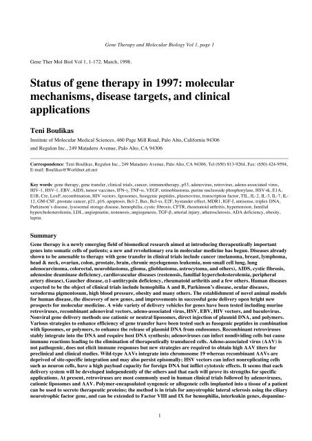
apoptosis (Figure 1). The E1A oncogene of adenovirus<br />
exerts its effect via p53 protein (Debbas and White, 1993;<br />
White, 1993). Indeed, expression of E1A increases the<br />
half-life of p53 resulting in accumulation of p53 molecules<br />
in adenovirus-infected cells leading to apoptosis. Although<br />
induction of host DNA synthesis by E1A provides a<br />
suitable environment for virus replication, the induction of<br />
apoptosis by the same protein impairs virus production<br />
since virus-infected cells are eliminated (see Han et al,<br />
1996 for references). p53-deficient cells are transformed<br />
by E1A because of absence of the pathway for induction<br />
of apoptosis by p53 (Lowe et al, 1994).<br />
E1A represses HER-2/neu transcription and functions<br />
as a tumor suppressor gene in HER-2/neu-overexpressing<br />
cancer cells. Transfer the E1A gene into cancer cells that<br />
overexpress HER-2/neu is an interesting aspect of gene<br />
<strong>therapy</strong> (see E1A in gene <strong>therapy</strong>; Yu et al, 1995; Chang<br />
et al, 1996; Ueno NT et al, 1997; Rodriguez et al, 1997;<br />
Xing et al, 1997).<br />
The E1B oncogene products inhibit apoptosis induced<br />
by E1A expression thus preventing premature death of<br />
host cells during adenovirus infection. This gives an<br />
advantage to virus for its proliferation and E1B proteins<br />
(19 kDa and 55 kDa) are necessary for transformation of<br />
primary rodent cells by E1A. E1A alone is unable to<br />
transform primary rodent cells (White, 1993).<br />
The E1B 19K protein of adenovirus is the putative<br />
viral homolog of the cellular Bcl-2 protein; using the yeast<br />
two-hybrid system for the identification of proteins<br />
interacting with E1B, Han and coworkers (1996) have<br />
identified Bax as one of the seven 19k-interacting clones.<br />
The 50-78 amino acid domain of Bax contains a conserved<br />
region homologous to Bcl-2 which is able to interact<br />
specifically with either Bcl-2 or E1B. In p53 mutant cells<br />
expression of Bax induced apoptosis; inhibition of<br />
apoptosis by Bcl-2 may proceed via its ability to bind the<br />
death-promoting Bax protein (Han et al, 1996). The bax<br />
gene is upregulated by p53.<br />
Expression of p53 and of adenovirus E1A induce<br />
apoptosis (Debbas and White, 1993; Lowe and Rudley,<br />
1993). A number of proteins when expressed at sufficient<br />
amounts block apoptosis; these include Bcl-2 and E1B 19<br />
kDa protein of adenovirus (Debbas and White, 1993;<br />
Chiou et al, 1994). All four protein molecules act<br />
upstream of Bax which is a potent inducer of apoptosis:<br />
both the cellular Bcl-2 and the 19 kDa protein E1B of<br />
adenovirus are able to interact with Bax inhibiting its<br />
involvement in induction of apoptosis (Han et al, 1996;<br />
Figure 1). E1A acts upstream of p53 by increasing the<br />
half-life of p53 resulting in an accumulation of p53
molecules in the nucleus (Lowe and Ruley, 1993);<br />
increased levels of p53 are then believed to upregulate the<br />
bax gene.<br />
The transcription factor E2F was originally identified<br />
as an activator of the adenovirus E2 gene and is implicated<br />
in the regulation of DNA replication (Shirodkar et al.,<br />
1992). Following infection of cells with adenovirus, the<br />
DNA binding activity of E2F increases and as a<br />
consequence transcription of the E2 gene of adenovirus<br />
increases (Kovesdi et al., 1987). These changes in E2F are<br />
mediated by E1A protein of adenovirus. RB forms specific<br />
complexes with E2F keeping E2F in a form unable to<br />
upregulate its target regulatory sequences. E2F can form<br />
specific complexes also with cyclin A during S-phase in<br />
NIH 3T3 cells (Mudryj et al., 1991). Both types of<br />
complexes, E2F-RB and E2F-cyclin A, can be dissociated<br />
by the adenovirus E1A protein (Chellappan et al., 1991;<br />
Bagchi et al., 1990; reviewed by White, 1998 this volume)<br />
but also by phosphorylation of RB at G1/S causing release<br />
of E2F and stimulation in transcription of genes required<br />
<strong>Gene</strong> Therapy and <strong>Molecular</strong> <strong>Biology</strong> Vol 1, page 9<br />
9<br />
for DNA replication (myc, DHFR). These events<br />
contribute to the uncontrolled proliferation of adenovirustransformed<br />
cells (Mudryj et al., 1990, 1991). Release of<br />
E2F from RB induced by E1A is critical for<br />
transformation of cells by E1A (for references see Hiebert<br />
et al, 1995).<br />
C. Strategies of adenoviruses to enter the<br />
cell<br />
In order to enter the host cell the adenovirus first<br />
attaches with a high affinity to a cell surface receptor,<br />
whose nature still remains elusive, using the head domains<br />
of the protruding viral fibers; the fibronectin-binding<br />
integrin on the cell surface then associates with the penton<br />
base protein on the adenovirus triggering endocytosis of<br />
the virus particle via coated pits and coated vesicles<br />
(Svensson and Persson, 1984; Greber et al, 1996). The<br />
third step in adenovirus entry into the host cell includes
<strong>Boulikas</strong>: An overview on gene <strong>therapy</strong><br />
Figure 1. Role of E1A and E1B19-kDa proteins of adenovirus in apoptosis.<br />
penetration of the adenoviral particles by acid-catalyzed<br />
rupture of the endosomal membrane involving the penton<br />
protein and the integrins and allowing escape to the<br />
cytoplasmic compartment; a decrease in endosome pH<br />
during internalization expose hydrophobic domains of<br />
these adenoviral capsid proteins which permits these<br />
proteins to insert into the vesicle membrane in a fashion<br />
that ultimately disrupts its integrity (Seth et al, 1984). At<br />
the final step the adenoviral particle is attached to the<br />
cytoplasmic side of pore complexes and the DNA is<br />
released to the interior of pore annuli entering the<br />
nucleoplasm.<br />
These highly ordered processes are accompanied by<br />
losses or protease degradation of specific proteins on the<br />
viral particles; the fibers and some of the penton base<br />
complexes on the adenovirus surface are already lost<br />
during the process of endocytosis; a viral protease,<br />
L3/p23, located inside the capsid at 10 copies per virion,<br />
plays a key role in the stepwise dismantling and in the<br />
weakening of the capsid structure culminating with the<br />
release of the adenovirus DNA by degrading of the viral<br />
capsid protein VI (Greber et al, 1996). The mechanism of<br />
disruption of endosomes by the adenoviral particles has<br />
been exploited to augment efficiency of transfection with<br />
transferrin-polylysine-DNA complexes (see fusogenic<br />
peptides and Curiel et al, 1991; Cotten et al, 1992; Wagner<br />
et al, 1992b; Cristiano et al, 1993; Morishita et al, 1993;<br />
Harries et al, 1993; Curiel, 1994).<br />
To overcome one of the major limitations to the<br />
clinical utility of adenoviruses which is the low efficiency<br />
of gene transfer achieved in vivo, Arcasoy et al (1997)<br />
found that the presence of the polycations polybrene,<br />
protamine, DEAE-dextran, and poly-L-lysine significantly<br />
increased the transfection efficiency in cell culture using<br />
the lacZ gene; because the polyanion heparin did not<br />
10<br />
significantly alter gene transfer efficiency, but completely<br />
abrogated the effects of polycations it supports the idea<br />
that the negative charges presented by membrane<br />
glycoproteins reduce the efficiency of adenovirusmediated<br />
gene transfer, an obstacle that can be overcome<br />
by polycations.<br />
D. Advantages and drawbacks of<br />
adenoviral vectors in gene delivery<br />
Adenoviruses possess a linear double-stranded genome<br />
which can be manipulated to accommodate up to 7.5 kb of<br />
DNA. Adenoviruses have the advantage of being able to<br />
infect nondividing cells. Other advantages are the rarity of<br />
recombination events between adenoviral vectors and the<br />
host chromosomes, the absence of induction of human<br />
malignancies by adenoviruses, and the relative safety of<br />
their use as vaccines (e.g. Ballay et al, 1985; Haj-Ahmad<br />
and Graham, 1986). For safety, replication-deficient,<br />
infectious adenoviruses are being used in somatic gene<br />
transfer; for example deletion in a portion of the E3 region<br />
of the virus permits encapsidation whereas deletion of a<br />
portion of the E1A coding sequence impairs viral<br />
replication (Gilardi et al, 1990; Rosenfeld et al, 1991).<br />
E. Deletion of adenoviral DNA sequences<br />
for gene delivery<br />
First generation recombinant adenoviruses were<br />
rendered defective by deletion of sequences spanning the<br />
E1A and E1B genes; these adenoviruses expressed low<br />
levels of early and late viral genes responsible for<br />
activating destructive cellular immune responses. Further<br />
deletion of other essential genes and growth in new<br />
packaging cell lines or incorporation of temperature
sensitive mutations which allow propagation of the virus<br />
in available packaging cell lines at permissive<br />
temperatures are strategies for improving the therapeutic<br />
efficacy of recombinant adenoviruses and for minimizing<br />
the immune response elicited to the host (Fisher et al,<br />
1996).<br />
E1-defective, recombinant adenoviruses can be<br />
replication-enabled by the codelivery of a plasmid<br />
encoding the deleted E1 functions, a strategy now<br />
designated as “conditional replication-enablement system<br />
for adenovirus” (CRESA); when the original replicationenabling<br />
plasmid was replaced by two separate plasmids<br />
that encoded the necessary E1A and E1B functions the<br />
E1-defective adenovirus could become conditionally<br />
replication-enabled by an RNA transcript encoding the<br />
required E1 functions. The RNA transcript of E1A<br />
enhanced the therapeutic efficacy of the E1-defective<br />
adenovirus: subcutaneous human tumor nodules<br />
containing a fraction of cells cotransduced with the<br />
replication-enabling RNA + DNA and an HSV-tk<br />
adenovirus were reduced to a greater extent than control<br />
nodules generated from the same fraction of cells<br />
cotransduced with the HSV-tk adenovirus and an<br />
irrelevant plasmid (Dion et al, 1996).<br />
A new type of recombinant adenovirus, (called deltarAd),<br />
deprived of all viral open reading frames and<br />
retaining only the essential cis elements (i.e., ITRs and<br />
contiguous packaging sequence), was propagated in 293<br />
cells in the presence of E1-deleted helper virus (Fisher et<br />
al, 1996). This adenovirus was packaged as concatamers<br />
into virions and was used to deliver successfully the CFTR<br />
gene to human airway epithelial cells in culture derived<br />
from a cystic fibrosis patient. The new delivery system<br />
needs improvements in its production and purification to<br />
allow its evaluation and use in vivo.<br />
F. Immune response to adenoviruses<br />
eliminate therapeutic cells<br />
Adenoviruses can achieve high levels of gene transfer<br />
(Haffe et al, 1992; Morsy et al, 1993; Herz and Gerard;<br />
Wilson, 1995; Kozarsky et al, 1996). However, the<br />
duration of transgene expression is limited (i) by clearance<br />
of the infected cells because of the cellular and humoral<br />
immune response (including those mediated by cytotoxic<br />
T lymphocytes) to adenoviral antigens (Yang Y et al,<br />
1994, 1995) and (ii) by loss of adenoviral episomes in<br />
progeny cells (Feng et al, 1997). To circumvent this<br />
problem adenoviral/retroviral chimeric vectors were<br />
constructed where the nonintegrative adenoviral vector<br />
was able to induce target cells to function as transient<br />
retrovirus producer cells and the retroviral particles were<br />
able to transduce neighboring cells; thus the recombinant<br />
adenovirus became integrative via the intermediate<br />
generation of a retroviral producing cell (Feng et al, 1997).<br />
First generation adenovirus-mediated gene transfer of<br />
<strong>Gene</strong> Therapy and <strong>Molecular</strong> <strong>Biology</strong> Vol 1, page 11<br />
11<br />
CFTR to the mouse lung resulted in the expression of viral<br />
proteins leading to the elimination of the therapeutic cells<br />
expressing CFTR by cellular immune responses and<br />
repopulation of the lung with nontransgene containing<br />
cells; second generation E1-deleted viruses, also crippled<br />
by a temperature sensitive mutation in the E2A gene,<br />
displayed substantially longer recombinant gene<br />
expression and induced a lower inflammatory response<br />
(Yang et al, 1994).<br />
In order to circumvent the elimination of adenovirustransduced<br />
cells by immune responses and for achieving<br />
persistence of transgene expression strategies to reduce the<br />
potential for viral gene expression have been developed;<br />
for example, an E4 modified adenovirus which was<br />
replication defective in cotton rats and displayed a reduced<br />
potential for viral gene expression in vivo was engineered<br />
(Armentano et al, 1997). Vectors containing a wild-type<br />
E4 region, E4 open reading frame 6, or a complete E4<br />
deletion were compared in the lungs of BALB/c mice for<br />
persistence of CFTR or lacZ expression; expression was<br />
transient from the E1a promoter with all vectors but<br />
persisted from the CMV promoter only with a vector<br />
containing a wild-type E4 region; thus, transient<br />
expression from adenoviral vectors may result from the<br />
down-regulation of a promoter and not necessarily from<br />
immune response-related factors (Armentano et al, 1997).<br />
The elimination of therapeutically important cells from<br />
the body after recombinant adenovirus-mediated delivery<br />
seems to be a great limiting factor for the use of<br />
adenoviruses in long-term gene <strong>therapy</strong> (Dai et al, 1995).<br />
This problem can be partially circumvented by daily<br />
administration of the immunosupressant cyclosporin A<br />
prohibiting the elimination of virally-transduced cell by<br />
activated T lymphocytes (Fang et al, 1995). A different<br />
approach to suppress elimination of therapeuticallytransduced<br />
cells after intra-articular delivery of genes to<br />
treat RA is by pretreatment of the joints with the anti-T<br />
cell receptor monoclonal antibody H57, a treatment which<br />
resulted in a significant reduction in lymphocytic<br />
infiltration and a persistence of transgene expression<br />
(Sawchuk et al, 1996).<br />
The prokaryotic Cre-LoxP recombination system was<br />
adapted to generate recombinant adenoviruses with<br />
extended deletions in the viral genome in order to<br />
minimize expression of immunogenic and/or cytotoxic<br />
viral proteins. An adenovirus was produced with a 25-kb<br />
deletion that lacked E1, E2, E3, and late gene expression;<br />
this vector exhibited viral titers similar to those achieved<br />
with first-generation (E1a-deleted) vectors which was<br />
efficient for gene transfer to cell culture but gene<br />
expression declined to undetectable levels much more<br />
rapidly than that sustained from first-generation vectors.<br />
Vectors deleted only at E1a were sustaining a better<br />
reporter gene expression because of their ability to<br />
replicate (Lieber et al, 1996).
A clinical protocol proposed recently for the <strong>therapy</strong> of<br />
amyotrophic lateral sclerosis uses a semipermeable<br />
membrane to enclose the ex vivo modified xenogenic BKH<br />
cells which is implanted intrathecally to provide human<br />
ciliary neurotrophic factor; the membrane prevents<br />
immunologic rejection of the cells interposing a virus<br />
impermeable barrier between the transduced cells and the<br />
host (Deglon et al, 1996; Pochon et al, 1996); the method<br />
has been applied before for cross-species transplantation<br />
of a polymer-encapsulated dopamine-secreting cell line to<br />
treat Parkinson's disease and for the delivery of nerve<br />
growth factor in rat and primate models of the Alzheimer's<br />
disease (Kordower et al, 1994; see Pochon et al, 1996 for<br />
more references). Evidently, similar approaches could be<br />
used to protect adenovirus- and retrovirus-transduced<br />
syngeneic cells from immunologic rejection provided that<br />
the therapeutic protein is secreted.<br />
A new area of investigation is directed toward surface<br />
modification of recombinant adenoviruses to render them<br />
safer and to minimize the strong immune responses<br />
against the virus and virus-infected cells; to this end<br />
Fender et al (1997) proposed a dodecahedron made of<br />
adenovirus pentons or penton bases and having only one<br />
or two adenovirus proteins instead of the 11 contained in<br />
an adenovirus virion; the penton is a complex of two<br />
oligomeric proteins, a penton base and fiber, involved in<br />
Figure 2. Localization of a recombinant adenoviral<br />
vector carrying 6.3 kb of dystrophin cDNA by in situ PCR<br />
following intramuscular injection to immunosuppressed<br />
mdx mice. Shown are transverse cryostat sections of mdx<br />
tibialis anterior muscle. Panel A shows a strong in situ<br />
hybridization signal (an E4 adenoviral sequence was<br />
amplified and an E4 probe was used) in myonuclei of an<br />
immunosuppressed animal injected with E1, E3-deleted<br />
adenovirus at 30 days postinjection (magnification 650x).<br />
Panel B was produced without Taq polymerase during PCR<br />
as a negative control. Panel C shows an uninjected muscle<br />
<strong>Boulikas</strong>: An overview on gene <strong>therapy</strong><br />
12<br />
the cell attachment, internalization, and liberation of virus<br />
from endosomes.<br />
It is certain that great improvements in adenoviral gene<br />
delivery will solve many of the current problems and<br />
permit a higher therapeutic efficacy in the near future.<br />
G. Examples of adenoviral gene transfer<br />
Recombinant adenovirus vectors have been used: for<br />
the transfer of factor IX gene in hemophilia B dogs via<br />
vein injection (Kay et al, 1994) and in mice (Smith et al,<br />
1993); for the transfer of genes into neurons and glia in the<br />
brain (le Gal la Salle, 1993); for the transfer of the gene of<br />
ornithine transcarmylase in deficient mouse and human<br />
hepatocytes (Morsy et al, 1993); for the transfer of the<br />
VLDL receptor gene for treatment of familial<br />
hypercholesterolaemia in the mouse model (Kozarsky et<br />
al, 1996); for the transfer of low density lipoprotein<br />
receptor gene in normal mice (Herz and Gerard, 1993);<br />
and for the ex vivo transduction of T cells from ADAdeficient<br />
patients (Blaese et al, 1995; Bordignon et al,<br />
1995). The adenovirus major late promoter was linked to a<br />
human α1-antitrypsin gene for its transfer to lung epithelia<br />
of cotton rat respiratory pathway as a model for the<br />
treatment of α1-antitrypsin deficiency; both in vitro and in<br />
vivo infections
processed as described in panel A showing no hybridization<br />
signal. From Zhao JE, Lochumuller H, Nalbantoglu J,<br />
Allen C, Prescott S, Massie B, Karpati G (1997) Study of<br />
adenovirus-mediated dystrophin minigene transfer to<br />
skeletal muscle by combined microscopic display of<br />
adenoviral DNA and dystrophin. Hum <strong>Gene</strong> Ther 8, 1565-<br />
1573. With kind permission of the authors (George Karpati,<br />
Montreal Neurological Institute, Canada) and Mary Ann<br />
Liebert, Inc.<br />
have shown production and secretion of α1-antitrypsin by<br />
the lung cells (Rosenfeld et al, 1991).<br />
A transductional preference of adenovirus-polylysine-<br />
DNA complexes and E1A/B-deleted replication-deficient<br />
adenoviruses was demonstrated for the prostate carcinoma<br />
cell lines DU145, LNCaP, and PC-3 over primary human<br />
bone marrow cells and the leukemia cell line KG-1; this<br />
finding led to a strategy to purge bone marrow of a<br />
specific subset of prostate carcinoma cells (Kim et al,<br />
1997).<br />
Figure 2 shows the localization of a recombinant<br />
adenoviral vector carrying 6.3 kb of dystrophin cDNA,<br />
driven by the CMV promoter, by in situ PCR following<br />
intramuscular injection to immunosuppressed mdx mice.<br />
Figure 3. shows a comparison of the persistence of<br />
dystrophin expression and adenoviral genomes in<br />
immunosuppressed versus immunocompetent mdx mice.<br />
The maximum number of fibers containing recombinant<br />
adenovirus was maintained until 60 days in<br />
<strong>Gene</strong> Therapy and <strong>Molecular</strong> <strong>Biology</strong> Vol 1, page 13<br />
13<br />
immunosuppressed mice but for only 10 days in<br />
immunocompetent animals. Thus, optimization of<br />
immunosuppression could assure successful long term<br />
dystrophin gene transfer for gene <strong>therapy</strong> of Duchenne<br />
muscular dystrophy (Zhao et al, 1997).<br />
A number of RAC-approved protocols for gene<br />
transfer to humans use recombinant adenoviruses<br />
(Appendix 1, protocols 118-157). <strong>Gene</strong>s transferred to<br />
patients with recombinant adenoviruses include p53<br />
(#130, 131, 147, 148, 152-156), RB (#140), CFTR (#118-<br />
123, 125, 128, 129), HSV-tk (126, 127, 132, 136, 139,<br />
141, 143, 145, 146), cytosine deaminase (#134, 151),<br />
VEGF (#157), IL-2 (#135), GM-CSF (#149, 150), antierbB-2<br />
single chain antibody (#133), ornithine<br />
transcarbamylase (#137), and GP100 melanoma antigen<br />
(#142).
<strong>Boulikas</strong>: An overview on gene <strong>therapy</strong><br />
Figure 3. Comparison of the persistence of dystrophin expression and adenoviral genomes in immunosuppressed versus<br />
immunocompetent mdx mice. Shown are combined dystrophin immunostaining and in situ PCR in tibialis anterior muscles of mdx mice<br />
at 10 days (A and C) and 60 days (B and D) postinjection. In A and B, FK506 was used as an immunosuppressant, whereas in C and D<br />
no immunosuppression was employed. At 10 days there was no significant difference in adenovirus positive nuclei (arrows) fibers<br />
between the immunosuppressed and the immunocompetent groups. At 60 days, however, there was a dramatic decline in the number of<br />
positive nuclei in the immunocompetent muscle. Magnification 650X. From Zhao JE, Lochumuller H, Nalbantoglu J, Allen C, Prescott<br />
S, Massie B, Karpati G (1997) Study of adenovirus-mediated dystrophin minigene transfer to skeletal muscle by combined microscopic<br />
display of adenoviral DNA and dystrophin. Hum <strong>Gene</strong> Ther 8, 1565-1573. With kind permission of the authors (George Karpati,<br />
Montreal Neurological Institute, Canada) and Mary Ann Liebert, Inc.<br />
IV. <strong>Gene</strong> delivery with Adeno-<br />
Associated Virus (AAV)<br />
A. Replication of AAV and rAAV: the role<br />
of the inverted terminal repeats<br />
AAVs are replication-defective parvoviruses, not<br />
associated with any human disease (nonpathogenic),<br />
requiring cotransfection with a helper virus to produce<br />
infectious virus particles; they can replicate in cell culture<br />
only in the presence of coinfection with adenovirus or<br />
herpes virus. Five serotypes of distinct AAV isolates have<br />
been recovered from human and other primates. AAV<br />
infections in humans are asymptomatic acquired with<br />
other viral infections such as adenovirus or HSV<br />
infections; 80-90% of adults are seropositive for<br />
antibodies against AAV (for references see Clark et al,<br />
1995; Berns and Linden, 1995).<br />
The replication of the AAV is dependent on two copies<br />
of a 145-bp inverted terminal repeat (ITR) sequence that<br />
flanks the AAV genome which is the primary cis-acting<br />
element required for productive infection and the<br />
generation of recombinant AAV (rAAV) vectors.<br />
In the absence of helper virus, the AAV particle can<br />
penetrate cells and find its way to the cell nucleus where<br />
the linear genome is uncoated and becomes integrated at a<br />
specific site on chromosome 19q13.3; several copies of<br />
AAV may integrate in tandem arrays. Thus, the AAV<br />
establishes a latent infection; the integrated viral genome<br />
can be activated and rescued by superinfection with helper<br />
virus (either adenovirus or any type of herpes virus).<br />
Inverted repeats at the ends of the viral DNA serve for the<br />
integration appearing near the junctions with cellular DNA<br />
sequences (Bohenzky et al, 1988).<br />
Adenovirus establishes foci called replication centers<br />
within the nucleus, where adenoviral replication and<br />
transcription occur; AAV was colocalized with the<br />
adenovirus replication centers using in situ hybridization<br />
and immunocytochemistry; AAV may, thus, utilize<br />
adenovirus and cellular proteins for its own replication;<br />
the rAAV genome was faintly detectable in a perinuclear<br />
distribution after successfully entering the cell; however,<br />
14<br />
rAAV was mobilized to replication centers when the cell<br />
was subsequently infected with adenovirus (Weitzman et<br />
al, 1996).<br />
Xiao et al (1997) have engineered the pDD-2 plasmid<br />
containing two copies of the D element, a unique sequence<br />
adjacent to the AAV nicking site, flanking a single ITR (a<br />
total of only 165 bp of AAV sequence); this modified<br />
hairpin was sufficient to sustain replication of the plasmid<br />
vector when Rep and adenovirus helper functions were<br />
supplied in trans. This plasmid has a significant prospect<br />
in gene transfer because is replicated more efficiently than<br />
infectious AAV clones; as a prelude to its replication the<br />
input circular plasmid was converted into a linear substrate<br />
by resolution of the AAV terminal repeat through a<br />
Holliday-like structure, a process most likely mediated by<br />
host factors. Linear monomer, dimer, and other highermolecular-weight<br />
replicative intermediates were generated<br />
during the replication of pDD-2, a feature characteristic of<br />
AAV replication. The replicative intermediates of this<br />
plasmid substrate were competent for AAV DNA<br />
replication, encapsidation, infection, integration, and<br />
subsequent rescue from the chromosome when<br />
superinfected with Ad and wild-type AAV (Xiao et al,<br />
1997). The elucidation of the important role of this 165-bp<br />
ITR sequence for AAV replication and the entire life cycle<br />
invigorates the important role of inverted repeats at the<br />
origin of replication not only of viruses but also of cellular<br />
origins of replication (<strong>Boulikas</strong>, 1996e).<br />
B. Packaging capabilities of AAVs<br />
AAVs posses a 4.7 kb single-stranded DNA genome.<br />
Hermonat et al (1997) have examined the maximum<br />
amount of DNA which can be inserted into the wild-type<br />
AAV genome without compromising packaging into an<br />
infectious virus particle; the maximum effective packaging<br />
capacity of AAV, examined as increments of 100 bp<br />
ligated at map unit 96 of AAV, is approximately 900 bp<br />
larger than wild type. Thus, wtAAV <strong>therapy</strong> vectors can<br />
be generated carrying a foreign gene of 900 bp or less with<br />
the advantages of wtAAV such as the ease in which high<br />
titers of infectious virus can be generated and the ability to<br />
specifically integrate in chromosome 19.
On the contrary, the payload capacity of recombinant<br />
AAV, which has been deprived of its viral genes and bears<br />
only the ITRs is in the order of 4.5-4.7 kb; this means that<br />
a cDNA up to this size can be inserted into a rAAV; for<br />
example the size of the CFTR cDNA is 4.5 kb and thus,<br />
the combined length of the promoter that drives CFTR<br />
expression and ITRs needs to be kept under 500 bp (Dong<br />
et al, 1996).<br />
Similar results were reported by Dong et al (1996) who<br />
have estimated that the optimal size of AAV vector is<br />
between 4.1 and 4.9 kb; the packaging efficiencies were<br />
sharply reduced above 5.2 kb and below 4.1 kb; two<br />
copies of the vector were packaged into each virion when<br />
vectors of 2.2-2.5 kb were provided.<br />
C. Integration of wtAAV but not of rAAV<br />
is site-specific<br />
Wild-type AAV is able to undergo targeted integration<br />
on chromosome 19 after infection in 15 out of 22 clones<br />
examined (Kotin et al, 1990, 1992). Of 51 integrations<br />
examined by fluorescence in situ hybridization (FISH) 48<br />
(94%) were to chromosome 19 after infection of IB3-1<br />
bronchial epithelial cells with wild-type AAV (Kearns et<br />
al, 1996). Site-specific integration has been reported for<br />
other viruses including avian leukosis virus (ALV)<br />
integrating adjacent to cellular oncogenes in tumors;<br />
however, the mechanism of ALV integration involves a<br />
process of selection of cells able to form tumors by<br />
overexpression of the oncogene due to virus integration<br />
rather than exclusive integration of the ALV at unique<br />
sites of the genome (Hayward et al, 1981). RSV also<br />
appears to be integrated at a limited number of sites (Shih<br />
et al, 1988). Adenovirus integration, a more rare event<br />
compared to the majority of episomal molecules, may also<br />
occur at a number of preferred sites (Jessberger et al,<br />
1989). A larger number of recombinase molecules than<br />
those known today may be present in mammalian cell<br />
nuclei and promote site-specific integration and<br />
recombination events.<br />
Although the human wild-type AAV (wtAAV) is<br />
unique in its ability to target viral integration to a specific<br />
site on chromosome 19, the recombinant AAV (rAAV)<br />
vectors have lost the site-specific integration and targeting<br />
ability; furthermore, rAAVs have incapacitated ability to<br />
integrate, and can be found as episomes. When wtAAV-2<br />
was used to infect IB3-1 bronchial epithelial cells all<br />
metaphase spreads examined by fluorescence in situ<br />
hybridization (FISH) had integrated copies and 94% of the<br />
integrations were to chromosome 19; furthermore, 36 of<br />
56 metaphase spreads had a single copy of wtAAV<br />
integrated and 20 of 56 showed two sites within<br />
chromosome 19 (Kearns et al, 1996). On the contrary,<br />
when a recombinant AAV containing the CFTR cDNA<br />
was used to infect the same cells, examination of 67<br />
<strong>Gene</strong> Therapy and <strong>Molecular</strong> <strong>Biology</strong> Vol 1, page 15<br />
15<br />
metaphase chromosome spreads identified four<br />
integrations (only 6% of total) to different chromosomes.<br />
No integration was to chromosome 19. When these studies<br />
were repeated on the A35 epithelial cell line selected for<br />
stable CFTR expression, the episomal AAV-CFTR<br />
sequences were abundant in the low molecular weight<br />
DNA fraction (Kearns et al, 1996).<br />
Yang et al (1997) have cloned over 40 AAV and<br />
rAAV integration junctions to determine the terminalrepeat<br />
sequences that mediate integration. These studies<br />
have shown that in both immortalized and normal diploid<br />
human cells, wt AAV targeted integration to chromosome<br />
19 in head-to-tail tandem arrays; the majority of the<br />
junction sequences were involving incomplete copies of<br />
the AAV inverted terminal repeats (ITRs); inversions of<br />
genomic and/or viral DNA sequences at the wt integration<br />
site took place. The viral integration event was found to be<br />
mediated by terminal repeat hairpin structures and cellular<br />
recombination pathways. In contrast, rAAV provirus<br />
integrated on chromosome 2 and at the same locus in two<br />
independent cell lines, in both the flip and flop<br />
orientations; genomic rearrangements took place at the<br />
integration site of rAAV, mainly involving deletions<br />
and/or rearrangement-translocations.<br />
Similar data were reported by Rutledge and Russell<br />
(1997): recombinant AAV vectors were found to be<br />
integrated by nonhomologous recombination as singlecopy<br />
proviruses in HeLa cells and at random chromosomal<br />
locations; the recombination junctions were scattered<br />
throughout the vector terminal repeats with no apparent<br />
site specificity; the flanking HeLa DNA at integration sites<br />
was not homologous to AAV or to the site-specific<br />
integration locus of wild-type AAV. Furthermore, vector<br />
proviruses with nearly intact terminal repeats were excised<br />
from the genomic HeLa DNA and were amplified after<br />
infection of cells with wild-type AAV and adenovirus.<br />
The integration patterns of four recombinant AAV-2<br />
genomes in individual clonal isolates of the human<br />
nasopharyngeal carcinoma cell line (KB) were different;<br />
the difference between the recombinant AAV-2 genomes<br />
were in the combinations of the genes for resistance to<br />
tetracycline, to neomycin, to ampicillin, with the genes for<br />
AAV replication, and the AAV capsid genes. None of the<br />
KB cell clones examined had the proviral genome<br />
covalently linked to the specific-site of integration of the<br />
wt AAV on chromosome 19 (Ponnazhagan et al, 1997a,b).<br />
D. Drawbacks of AAV in gene <strong>therapy</strong> and<br />
their remedy<br />
<strong>Gene</strong> transfer with AAV vectors has typically been<br />
low. Difficulties in generating recombinant virions on a<br />
large scale sufficient for preclinical and clinical trials and<br />
in obtaining high-titer virus stocks after the initial<br />
transfection into producer cells is a limiting factor for the
widespread usage of AAV vectors; this obstacle is<br />
expected to be overcome in the near future. The high viral<br />
titers required for preclinical and clinical studies have<br />
been achieved by a new strategy developed by Tamayose<br />
et al (1996); AAV vector particles in cell lysates could be<br />
concentrated by sulfonated cellulose column<br />
chromatography to a titer higher than 10 8 cfu/ml or 5 x<br />
10 10 particles/ml. A method for transfecting cells at<br />
extremely high efficiency with a rAAV vector and<br />
complementation plasmid while simultaneously infecting<br />
those cells with replication competent adenovirus using<br />
adenovirus-polylysine-DNA complexes has been<br />
developed by Mamounas et al (1995).<br />
The difficulties in developing packaging cell lines for<br />
AAV relate to low levels of rep gene expression from the<br />
AAV-p5 promoter and to the propensity of Rep proteins to<br />
suppress continued growth of immortalized cell lines;<br />
expression of AAV rep under control of the LTR of the<br />
human HIV together with the development of cell<br />
populations containing rescuable AAV recombinant<br />
genomes increased 50-fold the packaging efficiency of<br />
AAV vectors (Flotte et al, 1995).<br />
After infection of cell cultures with recombinant AAV<br />
there is a decline in the percentage of cells expressing the<br />
transferred gene with time in culture. This decline was<br />
associated with ongoing losses of vector genomes (Malik<br />
et al, 1997). For example, transfer to cultures of K562<br />
human erythroleukemia cells of a truncated rat nerve<br />
growth factor receptor (tNGFR) cDNA as a cell surface<br />
reporter under control of the LTR of the Moloney murine<br />
leukemia virus showed that about 30% of cells expressed<br />
tNGFR on the surface early after transduction at a<br />
multiplicity of infection (MOI) of 13 infectious units (IU),<br />
which declined to 3% over 1 month of culture. At an MOI<br />
of 130 IU, nearly all cells expressed tNGFR immediately<br />
and the proportion of cells expressing tNGFR declined to<br />
62% over 2 months of culture (Malik et al, 1997).<br />
Another obstacle of rAAV vectors is the low rate of<br />
integration of rAAV into the host genome (which can be<br />
improved at high MOI). The efficiency of integration was<br />
about 2% at low MOI (1.3 IU) and increased to at about<br />
49% at an MOI of 130 (Malik et al, 1997).<br />
E. Advantages using AAV and<br />
improvements in AAV gene delivery<br />
AAV does not elicit an immune reaction and is a<br />
nonpathogenic virus to humans. AAV contains normally a<br />
single-stranded copy of its genome. Transduction with<br />
AAV can be enhanced in the presence of adenovirus gene<br />
products through the formation of double stranded, nonintegrated<br />
AAV genomes.<br />
AAV has been reported to have advantages over other<br />
viruses for gene transfer to hematopoietic stem cells due to<br />
their high titers and relative lack of dependence on cell<br />
<strong>Boulikas</strong>: An overview on gene <strong>therapy</strong><br />
16<br />
cycle for target cell integration. A robust CMV/LacZ<br />
reporter gene expression in primary human CD34 + CD2 −<br />
progenitor cells induced to undergo T-cell differentiation<br />
was obtained without toxicity or alteration in the pattern of<br />
T-cell differentiation. 70% to 80% of the cells isolated<br />
from either adult bone marrow or umbilical cord blood<br />
were efficiently transduced with AAV; however, the<br />
expression was transient without integration; this limits<br />
the potential use of AAV in gene <strong>therapy</strong> strategies for<br />
diseases such as AIDS (Gardner et al, 1997).<br />
<strong>Gene</strong> transduction by AAV vectors in cell culture can<br />
be stimulated over 100-fold by treatment of the target cells<br />
with agents that affect DNA metabolism, such as<br />
irradiation or topoisomerase inhibitors (Russell et al,<br />
1995); great improvements in transduction efficiency can<br />
also be achieved in vivo: previous γ-irradiation increased<br />
the transduction rate in mouse liver by up to 900-fold, and<br />
the topoisomerase inhibitor etoposide increased<br />
transduction by about 20-fold after direct liver injection or<br />
after systemic delivery via tail vein injection; up to 3% of<br />
hepatocytes could be transduced after a single systemic<br />
vector injection (Koeberl et al, 1997). This is a significant<br />
advantage compared to stealth liposomes which , although<br />
concentrating in the liver, spleen and tumors can transduce<br />
Kupffer cells but not hepatocytes after systemic delivery<br />
(Martin and <strong>Boulikas</strong>, 1998, following article).<br />
A combination of the adenovirus-5 capsid protein or<br />
the Fiber protein of adenovirus with liposomes, termed<br />
adenosomes (adenovirus protein-cationic liposome<br />
complexes) improved the efficiency of gene transfer. This<br />
complex was able to mediate efficient transfer of a<br />
AAV/CMV-LacZ construct to endothelial cells (Zhou et<br />
al, 1995).<br />
Clark et al (1996) have developed a sensitive assay<br />
system to determine infectivity of AAV vectors based on<br />
the replication of input rAAV genomes rather than<br />
transgene expression which depends on the type of<br />
promoter which drives the foreign gene; this system uses a<br />
cell line that expresses AAV helper functions (rep and<br />
cap) upon induction by adenovirus infection.<br />
F. Examples using AAV for gene transfer<br />
AAV will infect a broad number of mammalian cell<br />
lines and has been used as a cloning vector to transduce<br />
the Neo R gene into mammalian tissue culture cells<br />
(Hermonat and Muzyczka 1984). Antisense AAV vectors<br />
have been used to inhibit HIV replication (Chatterjee et al,<br />
1992), and to correct Fanconi's anemia in human<br />
hematopoietic cells (Walsh et al, 1994). AAVs transduce<br />
preferentially cells in S phase; topoisomerase inhibitors<br />
increase transduction efficiency (Russell et al, 1995).<br />
Using AAV, the genomic copy of a normal human βglobin<br />
gene under control of the DNase l-hypersensitive<br />
site 2 (HS-2) from the locus control region was expressed
in K562 human erythroleukemia cells, which normally<br />
lack the β-globin gene; following selection with G418 by<br />
virtue of the neo-resistance function which was provided<br />
in the rAAV vector, stable integration of the exogenous βglobin<br />
allele was determined (Zhou et al, 1996). Similar<br />
data were reported by Einerhand et al (1995) transferring a<br />
recombinant AAV-vector containing a human β-globin<br />
gene together with the DNase1 hypersensitive sites 4, 3<br />
and 2 of the human β-globin locus control region as an<br />
approach for the gene <strong>therapy</strong> of β-thalassemia and sickle<br />
cell anemia. The vector replicated to high titers and could<br />
efficiently transduce hematopoietic stem cells isolated<br />
from patients. In order to treat sickle cell anemia Lubovy<br />
et al (1996) have transferred lacZ with a recombinant<br />
AAV vector and stably transduced hematopoietic stem<br />
cells purified from normal and homozygous sickle cell<br />
anemia patients.<br />
AAV was able to promote delivery of functional levels<br />
of glial cell line-derived neurotrophic factor (GDNF), in a<br />
degenerative model of Parkinson's disease (Mandel et al,<br />
1997). AAV has also been used for the transduction of the<br />
mouse liver in vivo with Factor IX cDNA as a prelude to<br />
treatment of hemophiliacs (Snyder et al, 1997; Herzog et<br />
al, 1997) and for the human intratracheal instillation of<br />
CFTR cDNA into neonatal New Zealand white rabbits<br />
(Rubenstein et al, 1997), and to the lungs of rhesus<br />
macaques without eliciting inflammation (Conrad et al,<br />
1996).<br />
AAV has also been used for the transfer of the human<br />
multidrug resistance gene (hMDR1) cDNA to NIH-3T3<br />
cells followed by selection of successfully transfected<br />
cells based on the drug-resistant phenotype conferred by<br />
the P-glycoprotein efflux pump (see below and Lee et al,<br />
1997, this volume); integration of MDR1 sequences into<br />
the host cell genome was demonstrated by fluorescent in<br />
situ hybridization (FISH) but also the persistence of<br />
nonintegrated AAV-MDR1 episomal plasmids (Baudard<br />
et al, 1996).<br />
<strong>Gene</strong> Therapy and <strong>Molecular</strong> <strong>Biology</strong> Vol 1, page 17<br />
17<br />
Introduction of a human globin gene into murine<br />
hematopoietic bone marrow cells ex vivo with a<br />
recombinant AAV vector followed by transplantation of<br />
these cells into lethally irradiated congenic mice sustained<br />
a long-term repopulating ability: human globin gene<br />
sequences were detected in the bone marrow and spleen in<br />
primary recipient mice for at least 6 months.<br />
Kessler et al (1996) have shown that following a single<br />
intramuscular administration of a recombinant adenoassociated<br />
virus (rAAV) vector, carrying either the lacZ or<br />
the human erythropoietin gene into adult BALB/c mice<br />
leads to the local production of the foreign protein in the<br />
muscle for at least 32 weeks; furthermore, human<br />
erythropoietin was secreted and stimulated red blood cell<br />
production in the mouse for up to 40 weeks. This finding<br />
was extended by Fisher et al (1997) who arrived to the<br />
unexpected finding that intramuscular injection of highly<br />
purified recombinant AAV can sustain a high level of<br />
transgene expression in the absence of adenovirus after<br />
direct injection to the muscle in mice (Figure 4); this<br />
expands the potential of AAV for the treatment of<br />
inherited and acquired diseases. Using this approach no<br />
humoral or cellular immune responses were elicited after<br />
transfer of lacZ against the neoantigenic E. coli βgalactosidase.<br />
The rAAV genome was integrated at single<br />
sites as head-to-tail concatamers into nuclei of<br />
differentiated muscle fibers. Transfer of the lacZ gene<br />
using a highly purified preparation of AAV which was<br />
injected into the skeletal muscle of adult mice in the<br />
presence of E2a-deleted adenovirus to enhance<br />
transduction followed by direct visualization of the βgalactosidase<br />
by X-gal histochemistry revealed high<br />
transduction of muscle fibers by day 17 associated with<br />
inflammation (Fig 4a and b). Animals that received the<br />
same AAVlacZ in the absence of adenovirus demonstrated<br />
higher levels of transduction that persisted for 240 days<br />
(Fig 4c-h).
<strong>Boulikas</strong>: An overview on gene <strong>therapy</strong><br />
Figure 4. Purified recombinant AAV-mediated lacZ gene transfer to the muscle in adult mice sustains a high level of expression and<br />
is inflammation-free. Purified AAVlacZ (1x10 9 genomes in 25 µl) was injected into the tibialis anterior of 5-week-old C57BL/6 mice<br />
and tissue was harvested at days 3 (c), 17 (d), 30 (e), 64 (f), and 180 (g, h) post-injection and analyzed by X-gal histochemistry. Samples<br />
of AAVlacZ (1x10 9 genomes in 25 µl) were also supplemented with an E2a mutant adenovirus dl802 (5x10 10 A 260 particles) just prior to<br />
injection and tissue was harvested at days 3 (a) and 17 (b) post-injection. Magnification: a-g, X10; h, X5.<br />
Purified AAVlacZ (175 µl , 1x10 12 genomes/ml) was injected into the tibialis anterior of a male rhesus monkey. Biopsies were taken<br />
14 days post-injection and frozen sections were cut and stained for β-galactosidase activity (i and j); magnification: I, X5; j, X10.<br />
k and l: rAAV vector expressing human β-glucoronidase was injected into the tibialis anterior of 5-week-old C57BL/6 mice (1x10 9<br />
genomes in 25 µl). 30 days post-injection the muscle was harvested and frozen sections were cut and stained for β-glucoronidase. From<br />
Fisher KJ, Jooss K, Alston J, Yang Y, Haecker SE, High K, Pathak R, Raper SE, Wilson JM (1997) Recombinant adeno-associated virus<br />
for muscle directed gene <strong>therapy</strong>. Nat Med 3, 306-312. Reproduced with the kind permission of the authors and Nature America, Inc.<br />
AAV-mediated delivery of the lacZ gene by direct<br />
injection to brain tumors which were induced from human<br />
glioma cells in nude mice showed that 30-40% of the cells<br />
along the needle track expressed β-galactosidase;<br />
subsequent delivery of the HSV-tk/IL-2 genes to these<br />
tumors with AAV and administration of GCV to the<br />
animals for 6 days resulted in a 35-fold reduction in the<br />
mean volume of tumors compared with controls by a<br />
significant contribution from the bystander effect (Okada<br />
et al, 1996).<br />
A phase I clinical trial for CF is being conducted at<br />
Johns Hopkins Hospital using AAV (see Kearns et al,<br />
1996, and protocols #165, 166 in Appendix 1).<br />
V. Herpes Simplex Virus-1 (HSV-1)<br />
and miniviral vectors<br />
HSV-1 has a capacity of inserting up to 30 kb of<br />
exogenous DNA which is a clear advantage over the<br />
adenovirus (up to 7.5 kb of exogenous DNA). High titer<br />
viral stocks can be prepared from HSV-1. HSV-1 also<br />
displays a wide range of host cells and can infect<br />
nonreplicating cells such as neuron cells in which the<br />
vectors can be maintained indefinitely in a latent state.<br />
However, infection with HSV-1 is cytotoxic to cells<br />
because of residual viral proteins produced by the virus.<br />
Strategies to circumvent this drawback led to the<br />
development of viral vectors with a very large capacity for<br />
insertion (almost as large as the size of the virus) which<br />
depend on defective helper virus for replication and<br />
packaging into infectious virions (see below). A miniviral<br />
vector can combine the advantage of cloning the gene in<br />
bacterial plasmids, the high efficiency of virus-mediated<br />
18<br />
gene transfer, and the possibility to transfer large genomic<br />
DNA fragments including far upstream, downstream and<br />
intronic regulatory elements.<br />
The HSV-1 genome is a 152 kb double-stranded DNA<br />
containing three origins of replication and encoding at<br />
least 72 unique proteins; it consists of a unique long<br />
segment replicated from oriL and two repeats flanking the<br />
unique segment each replicated from oriS. Spaete and<br />
Frenkel (1982) have constructed plasmids containing the<br />
lytic viral origin of replication, foreign DNA inserts, and<br />
the terminal packaging signal sequences; in the presence<br />
of a wild-type helper virus such an amplicon was<br />
amplified into multimeric tandemly-repeated forms of the<br />
original vector by rolling-circle replication and was<br />
packaged into infectious HSV virions (Spaete and Frenkel,<br />
1982). However, the helper virus caused death of the<br />
infected cells due to lytic replication and this system is not<br />
amenable to gene <strong>therapy</strong>.<br />
To circumvent this bottleneck two strategies have been<br />
developed leading to replication-defective helper HSV: (i)<br />
a temperature-sensitive system permitted production of<br />
virion stocks at 31 o C whereas infection of cells at 37 o C<br />
caused inactivation of the helper virus which was<br />
incapable of entering the lytic cycle and allowed delivery<br />
of the miniviral vector to the target cell without causing its<br />
death. (ii) In a different system, the immediately early<br />
gene IE3 was deleted from the helper virus; IE3 encodes<br />
for a protein (ICP4) essential for early and late viral gene<br />
expression and replication; the helper cell line used for<br />
packaging had a genomic insertion of the IE3 gene of<br />
HSV which was functionally expressed allowing for<br />
complementation and for lytic infection using the IE3-
defective HSV virus (DeLuca and Schaffer, 1987; Geller<br />
and Freese, 1990).<br />
Two types of viral vectors have been used for gene<br />
transfer to cancer cells: replication-incompetent vectors<br />
expressing a gene product that leads to the destruction of<br />
the tumor or replication-competent vectors that are<br />
inherently cytotoxic to the tumor cells. In order to<br />
combine the two modes of action Miyatake et al (1997)<br />
used a defective HSV vector that consisted of a defective<br />
particle, containing tandem repeats of the HSV-tk gene,<br />
and a replication-competent, non-neurovirulent HSV<br />
mutant as a helper virus. When glioma GL261 cells were<br />
infected with the tk-defective vector/helper virus the HSV-<br />
TK activity was significantly higher than that in helper<br />
virus-infected cells which contained a single copy of<br />
HSV-tk; subcutaneous injection of these cells to C57BL/6<br />
mice inducing gliomas led to a significant decrease in<br />
tumor size after GCV treatment.<br />
An HSV-1 vector containing a 6.8-kb fragment of the<br />
rat tyrosine hydroxylase promoter (pTHlac) supported a<br />
seven- to 20-fold increase in reporter gene expression in<br />
catecholaminergic cell lines compared to noncatecholaminergic<br />
cell lines. Furthermore, 4 days after stereotactic<br />
injection into the midbrain of adult rats and for a duration<br />
of 6 weeks, pTHlac supported a 10-fold targeting of βgalactosidase<br />
expression to tyrosine hydroxylaseexpressing<br />
neurons in the substantia nigra pars compacta<br />
compared with pHSVlac; this long term expression was<br />
significant compared to that from pHSVlac which<br />
decreased approximately 30-fold between 4 days and 6<br />
weeks after gene transfer (Song et al, 1997); this study<br />
also shows the importance of large control regions in the<br />
order of 7 kb in sustaining cell type-correct gene<br />
expression, something feasible with HSV and liposomes<br />
but nor with recombinant retrovirus, adenovirus, or AAV.<br />
VI. HIV vectors for gene transfer<br />
Recent studies have succeeded in exploiting the deadly<br />
HIV-1 virus, after crippling some functions, as a gene<br />
delivery vehicle. An advantage of HIV vectors has been<br />
the broad range of tissues and cell types they can<br />
transduce, a property granted because lentiviral vectors are<br />
pseudotyped with vesicular stomatitis virus G<br />
glycoprotein. Human lentiviral (HIV)-based vectors can<br />
transduce non-dividing cells in vitro and deliver genes in<br />
vivo; expression of transgenes in the brain has been<br />
detected for more than six months. HIV vectors have been<br />
also used to introduce genes directly into liver and muscle;<br />
3-4% of the total liver tissue was transduced by a single<br />
injection of 1-3 x 10 7 infectious units (I.U.) of recombinant<br />
HIV with no inflammation or recruitment of lymphocytes<br />
at the site of injection. Whereas expression of green<br />
fluorescent protein (GFP), used as a surrogate for<br />
therapeutic protein, was observed for more than 22 weeks<br />
in the liver and for over 8 weeks in the muscle using<br />
<strong>Gene</strong> Therapy and <strong>Molecular</strong> <strong>Biology</strong> Vol 1, page 19<br />
19<br />
lentiviral vectors, little or no GFP could be detected in<br />
liver or muscle transduced with the Moloney murine<br />
leukemia virus (Mo-MLV), a prototypic retroviral vector<br />
(Kafri et al, 1997).<br />
The development of a stable noninfectious HIV-1<br />
packaging cell line capable of generating high-titer HIV-1<br />
vectors is another important step towards use of HIV<br />
vectors in gene <strong>therapy</strong> (Corbeau et al, 1996). A hybrid<br />
murine leukemia virus-based vector containing U3 and R<br />
sequences from HIV-1 in place of the MLV U3 and R<br />
regions gave single transcriptional unit retroviral vectors<br />
under the control of Tat; this vector has advantages for<br />
anti-HIV gene <strong>therapy</strong> (Cannon et al, 1996).<br />
Although replication-incompetent HIV vectors<br />
displayed a strict CD4 + T cell tropism for gene transfer, a<br />
feature important for AIDS <strong>therapy</strong>, it was thought to<br />
preclude HIV-based vectors for other gene transfer<br />
applications; a two-step gene transfer system, however,<br />
was developed to expand the host range of the HIV vector:<br />
in the first step, the CD4 gene was introduced into target<br />
cells using a replication-defective adenoviral vector; in the<br />
second step the CD4-transfected cells were incubated with<br />
HIV vectors which resulted in stable integration and HIVmediated<br />
gene transfer (Miyake et al, 1996).<br />
An HIV multiply attenuated vector in which the<br />
virulence genes env, vif, vpr, vpu, and nef were deleted<br />
was able to deliver genes in vivo into adult neurons<br />
(Zufferey et al, 1997).<br />
HIV-mediated gene transfer was used to transfer the<br />
GFP gene under control of CMV to retinal cells by<br />
injection into the subretinal space of eyes in rats; the GFP<br />
gene was efficiently expressed in both photoreceptor cells<br />
and retinal pigment epithelium; predominant expression in<br />
photoreceptor cells was achieved using the rhodopsin<br />
promoter. The transduction efficiency was high and<br />
photoreceptor cells in >80% of the area of whole retina<br />
were expressing GFP (Miyoshi et al, 1997).<br />
VII. Epstein-Barr virus (EBV) and<br />
baculovirus vectors<br />
EBV is an episomaly-replicating virus in synchrony<br />
with the cell cycle. EBV infects human cells causing<br />
mononucleosis; the presence of the unique latent origin of<br />
replication (oriP) in EBV allows for episomal replication<br />
of the virus in human cells without entering the lytic cycle.<br />
The presence of oriP and of the replication initiator protein<br />
EBNA1 cDNA on a vector allows episomal replication in<br />
human cells; in addition, plasmids containing only oriP<br />
can replicate episomally into cell lines expressing EBNA-<br />
1 (Sun et al, 1994; Banerjee et al, 1995).<br />
A hybrid HSV-1/EBV vector has been developed by<br />
Wang and Vos (1996), which combines (i) the HSV-1<br />
lytic oriS; (ii) an HSV-1 packaging sequence which allows
eplication and packaging in the presence of defective<br />
helper virus carrying a deletion in the IE3 gene in the E5<br />
cell line expressing the IE3 gene; (iii) the latent oriP of<br />
EBV and (iv) the EBNA-1 cDNA allow episomal<br />
replication of the infectious vector in the E5 cell line so<br />
that viral stocks of high titer can be made. Infection of<br />
tumor-derived fibroblast and epithelial cell lines in culture<br />
and local injection of human liver tumors in nude mice<br />
was used to demonstrate 95-99% efficiency of infection<br />
and transfer of the reporter β-galactosidase gene.<br />
<strong>Gene</strong>tically modified baculoviruses (Autographa<br />
californica nuclear polyhedrosis virus) were used to<br />
efficiently deliver genes into cultured hepatocytes of<br />
different origin; delivery into human hepatocytes with<br />
baculovirus vectors approached 100% efficiency in cell<br />
culture and expression levels were high when mammalian<br />
promoters were chosen. A number of drawbacks preclude<br />
their direct application in vivo; nevertheless gene transfer<br />
was feasible in ex vivo perfused human liver tissue<br />
(Sandig et al, 1996; Hofmann et al, 1998 this volume).<br />
VIII. Liposomal gene delivery<br />
Abbreviations:<br />
DC-CHOL: 3β [N-(N',N'dimethylaminoethane)carbamoyl]cholesterol<br />
DDAB: dimethyldioctadecyl ammonium bromide<br />
DMRIE: N-[1-(2,3-dimyristyloxy)propyl]-N,N-dimethyl-N-(2hydroxyethyl)<br />
ammonium bromide<br />
DMTAP: 1,2-dimyristoyl-3-trimethylammonium propane<br />
DOGS: Dioctadecylamidoglycylspermine (Transfectam, Promega)<br />
DOPE: dioleyl phosphatidylethanolamine<br />
DOSPA: 2,3-dioleoyloxy-N-[2(sperminecarboxamido)ethyl]-N,Ndimethyl<br />
-1-propanaminium trifluoroacetate<br />
DOTAP: N-(1-(2,3-dioleoyloxy)propyl)-N,N,Ntrimethylammonium<br />
chloride<br />
DOTMA: N-[1-(2,3-dioleyloxy) propyl]-n,n,n-trimethylammonium<br />
chloride<br />
DPTAP: 1,2- dipalmitoyl-3-trimethylammonium propane<br />
DSTAP: 1,2-disteroyl-3-trimethylammonium propane<br />
Lipofectin: DOTMA:DOPE 1:1 (GIBCO BRL)<br />
A. Immune responses and toxicity of<br />
cationic lipid-DNA complexes<br />
Cationic lipids have been widely used for gene<br />
transfer; a number of clinical trials (34 out of 220 total<br />
RAC-approved protocols as of December 1997) use<br />
cationic lipids (see Table 4 in Martin and <strong>Boulikas</strong>, 1998,<br />
this volume, pages 203-206). Although many cell culture<br />
studies have been documented, systemic delivery of genes<br />
with cationic lipids in vivo has been very limited. All<br />
clinical protocols use subcutaneous, intradermal,<br />
intratumoral, and intracranial injection as well as<br />
intranasal, intrapleural, or aerosol administration but not<br />
<strong>Boulikas</strong>: An overview on gene <strong>therapy</strong><br />
20<br />
i.v. delivery because of the toxicity of the cationic lipids<br />
and DOPE (see Table 4 in Martin and <strong>Boulikas</strong>, 1998, this<br />
volume, pages 203-206).<br />
Liposomes formulated from DOPE and cationic lipids<br />
based on diacyltrimethylammonium propane (dioleoyl-,<br />
dimyristoyl-, dipalmitoyl-, disteroyl-trimethylammonium<br />
propane or DOTAP, DMTAP, DPTAP, DSTAP,<br />
respectively) or DDAB were highly toxic when incubated<br />
in vitro with phagocytic cells (macrophages and U937<br />
cells), but not towards non-phagocytic T lymphocytes; the<br />
rank order of toxicity was DOPE/DDAB > DOPE/DOTAP<br />
> DOPE/DMTAP > DOPE/DPTAP > DOPE/DSTAP; the<br />
toxicity was determined from the effect of the cationic<br />
liposomes on the synthesis of nitric oxide (NO) and TNFα<br />
produced by activated macrophages (Filion and Phillips,<br />
1997).<br />
Another factor to be considered before i.v. injections<br />
are undertaken is that negatively charged serum proteins<br />
can interact and cause inactivation of cationic liposomes<br />
(Yang and Huang, 1997). Condensing agents used for<br />
plasmid delivery including polylysine, transferrinpolylysine,<br />
a fifth-generation poly(amidoamine)<br />
(PAMAM) dendrimer, poly(ethyleneimine), and several<br />
cationic lipids (DOTAP, DC-Chol/DOPE, DOGS/DOPE,<br />
and DOTMA/DOPE) were found to activate the<br />
complement system to varying extents. Strong<br />
complement activation was seen with long-chain<br />
polylysines, the dendrimer, poly(ethyleneimine), and<br />
DOGS; complement activation was considerably reduced<br />
by modifying the surface of preformed DNA complexes<br />
with polyethyleneglycol (Plank et al, 1996).<br />
B. Mechanism of liposome entry to cells<br />
Cationic lipids increase the transfection efficiency by<br />
destabilizing the biological membranes including plasma,<br />
endosomal, and lysosomal membranes; indeed, incubation<br />
of isolated lysosomes with low concentrations of DOTAP<br />
caused a striking increase in free activity of βgalactosidase,<br />
and even a release of the enzyme into the<br />
medium demonstrating that lysosomal membrane is deeply<br />
destabilized by the lipid; the mechanism of destabilization<br />
was thought to involve an interaction between cationic<br />
liposomes and anionic lipids of the lysosomal membrane,<br />
allowing a fusion between the lipid bilayers; the process<br />
was less pronounced at pH 5 than at pH 7.4 and anionic<br />
amphipathic lipids were able to prevent partially this<br />
membrane destabilization (Wattiaux et al, 1997).<br />
In contrast to DOTAP and DMRIE which were 100%<br />
charged at pH 7.4, DC-CHOL was only about 50%<br />
charged as monitored by a pH-sensitive fluorophore; this<br />
difference decreases the charge on the external surfaces of<br />
the liposomes and was proposed to promote an easier<br />
dissociation of bilayers containing DC-CHOL from the<br />
plasmid DNA and an increase in release of the DNA-lipid
complex into the cytosol from the endosomes (Zuidam and<br />
Barenholz, 1997).<br />
C. Tissue targets using cationic liposomes<br />
in vivo<br />
Although cationic lipids have been used widely for the<br />
delivery of genes very few studies have used systemic i.v.<br />
injection of cationic liposome-plasmid complexes because<br />
of the toxicity of the lipid component and certainly in<br />
animal models, not humans. Administration by i.v.<br />
injection of two types of cationic lipids of similar<br />
structure, DOTMA and DOTAP, has shown that the<br />
transfection efficiency was determined mainly by the<br />
structure of the cationic lipid and the ratio of cationic lipid<br />
to DNA; the luciferase and GFP gene expression in<br />
different organs was transient, with a peak level between 4<br />
and 24 hr, dropping to less than 1% of the peak level by<br />
day 4 (Song et al, 1997).<br />
Figure 5 shows the effect of cationic lipid:DNA ratio<br />
on transfection efficiency after i.v. tail injection.<br />
Luciferase activity was detected in all organs examined<br />
with the highest level in lung. In the absence of neutral<br />
lipid both DOTMA and DOTAP promoted a linear<br />
increase in luciferase activity in the lung with increasing<br />
lipid:DNA from 12:1 to 36:1 nmol lipid: µg of DNA.<br />
DOTMA was 10 times more efficient than DOTAP (10 6<br />
versus 10 7 relative luciferase units (RLU) per mg protein.<br />
Cholesterol (Chol) mixed with DOTMA (1:1 molar ratio)<br />
decreased the level of gene expression in the lung whereas<br />
cholesterol did not affect the transfection efficiency of<br />
DOTAP liposomes. Inclusion of DOPE into either<br />
DOTAP or DOTMA liposomes significantly decreased the<br />
transfection efficiency by 100-fold in the lung.<br />
When a group of four cationic lipids with identical<br />
head group but of different fatty acyl chains were tested<br />
for their transfection efficiencies (Figure 6); these<br />
included DOTAP, DMTAP, DPTAP, and DSTAP. The<br />
C 14 acyl chain-lipid DMTAP had a similar transfection<br />
efficiency as DOTAP which has 18 carbon atoms in the<br />
acyl chain and one double bond (C 18Δ 9); on the contrary,<br />
the transfection efficiencies of DPTAP (C 16) was 10-100<br />
fold lower and that of DSTAP (C 18) was 100 to 1000 fold<br />
lower.<br />
Confocal microscopy of lung tissue after injection of<br />
25 µg pCMV-GFP plasmid DNA complexed with<br />
<strong>Gene</strong> Therapy and <strong>Molecular</strong> <strong>Biology</strong> Vol 1, page 21<br />
21<br />
DOTMA liposomes to mice (Figure 7) has shown that the<br />
type of cells that express the transgene are the endothelial<br />
cells that have typical characteristics of neighboring<br />
multiple air-sac structures (Figure 7D).<br />
A number of different organs in vivo can be targeted<br />
after liposomal delivery of genes or oligonucleotides.<br />
Intravenous injection of cationic liposome-plasmid<br />
complexes by tail vein in mice targeted mainly the lung<br />
and to a smaller extend the liver, spleen, heart, kidney and<br />
other organs (Zhu et al, 1993). Intraperitoneal injection of<br />
a plasmid-liposome complex expressing antisense K-ras<br />
RNA in nude mice inoculated i.p. with AsPC-1 pancreatic<br />
cancer cells harboring K-ras point mutations and PCR<br />
analysis indicated that the injected DNA was delivered to<br />
various organs except brain (Aoki et al, 1995).<br />
A number of factors for DOTAP:cholesterol/DNA<br />
complex preparation including the DNA:liposome ratio,<br />
mild sonication, heating, and extrusion were found to be<br />
crucial for improved systemic delivery; maximal gene<br />
expression was obtained when a homogeneous population<br />
of DNA:liposome complexes between 200 to 450 nm in<br />
size were used. Cryo-electron microscopy showed that the<br />
DNA was condensed on the interior of invaginated<br />
liposomes between two lipid bilayers in these<br />
formulations, a factor that was thought to be responsible<br />
for the high transfection efficiency in vivo and for the<br />
broad tissue distribution (Templeton et al, 1997).<br />
Steps to improve for successful liposome-mediated<br />
gene delivery to somatic cells include persistence of the<br />
plasmid in blood circulation, port of entry and transport<br />
across the cell membrane, release from endosomal<br />
compartments into the cytoplasm, nuclear import by<br />
docking through the pore complexes of the nuclear<br />
envelope, expression driven by the appropriate<br />
promoter/enhancer control elements, and persistence of<br />
the plasmid in the nucleus for long periods. A number of<br />
strategies for liposomal delivery and for enhancing the<br />
efficiency of uptake by the cells and release from<br />
endosomal compartments of plasmid or oligonucleotide<br />
DNA are reviewed in the following article (Martin and<br />
<strong>Boulikas</strong>, 1998).
<strong>Boulikas</strong>: An overview on gene <strong>therapy</strong><br />
Figure 5. Effect of cationic lipid:DNA ratio on transfection efficiency after i.v. tail injection. Each mouse received 25 µg of pCMV-<br />
Luciferase plasmid DNA complexed with various amounts of liposomes indicated on the charts (at 1:1 ratio when two lipids were used).<br />
Luciferase activity was assayed 20 h after i.v. injection in up to 5 different tissues represented with different bar forms: the empty bar is<br />
lung, the large stripe bar is spleen, the small stripe is heart, gray bar is liver, and black bar is kidney. Four time points (12h, 24h, 36h,<br />
and 48h from i.v. injection) of luciferase activity are shown. Numbers +02 to +08 to the left of the figure indicates 10 2 to 10 8 relative<br />
luciferase units (RLU) per mg protein in the tissue. From Song YK, Liu F, Chu S, Liu D (1997) Characterization of cationic liposomemediated<br />
gene transfer in vivo by intravenous administration. Hum <strong>Gene</strong> Ther 8, 1585-1594 with the kind permission of the authors<br />
(Dexi Liu, University of Pittsburgh) and Mary Ann Liebert, Inc.<br />
Figure 6. Effect of fatty acyl chain composition on<br />
transfection efficiency. Luciferase activity was assayed 20 h<br />
post-injection in the lung, spleen, heart, liver, and kidney (in<br />
the order shown, see legend to previous figure for bar<br />
symbols). From Song YK, Liu F, Chu S, Liu D (1997)<br />
Characterization of cationic liposome-mediated gene transfer<br />
in vivo by intravenous administration. Hum <strong>Gene</strong> Ther 8,<br />
1585-1594 with the kind permission of the authors (Dexi<br />
Liu, University of Pittsburgh) and Mary Ann Liebert, Inc.<br />
22
<strong>Gene</strong> Therapy and <strong>Molecular</strong> <strong>Biology</strong> Vol 1, page 23<br />
Figure 7. Analysis of green fluorescence protein (GFP) expression in the lung using confocal microscopy. 25 µg pCMV-GFP<br />
plasmid DNA complexed with DOTMA liposomes were injected to mice and GFP expression in the lung was examined 14 h postinjection<br />
. (A): transmitted light image and (B): fluorescence image (green) were observed at low magnification (Bar 100 µm). C and D<br />
are the images obtained at higher magnification showing the localization of GFP in endothelial cells (Bar 25 µm). E and F are the<br />
images from control animals injected with pCMV-Luc plasmid rather than GFP plasmid. From Song YK, Liu F, Chu S, Liu D (1997)<br />
Characterization of cationic liposome-mediated gene transfer in vivo by intravenous administration. Hum <strong>Gene</strong> Ther 8, 1585-1594 with<br />
the kind permission of the authors (Dexi Liu, University of Pittsburgh) and Mary Ann Liebert, Inc.<br />
D. Cationic lipids in oligonucleotide<br />
transfer<br />
Encapsulation of oligonucleotides into liposomes<br />
increased their therapeutic index, prevented degradation in<br />
cultured cells and in human serum and reduced toxicity to<br />
cells (Thierry and Dritschilo, 1992; Capaccioli et al, 1993;<br />
Morishita et al, 1993; Williams et al, 1996; Lewis et al,<br />
1996); conjugation to a fusogenic peptide enhanced the<br />
biological activity of antisense oligonucleotides (Bongartz<br />
23<br />
et al, 1994). However, most studies have been performed<br />
in cell culture, and very few in animals in vivo; there is<br />
still an important number of improvements needed before<br />
these approaches can move to the clinic.<br />
Zelphati and Szoka (1997) have found that complexes<br />
of fluorescently labeled oligonucleotides with DOTAP<br />
liposomes entered the cell using an endocytic pathway<br />
mainly involving uncoated vesicles; oligonucleotides<br />
redistributed from punctate cytoplasmic regions into the<br />
nucleus; this process was independent of acidification of
the endosomal vesicles. The nuclear uptake of<br />
oligonucleotides depended on several factors such as<br />
charge of the particle where positively charged complexes<br />
were required for enhanced nuclear uptake; DOTAP<br />
increased over 100 fold the antisense activity of a specific<br />
anti-luciferase oligonucleotide. Physicochemical studies of<br />
oligonucleotide-liposome complexes of different cationic<br />
lipid compositions indicated that either<br />
phosphatidylethanolamine or negative charges on other<br />
lipids in the cell membrane are required for efficient<br />
fusion with cationic liposome-oligonucleotide complexes<br />
to promote entry to the cell (Jaaskelainen et al, 1994).<br />
Similar results were reported by Lappalainen et al<br />
(1997); digoxigenin-labeled oligodeoxynucleotides<br />
(ODNs) complexed with the polycationic DOSPA and the<br />
monocationic DDAB (with DOPE as a helper lipid) were<br />
uptaken by CaSki cells in culture by endocytosis. The<br />
nuclear membrane was found to pose a barrier against<br />
nuclear import of ODNs which accumulated in the<br />
perinuclear area. Although DOSPA/DOPE liposomes<br />
could deliver ODNs into the cytosol, they were unable to<br />
mediate nuclear import of ODNs; on the contrary<br />
oligonucleotide-DDAB/DOPE complexes with a net<br />
positive charge were released from vesicles into the<br />
cytoplasm; it was determined that DDAB/DOPE mediated<br />
nuclear import of the oligonucleotides.<br />
DOPE-heme (ferric protoporphyrin IX) conjugates,<br />
inserted in cationic lipid particles with DOTAP, protected<br />
oligoribonucleotides from degradation in human serum<br />
and increased oligoribonucleotide uptake into 2.2.15<br />
human hepatoma cells; the enhancing effect of heme was<br />
evident only at a net negative charge in the particles<br />
(Takle et al, 1997). Uptake of liposomes labeled with 111 In<br />
and composed of DC-Chol and DOPE was primarily by<br />
liver, with some accumulation in spleen and skin and very<br />
little in the lung after i.v. tail injection; preincubation of<br />
cationic liposomes with phosphorothioate oligonucleotide<br />
induced a dramatic, yet transient, accumulation of the lipid<br />
in lung which gradually redistributed to liver. The<br />
mechanism of lung uptake involved entrapment of large<br />
aggregates of oligonucleotides within pulmonary<br />
capillaries at 15 min post-injection via embolism; labeled<br />
oligonucleotide was localized primarily to phagocytic<br />
vacuoles of Kupffer cells at 24 h post-injection; nuclear<br />
uptake of oligonucleotide in vivo was not observed<br />
(Litzinger et al, 1996).<br />
Phosphorothioate oligonucleotides were found in most<br />
tissues 48 h after i.p. administration with highest<br />
concentrations in kidney and liver; complexation of the<br />
oligonucleotide with DOTMA did not affect neither the<br />
oligonucleotide uptake nor its tissue distribution in normal<br />
mice but increased the oligonucleotide cellular uptake (4-<br />
10 times) in LOX ascites tumors (Saijo et al, 1994).<br />
Triplex-forming ODNs were delivered to cells in<br />
culture using adenovirus-polylysine-ODN complexes<br />
<strong>Boulikas</strong>: An overview on gene <strong>therapy</strong><br />
24<br />
designed to take advantage of the receptor mediated<br />
endocytosis of adenoviruses to transfer the ODNs to the<br />
cell nucleus; nuclear uptake peaked at 4 h and intact ODN<br />
persisted in the nucleus with a half-life of 12 h<br />
(Ebbinghaus et al, 1996).<br />
E. Fusogenic peptides enhance gene<br />
transfer efficiency<br />
Enveloped viruses have evolved efficient mechanisms<br />
to release their genomes from the endosomes into the<br />
cytoplasm of the host cells; specific envelope proteins of<br />
the nucleocapsid are capable of destabilizing the<br />
endosomal membrane. Therefore, inactivated viruses have<br />
been used to enhance the transfer of plasmids. Addition of<br />
adenoviral particles capable of inducing endosome lysis<br />
(Blumenthal et al, 1986), mediated by a conformational<br />
change in the adenovirus penton protein induced at the<br />
lower pH of endosomes (Seth, 1994) can increase<br />
transfection efficiency 100-1000 fold using 10 9 adenoviral<br />
particles/ml and the transferrin receptor (Curiel et al,<br />
1991; Cotten et al, 1992; Wagner et al, 1992b; Cristiano et<br />
al, 1993; Morishita et al, 1993; Harries et al, 1993; Curiel,<br />
1994; reviewed by Ledley, 1995).<br />
Use of fusogenic peptides from influenza virus<br />
hemagglutinin HA-2 enhanced greatly the efficiency of<br />
transferrin-polylysine-DNA complex uptake by cells; in<br />
this case the peptide was linked to polylysine and the<br />
complex was delivered by the transferrin receptormediated<br />
endocytosis (Wagner et al, 1992a; Plank et al,<br />
1994). This peptide had the sequence:<br />
GLFEAIAGFIENGWEGMID GGGYC and was able to<br />
induce the release of the fluorescent dye calcein from<br />
liposomes prepared with egg yolk phosphatidylcholine<br />
which was higher at acidic pH; this peptide was also able<br />
to increase up to 10-fold the anti-HIV potency of antisense<br />
oligonucleotides, at a concentration of 0.1-1 mM, using<br />
CEM-SS lymphocytes in culture (Bongartz et al, 1994).<br />
This peptide changes conformation at the slightly more<br />
acidic environment of the endosome destabilizing and<br />
breaking the endosomal membrane (Murata et al, 1992;<br />
Bullough et al, 1994). Fusogenic peptides have been used<br />
by other investigators (Midoux et al, 1993; Kamata et al,<br />
1994). It is thought that several fusogenic peptides selfassemble<br />
following their conformational change forming a<br />
transmembrane channel (Bongartz et al, 1994).<br />
Sendai virosomes were effective for delivering AAV<br />
neuropeptide Y (NPY) cDNA constructs in vivo.<br />
Injections into brain neocortex of Sendai-virosome<br />
encapsulated rAAV construct expressing NPY increased<br />
NPY-like immunoreactivity in neurons but not glia;<br />
injections into the rat hypothalamic para-ventricular<br />
nucleus increased body weight and food intake for 21 days<br />
(Wu et al, 1996). Tomita et al (1996) have found that<br />
newborn mice can sustain expression of the insulin gene
delivered by Sendai virus-liposome complexes for at least<br />
8 weeks as assayed by reverse transcriptase PCR and<br />
radioimmunoassay, compared to 2 weeks in adult animals.<br />
A 27 residue peptide vector, containing the fusion<br />
sequence of HIV gp41 and the nuclear localization<br />
sequence of SV40 T antigen was used to deliver<br />
oligonucleotides to cell nuclei very rapidly in cell culture<br />
(1h). The complexes formed strongly increased the<br />
stability of the oligonucleotide to nucleases, enhanced<br />
passage through the plasma membrane, and led to<br />
endosomal internalization (Morris et at, 1997).<br />
Certain cationic lipids are endowed with a better<br />
ability to disrupt the endosomal membrane and promote<br />
release of the plasmid to the cytoplasm, a prelude for its<br />
nuclear import. Presentation of plasmid DNA to COS cell<br />
cultures using three different lipid formulations: (i)<br />
vectamidine (3-tetradecylamino-N-tert-butyl-N'tetradecylpropionamidi<br />
ne), (ii) DOTMA:DOPE<br />
(Lipofectin), and (iii) DMRIE-Chol (1:1) resulted in<br />
complex entry via endocytosis for all three cationic lipids<br />
as revealed using transmission electron microscopy.<br />
However, the endosomal membrane in contact with<br />
complexes containing vectamidine or DMRIE-Chol, but<br />
not Lipofectin, often exhibited a disrupted morphology (El<br />
Ouahabi et al, 1997).<br />
F. Plasmid condensation with spermine,<br />
polylysine, protamine, histones enhances the<br />
transfection efficiency<br />
DNA can be presented to cells in culture as a complex<br />
with polycations such as polylysine, or basic proteins such<br />
as protamine, total histones or specific histone fractions<br />
(Fritz et al, 1996), cationized albumin, and others (Smull<br />
and Ludwig, 1962). These molecules increase the<br />
transfection efficiency. In addition to HMG1, also histone<br />
H1 and HMG17 were identified as transfection-enhancing<br />
proteins in cell culture (Zaitsev et al, 1997). Histone H2A<br />
significantly enhanced in vitro DNA transfection whereas<br />
other histones and anionic liposomes did not (Balicki and<br />
Beutler, 1997). <strong>Gene</strong> transfer through the asialoglycoprotein<br />
receptor-mediated endocytosis pathway was<br />
enhanced with the histones H1, H2a, H2b, H3, and H4<br />
which were galactosylated with the crosslinker agent, 1ethyl-3-(3-dimethylaminopropyl)carbodiimide,<br />
conjugated<br />
to DNA and then used to transfect HepG2 cells, which<br />
display the asialoglycoprotein receptor (Chen et al, 1994).<br />
Plasmid DNA and HMG1 were efficiently coencapsulated<br />
in liposomes by agitation and sonication, and<br />
were co-introduced into cells by hemagglutinating virus of<br />
Japan (HVJ)-mediated membrane fusion; the presence of<br />
HMG1 enhanced 3-fold the transfection efficiency (Kato<br />
et al, 1991).<br />
The interaction of plasmid DNA with protamine<br />
sulfate followed by the addition of DOTAP cationic<br />
<strong>Gene</strong> Therapy and <strong>Molecular</strong> <strong>Biology</strong> Vol 1, page 25<br />
25<br />
liposomes offered a better protection of plasmid DNA<br />
against enzymatic digestion and gave consistently higher<br />
gene expression in mice via tail vein injection compared<br />
with DOTAP/DNA complexes; 50 µg of luciferaseplasmid<br />
per mouse gave 20 ng luciferase protein per mg<br />
extracted tissue protein in the lung which was detected as<br />
early as 1 h after injection, peaked at 6 h and declined<br />
thereafter. Intraportal injection of<br />
protamine/DOTAP/DNA led to about a 100-fold decrease<br />
in gene expression in the lung as compared with i.v.<br />
injection; endothelial cells were the primary locus of lacZ<br />
transgene expression (Li and Huang, 1997). Protamine<br />
sulfate enhanced plasmid delivery into several different<br />
types of cells in vitro using the monovalent cationic<br />
liposomal formulations (DC-Chol and lipofectin); this<br />
effect was less pronounced with the multivalent cationic<br />
liposome formulation, lipofectamine (Sorgi et al, 1997).<br />
Spermine has been found to enhance the transfection<br />
efficiency of DNA-cationic liposome complexes in cell<br />
culture and in animal studies; this biogenic polyamine at<br />
high concentrations caused liposome fusion most likely<br />
promoted by the simultaneous interaction of one molecule<br />
of spermine (four positively charged amino groups) with<br />
the polar head groups of two or more molecules of lipids.<br />
At low concentrations (0.03-0.1 mM) it promoted<br />
anchorage of the liposome-DNA complex to the surface of<br />
cells and enhanced significantly transfection efficiency<br />
(<strong>Boulikas</strong> et al, in preparation).<br />
Because the receptor for ecotropic viruses is a<br />
transporter for basic amino acids, use of a histone as a<br />
facilitator increased the efficiency of retroviral infection<br />
(Singh and Rigby, 1996). Polybrene is the usual agent<br />
employed during retroviral infection. For supernate<br />
infections, concentrations of 5-10 µg/ml of protamine<br />
provided essentially the same infection efficiency as<br />
polybrene; protamine displayed low toxicity on a range of<br />
cell types and increased 7-fold the efficiency of retroviral<br />
infection (Cornetta and Anderson, 1989).<br />
The polycations polybrene, protamine, DEAE-dextran,<br />
and poly-L-lysine significantly increased the efficiency of<br />
adenovirus-mediated gene transfer in cell culture; this was<br />
thought to act by neutralizing the negative charges<br />
presented by membrane glycoproteins which reduce the<br />
efficiency of adenovirus-mediated gene transfer (Arcasoy<br />
et al, 1997).<br />
G. Targeted gene delivery<br />
Targeting a specific cell type or animal tissue is an<br />
important goal of gene <strong>therapy</strong>. Many different approaches<br />
have been undertaken to achieve targeting. A recombinant<br />
adenovirus encoding an anti-erbB-2 intracellular singlechain<br />
antibody (sFv) displayed a genetic selectivity for the<br />
erbB-2-positive prostate carcinoma cell lines DU145 and
LNCaP; delivery of this recombinant adenovirus resulted<br />
in cytotoxicity to the DU145 and LNCaP, but not PC-3,<br />
cell lines and reduced the clonogenic capacity of DU145<br />
cells cultured alone or mixed with various ratios of<br />
irradiated human bone marrow. This finding led to a<br />
strategy for effectively reducing DU145 and erbB-2positive<br />
primary prostate tumor contamination in bone<br />
marrow cultures (Kim et al, 1997). Delivery of an antierbB-2<br />
single chain (sFv) antibody gene for previously<br />
treated ovarian and extraovarian cancer patients is in<br />
clinical trials using adenoviral gene delivery (protocol<br />
#133).<br />
A luciferase expression vector (pRSVLuc) noncovalently<br />
linked to a humanized HER2 antibody<br />
(rhuMAbHER2) covalently modified with poly-L-lysine<br />
bridges was able to direct gene transfer to HER2<br />
expressing cells in vitro (Foster and Kern, 1997).<br />
A targeting gene <strong>therapy</strong> approach for hematopoietic<br />
stem/progenitor cells has been directed to cell lines<br />
expressing the c-kit receptor; plasmid DNA containing a<br />
luciferase reporter gene was condensed with polylysine<br />
covalently linked to streptavidin (which binds biotinylated<br />
ligand) and with polylysine covalently linked to<br />
adenovirus (to achieve endosomal lysis) with the final<br />
addition of biotinylated steel factor; omission of the<br />
adenovirus endosomalytic agent from the vector resulted<br />
in the loss of gene expression (Schwarzenberger et al,<br />
1996).<br />
Systemic administration of a c-fos antisense, regulated<br />
by mouse mammary tumor virus (MMTV) control<br />
elements in a retroviral vector, showed expression only in<br />
breast epithelium although the vector could be detected in<br />
several tissues thus supporting targeting to MMTVregulated<br />
tissues (Arteaga and Holt, 1996).<br />
Liposomes coated with polyethyleneglycol (PEG) can<br />
be efficiently targeted to tumor cells that express folate<br />
receptors (KB cells) via conjugation of folate to a PEG<br />
spacer of 25 nm in length; shorter PEG spacers were not<br />
efficient in mediating binding of the liposomes to KB cells<br />
(Lee and Low, 1995).<br />
Neri and coworkers (1997) were able to target an<br />
angiogenesis-associated oncofetal fibronectin (B-FN)<br />
isoform by affinity-matured recombinant antibody<br />
fragments. B-FN is present in vessels of neoplastic tissues<br />
during angiogenesis but is absent from mature vessels and<br />
could provide a target for diagnostic imaging and <strong>therapy</strong><br />
of cancer. Phage display libraries were screened to isolate<br />
human antibody fragments able to recognize this isoform<br />
across species; imaging of F9 murine teratocarcinomas<br />
grafted in nude mice is shown on Figures 8 and 9.<br />
H. Targeted gene delivery with peptidedisplaying<br />
phages<br />
<strong>Boulikas</strong>: An overview on gene <strong>therapy</strong><br />
26<br />
Development of methods to display and select<br />
collections of peptides specific for binding a target provide<br />
valuable tools to identification of peptide drugs; peptides<br />
could be selected for binding biological targets including<br />
cell surface receptor molecules, DNA, antibodies, or<br />
whole cells. The technique of peptide-displaying phages<br />
has been developed for targeted gene delivery. Selection<br />
of cell surface-binding peptides, ideally specific for each<br />
type of cell in the human body, will be used for<br />
incorporation into gene delivery vehicles to achieve the<br />
long-searched tissue specificity of the vector (reviewed by<br />
Russell, 1996).<br />
Development of the random peptide library as a source<br />
of specific protein binding molecules (Devlin et al, 1990)<br />
and exposure of random peptides on the surface of phages<br />
(Cwirla et al, 1990) has been the catalyst for progress in<br />
this promising field. Libraries of random 8 to 12 amino<br />
acid peptides expressed on the N-terminus of the pIII<br />
protein of the fd phage or on the N-terminus of the pVIII<br />
major coat protein of the same phage have been selected<br />
that bind the extracellular domain of human IL-1 receptor;<br />
screening was against immobilized IL-1 receptor<br />
extracellular domain. Two families of peptides could act<br />
as antagonists blocking triggering of the IL-1 signaling<br />
pathway; because IL-1 levels become elevated in<br />
autoimmune and inflammatory disorders, these peptide<br />
antagonists of IL-1 receptor could provide novel drugs for<br />
these diseases (Yanofsky et al, 1996).<br />
Phages displaying known integrin-binding peptides<br />
have been shown to bind and enter mammalian cells (Hart<br />
et al, 1994). A peptide antagonist to thrombin receptor has<br />
been identified using phage display (Doorbar and Winter,<br />
1994). Production of cell-targeting ligands has been<br />
achieved by cell-binding peptides specific for different<br />
cell types in culture; these peptides are selected through<br />
six rounds of binding (and amplification of phage clones)<br />
to a particular cell type from random peptide-presenting<br />
phage libraries; the selected peptides are apparently<br />
recognizing specific surface receptor molecules. For<br />
example, the 20mer peptide<br />
KTLTLEAALRNAWLREVGLK has been selected for its<br />
high affinity for PEA10 mouse fibroblast cells binding<br />
1000 more efficiently to the cells than random peptides<br />
(Barry et al, 1996).<br />
IX. <strong>Gene</strong> delivery with polymers,<br />
peptides and other means<br />
A. Delivery of transferrin-polylysine-DNA<br />
complexes<br />
A number of polymers have been tested and shown to<br />
enhance significantly the transfection efficiency of<br />
plasmids but also of viruses; the enhancement in<br />
transfection results from a facilitation in the interaction of
plasmids with the cell surface, transport to endosomes,<br />
release to the cytoplasm, and in some cases nuclear import<br />
(reviewed by Behr, 1994).<br />
Figure 8. Role of antibody valence. →<br />
Targeting of fluorescently-labeled antibody<br />
fragments to the F9 murine teratocarcinoma<br />
grafted in nude mice using the monomeric<br />
scFv(CGS-1) and dimeric scFv(CGS-1) 2 directed<br />
to oncofetal fibronectin; the dimeric scFv(D1.3) 2<br />
with a binding specificity to lysozyme was used<br />
as a negative control. t: tumor; b: bladder. From<br />
Neri D, Carnemolla B, Nissim A, Leprini A,<br />
Querze G, Balza E, Pini A, Tarli L, Halin C, Neri<br />
P, Zardi L, Winter G (1997) Targeting by<br />
affinity-matured recombinant antibody fragments<br />
of an angiogenesis associated fibronectin<br />
isoform. Nat Biotechnol 15, 1271-1275.<br />
Reproduced with the kind permission of the<br />
authors (Dario Neri, Inst for Mol <strong>Biology</strong> and<br />
Biophysics, Zürich) and Nature America, Inc.<br />
<strong>Gene</strong> Therapy and <strong>Molecular</strong> <strong>Biology</strong> Vol 1, page 27<br />
27<br />
← Figure 9. Role of antibody affinity. Targeting of<br />
fluorescently-labeled antibody fragments to the F9<br />
murine teratocarcinoma grafted in nude mice using the<br />
affinity-matured scFv(CGS-2) and the lower affinity<br />
scFv(28SI) directed to the same epitope of oncofetal<br />
fibronectin; Tte dimeric scFv(D1.3) 2 with a binding<br />
specificity to lysozyme was used as a negative control.<br />
t: tumor; b: bladder. From Neri D, Carnemolla B,<br />
Nissim A, Leprini A, Querze G, Balza E, Pini A, Tarli<br />
L, Halin C, Neri P, Zardi L, Winter G (1997)<br />
Targeting by affinity-matured recombinant antibody<br />
fragments of an angiogenesis associated fibronectin<br />
isoform. Nat Biotechnol 15, 1271-1275. Reproduced<br />
with the kind permission of the authors (Dario Neri,<br />
Inst for Mol <strong>Biology</strong> and Biophysics, Zürich) and<br />
Nature America, Inc.
Curriel and coworkers (1991) have used the transferrin<br />
receptor on the surface of mammalian cells to deliver<br />
plasmid-polylysine-transferrin complexes to cells. These<br />
complexes are taken up by endosomes following receptor<br />
binding, a method which suffers from that the endocytosed<br />
DNA is trapped in the intracellular vesicle and is later<br />
largely destroyed by lysosomes; use of the capacity of the<br />
adenoviruses to disrupt endosomes as part of their entry<br />
mechanism to the cells have augmented over 1000-fold the<br />
efficiency of gene transfer. This method has been further<br />
developed in collaboration with Max Birnstiel; true<br />
chemical coupling rather than simple addition of<br />
replication-defective adenovirus particles has shown a<br />
further increase in transfection efficiency (Cotten et al,<br />
1992; Wagner et al, 1992a,b).<br />
A monoclonal antibody against the CE7 antigen<br />
(chCE7) covalently linked to polylysine in the presence of<br />
chloroquine was able to transfect NB cells as efficiently as<br />
DOTAP, transfectam, TF-X50, or lipofectamine;<br />
furthermore, transfection was not observed in cell lines<br />
negative for the CE7 antigen (Coll et al, 1997).<br />
B. Polyethylenimine (PEI, ExGen500)<br />
Polyethylenimine, H 2N-(CH 2-CH 2-NH) n-H, is an<br />
organic polymer with a potential for high cationic charge.<br />
PEI enhanced transfection efficiency in cell culture<br />
(Boussif et al, 1996). ExGen500 is a linear 22 kDa form of<br />
PEI, which was found to be more efficient than lipofectin,<br />
DOTAP and DOGS in delivering the luciferase reporter<br />
gene in both newborn and adult rabbit lungs (Ferrari et al,<br />
1997). The PEI 800 kDa and PEI 25 kDa branched<br />
polymers have also been used to transfer marker genes to<br />
the newborn and adult mouse brain (Boussif et al, 1995;<br />
Abdallah et al, 1996). Another advantage of PEI is that it<br />
yields high transfection efficiencies with a charge ratio of<br />
DNA:PEI close to neutral; this is an advantage as particles<br />
with a net positive charge (cationic lipid-DNA complexes)<br />
interact with circulating serum proteins or anionic<br />
components of the extracellular matrix in the various<br />
tissues hindering their bio-availability (Schwartz et al,<br />
1995).<br />
The high transfection efficiency of ExGen 500 was<br />
suggested to arise from the “proton sponge” effect which<br />
leads to osmotic swelling of endosomes which have<br />
uptaken the DNA complexes (Ferrari et al, 1997).<br />
Different cationic compositions may result in different<br />
targeting and transfection abilities to specific organs; the<br />
branched, 25-kD polyethylenimine polymer (PEI 25k) was<br />
superior over DOTAP and DOGS (Transfectam) in the<br />
efficiency of transfection of the kidney when complexes of<br />
these cations with luciferase plasmid were injected into the<br />
left renal artery of rats; luciferase activity peaked at 2<br />
days, was still significantly higher than controls at 7 days,<br />
<strong>Boulikas</strong>: An overview on gene <strong>therapy</strong><br />
28<br />
but was undetectable at 14 days post-injection (Boletta et<br />
al, 1997).<br />
Scanning force microscopy allowed plasmid DNA<br />
strands to be visualized without drying in incomplete<br />
condensates prepared with varying stoichiometries of<br />
lipospermine or polyethylenimine in physiological<br />
solution; discrete nucleation centers of condensation were<br />
observed often surrounded by folded loops of DNA using<br />
either condensing agent; increasing the amount of<br />
lipospermine or polyethylenimine led to further<br />
aggregation through folding rather than winding of the<br />
DNA (Dunlap et al, 1997).<br />
C. APL PolyCat57 and other polymers<br />
APL PolyCat57 is a synthetic polyamino derivative<br />
(nonpeptide, nonlipid polymer) with a glucose backbone<br />
which was used by Goldman and coworkers (1997) for<br />
gene transfer in vivo and in vitro. A variety of human<br />
carcinoma cell lines were transfected with an efficiency<br />
superior to that of Lipofectamine. The polymer-plasmid<br />
complex was resistant to inhibition by serum allowing for<br />
efficient gene transfer in vivo. The level of the luciferase<br />
and β-galactosidase reporter gene expression after<br />
intrathecal injection, evaluated in animal models bearing<br />
stereotactically implanted D54-MG human glioma cell<br />
xenografts, was comparable to that obtained with an<br />
adenoviral vector.<br />
Liposomes composed of the cationic peptide<br />
amphiphile N,N-dihexadecyl-N α-[6-(trimethyl<br />
ammonio)- hexanoyl]-L-alaninamide bromide comprising<br />
an L-alanine residue interposed between a charged head<br />
group and a double-chain segment were more effective<br />
and less toxic than lipofectin, and DOTAP for the<br />
transfection of COS-7 cells (Kato et al, 1996).<br />
D. Adenovirus-polymer complexes<br />
An adenovirus/DNA complex was constructed by<br />
chemically linking poly-L-lysine to the capsid of the<br />
replication-defective adenovirus dl312; this complex was<br />
then coupled with plasmid DNA via ionic interaction. This<br />
system was used to deliver the tumor suppressor protein<br />
p53 to the p53 - human lung cancer cell line H1299, both in<br />
vitro and in vivo, leading to induction of apoptosis;<br />
injection of the complex carrying the p53 gene to<br />
subcutaneous tumor sites 5 days after tumor cell<br />
implantation resulted in a significant inhibition of<br />
tumorigenicity as measured by the number and size of<br />
tumors that developed 21 days after treatment (Nguyen et<br />
al, 1997a,b).<br />
Complexes of cationic polymers and cationic lipids<br />
with adenovirus increased adenovirus uptake and<br />
transgene expression in cells that were inefficiently
infected by adenovirus alone; infection by both complexes<br />
was independent of adenovirus fiber and its receptor,<br />
occurred via a different cellular pathway than adenovirus<br />
alone, and enhanced gene transfer to the nasal epithelium<br />
of cystic fibrosis mice in vivo (Fasbender et al, 1997).<br />
E. Peptides in transfer of oligonucleotides<br />
Peptide/oligonucleotide complexes containing a<br />
peptide vector and single or double stranded<br />
oligonucleotides were delivered into cultured mammalian<br />
cells in less than 1 h with relatively high efficiency (90%)<br />
at a peptide/oligonucleotide ratio of 20/1. The peptide<br />
vector, termed MPG (27 residues), contained a<br />
hydrophobic domain derived from the fusion peptide of<br />
HIV gp41 and a hydrophilic domain derived from the<br />
nuclear localization sequence of SV40 T-antigen. The<br />
complexes involved electrostatic interactions between<br />
basic peptide residues and phosphate groups from the<br />
oligos, as well as additional peptide-peptide interactions<br />
yielding oligonucleotides most likely coated with several<br />
molecules of MPG; these complexes, which strongly<br />
increased the stability of the oligonucleotide to nucleases,<br />
enhanced passage through the plasma membrane, and did<br />
lead to endosomal internalization; such complexes are<br />
promising delivery systems for oligos (Morris et at, 1997).<br />
The cationic amphipathic peptide WEAKLAKALA<br />
KALAKHLAKALAKALKACEA was synthesized by<br />
Wyman et al (1997) to display hydrophobic leucine<br />
residues on one side and hydrophilic lysine residues on the<br />
other after coiling to an amphipathic α-helix at pH 7.5;<br />
this peptide was suited for oligonucleotide nuclear<br />
delivery when complexes were prepared at a 10/1 (+/-)<br />
charge ratio and was endowed with the additional property<br />
of destabilizing membranes in cell culture.<br />
F. Plasmoviruses<br />
Plasmoviruses are plasmids capable of expressing all<br />
the viral genes required for generating infectious particles<br />
and packaging a defective genome; transfected as<br />
plasmids, plasmoviruses transform the transduced cells<br />
into packaging cells; the cells then release infectious<br />
replication-defective retrovirus particles of typical type C<br />
as revealed by electron microscopy, with the gag proteins<br />
correctly processed in the released particles and containing<br />
the transgene to be transferred. Released particles are<br />
capable of infecting nearby cells and to propagate the<br />
transgene in the culture, resulting in stable integration of<br />
plasmovirus proviral DNA into the host genome of<br />
infected cells. Nonintegrated plasmovirus DNA was not<br />
toxic for the cells. Plasmoviruses have been used for the<br />
propagation of the HSV-tk gene in cell culture resulting in<br />
a major improvement in therapeutic efficacy after<br />
ganciclovir treatment, when compared to that of<br />
<strong>Gene</strong> Therapy and <strong>Molecular</strong> <strong>Biology</strong> Vol 1, page 29<br />
29<br />
plasmovirus constructs that cannot propagate (Morozov et<br />
al, 1997).<br />
G. Particle-mediated gene transfer<br />
(PMGT) or gene gun<br />
The particle-mediated gene transfer (PMGT)<br />
technique, unlike retroviral transfection, does not require<br />
tumor cell proliferation in vitro for gene transfer; instead,<br />
tumor tissue can be dissociated into small tissue clumps or<br />
cell aggregates and then immediately transfected using the<br />
gene gun; plasmid-coated gold particles are delivered to<br />
tumor cells using helium pressure with a hand-held gene<br />
delivery device overcoming the cumbersome exposure of<br />
the patient to viral antigens. PMGT with gold particles<br />
coated with human GM-CSF plasmid DNA is being used<br />
to transfect melanoma or renal carcinoma tissue from<br />
patients; tumor cells are then lethally-irradiated and<br />
patients are intradermally vaccinated to elicit anti-tumor<br />
immune responses (Mahvi et al, 1997).<br />
<strong>Gene</strong> gun-mediated DNA delivery into the epidermis<br />
overlying an established intradermal murine tumor was<br />
used to compare the antitumor effect of several transgene<br />
expression plasmids encoding the cytokines IL-2, IL-4,<br />
IL-6, IL-12, IFN-γ, TNF-α, and GM-CSF; IL-12 was<br />
superior (see IL-12) (Rakhmilevich et al, 1997).<br />
X. Direct injection of naked plasmid<br />
DNA<br />
Naked plasmid has been injected to various tissues and<br />
has shown transfection efficiency. Muscle has been the<br />
classical tissue in a number of studies. Intramuscular (i.m.)<br />
administration of expression plasmids may directly deliver<br />
the plasmid to the cytoplasm by damaging the myofibril<br />
along the injected area. Direct i.m. injection of naked<br />
VEGF plasmid DNA was used in rabbits to optimize<br />
treatment of acute limb ischemia; after ligation of distal<br />
external iliac artery in New Zealand White rabbits, direct<br />
injection of 500 µg of a VEGF 165 expression vector into<br />
the ischemic thigh muscles resulted in more<br />
angiographically recognizable collateral vessels at 30 days<br />
posttransfection (Tsurumi et al, 1996, 1997). Injection,<br />
guided by intense illumination along the longitudinal axis<br />
of the mouse quadriceps muscle and parallel to the<br />
myofibers, yielded 200-fold higher levels of luciferase<br />
expression than perpendicular injection (Levy et al, 1996).<br />
Other tissues including skin, liver, brain and the gastric<br />
submucosa have been successfully transduced with<br />
reporter gene cDNA using naked plasmid delivery. Skin<br />
from transglutaminase 1 (TGase1)-deficient patients<br />
suffering with lamellar ichthyosis was regenerated on<br />
nude mice; repeated in vivo direct injections of naked<br />
DNA using a TGase1 expression plasmid showed
estoration of TGase1 expression in the correct tissue<br />
location (Choate and Khavari, 1997).<br />
A naked luciferase expression vector injected<br />
intracerebrally in mice provided expression of the<br />
luciferase transgene, in both neurons and glia cells<br />
(Schwartz et al, 1996). Naked plasmid DNA in hypertonic<br />
solutions, injected intraportally in mice whose hepatic<br />
veins were transiently occluded, resulted in high levels of<br />
luciferase and β-galactosidase expression in 1% of the<br />
hepatocytes throughout the entire liver using 100 µg DNA<br />
(Budker et al, 1996).<br />
A single injection through the tail vein of a naked<br />
endothelium-derived nitric oxide cDNA plasmid caused a<br />
significant reduction of systemic blood pressure for 5 to 6<br />
weeks in spontaneously hypertensive rats (Lin et al, 1997).<br />
In vivo delivery of a luciferase gene under control of the<br />
human cytomegalovirus immediate early gene promoter<br />
after intravenous injection (50 µg DNA) via the tail vein<br />
into ICR mice has shown that the DNA was degraded with<br />
a half-life of less than 5 min from the blood; plasmid DNA<br />
was differentially retained in the lung, spleen, liver, heart,<br />
kidney, bone morrow, and muscle up to 24 h postinjection;<br />
femtogram levels of plasmid were detected only in muscle<br />
at 6 months post infection (Lew et al, 1995). pCAT was<br />
rapidly degraded after incubation with mouse whole blood<br />
in vitro with a half-life of approximately 10 min and much<br />
faster after intravenous injection; i.v. injection of<br />
radioactively-labeled pCAT showed rapid elimination<br />
<strong>Boulikas</strong>: An overview on gene <strong>therapy</strong><br />
30<br />
from the plasma due to extensive uptake by nonparenchymal<br />
cells in the liver, a process thought to be<br />
mediated via scavenger receptors on these cells (Kawabata<br />
et al, 1995).<br />
Direct injection of plasmids carrying reporter genes<br />
into the gastric submucosa of adult rats resulted in<br />
transient expression (1-3 days and in exceptional cases for<br />
up to 21 days) in smooth muscle cells of the muscularis<br />
mucosae and the muscular layer and mesenchymal cells in<br />
the lamina propria. These studies indicate that the<br />
gastrointestinal nonepithelial tissue, a useful target for in<br />
vivo gene transfer, can be transfected with naked DNA<br />
(Takehara et al, 1996).<br />
Clinical protocols #158-161 use naked plasmid DNA.<br />
Protocol #158 proposes transferring the carcinoembryonic<br />
antigen to autologous tumor cells in patients with<br />
metastatic colorectal cancer for cancer immuno<strong>therapy</strong><br />
(Appendix 1, page 170). Protocols 159 and 160 use an<br />
intraarterial angioplasty catheter to deliver VEGF cDNA<br />
plasmid to patients with peripheral artery disease or<br />
restenosis. Plasmid DNA coding for tumor idiotype is<br />
being used for intramuscular injection for immumo<strong>therapy</strong><br />
of non-Hodgkin’s B-cell lymphoma (protocol #161).<br />
Table 1 summarizes the advantages and disadvantages<br />
of the principal gene delivery methods.<br />
Table 1. Advantages and drawbacks of delivery systems<br />
<strong>Gene</strong> deliv.<br />
system<br />
Advantages Drawbacks<br />
Murine Very safe; may achieve high efficiency of transduction; infects Loss in expression soon after infection; low efficiency<br />
retroviral only dividing cells; integrates into host DNA.<br />
in vivo; up to 8 kb of DNA; high titers required for in<br />
vectors<br />
vivo gene delivery; immunogenicity.<br />
Recombinant Infect nondividing cells; rarity of recombination events Induction of immune responses that eliminates<br />
adenoviruses between adenoviral vectors and the host chromosomes; high therapeutic cells; may induce unwanted infections to<br />
efficiency of transduction; adenovirus vectors efficiently humans; only up to 7.5 kb of exogenous DNA can be<br />
escape from the endosome and enter the nucleus; episomalyreplicating<br />
virus.<br />
inserted; loss of adenoviral episomes in progeny cells.<br />
AAV Does not stimulate inflammation or immune reaction; enters Low efficiency of gene transfer; only up to 4.1 and 4.9<br />
nondividing cells and does not replicate; nonpathogenic virus. kb can be incorporated; wt AAV integrates on<br />
chromosome 19 but recombinant AAV integrates at<br />
different sites (e.g. chromosome 2); integration may<br />
cause inactivation of the transgene by chromatin<br />
effects.<br />
HSV-1 Can take up to 30 kb of exogenous DNA; high titer viral<br />
stocks; wide range; can infect nonreplicating cells<br />
Infection with HSV-1 is cytotoxic.<br />
Baculovirus Specificity for hepatocytes; high efficiency of infection Not applicable in vivo at present.<br />
EBV Episomaly-replicating virus wt EBV infects human cells causing mononucleosis.<br />
HIV-1 Transduces non-dividing cells; broad range of tissues and cell<br />
types; no inflammation; sustains expression of GFP for 8-22<br />
weeks in muscle and liver after injection to animals<br />
Start up technology, not broadly tested.<br />
Hybrid High efficiency of infection (95-99% after intratumoral liver Not broadly tested.<br />
HSV/EBV injection)<br />
Cationic lipids High efficiency of transfection via membrane destabilization Toxic, not suited for i.v. injection; can interact with
<strong>Gene</strong> Therapy and <strong>Molecular</strong> <strong>Biology</strong> Vol 1, page 31<br />
(cell membrane and endosomal); destabilize lysosomal<br />
membranes and promote release of plasmid in the cytoplasm.<br />
After escaping serum components and immune cells,<br />
crossing the cell membrane, released from endosomes to<br />
the cytoplasm and transported through the nuclear pores to<br />
the nucleus the transgene has to accomplish two additional<br />
tasks: (i) to be efficiently transcribed and (ii) its<br />
expression to last for long periods. These two very<br />
important factors depend on the DNA regulatory elements<br />
that drive the expression of the therapeutic gene. The use<br />
of mammalian gene expression vectors has revolutionized<br />
the field of direct gene delivery. The proper choice of<br />
promoter and enhancer elements linked to the gene of<br />
interest is decisive for the successful expression of the<br />
gene in the desired tissue or cell type in gene <strong>therapy</strong>.<br />
The majority of mammalian expression vectors make<br />
use of promoter/enhancer elements from pathogenic<br />
viruses including the immediately early promoter of the<br />
human cytomegalovirus (CMV), the Rous sarcoma virus<br />
(RSV) promoter, the enhancer/origin of replication of<br />
SV40, the adenovirus type 2 major late promoter (Ad-<br />
MLP), as well as promoters from the mouse mammary<br />
tumor virus (MMTV), human immunodeficiency virus<br />
(HIV), herpes simplex virus (HSV), Epstein-Barr virus<br />
(EBV), and others.<br />
Many studies have compared the strength of different<br />
promoters in driving a therapeutic gene both in cell culture<br />
and in vivo. I will mention a few sample studies here.<br />
Recombinant adenoviruses carrying the HSV-tk gene<br />
under control of the human cytomegalovirus (CMV)<br />
immediate early gene promoter or the adenovirus type 2<br />
31<br />
negatively charged serum proteins in vivo causing<br />
transgene inactivation; gene expression is transient; i.v.<br />
injection targets mainly the lung<br />
Not taken up by tumor cells but remain in the<br />
extracellular space.<br />
Low transfection; not widely applicable method; naked<br />
plasmid is cleared from blood rapidly.<br />
Not broadly tested.<br />
Stealth Non toxic, escape immune surveillance and concentrate into<br />
liposomes solid tumors by extravasation.<br />
Naked Suited for intramuscular injection and DNA vaccination; easy<br />
plasmid DNA to use; no viral antigens.<br />
<strong>Gene</strong> gun Easy to use (plasmid-coated gold particles are delivered to<br />
tumor cells using helium pressure); rapid, suited for gene<br />
transfer to tumor specimens from patients for immuno<strong>therapy</strong>.<br />
major late promoter (Ad-MLP) were compared for their<br />
XI. Promoters and enhancers for<br />
killing efficiency in combination with GCV treatment; the<br />
transgene expression<br />
rat 9L model for brain tumor and leptomeningeal<br />
metastases was used; the adenovirus containing the CMV<br />
A. Viral promoters<br />
promoter showed greater cell killing efficiency compared<br />
to the Ad-MLP promoter; animals with brain tumors<br />
showed significantly longer survival time and animals<br />
with leptomeningeal metastases had symptom-free periods<br />
(Vincent et al, 1997).<br />
Figure 10. Effect of different introns (A)<br />
and polyadenylation signals (B) on CAT<br />
expression. ELM cells were co-transfected<br />
with equimolar amounts of each plasmid using<br />
DMRIE:DOPE and CAT protein levels in cell<br />
lysates were assayed 48 h after transfection;<br />
pCMVβ was used as an internal control. SVI is<br />
the SV40 19S/16S intron; HI, hybrid intron,<br />
SV40 pA, SV40 late polyadenylation signal;<br />
BGH, bovine growth hormone polyadenylation<br />
signal; β-Glo, rabbit β-globin polyadenylation<br />
Doll et al (1996) have compared the efficiency of<br />
expression of the β-galactosidase gene flanked by the<br />
AAV ITRs in brain tumors and primary brain cell cultures<br />
driven by four different promoters. The human CMV<br />
immediate-early enhancer/promoter was always the<br />
strongest, generally by at least one order of magnitude,<br />
compared with the SV40 early enhancer/promoter, the JC<br />
polymovirus promoter, and the chicken β-actin promoter<br />
coupled to the CMV enhancer. High level of expression<br />
was usually seen within 24 h of transgene delivery by<br />
either transfection or infection, but dropped dramatically<br />
within days; all four promoters showed the same decline<br />
in sustaining gene expression of β-galactosidase with time<br />
(Doll et al, 1996).<br />
The type of regulatory elements on plasmid vectors,<br />
including promoter, enhancer, intron, and polyadenylation<br />
signals, were systematically evaluated by Yew et al (1997)<br />
by constructing a series of plasmids. Figure 10 shows the<br />
effect of different introns (panel A) and different poly(A)<br />
signals (panel B) on CAT expression. A hybrid intron (HI)<br />
appeared to be the most effective. There was a 4-fold<br />
increase in CAT expression from the bovine growth<br />
hormone (BGH) poly(A) signal vector compared to the<br />
SV40 poly(A) signal vector.
signal. The data are expressed as mean ±SD<br />
(n=3). From Yew NS, Wysokenski DM, Wang<br />
KX, Ziegler RJ, Marshall J, McNeilly D,<br />
Cherry M, Osburn W, Cheng SH (1997)<br />
Optimization of plasmid vectors for high-level<br />
expression in lung epithelial cells. Hum <strong>Gene</strong><br />
Ther 8, 575-584. Reproduced with kind<br />
permission of the authors (Nelson Yew,<br />
Genzyme Corp., Framingham, MA) and Mary<br />
Ann Liebert, Inc.<br />
Figure 11. Comparison of CAT<br />
expression from different promoters in<br />
vitro. ELM cells (solid bars) or CFT1<br />
cells (stippled bars), a human airway<br />
epithelial cell line derived from a CF<br />
patient, were transfected as described in<br />
Figure 10. CAT ELISA assays were<br />
carried out 48 h after transfection (an<br />
average of 6 assays). CAT protein levels<br />
were normalized to pCF1-CAT (in A) or<br />
pCMVHICAT (in B). A. Expression<br />
from plasmids containing the BGH<br />
poly(A) signal. SPC, Surfactant protein<br />
C promoter; NOS, nitric oxide synthase<br />
promoter; UbB, ubiquitin B promoter;<br />
MUC1, mucin 1 promoter; IL8,<br />
interleukin 1 promoter; CE, CMV<br />
enhancer; pCAT control is a<br />
promoterless CAT plasmid. B.<br />
Expression from plasmids containing the<br />
SV40 poly(A) signal. CC10, Clara cell<br />
10 kDa protein promoter; E1a,<br />
adenovirus E1a promoter. The data are<br />
expressed as mean ±SD (n=3-12). From<br />
Yew NS, Wysokenski DM, Wang KX,<br />
Ziegler RJ, Marshall J, McNeilly D,<br />
Cherry M, Osburn W, Cheng SH (1997)<br />
Optimization of plasmid vectors for<br />
high-level expression in lung epithelial<br />
cells. Hum <strong>Gene</strong> Ther 8, 575-584.<br />
Reproduced with kind permission of the<br />
authors (Nelson Yew, Genzyme Corp.,<br />
Framingham, MA) and Mary Ann<br />
Liebert, Inc.<br />
Figure 11 compares the strength of different<br />
promoters from CAT constructs containing the bovine<br />
growth hormone (BGH) poly(A) signal (panel A) or the<br />
SV40 poly(A) signal (panel B) and the hybrid intron.<br />
The promoters were chosen for lung targeting. CMV<br />
yielded the highest expression in vitro. To determine<br />
whether or not incorporating two CMV enhancers could<br />
produce higher levels of CAT expression than one, a<br />
<strong>Boulikas</strong>: An overview on gene <strong>therapy</strong><br />
32<br />
second CMV enhancer (from -118 to -522 relative to the<br />
transcription start site) was inserted 186 bp upstream of<br />
the CMV promoter and its associated enhancer; in the<br />
context of the SV40 poly(A) signal the second CMV<br />
enhancer (CE in Figure 11) increased expression 3-fold;<br />
however, when the BGH poly(A) signal was present, the<br />
second copy of CMV did not increase CAT expression<br />
(Figure 12).
B. Transcription factor binding sites<br />
within the CMV promoter<br />
Because of its wide use and the more potent effect,<br />
the CMV IE enhancer/promoter deserves some special<br />
attention. In order to understand the potent effect of the<br />
CMV promoter in the expression of foreign genes we<br />
need to understand the transcription factors (TFs) that<br />
activate this regulatory region; TFs in the transfected cell<br />
will be responsible for binding to the CMV promoter<br />
leading to the activation of the transgene. At present not<br />
all TF regulatory circuits leading to activation of CMV<br />
have been deciphered. Figure 13 shows two CMV<br />
<strong>Gene</strong> Therapy and <strong>Molecular</strong> <strong>Biology</strong> Vol 1, page 33<br />
33<br />
promoters retrieved from Genbank which are being used<br />
in expression vectors.<br />
The CMV IE promoter includes the 10-bp<br />
palindromic sequence CCATATATGG (Figure 13)<br />
which resembles the core motif of serum response<br />
elements and proved to bind specifically to the cellular<br />
nuclear protein serum response factor (SRF). Reporter<br />
gene constructs containing four tandem copies of these<br />
elements displayed up to 13-fold increased basal<br />
enhancer activity and 18-fold tetradecanoyl phorbol<br />
acetate responsiveness in U937 cells (Chang et al, 1993).<br />
Figure 12. Effect of a second CMV enhancer region on CAT expression from the CMV promoter. Plasmids were transfected into ELM<br />
cells and the cells were harvested 48 h after transfection. Expression was normalized to pCMVHICAT (in B). A. CAT protein levels in<br />
cell lysates. RE, RSV LTR enhancer. B. Levels of CAT RNA. Total RNA was isolated from the transfected cells and a quantitative<br />
RNA protection assay was performed. The data are expressed as mean ±SD (n=3-9). From Yew NS, Wysokenski DM, Wang KX,<br />
Ziegler RJ, Marshall J, McNeilly D, Cherry M, Osburn W, Cheng SH (1997) Optimization of plasmid vectors for high-level expression<br />
in lung epithelial cells. Hum <strong>Gene</strong> Ther 8, 575-584. Reproduced with kind permission of the authors (Nelson Yew, Genzyme Corp.,<br />
Framingham, MA) and Mary Ann Liebert, Inc.<br />
Two multicopy basal enhancer motifs within the<br />
simian CMV IE enhancer, namely, 11 copies of the 16-bp<br />
cyclic AMP response element (CRE) and 3 copies of<br />
novel 17-bp serum response factor (SRF) binding sites
eferred to as the SNE (SRF/NF-κB-like element), as well<br />
as four classical NF-κB sites within the human CMV<br />
promoter, contributed to TPA responsiveness; the SNE<br />
sites of the simian CMV promoter contain potential<br />
overlapping core recognition binding motifs for SRF, Rel/<br />
NF-κB, ETS, and YY1 class transcription factors but fail<br />
to respond to either serum or tumor necrosis factor α; the<br />
TPA responsiveness of both human and simian CMV<br />
elements proved to involve synergistic interactions<br />
between the core SRF binding site (CCATATATGG) and<br />
the adjacent inverted ETS binding motifs (TTCC TTCC), which<br />
correlated directly with formation of a bound tripartite<br />
complex containing both the cellular SRF and ELK-1<br />
proteins. This protein complex was more abundant in U-<br />
937, K-562, and HeLa cell extracts than in Raji, HF,<br />
<strong>Boulikas</strong>: An overview on gene <strong>therapy</strong><br />
34<br />
BALB/c 3T3, or HL-60 cells. A 40-fold stimulation of<br />
chloramphenicol acetyltransferase activity mediated by<br />
four tandem repeats of the SNE could be induced within 2<br />
h (and up to 250-fold within 6 h) after addition of TPA in<br />
DNA-transfected U-937 cells, indicating that the<br />
stimulation appeared likely to be a true protein kinase Cmediated<br />
signal transduction event rather than a<br />
differentiation response (Chan et al, 1996). These studies<br />
demonstrate that different cell types are expected to<br />
sustain different levels of expression from CMV and that,<br />
for cell culture transfections, PKC transduction pathways<br />
are likely to stimulate transgene expression from CMV<br />
promoters. These findings have important implications for<br />
promoter choice in gene <strong>therapy</strong>.<br />
7TCAATATTGGCCATTAGCCATATTATTCATTGGTTATATAGCATAAATCAATATTGGC<br />
TATTGGCCATTGCATACGTTGTATCTATATCATAATATGTACATTTATATTGGCTCATGTC<br />
CAATATGACCGCCATGTTGGCATTGATTATTGACTAGTTATTAATAGTAATCAATTACGG<br />
GGTCATTAGTTCATAGCCCATATATGGAGTTCC TTCCGCGTTACATAACTTACGGTAAATGGCC<br />
CGCCTGGCTGACCGCCCAACGACCCCCGCCCATTGACGTCAATAATGACGTATGTTCCCA<br />
TAGTAACGCCAATAGGGACTTTCCATTGACGTCAATGGGTGGAGTATTTACGGTAAACT<br />
GCCCACTTGGCAGTACATCAAGTGTATCATATGCCAAGTCCGCCCCCTAttgacgtcaaTGACG<br />
GTAAATGGCCCGCCTGGCATTATGCCCAGTACATGACCTTACGGGACTTTCCTACTTGGC<br />
AGTACATCTACGTATTAGTCATCGCTATTACCATGGTGATGCGGTTTTGGCAGTACACCA<br />
ATGGGCGTGGATAGCGGTTTGACTCACGGGGATTTCCAAGTCTCCACCCCATTGACGTCA<br />
ATGGGAGTTTGTTTTGGCACCAAAATCAACGGGACTTTCCAAAATGTCGTAATAACCCC<br />
GCCCCGTTGACGCAAATGGGCGGTAGGCGTGTACGGTGGGAGGTCTATATAAGCAGAGC<br />
TCGTTTAGTGAACCGTCAGATCACTAGAAGCTTTATTGCGGTAGTTTATCACAGTTAAATT<br />
GCTAACGCAGTCAGTG803<br />
Figure 13A. The CMV promoter sequence from plasmid pRL-CMV, 4079 bp (nucleotides 7-803, Promega) (LOCUS AF025843)<br />
37TCTGCTTATATAGACCTCCCACCGTACACGCCTACCGCCCATTTGCGTCAATGGGGC<br />
GGAGTTGTTACGACATTTTGGAAAGTCCCGTTGATTTTGGTGCCAAAACAAACTCCCATT<br />
GACGTCAATGGGGTGGAGACTTGGAAATCCCCGTGAGTCAAACCGCTATCCACGCCCAT<br />
TGATGTACTGCCAAAACCGCATCACCATGGTAATAGCGATGACTAATACGTAGATGTACT<br />
GCCAAGTAGGAAAGTCCCATAAGGTCATGTACTGGGCATAATGCCAGGCGGGCCATTTA<br />
CCGTCATTGACGTCAATAGGGGGCGTACTTGGCATATGATACACTTGATGTACTGCCAA<br />
GTGGGCAGTTTACCGTAAATACTCCACCCATTGACGTCAATGGAAAGTCCCTATTGGCGT<br />
TACTATGGGAACATACGTCATTATTGACGTCAATGGGCGGGGGTCGTTGGGCGGTCAGCC<br />
AGGCGGGCCATTTACCGTAAGTTATGTAACGCGGAACTCCATATATGGGCTATGAACTAA<br />
TGACCCCGTAATTGATTACTATTAATAACTAGTCAATAATCAATGTCAACATGGCGGTAA<br />
TGTTGGACATGAGCCAATATAAATGTACATATTATGATATGGATACAACGTATGCAATGG<br />
GCCAA695<br />
Figure 13B. The CMV IE promoter (nucleotides 37-695, 658 bp) from the expression vector pCMVtkLUC + (ACCESSION<br />
AF027129). ttgacgtcaa is the binding site of HB16; GGGACTTTCC is the binding site for HIVEN 86A (two sites); CCATATATGG<br />
is the SRF binding site; TTCC is the ETS core motif; three CATTGACG motifs in each sequence are in bold-face (see <strong>Boulikas</strong>, 1994<br />
for more references).
A closely related family of ubiquitous DNA binding<br />
proteins, called MDBP, binds with high affinity to two 14<br />
base pair (bp) sites within the human cytomegalovirus<br />
immediate early gene 1 (CMV IE1) enhancer; these<br />
MDBP sites did not require cytosine methylation for<br />
optimal binding; mutation of one of the enhancer MDBP<br />
sites to prevent MDBP recognition modestly increased the<br />
function of a neighboring CREB binding site in a transient<br />
transfection assay (Zhang et al, 1995). Furthermore, the<br />
CMV promoter competed with the Egr1 promoter for<br />
transcription factors or co-factors which might be required<br />
for activation by WT1; WT1 was converted from an<br />
activator to a repressor by co-transfection of an excess of<br />
the parental CMV-based vector (Reddy et al, 1995).<br />
C. Tissue-specific promoters in gene<br />
<strong>therapy</strong><br />
A number of studies have used tissue-specific<br />
promoters and enhancers from mammalian genes in order<br />
to attain a cell type-specific expression of the transgene.<br />
The discovery of genes which are expressed at high levels<br />
in specific tumor cell types has prompted the idea of the<br />
use of their promoter or enhancer DNA sequences to<br />
express in this particular cancer cell type therapeutically<br />
important genes (Venkatesh et al, 1990; Brady et al, 1994;<br />
Dimaio et al, 1994; Osaki et al, 1994; Pang et al, 1995).<br />
Examples include the expression of the suicidal CD<br />
gene under control of the regulatory regions of the tumor<br />
marker gene carcinoembryonic antigen (Richards et al,<br />
1995), the expression of HSV-tk gene, under control of αfetoprotein<br />
enhancer and albumin promoter, into adult<br />
liver cells in transgenic animals (Su et al, 1996), the<br />
expression of β-galactosidase in tyrosine hydroxylaseexpressing<br />
neurons in the substantia nigra midbrain of<br />
adult rats using the tyrosine hydroxylase promoter (Song<br />
et al, 1997), and the expression of the lacZ marker gene<br />
under control of the murine pancreatic amylase promoter<br />
in the pancreas in neonatal and adult mice (Dematteo et al,<br />
1997). Transduction of the human LDL-R cDNA under<br />
the transcriptional control of the liver-type pyruvate kinase<br />
promoter allowed high and tissue specific expression of<br />
the gene in primary hepatocytes (Pages et al, 1996b).<br />
Fibroblasts, infected with recombinant retroviruses and<br />
selected with G418 for the expression of the vector<br />
carrying the therapeutic gene, have been used for the ex<br />
vivo treatment in animal models; when the therapeutic<br />
gene was either under control of the viral LTR or an<br />
heterologous internal promoter, expression of the<br />
transgene from the integrated retrovirus was shut off<br />
(Scharfmann et al, 1991). The use of the dihydrofolate<br />
reductase housekeeping gene promoter which is expressed<br />
in all cell types, led to sustained expression, albeit at very<br />
low levels (Scharfmann et al, 1991); it appears that the<br />
<strong>Gene</strong> Therapy and <strong>Molecular</strong> <strong>Biology</strong> Vol 1, page 35<br />
35<br />
combination of a suitable enhancer and promoter for a<br />
particular cell type and the method of introduction of the<br />
transgene is crucial for sustained expression.<br />
Combination of the mouse muscle creatine kinase<br />
enhancer with the human cytomegalovirus promoter to<br />
drive the expression of the canine factor IX gene in ex vivo<br />
infected mouse primary myoblasts led to the expression of<br />
factor IX and its secretion in the blood of mice<br />
transplanted with these myoblasts for over 6 months;<br />
however, the levels of factor IX protein secreted into the<br />
plasma (10 ng/ml for 10 7 injected cells) were not sufficient<br />
to be of therapeutic value (Dai et al, 1992).<br />
Joki and coworkers (1995) have used the promoter of<br />
the early growth response gene 1 (EGR-1, also known as<br />
Zif/268, TIS-8, NFGI-A, or Krox-24) to confer selective<br />
expression of the luciferase gene in glioma cell lines<br />
exposed to ionizing radiation; a 10-fold higher activity in<br />
luciferase activity was found after irradiation of the cells<br />
which was detectable at 1-3 h after stimulation with 20 Gy<br />
(stereotactic radiosurgery during treatment of isolated<br />
brain metastases, arteriovenous malformations,<br />
meningiomas, craniopharyngiomas, and glioblastomas<br />
uses a single dose of 20-30 Gy). Transfection of the HSVtk<br />
gene under control of the EGF-1 promoter rendered<br />
irradiated, but not nonirradiated, cells sensitive to GCV.<br />
Irradiation induces DNA repair, cell cycle arrest, and<br />
reinitiation of DNA synthesis in surviving cells; γradiation<br />
also induces higher levels of a number of<br />
proteins including p53, AP-1, NF-κB, TNF, IL-1, and<br />
EGF-1. Therefore, use of the EGF-1 promoter can activate<br />
gene expression selectively in radiation fields and could be<br />
used to drive the expression of cytotoxic genes (HSV-tk)<br />
for the killing of cancer cells.<br />
Peptides containing the three zinc fingers of Zif268<br />
could efficiently repress activated transcription from<br />
promoter constructs prepared with Zif268 binding sites<br />
inserted at various positions with respect to the TATA box<br />
(Kim and Pabo, 1997); such strategies could find<br />
important applications in gene <strong>therapy</strong> leading to<br />
construction of artificial promoters able to activate or<br />
repress transcription of transferred genes. A potent hybrid<br />
CAG promoter was used to drive the HSV-tk gene and<br />
showed effective eradication of pancreatic tumors in<br />
animal xenografts (Aoki et al, 1997).<br />
D. <strong>Molecular</strong> switch systems<br />
The ability to regulate gene expression via exogenous<br />
stimuli will facilitate the study of gene functions in<br />
mammalian cells. <strong>Molecular</strong> switch systems have been<br />
devised (Wang et al, 1994) allowing the researcher to turn<br />
on or off individual genes; the switch used by Delort and<br />
Capecchi (1996) is composed of three elements: (i) the<br />
inducible UAS promoter, a synthetic promoter containing
five GAL4 response elements, normally absent from<br />
mammalian genomes; (ii) the synthetic hybrid steroid<br />
receptor (TAXI), composed of the GAL4 DNA -binding<br />
domain, a truncated human progesterone receptor, and the<br />
acidic region from VP16 protein of HSV; the hybrid<br />
molecule activates transcription from the UAS promoter<br />
when bound to an inducer drug, and (iii) the synthetic<br />
nontoxic drug inducer RU486 which is permeable to<br />
blood-brain and placental barriers; this model allows up to<br />
100-fold induction of a gene linked to this system and can<br />
be finely tuned to lower levels of induction (Delort and<br />
Capecchi, 1996).<br />
Transient cotransfection of HeLa cells with the UAS-<br />
CAT and the hybrid receptor expression vector showed<br />
that the hybrid TAXI protein bound to the UAS promoter<br />
only after treatment with RU486 but not progesterone; the<br />
TAXI/UAS system was successfully used in transgenic<br />
mice to regulate the expression of a human growth<br />
hormone gene; the ex vivo approach, however, did not<br />
sustain long-term expression of the transgene. This system<br />
might allow physicians to alter the level of expression of<br />
foreign genes during somatic cell transfer in response to<br />
the clinical state of the patient (Delort and Capecchi,<br />
1996).<br />
Iida et al (1996) have modified the tetracyclinecontrolled<br />
inducible system by the addition of the ligandbinding<br />
domain of the estrogen receptor to the carboxy<br />
terminus of the tTA transactivator; a single retroviral<br />
vector could transduce both the transactivator gene and the<br />
gene of interest controlled by the tTA-inducible promoter<br />
into mammalian cells; cell lines expressing the<br />
transactivator were established where the expression of a<br />
gene (the toxic G protein of vesicular stomatitis virus)<br />
depended on the removal of tetracycline and the addition<br />
of estrogen.<br />
A different genetic switch used consisted of the<br />
cytochrome P450 1A1 promoter driving the expression of<br />
the human apolipoprotein E (apoE) gene in transgenic<br />
mice; this switch system was induced by βnaphthoflavone;<br />
the inducer could pass transplacentally<br />
and via breast milk from an injected mother to her<br />
suckling neonatal pups, giving rise to the induction of<br />
human apoE in neonate plasma and lowering the<br />
cholesterol levels in hypercholesterolemic pups (Smith et<br />
al, 1995).<br />
XII. DNA recombination in gene<br />
<strong>therapy</strong><br />
A. Mechanisms of DNA recombination<br />
<strong>Gene</strong>tic recombination, i.e., exchange of segments of<br />
DNA between two molecules of DNA, is a very frequent<br />
event. It often occurs during meiosis and also between<br />
homologous chromosomes in mitosis. Homologous<br />
recombination involving double-strand DNA breaks<br />
(DSBs), has similarities to mechanisms of repair of DSB<br />
<strong>Boulikas</strong>: An overview on gene <strong>therapy</strong><br />
36<br />
lesions by cells. Specific recombinases have played and<br />
continue to play an important role in molecular evolution<br />
and genome shuffling; deregulation in recombination<br />
procecess is connected to chromosomal aberrations<br />
(inversions, translocations) in cancer. The double-strandbreak<br />
repair model was put forward by Szostak and<br />
collaborators (1983) to explain genetic recombination in<br />
yeast. Recent studies (reviewed by Stahl, 1996) have<br />
isolated the recombination intermediate molecules<br />
predicted by the DSB repair model; in this model, a 5’-3’<br />
exonuclease is responsible for the removal of segments of<br />
single strands starting bidirectionally from the DSB<br />
followed by invasion, repair synthesis and ligation to give<br />
the joint molecule which is then reduced to a pair of<br />
duplexes by a Holliday junction resolvase.<br />
The development of mature lymphocytes in mammals<br />
results from a complex combination of genetically<br />
preprogrammed events and interactions with antigens.<br />
Shared in its general mechanisms by both B (bone<br />
marrow) and T (thymus) lymphocytes this developmental<br />
program involves a series of cell migration gene<br />
rearrangements, cell-to-cell contacts, as well as positive<br />
and negative selection processes; recombination<br />
mechanisms take place at the immunoglobulin and the T<br />
cell receptor genes to generate a large number of<br />
immunoglobulin genes in different lymphocyte clones.<br />
One site-specific recombination event brings together the<br />
V and the J segments of the light chain immunoglobulin<br />
genes. In the case of the heavy chain genes, one<br />
recombination event joins a V to a D segment,<br />
sequentially followed in a time frame by the joining of the<br />
recombined V-D segment to a J segment. Recent studies<br />
have shown that the mechanism of V(D)J recombination is<br />
a two-step process involving: (i) site-specific DNA<br />
cleavage at the 7mer sequence and at the first nucleotide<br />
of the coding sequence, implicating the RAG-1 and RAG-<br />
2 proteins which are necessary and sufficient for this step<br />
(van Gent et al, 1996); (ii) joining of broken ends in a<br />
mechanism similar to the repair of double strand breaks.<br />
The murine SCID locus has provided crucial information<br />
in the elucidation of the second step in V(D)J<br />
recombination: thymocytes in SCID mice are able to<br />
catalyze joining of signal ends but display an<br />
accumulation of hairpin coding ends (Zhu et al, 1996).<br />
The murine SCID locus has been mapped to the gene<br />
encoding the catalytic subunit of DNA-dependent protein<br />
kinase (DNA-PK) (Kirchgessner et al, 1995).<br />
Group I introns from a variety of organisms contain<br />
long open reading frames (ORFs) that encode site-specific<br />
DNA endonucleases which promote integration of their<br />
DNA into cognate sites via homologous recombination.<br />
These endonucleases typically cleave intron-lacking DNA<br />
near the site of intron insertion (exon-exon junction)<br />
creating a staggered DSB which facilitates intron invasion<br />
(intron homing). This mechanism has been demonstrated<br />
in mitochondria, chloroplasts and nuclei of eukaryotic
cells. I-CreI is a member of this class of molecules that<br />
promotes homing of the chloroplast 23S rRNA intron in<br />
Chlamydomonas reinhardtii ; I-CreI contains once the<br />
LAGLI-DADG motif (whereas other members of the<br />
family contain two copies of this motif separated by 90-<br />
120 amino acids); this motif is important for the<br />
endonuclease activity of the molecule. DNA cleavage by<br />
I-CreI requires Mg 2+ or Mn 2+ and is inhibited by<br />
monovalent cations, has an optimum for catalytic activity<br />
of 50-55 o C, is stabilized by DNA and binds to 12 nt on<br />
each target strand (Wang et al, 1997).<br />
B. Aberrant recombinations can result in<br />
human disease<br />
Mammals carry about 1,000,000 copies of Alu<br />
sequences and 10,000 to 100,000 copies of complete and<br />
truncated versions of the L1 class of LINEs. Such<br />
sequences promote homologous recombination causing<br />
translocations of genes and have been hold responsible for<br />
a number of human disorders. Alu sequences are found in<br />
introns. One type of mutation in the LDL receptor gene<br />
responsible for familial hypercholesterolemia has incurred<br />
by Alu-Alu recombination deleting several exons and thus<br />
producing a truncated receptor molecule with loss of<br />
function (Lehrman et al, 1987). De novo insertions of an<br />
L1 element into the factor VIII gene can cause hemophilia<br />
A in humans (Kazazian et al, 1988).<br />
Foreign DNA transferred to host cells may be rejected<br />
(degraded), integrated at random sites by illegitimate<br />
recombination, integrated at homologous sites by<br />
legitimate recombination, or remain extrachromosomal<br />
and replicate autonomously. The homologous<br />
recombination between chromosomal DNA and<br />
transfected DNA sequences, an event termed "gene<br />
targeting," can be used to correct mutated genes in<br />
cultured cells.<br />
It has been known for a long time that the translocation<br />
of an active gene to the neighborhood of heterochromatin<br />
(transcriptionally inert part of the genome) results in<br />
silencing of the translocated gene, a process known as<br />
"position effect variegation", first described in Drosophila<br />
(Lewis, 1950; Wilson et al, 1990).<br />
Chromosomal translocations seem to contribute to<br />
tumorigenesis either by activating proto-oncogenes to<br />
oncogenes or by inactivating tumor suppressor genes.<br />
Mammalian chromosomes contain a number of breaksusceptible<br />
or fragile sites where breakage can be induced<br />
reproducibly by experimental manipulations. Such fragile<br />
sites might lie in the neighborhood of transposable<br />
<strong>Gene</strong> Therapy and <strong>Molecular</strong> <strong>Biology</strong> Vol 1, page 37<br />
37<br />
elements, hypervariable minisatellites or other DNA<br />
structural peculiarities such as Z-DNA, and other hotspots<br />
of recombination (reviewed by Haluska et al., 1987).<br />
Nonrandom chromosome rearrangements, observed in<br />
a variety of human and animal tumors are associated with<br />
the enhanced expression or deregulation of cellular<br />
oncogenes. For example, the human c-myc oncogene<br />
becomes active following its translocation close to the<br />
enhancer sequence within the immunoglobulin heavy<br />
chain gene locus (Hayday et al., 1984). The chromosomal<br />
translocation (17;19) in acute lymphoblastic leukemia<br />
produces a chimeric transcription factor consisting of the<br />
amino-terminal portion of the helix-loop-helix proteins<br />
E12/E47 fused to the DNA binding and leucine zipper<br />
dimerization motifs of the liver-specific protein factor Hlf<br />
(Hepatic leukemia factor), normally not expressed in<br />
lymphoid cells (Hunger et al., 1992). In pre-B cell acute<br />
lymphoblastic leukemia (ALL) the t(1;19) translocation<br />
brings together two gene fragments encoding for<br />
transcription factors and results in the synthesis of a<br />
chimeric transcription factor composed of truncated E2A<br />
and Pbx1 (Kamps et al., 1991).<br />
C. Exploitation of recombinases in gene<br />
<strong>therapy</strong>: the Cre/LoxP system<br />
A strategy has recently been developed which<br />
facilitates culturing of human cells derived from primary<br />
tumors. This method is based on the transient expression<br />
of T antigen of SV40 which has been shown to<br />
immortalize primary cells of human and murine origin and<br />
on the use of bacteriophage recombinase Cre which<br />
catalyzes sequence-specific recombination at the LoxP<br />
sequence inducing permanent deletion of T antigen<br />
cDNA; indeed, if two LoxP sequences were provided as<br />
direct repeats the intervening sequence could be deleted<br />
during Cre recombination and lost from cells. This is an<br />
advantage for primary cell cultures because the continuous<br />
expression of SV40 large T antigen may alter the<br />
antigenicity of the cells and induce other type of mutations<br />
not associated with the original tumor; according to this<br />
strategy large T antigen can be expressed in a timedependent<br />
way (Li et al, 1997).<br />
Figure 14 shows the structure of the two retroviral<br />
vectors used by Li et al (1997) to facilitate culturing of<br />
primary tumor cells and Figure 15 the change in<br />
morphology as a result of Cre-Puro retrovirus-induced loss<br />
of expression of T antigen in T antigen-immortalized<br />
primary cell cultures of mouse breast cancer cells.
<strong>Boulikas</strong>: An overview on gene <strong>therapy</strong><br />
Figure 14. Structure of retroviral vectors LoxP-HyTK-large T and Cre-puro allowing expression of T antigen and Cre/LoxPcatalyzed<br />
deletion of T antigen cDNA. The 4 small arrows indicate the primers used for PCR analysis; the large arrow above the NLS-<br />
CRE box indicates the Cre recombinase mediating LoxP specific deletion. LTR are the long terminal repeats of Moloney Murine<br />
Leukemia virus; LOX is the 34 bp sequence identical to the recognition site of Cre recombinase; HyTK is the hygromycin/thymidine<br />
kinase fusion gene; NLS-CRE is the Cre recombinase gene targeted to the nucleus by a nuclear localization signal (NLS); SV40 is the<br />
SV40 promoter; Puro is a puromycin selection marker. From Li LP, Schlag PM, Blankenstein T (1997) Transient expression of SV 40<br />
large T antigen by Cre/LoxP-mediated site-specific deletion in primary human tumor cells. Hum <strong>Gene</strong> Ther 8, 1695-1700. Reproduced<br />
with the kind permission of the authors and Mary Ann Liebert, Inc.<br />
38
<strong>Gene</strong> Therapy and <strong>Molecular</strong> <strong>Biology</strong> Vol 1, page 39<br />
Figure 15. Large T antigen-immortalized breast cancer cells change morphology and lose expression of T antigen after infection<br />
with Cre-Puro retrovirus: after prolonged culture the cells were infected with Cre-Puro retrovirus (B, D) or mock-infected (A, C),<br />
selected for Puromycin resistance (A, B, C, D) and resistance to ganciclovir (B, D) and analyzed by light microscopy (A, B) and staining<br />
with a large T-antibody (C, D). From Li LP, Schlag PM, Blankenstein T (1997) Transient expression of SV 40 large T antigen by<br />
Cre/LoxP-mediated site-specific deletion in primary human tumor cells. Hum <strong>Gene</strong> Ther 8, 1695-1700. Reproduced with the kind<br />
permission of the authors and Mary Ann Liebert, Inc.<br />
Reversible immortalization of primary cells was<br />
achieved by Westerman and Leboulch (1996) using<br />
retrovirus-mediated transfer of an oncogene that could be<br />
subsequently excised by site-specific Cre/LoxP<br />
recombination; the FLP/FRT recombination was not<br />
efficient in primary cells. Pure populations of cells in<br />
which the oncogene was permanently excised were<br />
obtained which reverted to their preimmortalized state.<br />
Using the Cre/LoxP recombination strategy primary cells<br />
could be cultured and expanded; the method was proposed<br />
to be applicable for facilitating gene transfer to cells<br />
unresponsive to exogenous growth factors.<br />
A retroviral vector, containing both a neomycin<br />
resistance expression unit flanked by loxP sites and GM-<br />
CSF cDNA, was used to transduce the human<br />
hematopoietic K-562 cell line. Superinfection of K562 cell<br />
clones with a retrovirus containing a Cre recombinase<br />
expression unit and molecular analyses of 30 doubly<br />
transduced subclones showed a strict correlation between<br />
Cre expression and LoxP-flanked selectable cassette<br />
excision; excision of the selectable cassette resulted in a<br />
significant increase of GM-CSF transcription driven by<br />
the retroviral promoter (Fernex et al, 1997).<br />
Novel retroviral vectors for gene transfer were<br />
developed by Bergemann et al (1995) by inserting two<br />
LoxP sites into a retroviral vector also containing the<br />
HSV-tk gene; Cre expression in cells infected with this<br />
vector was followed by BrdU selection for cells in which<br />
site-specific recombination took place. Furthermore,<br />
replacement of the enhancer/promoter elements in both<br />
LTRs by Lox sequences led to the development of<br />
retroviral suicide vectors for gene <strong>therapy</strong>. Vanin et al<br />
(1997) have used the Cre/LoxP recombinase system to<br />
generate high-titer retroviral producer cell lines;<br />
incorporation of LoxP sites at the flanks of a Neo R -HSV-tk<br />
cassette in the proviral DNA allowed excision of these<br />
selectable markers through expression of Cre recombinase<br />
and the production of a high-titer producer cell line<br />
containing a single LoxP site flanked by the viral LTRs.<br />
Retransfection of this cell line with a plasmid containing a<br />
gene of interest flanked by LoxP sites and the Cre<br />
expression vector allowed insertional LoxP/LoxP<br />
recombination of the gene into the favorable preexisting<br />
site in the genome and the generation of a new line with a<br />
39<br />
titer equivalent to that of the parental producer cell line.<br />
The Cre/LoxP recombinase strategy has been used to<br />
generate retroviral vectors with the ability to excise<br />
themselves after inserting a gene into the genome (Russ et<br />
al, 1996).<br />
Bushman and Miller (1997) fused retroviral integrase<br />
enzymes to sequence-specific DNA-binding domains and<br />
investigated target site selection by the resulting proteins.<br />
A fusion protein composed of HIV integrase linked to the<br />
DNA-binding domain of λ repressor was able to direct<br />
selective integration of retroviral cDNA in vitro into target<br />
DNA containing λ repressor binding sites. A fusion of<br />
HIV integrase to the DNA binding domain of the zinc<br />
finger protein Zif268 also directed increased integration<br />
near Zif268 recognition sites.<br />
Introduction of foreign DNA into cell nuclei with<br />
recombinase cDNA and appropriate sequences to promote<br />
recombination may promote nor only insertion of a<br />
therapeutic gene into a specific chromosomal site but also<br />
chromosomal rearrangements that could convert<br />
therapeutically transduced cells into malignant. There is a<br />
great deal of knowledge to be derived from these very<br />
promising strategies of gene <strong>therapy</strong> before they can be<br />
successfully applied to humans.<br />
XIII. Fate of the transgene in the<br />
nucleus<br />
A. How to sustain transgene expression?<br />
A major drawback in gene <strong>therapy</strong> applications is loss<br />
in gene activity within a few days from gene transfer<br />
although all previous steps were successful. In other<br />
words, the transferred gene is transiently expressed for 1-4<br />
days and its expression thereafter declines dramatically.<br />
This is due (i) to the degradation of the gene in the<br />
nucleus; (ii) the dilution of the plasmid during replication<br />
of the cells from its inability to replicate; (iii) its<br />
inactivation by position effects from chromatin<br />
surroundings after its integration into the chromosomal<br />
DNA; (iv) the elimination of the therapeutic cells<br />
expressing the transgene by the immune system of the<br />
organism either because of the antigenicity of the<br />
expressed protein or because of the antigenicity of viral
proteins, an effect often associated with adenoviral and<br />
retroviral gene delivery.<br />
A number of strategies are being pursued to solve<br />
these problems. Sustaining the expression of a transgene<br />
into somatic cells for, lets say, 6 months would mean than<br />
a gene <strong>therapy</strong> treatment would need to be repeated twice<br />
a year, for example to a hemophilia patient or to a patient<br />
who has undergone balloon treatment after coronary heart<br />
disease and is being treated via arterial gene transfer.<br />
An approach to sustain expression of the transgene is<br />
via episomal replication of the plasmid carrying the<br />
transgene for long periods of time, maintaining the<br />
plasmid in high copy numbers, and in a form replicating in<br />
synchrony with the cell cycle; even better a plasmid can be<br />
replicated continuously independently of the cell cycle, an<br />
approach to find application in the transfection of<br />
nondividing cells by plasmids (which to date is a virtue of<br />
adenoviruses, AAV, and HIV-1 vectors; see Table 1).<br />
A way to sustain expression of the transgene could be<br />
achieved via targeted integration into one or several<br />
different chromosomal locations and the insulation of the<br />
transgene from neighboring chromatin domains using<br />
special classes of DNA sequences able to act as insulators<br />
and maintain independent realms of gene activity (such as<br />
matrix-attached regions, MARs). In this case flanking of<br />
the foreign gene by two MAR sequences is expected to<br />
insulate it against position effect variegation and prevent<br />
inactivation of the gene at the chromatin level by<br />
chromatin condensation or other mechanisms propagated<br />
from the neighboring domains at the integration site<br />
(<strong>Boulikas</strong>, 1995b).<br />
Several studies have shown that linearization of<br />
plasmids with restriction enzymes favor highly their<br />
integration into the host's genome compared with<br />
supercoiled, covalently-closed plasmid DNA. Free ends of<br />
DNA are known to promote recombination and a number<br />
of nuclear proteins including p53, poly(ADP-ribose)<br />
polymerase, ligases I and II, Ku antigen, DNA-dependent<br />
protein kinase are known to bind to free ends of DNA,<br />
whereas other molecules such as helicases and<br />
endonucleases are known to function during repair of<br />
lesions in DNA inducing the appearance of strand breaks;<br />
especially important in this aspect are members of the<br />
RAD50-57 family of proteins involved in recombination<br />
and in repair of double-strand breaks.<br />
B. Episomal plasmids for gene transfer<br />
Integration or replication of a foreign gene introduced<br />
as a plasmid into mammalian cells is a very rare event;<br />
plasmid DNA resides transiently in the nucleus as an<br />
episomal, extrachromosomal element for short periods of<br />
time after transfection of cells in culture (usually up to one<br />
or very few days) during which transcription can take<br />
place; after that the episomal DNA is degraded and lost<br />
permanently from the cells.<br />
<strong>Boulikas</strong>: An overview on gene <strong>therapy</strong><br />
40<br />
Viral origins of replication have been introduced into<br />
the same plasmid as the reporter gene and found to<br />
increase the persistence of expression. A polyoma virusbased<br />
plasmid containing the polyoma virus origin of<br />
replication and the T antigen gene, as well as the neo R<br />
gene was maintained extrachromosomally in mouse<br />
embryonic stem (ES) cells at 10-30 copies per cell for at<br />
least 74 cell generations in the presence of G418<br />
(Gassmann et al, 1995).<br />
Prolonged episomal persistence may be an advantage<br />
for gene <strong>therapy</strong> of nondividing cells. A limited number of<br />
studies in gene transfer have used plasmids able to<br />
replicate episomally. Most of the plasmids used contain<br />
viral origins of replication but also the gene of the<br />
replication initiator protein that after its expression in the<br />
host will interact with the origin of the plasmid to maintain<br />
a relatively high copy number of plasmids which will<br />
persist for some time. The advantage using episomal<br />
replication of plasmids is enormous in somatic human<br />
gene <strong>therapy</strong> as it can sustain expression of a transgene for<br />
a few months after a single injection of the plasmid as<br />
compared to the loss of expression after about 1-10 days<br />
(maximum at day 2) following injection of nonepisomal<br />
plasmids (Zhu et al, 1993). Thierry and coworkers (1995)<br />
have succeeded in sustaining the expression of the<br />
luciferase reporter gene in mice for up to 3 months after a<br />
single intravenous injection of a plasmid including the<br />
human papovavirus BKV early region and origin of<br />
replication, the large tumor antigen (T antigen) as the<br />
replication initiator protein, and the late viral capsid<br />
proteins in the same construct harboring the luciferase<br />
gene; this plasmid was shown to be replicated<br />
extrachromosomally for 2 weeks in the lung.<br />
Episomal replication of a hybrid HSV-1/EBV vector<br />
was achieved when the latent oriP of EBV and the EBNA-<br />
1 cDNA, which encodes for the replication initiator<br />
protein of EBV, were included in the vector (Wang and<br />
Vos, 1996).<br />
Expression of viral replication initiator proteins (e.g. T<br />
antigen) is oncogenic. Of special interest in human gene<br />
<strong>therapy</strong> is to determine human DNA sequences able to<br />
sustain the extrachromosomal replication of plasmids into<br />
permissive human cells for longer periods. Such DNA<br />
sequences known to act as origins of replication, although<br />
poorly understood, have been found in human, monkey,<br />
and other mammalian genomes and could be used to<br />
sustain the replication of the plasmid thus increasing its<br />
copy number in the cell and the time of its persistence (see<br />
page 122-123).<br />
To this end, a technology has been developed in our<br />
laboratory that permits us to isolate human origins of<br />
replication (ORIs) and to include selected ORIs together<br />
with the cDNA of the replication initiator protein<br />
responsible for activating this particular ORI, in plasmids<br />
with therapeutic genes (<strong>Boulikas</strong> et al, in preparation).
C. Considerations of chromatin structure<br />
of plasmids during gene delivery<br />
Almost all supercoiled plasmids used in gene transfer,<br />
as produced in bacteria, are under negative supercoiling.<br />
Immediately after their import into nuclei plasmids are<br />
packaged into nucleosomes that absorb and constrain part<br />
or most likely all of the negative supercoils. This is true<br />
assuming that no cuts on the DNA are introduced during<br />
its passage through the cell membrane barrier to<br />
cytoplasmic lysosomes before entering nuclei; if DNA is<br />
cut the supercoils on the plasmid will be relaxed. Nicked<br />
DNA might be repaired and ligated in nuclei by DNA<br />
ligases and be subject to the same constrains as<br />
chromosomal DNA. Use of linear plasmids is expected to<br />
stimulate recombination during repair of double strand<br />
breaks (also would increase degradation of the plasmid in<br />
the nucleus and loss of the transgene) ultimately resulting<br />
in plasmid integration at variable chromosomal loci,<br />
determined to some extend by the nature of the free ends<br />
of DNA and the short terminal sequence of the DNA at the<br />
ends as well as the type of recombinase molecules in the<br />
cell type used.<br />
Treatment of cell cultures with sodium butyrate<br />
inducing hyperacetylation of core histones would reverse<br />
in part the relieving of the negative torsional strain by the<br />
wrapping of the plasmid around histone octamers and will<br />
provide DNA in a negatively superhelical of underwound<br />
form able to sustain transcription of the template (Schlake<br />
et al, 1994).<br />
D. Overcoming the influences of<br />
chromosomal surroundings at plasmid<br />
integration sites<br />
Use of two MARs each flanking the reporter gene on<br />
either side is expected to form a minidomain after<br />
integration of the foreign gene into a chromosomal site.<br />
MARs potentiate the effect of promoters and enhancers<br />
when two MAR elements are placed one upstream and the<br />
other downstream from control elements but not between<br />
them. MARs will (i) shield reporter genes from the<br />
influences of chromosomal surroundings that most often<br />
cause inactivation of foreign genes. This effect of<br />
chromatin structure on neighboring sequences is known as<br />
position effect variegation. Indeed about 85% of the<br />
chromosomal sites are transcriptionally inactive assuming<br />
that 15% of the genomic DNA is transcribed; however,<br />
even integration of a foreign gene into an active chromatin<br />
<strong>Gene</strong> Therapy and <strong>Molecular</strong> <strong>Biology</strong> Vol 1, page 41<br />
41<br />
locus may not warrantee its transcriptional activation as<br />
other parameters, such as proximity of the integration site<br />
to the natural promoter and enhancer elements of the<br />
active chromatin domain, or orientation of the integrated<br />
gene with respect to the active gene in the chromosomal<br />
DNA may determine its level of expression. (ii) MARs<br />
will maintain a supercoiled DNA topology within the<br />
domain thus increasing the negative supercoiling at local<br />
promoter and enhancer sites, a prerequisite for efficient<br />
transcription (see <strong>Boulikas</strong>, 1995b).<br />
XIV. Transfer of reporter genes<br />
A. Transfer of the β-galactosidase (lacZ)<br />
reporter gene<br />
Before a gene <strong>therapy</strong> preclinical study or even gene<br />
transfer to cells in culture begins it is essential to test the<br />
variables and pinpoint the conditions leading to the<br />
success of the operation using reporter gene transfer.<br />
LacZ, encoding the β-galactosidase (β-Gal) from E. coli is<br />
one of the most commonly used reporter genes. A staining<br />
procedure for this enzymatic activity can result in the<br />
generation of blue color using X-Gal as a substrate leading<br />
to the direct visualization of its activity, for example, in<br />
thin sections through animal tissues.<br />
Transfer of the reporter β-galactosidase gene to human<br />
liver tumors in nude mice was performed by Wang and<br />
Vos (1996) using a hybrid HSV-1/EBV vector which<br />
replicates episomally when the latent oriP of EBV and the<br />
EBNA-1 cDNA were included.<br />
Many mammalian tissues, especially intestine, kidney,<br />
epididymis, and lung contain endogenous β-Gal, a<br />
lysosomal enzyme participating biochemically in<br />
glycolipid digestion. Weiss et al (1997) were able to detect<br />
mammalian β-Gal activity on histochemical preparations<br />
of mouse, rat and baboon lung tissue (Figure 16) and also<br />
to distinguish between the endogenous and bacterial β-Gal<br />
activity in airway epithelial cells in the transgenic Rosa-26<br />
mouse in based upon the differences in pH optima<br />
between the mammalian and bacterial enzymes (Figure<br />
17). Time and temperature of exposure to X-Gal could not<br />
be used to distinguish between endogenous and exogenous<br />
β-Gal activity; thus, exposure of tissue preparation to pH<br />
8.0-8.5, which minimized detection of the endogenous<br />
activity allowed unambiguous discrimination and was the<br />
method of choice to detect reporter β-Gal activity.
<strong>Boulikas</strong>: An overview on gene <strong>therapy</strong><br />
Figure 16. Endogenous mammalian β-Gal activity was detected in minced lung preparations from a variety of species following<br />
incubation with X-Gal in PBS. Representative fields from paraffin-embedded sections are shown. Original magnification 400X. Airway<br />
epithelium, alveolar macrophages and several unidentified cell types are stained blue and are, therefore, positive for β-Gal activity. From<br />
Weiss DJ, Liggitt D, Clark JG (1997) In situ histochemical detection of β-galactosidase activity in lung: assessment of X-Gal reagent in<br />
distinguishing lacZ gene expression and endogenous β-galactosidase activity. Hum <strong>Gene</strong> Ther 8, 1545-1554. With the kind permission<br />
of the authors (Daniel Weiss, Fred Hutchinson Cancer Research Center) and Mary Ann Liebert, Inc.<br />
42
<strong>Gene</strong> Therapy and <strong>Molecular</strong> <strong>Biology</strong> Vol 1, page 43<br />
Figure 17. Endogenous mammalian β-Gal activity was not detected following incubation with X-Gal at pH>7.5 whereas bacterial β-<br />
Gal activity was detected at pH 8.0-8.5 in airway epithelial cells in the transgenic Rosa-26 mouse. Original magnification 250X. From<br />
Weiss DJ, Liggitt D, Clark JG (1997) In situ histochemical detection of β-galactosidase activity in lung: assessment of X-Gal reagent in<br />
distinguishing lacZ gene expression and endogenous β-galactosidase activity. Hum <strong>Gene</strong> Ther 8, 1545-1554. With the kind permission<br />
of the authors (Daniel Weiss, Fred Hutchinson Cancer Research Center) and Mary Ann Liebert, Inc.<br />
Table 2. Reporter gene transfer in vivo<br />
<strong>Gene</strong><br />
delivered<br />
CAT and<br />
luciferase<br />
Luciferase/<br />
CMV<br />
construct<br />
and lacZ<br />
Luciferase/<br />
CMV<br />
lacZ and<br />
HSV-tk/IL-<br />
2<br />
Human<br />
disease<br />
Vector Method/Goal Results Reference<br />
none DOTM Expression of reporter genes in CMV promoter was most effective;<br />
Zhu et al, 1993<br />
A: mouse tissues after a single expression in vascular endothelia,<br />
DOPE intravenous injection.<br />
extravascular and parenchymal cells present<br />
in lung, spleen, and heart; lower expression<br />
in all other tissues; persisted for 9 weeks.<br />
none Transfe To show effectiveness in gene Transient expression of luciferase in striatal Schwartz et al,<br />
ctam transfer after intracranial injection parenchymal cells; lipospermine:DNA 1995<br />
(DOGS of liposome-plasmid complexes charge ratio of 2 or smaller was most<br />
:DOPE<br />
)<br />
into the newborn mouse.<br />
effective in vivo.<br />
none Cationi Distribution of luciferase gene Use of viral origins of replication and T Thierry et al,<br />
c expression among tissues and antigen cDNA in vector sustained expression 1995<br />
liposo persistence of expression after up to 3 months primarily in the lung in mice.<br />
mes systemic injection.<br />
glioma AAV Direct injection of tumors induced 30-40% of the cells along the needle track Okada et al,.<br />
from human glioma cells into the expressed β-galactosidase; administration of 1996<br />
brains of nude mice.<br />
GCV to the HSV-tk/IL-2 treated animals for<br />
6 days, resulted in a 35-fold reduction in the<br />
mean volume of tumors compared with<br />
43
lacZ and<br />
human<br />
erythropoie<br />
tin<br />
<strong>Boulikas</strong>: An overview on gene <strong>therapy</strong><br />
none AAV Single intramuscular injection into<br />
adult BALB/c mice.<br />
lacZ Retinal<br />
degeneration;<br />
retinitis<br />
pigmentosa<br />
lacZ Inherited<br />
hearing<br />
disorders<br />
lacZ Parkinson’s<br />
disease<br />
lacZ Leptomening<br />
eal cancer<br />
lacZ vascular<br />
disorders<br />
human<br />
placental<br />
alkaline<br />
phosphatas<br />
e (AP)<br />
AAV Subretinal injection of recombinant<br />
AAV particles encoding lacZ.<br />
AAV To assess the feasibility of<br />
introducing genetic material<br />
directly into the peripheral auditory<br />
system; infusion into the cochlea of<br />
guinea pigs.<br />
HSV-1 Stereotactic injection into the<br />
midbrain of adult rats.<br />
AAV To test the feasibility of AAVmediated<br />
gene <strong>therapy</strong>.<br />
AAV To develop gene therapies for<br />
vascular disorders by gene transfer<br />
into isolated segments of normal<br />
and balloon-injured rat carotid<br />
arteries.<br />
lung disease AAV To assess the ability of AAV<br />
vectors to transduce airway cells;<br />
AP gene was delivered to one lobe<br />
of the rabbit lung using a balloon<br />
catheter.<br />
A phase I study involving six patients with inoperable<br />
lung cancer and an endobronchial lesion accessible by<br />
bronchoscopy was initiated to evaluate the feasibility,<br />
tolerance, and clinical effects using adenoviral delivery of<br />
the Escherichia coli lacZ marker gene driven by the RSV<br />
promoter; biopsy specimens of the tumor and surrounding<br />
mucosa in 5 patients were tested positive for βgalactosidase<br />
expression (Tursz et al, 1996).<br />
B. Transfer of the luciferase and green<br />
fluorescent protein (GFP) reporter genes<br />
A synthetic polyamino derivative was used by<br />
Goldman and coworkers (1997) to transfer the luciferase<br />
and β-galactosidase reporter genes in animal models<br />
bearing stereotactically implanted human glioma cell<br />
xenografts. The luciferase reporter gene was transferred in<br />
both newborn and adult rabbit lungs using<br />
polyethylenimine (Ferrari et al, 1997).<br />
Thierry and coworkers (1995) have succeeded in<br />
sustaining the expression of the luciferase reporter gene in<br />
44<br />
controls.<br />
Protein expression was detected in myofibers Kessler et al,<br />
for at least 32 weeks; dose-dependent 1996<br />
secretion of erythropoietin and corresponding<br />
increases in red blood cell production in mice<br />
persisted for up to 40 weeks.<br />
Successful transduction of all layers of the Ali et al, 1996<br />
neuroretina as well as the retinal pigment<br />
epithelium; the efficiency of transduction of<br />
photoreceptors was significantly higher than<br />
that achieved with an equivalent adenoviral<br />
vector.<br />
Thin sections of cochleae showed intense Lalwani et al,<br />
immunohistochemical reactivity in the spiral 1996<br />
limbus, spiral ligament, spiral ganglion cells<br />
and the organ of Corti but much weaker<br />
staining in the contralateral ear.<br />
A 6.8-kb fragment of the rat tyrosine Song et al,<br />
hydroxylase promoter supported a 7- to 20- 1997<br />
fold increase in reporter gene expression in<br />
catecholaminergic tyrosine hydroxylaseexpressing<br />
neurons in the substantia nigra.<br />
Successful transduction of medulloblastoma Rosenfeld et al,<br />
(DAOY) cells in a nude rat model of 1997<br />
leptomeningeal disease.<br />
Comparable gene transfer into medial and Rolling et al,<br />
adventitial cells, with significantly higher 1997<br />
efficiency of transduction in injured<br />
compared with normal vessels.<br />
AP staining was almost exclusively in the<br />
epithelial and smooth muscle cells in the<br />
bronchus at the region of balloon placement;<br />
staining was in ciliated cells but was also in<br />
basal cells and airway smooth muscle cells.<br />
Halbert et al,<br />
1997<br />
mice for up to three months using episomal vectors (Table<br />
2).<br />
GFP has been used as a reporter molecule for gene<br />
expression because it fluoresces green after blue-light<br />
excitation. However, many attempts by Hanazono et al<br />
(1997) to isolate stable retroviral producer cell clones<br />
secreting vectors containing GFP (after transfer of the<br />
neoR gene and selection in G418) have failed because<br />
stable GFP-clones were undergoing major rearrangements<br />
or other mutations which abrogated GFP expression and<br />
prevented vector production.<br />
Additional studies using reporter gene transfer are<br />
summarized on Table 2.<br />
DIVISION TWO: GENE THERAPY<br />
OF CANCER<br />
XV. Cancer immuno<strong>therapy</strong> and tumor<br />
vaccines
A. The molecular basis of cancer<br />
immuno<strong>therapy</strong><br />
Many human tumors are nonimmunogenic or weakly<br />
immunogenic. The immune system, evolved to rid the<br />
body of unwanted intruders, could be instructed and<br />
reinforced to eliminate cancer cells. Increasing the<br />
immunogenicity of tumors by causing local cytokine<br />
production or by increase in the expression in MHC<br />
antigen can lead to local antitumor effect (Tepper et al,<br />
1989; Fearon et al, 1990). Indeed, immune surveillance is<br />
of the major defense mechanisms against cancer;<br />
immunosuppressed individuals are more prone to cancer<br />
and nude mice, lacking immune response, are exploited in<br />
the lab to elicit tumors after injection of tumorigenic cells,<br />
a response that, in many cases, cannot be elicited in<br />
normal mice.<br />
Cells undergoing malignant transformation are<br />
believed to be eliminated from the body by white blood<br />
cells including natural killer cells (NK), lymphokineactivated<br />
killer cells (LAK), cytotoxic T lymphocytes<br />
(CTL), tumor-infiltrating lymphocytes (TIL), and<br />
activated macrophages; since established cancers in the<br />
human body may escape this potential defense mechanism<br />
of immunologic surveillance, cancer patients have been<br />
treated with IL-2 to stimulate their cellular immune<br />
mechanisms to kill cancer cells; lengthy and complete<br />
remissions, however, were at a low rate and complications<br />
were encountered by the toxicity caused by the systemic<br />
administration of IL-2 (Rosenberg, 1992).<br />
Transfection of the IL-2 gene into human melanoma<br />
cells increased cellular immune response (Uchiyama et al,<br />
1993). This and similar approaches have established the<br />
foundation of the ex vivo cancer immuno<strong>therapy</strong> by<br />
transfer of autologous (cancer patient’s) cells after<br />
transduction in vitro with cytokine genes (see below). The<br />
ultimate goal is the activation of tumour-specific T<br />
lymphocytes capable of rejecting tumour cells from<br />
patients.<br />
B. Cancer immuno<strong>therapy</strong> with tumor<br />
infiltrating lymphocytes (TILs)<br />
Ex vivo approaches in immuno<strong>therapy</strong> have been<br />
aimed at isolating T cells directly from tumors (known as<br />
tumor infiltrating lymphocytes or TILs), stimulate TILs to<br />
proliferate in cell culture with IL-2 followed by their<br />
reintroduction into the blood stream of advanced cancer<br />
patients (Rosenberg et al, 1988). The adoptive transfer of<br />
TILs was 50-100 times more potent than that of<br />
lymphokine-activated killer (LAK) cells isolated from the<br />
patient's tumors. Treatment of 20 patients with TILs after<br />
their expansion in vitro, plus IL-2, resulted in objective<br />
regression of metastatic melanomas in lungs, liver, bone,<br />
skin, and subcutaneous sites which lasted for several<br />
months (Rosenberg et al, 1988).<br />
<strong>Gene</strong> Therapy and <strong>Molecular</strong> <strong>Biology</strong> Vol 1, page 45<br />
45<br />
TILs were also transfected in vitro with the bacterial<br />
neomycin-resistance gene and were reintroduced into<br />
patients with advanced cancer in order to follow their<br />
persistence in blood circulation with PCR methods<br />
(Aebersold et al, 1990). Such “gene marking” clinical<br />
protocols using TILs are numbers 1, 3, 9. 13, 57, and 169<br />
in Appendix 1, pages 159-172. Having shown safety in<br />
the NeoR-modified TIL protocol, the gene for tumor<br />
necrosis factor (TNF) was added to the vector for <strong>therapy</strong><br />
of malignant melanoma in advanced cancer patients; the<br />
first patient began treatment in January 1991. TNF,<br />
however, is effective as an anticancer agent in mice at 400<br />
mg/kg body weight, but in humans, TNF is toxic at 8<br />
mg/Kg and so far of no proven therapeutic value at this<br />
low concentration (reviewed by Anderson, 1992). In a<br />
similar approach, the TNF gene was replaced by the gene<br />
of interleukin-2 (IL-2) in order to develop locally high<br />
doses of IL-2 at the tumor site by immunization with TIL<br />
cells from the patient producing systemic antitumor<br />
immunity (Rosenberg et al, 1992).<br />
TNF-α, (also IL-1β, IFN-γ, and vitamin D3) after<br />
binding to their transmembrane receptors stimulate the<br />
production of the second messager ceramide from<br />
sphingomyelin in the plasma membrane by activating<br />
sphingomyelinase; this results in a cascade of signal<br />
transduction events that result in down regulation of c-myc<br />
and induction of apoptosis, to terminal differentiation, or<br />
to RB-mediated cell cycle arrest (see apoptosis further<br />
below).<br />
IL-2 stimulates the differentiation of precursor<br />
lymphocytes into LAK cells and further stimulates LAK<br />
cell proliferation; LAK cells are probably produced by<br />
activation of NK cells or from activated T cells by IL-2.<br />
Administration of IL-2 plus amplified LAK cells into mice<br />
models led to marked regression of disseminated cancers<br />
and leukemia. LAK cells are able to destroy tumor cells<br />
that express only weakly histocompatibility antigens. IL-2,<br />
however, has several pleiotropic effects: stimulation of B<br />
cell proliferation; activation of HLA class II antigen<br />
expression on endothelial cells, TILs, and melanoma cells;<br />
and enhanced production and release of TNF-α, and IFN-γ<br />
(see Cassileth et al, 1995 and the references cited therein).<br />
However, the use of large numbers of adoptively<br />
transferred, broadly cytotoxic LAK cells in combination<br />
with IL-2 has been effective for only small subsets of<br />
cancer patients (reviewed by Wiltrout et al, 1995).<br />
TILs, which could potentially kill tumor cells, are<br />
found in many tumors but remain suppressed or anergic;<br />
this anergy may arise from the absence of lymphokines<br />
which provide signals for TIL cell activation and<br />
stimulation to proliferation although ligands may be bound<br />
to the variable region of the T cell receptor; indeed,<br />
nonimmunogenic tumors are rejected by syngeneic mice<br />
upon transfection by IL-2 or IL-4 genes; IL-2 lymphokine<br />
production by the tumor cells bypasses T helper function
in the generation of an antitumor response rendering the<br />
tumor cells immunogenic; nontransfected tumors are not<br />
rejected by the animal and grow causing its death (Tepper<br />
et al, 1989; Fearon et al, 1990).<br />
Ex vivo gene <strong>therapy</strong> trials using cytokine gene<br />
transfer (see below) circumvent the problem of toxicity of<br />
IL-2 administration; for non-gene transfer therapies, white<br />
blood cells drawn from patients are fractionated, cultured,<br />
stimulated with IL-2 or other cytokines, and reintroduced<br />
in much higher numbers into the blood of the patient.<br />
C. Cancer immuno<strong>therapy</strong> with cytokine<br />
genes<br />
The combination of immuno<strong>therapy</strong> with conventional<br />
treatments such as radio- and chemo<strong>therapy</strong> may be<br />
necessary to eradicate minimal residual disease. Advanced<br />
therapies involve the transfection of lymphocytes in<br />
culture with cytokine genes followed by selection of the<br />
successfully transfected cells with a selectable marker<br />
such as the bacterial neomycin-resistance gene (Cassileth<br />
et al, 1995). Numerous phase I clinical trials employing<br />
either syngeneic genetically modified or allogenic tumor<br />
vaccines are in progress (see immuno<strong>therapy</strong> in Appendix<br />
1, page 159-172). The development of tumor cells<br />
transduced with cytokine genes and their exploitation as<br />
tumor vaccines in patients with cancer is a very promising<br />
field (reviewed by Jaffee and Pardoll, 1997).<br />
Cytokine genes used for cancer immuno<strong>therapy</strong><br />
include those of IL-2, IL-4, IL-7, IL-12, IFNs, GM-CSF,<br />
TNF-α in combination with genes encoding costimulatory<br />
molecules, such as B7-I. The major goal of the<br />
use of immunostimulatory cytokines is the activation of<br />
tumour-specific T lymphocytes capable of rejecting<br />
tumour cells from patients with low tumour burden or to<br />
protect patients from a recurrence of the disease. As<br />
distant metastasis is the major cause for therapeutic<br />
failures in clinical oncology, treatment of patients having a<br />
low tumor volume with immuno<strong>therapy</strong> could protect the<br />
patient from recurrence of disease. Treatment of rodent<br />
tumor models with little or no intrinsic immunogenicity<br />
with this approach resulted in regression of preexisting<br />
tumors and cure of the animals from their disease;<br />
furthermore, in some instances cured animals had retained<br />
immunological memory and resisted a second challenge<br />
with the parental tumor cells (reviewed by Gilboa, 1996;<br />
Mackensen et al, 1997).<br />
The transduction of the tumor cells of the patient with<br />
cytokine genes ex vivo and the development of tumor<br />
vaccines depends on the establishment of primary cell<br />
culture from the solid tumor. Although malignant<br />
melanomas are easy to culture, it is difficult to establish<br />
cell lines from most other primary human tumors using<br />
convenient methods; primary tumor cultures are being<br />
used (i) for the transduction of autologous cells from the<br />
<strong>Boulikas</strong>: An overview on gene <strong>therapy</strong><br />
46<br />
cancer patient with cytokine genes to develop cancer<br />
vaccines after intradermal implantation to the patient; (ii)<br />
for characterization of tumor-specific cytotoxic T<br />
lymphocytes in order to identify specific antigens on the<br />
human primary culture; (iii) for extensive phenotypic<br />
characterization of the tumor in cell culture. The Cre/LoxP<br />
system (see recombinases in gene <strong>therapy</strong>) has been used<br />
to facilitate the establishment of primary cell lines from<br />
human tumors (Li et al, 1997).<br />
Human gene <strong>therapy</strong> protocols 3 and 10 (Appendix 1)<br />
use immunization of cancer patients with autologous<br />
cancer cells transduced with the gene for tumor necrosis<br />
factor (TNF).<br />
D. Cancer immuno<strong>therapy</strong> with the IL-2<br />
gene<br />
Active immunization with pancreatic tumor cells<br />
genetically engineered to secrete IL-2 were shown to<br />
inhibit pancreatic tumor growth in vivo; this was shown<br />
using a poorly immunogenic subcutaneous model of<br />
murine ductal pancreatic cancer by implanting tumor<br />
Panc02 cells in C57BL/6 mice; whereas 90% of animals<br />
vaccinated with irradiated parental Panc02 and<br />
subsequently challenged with parental Panc02 cells<br />
developed tumors by 48 days only 40% of animals<br />
vaccinated with irradiated Panc02 cells engineered to<br />
secrete IL-2 and challenged with parental Panc02 cells<br />
developed tumors by 48 days (Clary et al, 1997).<br />
According to a RAC-approved clinical protocol the<br />
gene for human interleukin-2 (IL-2) was transduced into a<br />
cell line established from the neoplastic cells of a patient<br />
with malignant melanoma; this procedure established an<br />
IL-2-secreting cell line with integration of the IL-2 gene<br />
into genomic DNA. The IL-2-secreting cells were<br />
irradiated, in a manner sufficient to inactivate 100% of the<br />
cells but insufficient to completely inhibit IL-2 synthesis,<br />
and administered to 12 patients with metastatic malignant<br />
melanoma in a Phase I toxicity study. These cells have the<br />
capacity to induce an antimelanoma response as shown in<br />
animal studies (Das Gupta et al, 1997).<br />
A significant number of RAC-approved clinical<br />
protocols use IL-2 cDNA transfer. These include protocols<br />
11, 16, 19, 20, 46, 48, 50, 61, 71, 102, and 135, in<br />
Appendix 1 and protocols 190, 197, 198, 200, 204, 211,<br />
213, 215, and 219 using cationic lipids for gene transfer<br />
(Table 4 in Martin and <strong>Boulikas</strong>, 1998, this volume, pages<br />
203-206).<br />
E. Cancer immuno<strong>therapy</strong> with the IL-3<br />
gene<br />
IL-3 was found to enhance the development of<br />
cytotoxic T lymphocytes; during antitumor response,<br />
macrophages could ingest whole tumor cells, cell
fragments, or heat shock proteins complexed to antigenic<br />
peptides and then process the tumor antigens for<br />
presentation; IL-3 stimulated antigen-presenting cells<br />
(APCs), which are macrophage-like, within the tumor<br />
leading to generation of cytotoxic T lymphocytes (CTLs).<br />
This constitutes a plausible pathway for enhancement in<br />
tumor rejection by IL-3 stimulation (Pulaski et al, 1996).<br />
IL-3 signaling proceeding either via the JAK-STAT or<br />
the Ras-Raf pathways, stimulates a number of genes such<br />
as the DUB-1 encoding a deubiquitinating enzyme the<br />
overexpression of which leads to G1 arrest (Zhu et al,<br />
1996); deubiquitination might be an additional mechanism<br />
to couple extracellular signaling to cell growth. IL-3<br />
signaling leads to stimulation in myeloid cell proliferation.<br />
F. Cancer immuno<strong>therapy</strong> with the IL-7<br />
gene<br />
Primary cell cultures from 45 patients with malignant<br />
melanoma were transfected via electroporation with the<br />
gene encoding for human interleukin-7 (IL-7) resulting in<br />
the production of biologically active IL-7 without altering<br />
the expression of HLA class I and II, ICAM-1, and of a<br />
melanoma-associated antigen. Irradiation of the<br />
transfected cells with 10,000 cGy, which arrested tumor<br />
cell growth in vitro, did not affect the ability of the cells to<br />
secrete IL-7 in the culture medium; this approach, which<br />
does not use retroviruses, could be applicable in<br />
vaccination protocols for melanoma patients (Finke et al,<br />
1997).<br />
Transfer of the IL-7 cDNA for cancer immuno<strong>therapy</strong><br />
is being used in a human clinical trial (protocol 70,<br />
Appendix 1).<br />
G. Cancer immuno<strong>therapy</strong> with the IL-12<br />
gene<br />
IL-12 gene <strong>therapy</strong> is one of the more novel and<br />
promising approaches in cancer <strong>therapy</strong>. IL-12 is a<br />
heterodimeric cytokine composed of two subunits, p40<br />
and p35, that requires the simultaneous expression of both<br />
the p35 and p40 chain genes from the same cell for<br />
production of biologically active IL-12. Coordinate<br />
expression of the IL-12 p40 and p35 genes in several solid<br />
tumor models has been found to induce strong and specific<br />
antitumor immune responses. A variety of biological<br />
functions have been attributed to IL-12 including the<br />
induction of IFN-γ and the promotion of predominantly<br />
Th1-type immune responses to antigens (Tahara et al,<br />
1996).<br />
The local secretion of IL-12 achieved by gene<br />
transduction suppressed tumor growth and promoted the<br />
acquisition of specific antitumor immunity in mice. This<br />
was shown by intradermal inoculation of mice with<br />
NIH3T3 cells transduced with expression plasmids or a<br />
<strong>Gene</strong> Therapy and <strong>Molecular</strong> <strong>Biology</strong> Vol 1, page 47<br />
47<br />
retroviral vector expressing the murine IL-12 gene<br />
admixed with murine melanoma BL-6 cells; CD4 + and<br />
CD8 + T cells, as well as NK cells, were responsible for the<br />
observed antitumor effects resulting from IL-12 paracrine<br />
secretion. Transduction of tumor cells with B7.1 gene<br />
enhanced the antitumor immune response (Tahara et al,<br />
1996).<br />
The antitumor effect of several transgene expression<br />
plasmids encoding the cytokines IL-2, IL-4, IL-6, IL-12,<br />
IFN-γ, TNF-α, and GM-CSF was tested using the gene<br />
gun-mediated DNA delivery into the epidermis overlying<br />
an established intradermal murine tumor; this study<br />
showed that IL-12 gene <strong>therapy</strong> was much more effective<br />
than treatment with any other tested cytokine gene for<br />
induction of tumor regression as determined from the<br />
increased CD8 + T cell-mediated cytolytic activity in the<br />
draining lymph nodes of tumor-bearing mice; treated<br />
animals were able to eradicate not only the treated but also<br />
the untreated solid tumors at distant sites; elevated<br />
systemic levels of IFN-γ, were found after IL-12 gene<br />
<strong>therapy</strong>. This approach is providing a safer alternative to<br />
IL-12 protein <strong>therapy</strong> for clinical treatment of cancers<br />
(Rakhmilevich et al, 1997).<br />
Lieu et al (1997) have evaluated three IL-12 retroviral<br />
vector designs for their level of IL-12 expression in<br />
leukemia/lymphoma cells; these retroviral vectors were<br />
based on the murine stem cell virus (MSCV) which<br />
efficiently transduces functional genes into normal<br />
hematopoietic cells. MSCVpac-mlL-12 and MIPV-mIL-<br />
12 contained an encephalomyocarditis virus internal<br />
ribosome entry site for internal translation of bicistronic<br />
mRNA transcripts, while MDCVpac-mIL-12 carried an<br />
expression cassette in the U3 region of the 3' LTR. The<br />
MSCVpac-mIL-12 vector was more efficient and directed<br />
robust expression of both p40 and p35 IL-12 genes in<br />
several murine tumor cell lines of hematopoietic origin,<br />
including a T-cell lymphoma, a B-cell lymphoma, and a<br />
plasmacytoma/myeloma.<br />
Adenoviral delivery of the IL-12 gene was effective<br />
against breast tumors (Bramson et al, 1996) or metastatic<br />
colon carcinoma (Caruso et al, 1996) in animal models:<br />
mice bearing breast tumors, injected intratumorally with a<br />
single dose of an adenovirus expressing IL-12 showed<br />
regressions in greater than 75% of the treated tumors; this<br />
effect was accompanied with a maximum expression of<br />
IL-12 within the tumor between 24 and 72 hr postinjection<br />
which lasted for 9 days and an elevation in IFN-γ<br />
within the tumor; local production of IL-12 also stimulated<br />
IFN-γ production in tumor-draining lymph node cells<br />
(Bramson et al, 1996). Whereas intratumoral adenoviral<br />
transfer of the HSV-tk and the murine IL-2 genes resulted<br />
in substantial hepatic tumor regression, induced an<br />
effective systemic antitumoral immunity in the host and<br />
prolonged the median survival time of the treated animals<br />
from 22 to 35 days a recombinant adenovirus expressing<br />
the murine IL-12 gene was much more effective:
intratumoral administration of the IL-12 vector alone<br />
increased significantly survival time of the animals; 25%<br />
of the treated animals lived over 70 days (Caruso et al,<br />
1996).<br />
The immunological host response to syngeneic murine<br />
mammary carcinoma cell line variants, genetically<br />
modified to express B7-1 or secrete GM-CSF and IL-12,<br />
was examined by Aruga et al (1997). The mammary<br />
adenocarcinoma MT-901 subline was weakly<br />
immunogenic by immunization/challenge experiments and<br />
induced tumor-specific T-cell responses in lymph nodes<br />
draining progressive subcutaneous tumors; however,<br />
tumor clones from this cell line expressing B7-1 or<br />
secreting GM-CSF exhibited reduced tumorigenicity and<br />
resulted in significantly enhanced T-cell reactivity to<br />
tumor-draining lymph node (TDLN) cells as compared to<br />
wild-type TDLN cells. In contrast, transduction with the<br />
IL-12 gene led to complete tumor growth inhibition.<br />
An adenovirus vector, AdIL12-B7-1, encoding the two<br />
IL-12 subunits in early region 1 (E1) and the B7-1 gene in<br />
E3 of adenovirus under control of the murine CMV<br />
promoter was used to treat mice tumors derived from a<br />
transgenic mouse mammary adenocarcinoma. A single<br />
intratumoral injection with a low dose (2.5 x10 7<br />
pfu/mouse) mediated complete regression in 70% of<br />
treated animals whereas a similar dose of recombinant<br />
virus encoding IL-12 or B7-1 alone resulted in only a<br />
delay in tumor growth. Coinjection of two different<br />
viruses expressing either IL-12 or B7-1 induced complete<br />
tumor regression in only 30% of animals treated (Putzer et<br />
al, 1997).<br />
Human peripheral blood lymphocytes (HuPBLs),<br />
injected s.c. in mixture with human lung tumor cells into<br />
severe combined immunodeficient (SCID) mice, engrafted<br />
and displayed antitumor cytotoxic activity; this antitumor<br />
activity was dependent upon both CD8 +<br />
T cells and CD56 +<br />
natural killer cells in the donor HuPBLs. IL-12 enhanced<br />
the human peripheral blood lymphocyte-mediated tumor<br />
suppression; this implies that transfer of the IL-12 gene<br />
has a prospect in this type of immuno<strong>therapy</strong>. This could<br />
be significant under the light of studies showing that PBLs<br />
isolated from a lung cancer patient also suppressed the<br />
growth of the patient's (autologous) tumor when<br />
coinjected s.c. with the tumor cells into SCID mice<br />
(Iwanuma et al, 1997).<br />
Tumor cell vaccines were transduced with IL-12 or IL-<br />
2 genes and the antitumor response induced in mice<br />
bearing lung metastases of the BALB/c colon carcinoma<br />
C51 were compared by Rodolfo et al (1996). The cells<br />
used for transduction with the IL-12 or IL-2 genes were<br />
the histologically related, and antigenically cross-reacting<br />
C26 tumor cells which were irradiated and injected s.c.<br />
Vaccination with C26/IL12 cells cured 40% of mice,<br />
while vaccination with C26/IL2 cells reduced the number<br />
of metastatic nodules without affecting survival; both cell<br />
<strong>Boulikas</strong>: An overview on gene <strong>therapy</strong><br />
48<br />
vaccination regimens showed similar antitumor CTL<br />
activation in mice. Both treatments induced antibodies<br />
directed against tumor-associated antigens, but only sera<br />
from mice treated with C26/IL12 contained antibodies that<br />
lysed tumor cells. The better therapeutic efficacy of<br />
vaccination with C26/IL12 was found to be associated,<br />
among other factors, with an early infiltration of the<br />
metastatic lungs by activated T lymphocytes (Rodolfo et<br />
al, 1996).<br />
Transfer of the IL-12 cDNA for cancer<br />
immuno<strong>therapy</strong> is being used in human clinical trials<br />
(protocols 62, 111, 180, and 183, Appendix 1).<br />
H. Adoptive immuno<strong>therapy</strong> with GM-<br />
CSF<br />
1. Cell culture experiments<br />
The human hematopoietic growth factor, granulocytemacrophage<br />
colony-stimulating factor (GM-CSF), is<br />
important in the management and gene <strong>therapy</strong> of a<br />
variety of malignant disorders of the human hematopoietic<br />
system. Infection of COS-1 monkey kidney cells with a<br />
recombinant AAV vector containing the GM-CSF gene<br />
resulted in the release of recombinant GM-CSF protein<br />
into the supernatant; the released GM-CSF was able to<br />
sustain the active proliferation of the GM-CSF-dependent<br />
human megakaryocytic leukemia cell line, M07e, (Luo et<br />
al, 1995).<br />
2. Animal studies<br />
The Dunning rat R3327-MatLyLu prostate tumor<br />
model (an anaplastic androgen-dependent,<br />
nonimmunogenic tumor that metastasizes to the lymph<br />
nodes and the lung) has been used for GM-CSF <strong>therapy</strong>;<br />
IL-2- or GM-CSF-secreting human tumor cell preparations<br />
(tumor vaccines) were used for the treatment of advanced<br />
human prostate cancer in rats. All animals with<br />
subcutaneously established tumors were cured; the cancer<br />
vaccine induced immunological memory that protected the<br />
animals from subsequent tumor challenge; GM-CSF was<br />
less effective than IL-2 (Vieweg et al, 1994). Using the<br />
Dunning rat prostate carcinoma model, animals with<br />
hormone refractory prostate cancer treated with irradiated<br />
prostate cancer cells genetically engineered to secrete<br />
human GM-CSF showed longer disease-free survival<br />
compared to untreated control rats.<br />
To further test the clinical feasibility of the prostate<br />
cancer cell vaccine, cancer cells from patients with stage<br />
T2 prostate cancer undergoing radical prostatectomy were<br />
successfully transduced with MFG-GM-CSF, achieving a<br />
significant human GM-CSF secretion in each of 10<br />
consecutive cases (Sanda et al, 1994).<br />
Continuous secretion of GM-CSF and activation of<br />
macrophages may contribute to the antitumor effects of a
ecombinant vaccinia virus expressing the gene for murine<br />
GM-CSF injected to solid melanoma tumors twice weekly<br />
for 3 weeks; this injection regimen resulted in growth<br />
inhibition of the subcutaneous tumor and enhanced the<br />
survival of the animals (Ju et al, 1997).<br />
A recent effort has been toward potentiation of Tlymphocyte-mediated<br />
antitumor effects. T-lymphocyte<br />
response incapacitation in the murine renal cancer model<br />
could arise from an impairment of critical nuclear<br />
transcription factors. A vaccine-oriented gene <strong>therapy</strong><br />
approach used T cells and antigen-presenting dendritic<br />
cells which were recruited through the use of antigen,<br />
chemokines and GM-CSF and further potentiated by<br />
fibroblasts expressing IL-2, IL-4, IL-7, or IL-12; the goal<br />
of this approach was to optimize MHC class I- and class<br />
II-dependent pathways for induction of T-lymphocytemediated<br />
responses to cancer in animal models (Wiltrout<br />
et al, 1995).<br />
Chen et al (1996) found that adenoviral delivery of a<br />
combination of HSV-tk, mouse IL-2, and mouse GM-CSF<br />
is much more effective for the treatment of metastatic<br />
colon carcinoma in the mouse liver than HSV-tk alone or<br />
HSV-tk combined only with IL-2; a fraction of the<br />
animals developed long-term antitumor immunity and<br />
survived for more than 4 months without tumor recurrence<br />
in the three gene combination regimen; thus, local<br />
expression of GM-CSF in the hepatic tumors and<br />
prolonged IL-2 expression were necessary to generate<br />
persistent antitumor immunity.<br />
A gene gun device was used to accelerate and<br />
introduce gold particles coated with GM-CSF cDNA<br />
plasmids into mouse and human tumor cells. Transfected<br />
and irradiated murine B16 melanoma cells produced about<br />
100 ng/ml murine GM-CSF/million cells per 24 hr in vitro<br />
for at least 10 days. Toward development of a tumor<br />
vaccine, irradiated B16 tumor cells expressing murine<br />
GM-CSF cDNA were then injected into mice. Subsequent<br />
challenge of these mice with nonirradiated, nontransfected<br />
B16 tumor cells showed that 58% of the animals were<br />
protected from the tumor by the prior vaccine treatment<br />
compared to only 2% of control animals inoculated with<br />
irradiated B16 cells transfected with the luciferase gene<br />
(Mahvi et al, 1996).<br />
Human tumor tissue transfected within 4 hr of surgery<br />
produced significant levels of transgenic human GM-CSF<br />
protein in vitro. Human GM-CSF was readily detectable in<br />
serum and at the injection site following subcutaneous<br />
implantation of these transfected tumor cells into nude<br />
mice (Mahvi et al, 1996).<br />
The autocrine secretion of GM-CSF by transduced<br />
tumor cells was found to serve as an effective immune<br />
adjuvant in the host response to a weakly immunogenic<br />
murine mammary carcinoma tumor: transfer of activated<br />
lymph node cells derived from mice inoculated with GM-<br />
CSF-secreting (240 ng/million cells/24 hours) murine<br />
<strong>Gene</strong> Therapy and <strong>Molecular</strong> <strong>Biology</strong> Vol 1, page 49<br />
49<br />
mammary carcinoma cells resulted in the prolonged<br />
survival of animals with macroscopic metastatic disease;<br />
this was not evident utilizing lymph node cells from mice<br />
inoculated with wild-type tumor (Aruga et al, 1997).<br />
3. Clinical trials<br />
Autologous cells (sensitized T cells) geneticallymodified<br />
to secrete GM-CSF have been used for adoptive<br />
immuno<strong>therapy</strong> on humans. GM-CSF has been used for<br />
the treatment of advanced melanoma or renal cell cancers<br />
(Chang et al, 1996). The steps included retrieval of tumor<br />
from the patient for use as a vaccine; the tumor cell line<br />
was transduced with a retroviral/GM-CSF vector; cells<br />
were reintroduced into the patient (tumor vaccination).<br />
Removal of draining lymph nodes after 7-10 days and<br />
activation of lymph node cells with a monoclonal antibody<br />
directed against CD3 and expansion of the cell population<br />
with IL-2 gave anti-CD3 + /IL2-activated cells which were<br />
exquisitely tumor-specific and mediated the regression of<br />
established tumors in animal models (Figure 18).<br />
According to a phase I clinical trial cancer patients are<br />
intradermally vaccinated with lethally-irradiated tumor<br />
cells that have been transfected by particle-mediated gene<br />
transfer with gold particles coated with human GM-CSF<br />
plasmid DNA; this is based on preclinical studies showing<br />
that vaccination of mice with irradiated, GM-CSFtransfected<br />
melanoma cells provided protection from<br />
subsequent challenges with non-irradiated, non-transfected<br />
tumor cells. Human tumor immuno<strong>therapy</strong> studies in<br />
course use patients' fresh specimens of melanoma or renal<br />
carcinoma; cells are dissociated, lethally-irradiated and<br />
transfected with GM-CSF plasmid DNA-coated gold<br />
particles resulting in the subsequent production of<br />
biologically active GM-CSF protein by the patient’s cells.<br />
Patient’s cells are used intradermally as a vaccine to elicit<br />
anti-tumor immune responses. Surgical excision of the<br />
vaccination sites will assess GM-CSF production and<br />
infiltration of immune effector cells; patients are being<br />
subjected to an intradermal injection in their opposite<br />
extremity of 5 million irradiated cryopreserved tumor cells<br />
taken from the patient at the time of vaccine preparation to<br />
asses immune reactions (DTH testing); if a positive<br />
reaction is noted on day 28 the DTH site will be surgically<br />
removed (Mahvi et al, 1997).
Figure 18. A clinical protocol for adoptive immuno<strong>therapy</strong><br />
of advanced melanoma patients. Adapted from Chang et al<br />
(1996).<br />
A number of RAC-approved human gene <strong>therapy</strong><br />
protocols use GM-CSF cDNA transfer. These are<br />
protocols 35, 53, 63, 113, 149, 150, 162, and 181 in<br />
Appendix 1.<br />
I. Cancer immuno<strong>therapy</strong> with the IFN-γ<br />
gene<br />
Solid tumors in nude mice have been successfully<br />
eradicated with treatment with tumor cell lines stably<br />
transfected with an IFN gene. A number of human tumor<br />
cell lines including 293, HeLa, K562, and Eskol (a<br />
malignant immunoblastic lymphoma) were infected with a<br />
rAAV carrying a synthetic type I interferon gene and the<br />
bacterial neomycin-resistant gene and geneticin-resistant<br />
cells were selected; when injected into nude mice, 293,<br />
K562, and Eskol cells failed to form tumors for a duration<br />
of up to 3 months; on the contrary, mice receiving<br />
nontransduced cells developed tumors within 7 to 10 days;<br />
in addition, treatment of an established Eskol tumor with<br />
transduced 293 cells resulted in tumor regression (Zhang<br />
et al, 1996).<br />
Three RAC-approved human gene <strong>therapy</strong> protocols<br />
use IFN-γ cDNA transfer. These are protocols 36, 54, and<br />
71 in Appendix 1.<br />
J. Immuno<strong>therapy</strong> with synthetic tumor<br />
peptide vaccines<br />
Progress in the identification of tumor-specific<br />
antigens, that is proteins expressed at high levels by a<br />
specific tumor cell type such as prostate or breast cancer,<br />
most of which are surface glycoproteins easily<br />
recognizable by the immune system, as well as the<br />
<strong>Boulikas</strong>: An overview on gene <strong>therapy</strong><br />
50<br />
deciphering of the mechanisms for enhancing the response<br />
of cytotoxic T cell lymphocytes have advanced the<br />
potential for developing cancer vaccines.<br />
Cancer immunotherapies based on synthetic tumor<br />
peptide vaccines have been developed. Tumor-specific<br />
CD8 +<br />
cytotoxic T lymphocytes (CTLs) recognize short<br />
peptide epitopes presented by MHC class I molecules that<br />
are expressed on the surface of cancer cells. Bone marrowderived<br />
dendritic cells, grown in vitro in media containing<br />
combinations of GM-CSF + IL-4, when pulsed with<br />
synthetic tumor peptides (which are loaded on the surface<br />
of the dendritic cells) became potent antigen-presenting<br />
cells (APCs) capable of generating a protective antitumor<br />
immune response. Injection of these cells into naive mice<br />
protected the mice against a subsequent lethal tumor<br />
challenge; in addition, treatment of mice bearing C3<br />
sarcoma or 3LL lung carcinoma tumors with the same<br />
type of cells resulted in sustained tumor regression in over<br />
80% of the animals (Mayordomo et al, 1995).<br />
One of the obstacles of this method has been the<br />
difficulty in obtaining sufficient numbers of APCs;<br />
dendritic APCs have been isolated from CD34 +<br />
hematopoietic progenitor cells drawn from cord blood and<br />
expanded in cell culture in the presence of GM-CSF and<br />
TNF-α; TNF-α inhibits the differentiation of dendritic<br />
cells into granulocytes. Human peripheral blood<br />
mononuclear cells or mouse bone marrow cells depleted of<br />
lymphocytes could also yield dendritic cells when cultured<br />
in the presence of GM-CSF + IL-4 (Mayordomo et al,<br />
1995).<br />
K. DNA vaccines<br />
Vaccines may be one of the first successful<br />
applications of foreign genes into mammalian cells under<br />
control of heterologous promoters and enhancers (Felgner<br />
and Rhodes, 1991; Thompson, 1992; Gilboa and Smith,<br />
1994). Vaccination with DNA has been shown to be a<br />
promising approach for immunization against a variety of<br />
infectious diseases (Wang et al, 1993; Michel et al, 1995;<br />
Huygen et al, 1996; Kuhober et al, 1996). The method<br />
consists in introducing the gene of a viral or bacterial<br />
antigen which is uptaken and expressed by the host’s cells<br />
to elicit an antigen-specific immune response. DNA<br />
coding for an antigen can be directly injected into muscle<br />
or skin and stimulate an immune response against the<br />
expressed antigen; the gene can either code for surface<br />
molecules, which are often used for conventional peptide<br />
vaccines, or from internal microbial proteins.<br />
During this approach the antigens are produced<br />
intracellularly where they are correctly folded and can be<br />
presented to the immune system to stimulate cytotoxic T<br />
cells; the method is safe and simple and has shown<br />
promising results on animals (reviewed by Moelling,<br />
1997). For example, mice injected intramuscularly with an
HIV-1 envelope DNA construct developed anti-HIV<br />
envelope immune responses (Wang et al, 1993);<br />
intramuscular injection of plasmid DNA expression<br />
vectors encoding the three envelope proteins of the<br />
hepatitis B virus (HBV) induced humoral responses in<br />
C57BL/6 mice specific to several antigenic determinants<br />
of the viral envelope (Michel et al, 1995). Immunization<br />
of mice with plasmid DNA constructs encoding one of the<br />
secreted components of Mycobacterium tuberculosis,<br />
antigen 85 gene induced substantial humoral and cellmediated<br />
immune responses (Huygen et al, 1996).<br />
Because immunization of cancer patients with tumor<br />
antigen proteins is a very promising approach used<br />
extensively in cancer <strong>therapy</strong> (e.g. Karanikas et al, 1997)<br />
many of these approaches could be transferred to the DNA<br />
level using the gene encoding the tumor antigen.<br />
As an extension, this method could find application<br />
using human tumor antigen genes rather than<br />
bacterial/viral antigen genes, that is genes encoding for<br />
proteins expressed in tumor but not in normal cells leading<br />
to development of tumor vaccines (Graham et al, 1996;<br />
Okamoto et al, 1997); this method mimics the infection of<br />
the cell in the host by a pathogenic virus resulting in the<br />
intracellular processing of the viral proteins and their<br />
presentation on the cell surface. Human tumor antigens<br />
are, however, weakly immunogenic compared to microbial<br />
antigens a problem connected with polymorphism in the<br />
major histocompatibility complex proteins of the host and<br />
in antigen presentation.<br />
Development of a fusigenic viral liposome vector was<br />
made possible using the HVJ (hemagglutinating virus of<br />
Japan, a Sendai virus) renowned for its cell fusion ability;<br />
plasmid DNA containing the human tumor antigen genes<br />
MAGE-1 and MAGE-3 was mixed with HMG-1<br />
nonhistone protein (to increase nuclear import and<br />
expression of the plasmid after transfection) and was<br />
encapsulated into anionic liposomes (phosphatidylserine,<br />
phosphatidylcholine, cholesterol) followed by the addition<br />
of inactivated HVJ; intramuscular injection into mice<br />
resulted in production of MAGE-1 and -3 IgG antibodies<br />
(Okamoto et al, 1997).<br />
XVI. <strong>Gene</strong> <strong>therapy</strong> of cancer and<br />
candidate genes<br />
A. Mechanisms of carcinogenesis<br />
Whereas for inborn errors of metabolism transfer of a<br />
single gene can correct the disorder, cancer is a complex<br />
disease involving mutations in a number of protooncogenes<br />
and tumor suppressor genes as well as an<br />
imbalance and disarray in phosphorylation events and<br />
regulatory circuits of the cell cycle. As a result of<br />
transformation, tumor cells acquire a proliferation<br />
advantage compared with normal cells, most of which are<br />
quiescent in the adult organism; cancer cells acquire<br />
<strong>Gene</strong> Therapy and <strong>Molecular</strong> <strong>Biology</strong> Vol 1, page 51<br />
51<br />
partial independence from regulatory signals from<br />
neighboring cells for restricted cell growth. A crucial step<br />
in cancer development is the nonelimination of pre-cancer<br />
cells by apoptosis (usually a subsequence of a mutation in<br />
the p53 gene); such cells acquire a number of unrepaired<br />
damage in their DNA, such as strand breaks, which induce<br />
chromosomal translocations and result in clonal expansion<br />
of this cell population.<br />
Tumor cells are able to survive after DNA damage,<br />
and display an increase in mutation rate; cancer cell<br />
populations are heterogenous with respect to<br />
translocations, loss of heterozygosity, point mutations and<br />
transpositions in various genes. Whenever the mutated cell<br />
acquires an advantage for rapid growth over other cells in<br />
the tumor mass, escaping cell cycle checkpoints, it may<br />
replace the original population, a phenomenon known as<br />
tumor progression; this may lead to appearance of a more<br />
malignant phenotype. As a result, tumor cells are of<br />
different genotypes and clones obtained from the same<br />
solid tumor may differ in the level of malignancy.<br />
A number of candidate genes, when become mutated<br />
or overexpressed, may lead to tumor phenotype: p53, RB,<br />
and p21 appear to be the most important. The deregulation<br />
of other genes is connected to tumor progression whereas<br />
different groups of genes are associated with tumor cell<br />
metastasis. These facts make a single gene transfer<br />
approach to tumor cell mass to inhibit its growth or change<br />
its phenotype from malignant to normal very challenging.<br />
B. Human clinical trials<br />
The genes used for cancer gene <strong>therapy</strong> in human<br />
clinical trials include a number of tumor suppressor genes<br />
(p53, RB, BRCA1, E1A), antisense oncogenes (antisense<br />
c-fos, c-myc, K-ras), suicide genes (HSV-tk, in<br />
combination with ganciclovir, cytosine deaminase in<br />
combination with 5-fluorocytosine) which have been very<br />
effective in eradicating solid tumors in animals. Also the<br />
cytokine genes (IL-2, IL-7, IFN-γ, GM-CSF) are being<br />
used for the ex vivo treatment of cancer cells isolated from<br />
human patients and are able to elicit an immunologic<br />
regression especially on immunoresponsive malignancies<br />
(melanomas, colorectal carcinomas, renal cell carcinomas)<br />
(Culver, 1996). Future directions might be toward use of<br />
genes involved in the control of tumor progression and<br />
metastasis. Discovery of new genes which are over- or<br />
under-expressed during transformation and metastasis is a<br />
promising approach for the identification of novel gene<br />
targets in cancer gene <strong>therapy</strong> (Georgiev et al, 1998, this<br />
volume).<br />
Diseases amenable to <strong>therapy</strong> with gene transfer in<br />
clinical trials (Appendix 1 and Table 4 in Martin and<br />
<strong>Boulikas</strong>, this volume) include cancer (melanoma, breast,<br />
lymphoma, head and neck, ovarian, colon, prostate, brain,<br />
chronic myelogenous leukemia, non-small cell lung, lung<br />
adenocarcinoma, colorectal, neuroblastoma, glioma,
glioblastoma, astrocytoma, and others), AIDS, cystic<br />
fibrosis, adenosine deaminase deficiency, cardiovascular<br />
diseases (restenosis, familial hypercholesterolemia,<br />
peripheral artery disease), Gaucher disease, Hunter<br />
syndrome, chronic granulomatous disease, PNP<br />
deficiency, α1-antitrypsin deficiency, leukocyte adherence<br />
deficiency, partial ornithine transcarbamylase deficiency,<br />
Cubital Tunnel syndrome, Canavan disease and<br />
rheumatoid arthritis. Several RAC-approved protocols use<br />
gene marking rather than gene <strong>therapy</strong> . An important<br />
number of protocols in cancer use ex vivo immuno<strong>therapy</strong><br />
(Appendix 1, pages 159-172 & 203-206).<br />
XVII. <strong>Gene</strong> <strong>therapy</strong> strategies based on<br />
p53<br />
A. p53 as a tumor suppressor protein<br />
The p53 has been a fascinating subject in cancer<br />
biology since its discovery (Lane and Crawford, 1979;<br />
Linzer and Levine, 1979). Originally assigned in the<br />
constellation of oncogenes was later shown to exert<br />
suppressive effects on cell growth (Finlay et al, 1989);<br />
indeed, the mutated p53 has many characteristics of an<br />
oncogene (Will and Deppert, 1998, this volume).<br />
Mutations in the p53 gene contribute to the emergence of<br />
the malignant phenotype (Diller et al., 1990; Baker et al.,<br />
1990). Alterations in the p53 tumor suppressor gene<br />
appear to be involved, directly or indirectly, in the<br />
majority of human malignancies (Vogelstein, 1990). For<br />
example, human lung cancer cell lines and specimens<br />
showed allelic loss for chromosome regions 3p and 17p<br />
(p53 is assigned to 17p13); these specimens displayed<br />
homozygous deletions of p53, DNA rearrangements<br />
involving the p53 gene, or expression of truncated p53<br />
transcripts suggesting abnormal splicing, initiation, and<br />
termination arising from point or other mutations<br />
(Takahashi et al, 1989; Nigro et al, 1989).<br />
An interesting approach to unravel the molecular<br />
mechanism of action of p53 in restricting cell growth and<br />
in inducing apoptosis was the cloning of genes induced by<br />
p53 before the onset of apoptosis; this led to the<br />
identification of a group of 14 genes (out of 7,202<br />
transcripts examined) which were markedly increased in<br />
p53-expressing cells compared with control cells many of<br />
which were predicted to encode proteins that could<br />
generate oxidative stress or respond to oxidative stress<br />
(Polyak et al, 1997). Additional studies in this line have<br />
suggested that the induction of the apoptotic pathway by<br />
p53 involves (i) transcriptional induction of redox-related<br />
genes; (ii) formation of reactive oxygen species; and (iii)<br />
the oxidative degradation of mitochondrial components<br />
(Polyak et al, 1997).<br />
p53 can inhibit transformation of rat embryo<br />
fibroblasts mediated by adenovirus E1A plus activated ras<br />
and can also suppress focus formation mediated by myc<br />
<strong>Boulikas</strong>: An overview on gene <strong>therapy</strong><br />
52<br />
plus activated ras (Finlay et al, 1989; Eliyahu et al, 1989).<br />
Both alleles of p53 need to be mutated or altered for<br />
transformation. Introduction of a null mutation by<br />
homologous recombination in murine embryonic stem<br />
cells gave mice which appeared normal but were<br />
susceptible to a variety of neoplasms by 6 months of age<br />
(Donehower et al, 1992).<br />
The tumor suppressive activity of p53 seems to involve<br />
at least six independent pathways: (i) induction by p53 of<br />
the p21/Waf-1/Cip-1 gene which causes growth arrest both<br />
via inhibition of cyclin-dependent kinases and via<br />
inactivation of PCNA; PCNA is the accessory molecule to<br />
DNA polymerases α and δ and its absence causes arrest of<br />
DNA synthesis at the replication fork; (ii) induction of the<br />
death-promoting bax gene by p53 as a mechanism which<br />
eliminates oncogenic virus-infected and transformed cells;<br />
(iii) by a direct interaction of p53 with origins or<br />
replication preventing firing and initiation of DNA<br />
replication; (iv) via binding of p53 to a number of<br />
important molecules involved in transcription (TATA boxbinding<br />
protein or TBP, TFIIH); (v) by the role of p53 in<br />
DNA repair via its patrolling the genome for small<br />
insertion deletion mismatches or free ends of DNA; (vi)<br />
p53 is able to attract RPA, an accessory to DNA<br />
polymerases α and δ as well as TFIIH and RAD51 at the<br />
damaged DNA sites; TFIIH, RAD51, and RPA have a<br />
demonstrated role in DNA repair (Figure 19). Additional<br />
properties of p53 include the induction of Gadd45<br />
involved in the arrest of the cell cycle and induction of<br />
Mdm2 which, after exceeding a threshold value in the cell<br />
associates with p53 to restrict its regulatory functions;<br />
thus, Mdm2 acts as a feedback loop for p53 to moderate<br />
its apoptotic and cell cycle restrictive functions (Figure<br />
20).<br />
B. <strong>Gene</strong>s regulated by wild-type p53<br />
Protein p53 appears to be a transcription factor able to<br />
recognize specific regulatory regions in a number of genes<br />
via its central DNA-binding domain; the DNA sequencespecific<br />
binding of wt p53 is regulated by the C-terminal<br />
domain of p53 and is activated by a variety of<br />
posttranslational modifications (Hupp et al, 1992). p53 is<br />
phosphorylated and is constitutively expressed at low<br />
levels in most normal tissues (Lane and Crawford, 1979;<br />
Linzer and Levine, 1979).<br />
The sequence specificity of p53 has been determined<br />
using random synthetic oligonucleotides followed by<br />
selection by wtp53 and cloning; these studies revealed the<br />
10 bp motif RRRC AA<br />
GYYY (where R is purine and Y<br />
TT<br />
pyrimidine) as the binding and recognition site of wtp53<br />
recognition (El-Deiry et al., 1992); two such 10 bp motifs<br />
are required for p53 binding separated by up to 13 bp of<br />
random sequence. Since the 10 bp motif is a palindrome,<br />
the binding site of p53 comprises 4 copies of the half
inding sites A<br />
GYYY oriented in opposite directions,<br />
T<br />
which suggested that p53 binds either as a dimer to two<br />
<strong>Gene</strong> Therapy and <strong>Molecular</strong> <strong>Biology</strong> Vol 1, page 53<br />
53<br />
cruciforms or as a tetramer with each subunit interacting<br />
with one half site. The second possibility is favored since
<strong>Boulikas</strong>: An overview on gene <strong>therapy</strong><br />
Figure 19. Regulatory circuits involving p53. From <strong>Boulikas</strong> T (1997) <strong>Gene</strong> <strong>therapy</strong> of prostate cancer: p53, suicidal genes, and<br />
other targets. Anticancer Res 17, 1471-1506. With the kind permission of Anticancer Research.<br />
54
<strong>Gene</strong> Therapy and <strong>Molecular</strong> <strong>Biology</strong> Vol 1, page 55<br />
Figure 20. A summary of the apoptotic and cell cycle restrictive activities of p53. From <strong>Boulikas</strong> T (1997) <strong>Gene</strong> <strong>therapy</strong> of prostate<br />
cancer: p53, suicidal genes, and other targets. Anticancer Res 17, 1471-1506. With the kind permission of Anticancer Research.<br />
biophysical studies indicate that p53 exists as a tetramer in<br />
solution (Stenger et al., 1992).<br />
Increased levels of p53 upregulate the expression of<br />
specific genes including Cip-1/Waf-1/p21 (El-Deiry et al,<br />
1993), GADD45 (Kastan et al, 1992), cyclin G (Okamoto<br />
and Beach, 1994), and mdm2 (Perry et al, 1993; Barak et<br />
al, 1993; Momand et al, 1992) which is induced by UV<br />
damage in a p53-dependent pathway (Perry et al, 1993).<br />
Gadd45 inhibits cell cycle progression (Papathanasiou et<br />
al, 1991).<br />
Mdm2 acts as a feedback loop for the biological<br />
functions of p53 apparently to moderate the G1/S arrest or<br />
apoptosis triggered by p53 following severe damage to<br />
DNA. Mdm2 protein associates with p53 causing p53<br />
inactivation by preventing its sequence-specific binding to<br />
regulatory targets in DNA (Momand et al, 1992; Oliner et<br />
al, 1992). Elevated levels of Mdm2 mimic the effect of T<br />
antigen, E1B of adenovirus, E6 of HPV, which also<br />
inactivate p53 in a similar manner; overexpression of<br />
Mdm2 can block the induction of apoptosis by p53 (Chen<br />
et al, 1994).<br />
Additional genes up-regulated by p53 include human<br />
PCNA (Shivakumar et al, 1995), mouse muscle creatine<br />
kinase MCK (Zambetti et al, 1992), EGFR (Deb et al,<br />
1994), the potent promoter of the death pathway Bax<br />
(Miyashita and Reed, 1995), and thrombospondin-1<br />
(Dameron et al, 1994). Other cellular regulatory regions<br />
that interact with p53 include the RGC repeats in the<br />
ribosomal gene cluster (Farmer et al, 1992; Kern et al,<br />
1992).<br />
The PCNA promoter is up-regulated in the presence of<br />
moderate amounts of wt p53; however, at higher levels of<br />
wt p53 the PCNA promoter is inhibited whereas tumorderived<br />
p53 mutants activate the PCNA promoter<br />
(Shivakumar et al, 1995); it has been suggested that the<br />
moderate elevation in wt p53 seen after DNA damage<br />
induces PCNA to cope with its DNA repair activities<br />
(Shivakumar et al, 1995); this inhibition in DNA<br />
replication but stimulation in repair by p53 might be<br />
accomplished by an independent pathway involving<br />
induction of p21 (El-Deiry et al, 1993) which interacts<br />
with PCNA protein auxiliary to DNA polymerase δ to<br />
inhibit the replication but not the repair functions of<br />
PCNA (Li et al, 1994).<br />
The bax gene which induces apoptosis (Figure 21) is<br />
upregulated by p53 whereas the bcl-2 gene which inhibits<br />
apoptosis in B cells is down-regulated by p53 (Miyashita<br />
et al, 1994a,b; Miyashita and Reed, 1995). Initiated cancer<br />
cells may lead to tumor development only when a<br />
55<br />
dysfunction in their apoptotic pathway takes place; some<br />
of the mechanisms leading to inactivation of the apoptotic<br />
pathway in cancer cells may result from an up-regulation<br />
in the bcl-2 gene (a Bcl-2 chimeric factor is produced in<br />
leukemias as a result of a translocation) or downregulation<br />
of the bax gene. <strong>Gene</strong> <strong>therapy</strong> for cancer could<br />
involve restoration of the apoptotic pathway in cancer<br />
cells leading to their suicidal death (see below).<br />
Figure 21. Involvement of Bax and Bcl-2 proteins in<br />
apoptosis. Bax is a potent inducer of apoptosis; binding of Bcl-2<br />
to Bax (also binding of the E1B 19 kDa protein of adenovirus to<br />
Bax) prevents Bax from its apoptotic functions. From <strong>Boulikas</strong> T<br />
(1997) <strong>Gene</strong> <strong>therapy</strong> of prostate cancer: p53, suicidal genes, and<br />
other targets. Anticancer Res 17, 1471-1506. With the kind<br />
permission of Anticancer Research.<br />
Binding sites for p53 have been found at the origin of<br />
replication of polyomavirus with an inhibitory effect on<br />
virus replication in vitro (Miller et al, 1995) and at the<br />
SV40 ORI (Bargonetti et al, 1991) as well as in putative<br />
cellular origins of replication (Kern et al, 1991).<br />
A number of genes not containing p53 response<br />
elements may be repressed by p53 (Ginsberg et al, 1991;<br />
Mercer et al, 1991; Shiio et al, 1992; Seto et al, 1992).
C. Binding of p53 to viral oncoproteins<br />
p53 was first detected in rodent cells transformed with<br />
SV40 in a complex with T antigen (Lane and Crawford,<br />
1979; Linzer and Levine, 1987). Subsequent studies have<br />
shown that p53 can be complexed with adenovirus E1B<br />
(Sarnow et al, 1982; van den Heuvel et al., 1990) and the<br />
E6 oncoprotein of human papilloma virus (Werness et al.,<br />
1990). SV40 T antigen was unable to act as an initiator of<br />
SV40 DNA replication in vitro when complexed with wt<br />
murine p53 (Wang et al, 1989) thought to act by blocking<br />
the interaction of T antigen with DNA polymerase α<br />
(Gannon and Lane, 1987; Braithwaite et al, 1987).<br />
What appears to be important in understanding the<br />
involvement of p53 in tumorigenesis is that p53 is unable<br />
to transactivate the p53-inducible reporter genes in cells<br />
that express one of these viral oncoproteins (Yew and<br />
Berk, 1992). In addition, the growth suppressive effect of<br />
p53 protein may be mediated by its association with<br />
cellular proteins (Fields and Jang, 1990; Raycroft et al.,<br />
1990). Negative elements that could be required for an<br />
efficient growth shutdown leading to the reversible G 0<br />
state or to irreversible out-of-cycle conditions such as<br />
terminal differentiation, apoptosis, and senescence, may<br />
be affected by p53 (Bargonetti et al., 1991).<br />
D. Transcription repression by interaction<br />
of p53 with TBP<br />
Although p53 activates a number of promoters that<br />
contain p53-responsive elements, it represses transcription<br />
from many promoters that lack p53 binding sites; central<br />
to the promoter repression by p53 was thought to be its<br />
interaction with the TATA-box binding protein or TBP<br />
(Seto et al, 1992; Mack et al, 1993; Truant et al, 1993).<br />
This interaction may activate transcription when TBP<br />
interacts with a preformed p53-DNA complex or may<br />
repress transcription when p53 interacts with DNA-bound<br />
TBP (Deb et al, 1994). However, p53 acts as a repressor<br />
only in cells undergoing apoptosis and p53-mediated<br />
transcriptional repression is released by adenovirus E1B or<br />
cellular Bcl-2 (Shen and Shenk, 1994; Sabbatini et al,<br />
1995).<br />
Both wild-type and mutant p53 interact with C/EBP on<br />
the human hsp70 promoter (Agoff, 1993), with TFIIH<br />
(Xiao et al, 1994), holo-TFIID (Chen et al, 1993; Liu et al,<br />
1993) and the TAFII40 and TAFII60 subunits of TFIID<br />
(Thut et al, 1995).<br />
E. Inhibition of DNA replication by wildtype<br />
p53<br />
Several lines of evidence suggested inhibition in DNA<br />
replication by wild-type p53 but not by tumor-derived<br />
<strong>Boulikas</strong>: An overview on gene <strong>therapy</strong><br />
56<br />
mutant forms of p53. Indeed, SV40 T antigen was unable<br />
to act as an initiator of SV40 DNA replication in vitro<br />
when complexed with p53 (Wang et al, 1989); mutant p53<br />
was unable to cause inhibition in the initiating functions of<br />
T antigen in vitro (Friedman et al, 1990). Inhibition in<br />
DNA replication in vivo by p53 (Braithwaite et al, 1987)<br />
suggested that p53 might interact with cellular DNA<br />
replication initiator proteins or other components of the<br />
replication fork. p53 also interacts with replication protein<br />
A (RPA) implicated in DNA replication and in repair;<br />
interaction of p53 inhibits the replication functions of RPA<br />
(Dutta et al, 1993) although interaction of p53 with RPA<br />
via its acidic domains stimulates BPV-1 DNA replication<br />
in vitro (Li and Botchan, 1993). Immunolocalization of<br />
p53 (also of RB and host replication proteins) at foci of<br />
viral replication in HSV-infected cells (Wilcock and Lane,<br />
1991) provided further evidence for a direct interaction of<br />
p53 with proteins (or DNA sequences) at the replication<br />
fork.<br />
According to a second model, p53 can cause inhibition<br />
in DNA replication by a direct interaction with origins of<br />
replication at the DNA sequence level rather than via its<br />
interaction with replication initiator proteins. The potential<br />
role of p53 as a down-regulator of DNA replication in a<br />
DNA-binding-dependent manner has been suggested from<br />
replication assays of polyoma virus in vitro (Miller et al,<br />
1995) and from the inhibition in nuclear DNA replication<br />
by a form of p53, truncated at its C-terminus, which is<br />
constitutively active for DNA binding in transcription<br />
incompetent extracts from Xenopus eggs (Cox et al, 1995).<br />
In the experiments of Miller and coworkers (1995) wildtype<br />
p53 suppressed DNA replication in vitro when the<br />
p53 binding site (RGC) 16 from the ribosomal gene cluster<br />
was cloned on the late side of the polyomavirus (Py) core<br />
origin; when mutated p53-binding sites were used, the<br />
inhibition in Py replication was not observed. In addition,<br />
RPA (able to interact directly with p53) was unable to<br />
relieve the p53-mediated repression in Py replication.<br />
Furthermore, tumor-derived mutants of p53 that had lost<br />
their sequence-specific DNA-binding capacity were<br />
unable to inhibit Py replication of the construct with the<br />
wild-type oligomerized RGC sites in vitro.<br />
F. Differences in biological functions<br />
between wild-type p53 and tumor-derived p53<br />
mutants<br />
Tumor-derived mutant forms of p53 have lost their<br />
DNA sequence-specific binding capacities. For example<br />
the Trp-248 and His-273 mutants of p53 have poor DNAbinding<br />
abilities and are unable to activate transcription<br />
from constructs containing p53 binding sites (Farmer et al,<br />
1992).<br />
Wild-type (wt) p53 tumor suppressor protein<br />
negatively regulates cell growth (Hollstein et al, 1991;
Prives, 1994). Whereas the wild-type p53 acts as a tumor<br />
suppressor, several of the mutant forms display oncogenic<br />
activities (Levine, 1993; Prives and Manfredi, 1993;<br />
Deppert, 1994). Although the wt p53 has been postulated<br />
to repress growth by activating genes that repress growth<br />
(p21), many of the mutant forms have lost their DNA<br />
sequence-specific binding and transcriptional activation<br />
capacities (reviewed by Zambetti and Levine, 1993).<br />
According to one model (see Vogelstein and Kinzler,<br />
1992), wt p53 is a positive regulator for the transcription<br />
of genes that by themselves are negative regulators of<br />
growth control and/or invasion. Indeed, p53 upregulates<br />
the genes of p21/CIP1/WAF1 (ElDeiry et al, 1993) and<br />
GADD45 (Kastan et al, 1992) whose products interact<br />
with PCNA to inhibit its association with DNA<br />
polymerase δ thus causing arrest in DNA replication<br />
(Smith et al, 1994). This feature of p53 that is central to its<br />
ability to suppress neoplastic growth is lost by mutations<br />
on p53 that result in loss of its ability to bind to DNA or to<br />
interact with other transcription protein factors.<br />
Mutant p53 can transactivate genes that up-regulate<br />
cellular growth (Deb et al, 1992; Dittmer et al, 1993) such<br />
as PCNA (Shivakumar et al, 1995), EGFR (Deb et al,<br />
1994), multiple drug resistance (MDR1) (Chin et al, 1992;<br />
Zastawny et al, 1993), and human HSP70 in vivo<br />
(Tsutsumi-Ishi et al, 1995). These studies support the idea<br />
for an oncogene function of the mutant p53 protein<br />
compared with the tumor suppressor function of wt p53;<br />
mutation in the p53 gene may, thus, cause gain of new<br />
functions such as transforming activation and binding to a<br />
distinct class of promoters which are not normally<br />
regulated by wt p53 (Zambetti and Levine, 1993;<br />
Tsutsumi-Ishi et al, 1995). At the same time appearance of<br />
mutations in the p53 gene result in the loss of function of<br />
the wt p53 (Zambetti and Levine, 1993).<br />
The wild-type but not mutant p53 at low levels<br />
transactivates the human PCNA promoter in a number of<br />
different cell lines; the wild-type p53-response element<br />
from the PCNA promoter functions in either orientation<br />
when placed on a heterologous synthetic promoter; thus<br />
moderate elevation of p53 can induce PCNA, enhancing<br />
the nucleotide excision repair functions of PCNA<br />
(Shivakumar et al, 1995). Whereas low levels of wild-type<br />
p53 activate the PCNA promoter, higher concentrations of<br />
wt p53 inhibit the PCNA promoter, and tumor-derived p53<br />
mutants activate the promoter (Shivakumar et al, 1995).<br />
While the wtp53 is endowed with a 3'-to-5'<br />
exonuclease activity, associated with the central DNAbinding<br />
domain, and thought to function during repair,<br />
replication, and recombination, the 273 His<br />
mutant of p53<br />
has lost the exonuclease activity (Mummenbrauer et al,<br />
1996).<br />
<strong>Gene</strong> Therapy and <strong>Molecular</strong> <strong>Biology</strong> Vol 1, page 57<br />
57<br />
G. Involvement of p53 in repair and<br />
control of the cell cycle<br />
p53 controls the level of expression of the p21 gene,<br />
encoding a protein that inhibits the activity of cyclindependent<br />
kinases (CDKs); CDK activity is essential for<br />
the phosphorylation of RB at the G1/S checkpoint of the<br />
cell cycle resulting in the release of E2F transcription<br />
factor from RB-E2F complexes and in the up-regulation<br />
by the released E2F of genes required for DNA synthesis.<br />
p21 levels are reduced considerably in tumor cells that<br />
have lost the p53 protein or contain a nonfunctional<br />
mutated form of p53 (El-Deiry et al, 1993). In addition,<br />
the p21 inhibitor of cyclin-dependent kinases associates<br />
with PCNA thus blocking its ability to activate DNA<br />
polymerase δ; this could give rise to the abnormal control<br />
in DNA replication or to the loss of coordination between<br />
DNA replication and cell cycle progression seen in tumor<br />
cells. Thus the upregulation of the p21 gene by p53 acts in<br />
two different ways causing a cascade of events.<br />
p53 is linked directly to homologous recombination<br />
processes via its interaction with the RAD51/RecA protein<br />
(Stürzbecher et al, 1996).<br />
H. A proposal for an efficient killing of<br />
cancer cells using p53/PAX5 expression<br />
vectors<br />
Introduction of a null p53 mutation by homologous<br />
recombination in murine embryonic stem cells gave mice<br />
which appeared normal but were susceptible to a variety<br />
of neoplasms by 6 months of age (Donehower et al, 1992).<br />
Relevant to the issue that p53 is dispensable for embryonic<br />
development are the studies of Stuart and coworkers<br />
(1995) suggesting that during early embryo development<br />
p53 is not expressed because of the suppression of its gene<br />
by Pax5; at later stages of development Pax5 inactivation<br />
allows p53 to be expressed and exert its control on cell<br />
growth (Figure 22).<br />
A significant factor to be considered in approaches<br />
aimed at transferring the wt p53 gene to tumor cells is the<br />
impairment of the wt p53 functions by the endogenous<br />
mutant p53 expressed in tumor cells which is able to<br />
tetramerize with wt p53; optimal results will be expected<br />
if the endogenous mutant p53 gene is inhibited<br />
concurrently with overexpression of the wt p53 gene. It<br />
has been proposed (<strong>Boulikas</strong>, 1997) that effective<br />
suppression of tumor growth with p53 vectors could be<br />
achieved by the simultaneous transfer of wt p53 plus Pax5<br />
to cancer cells; Pax5 is a well established supressor of the<br />
p53 gene; its effect is exerted via a direct interaction of<br />
Pax5 with a control element in the first exon of the p53<br />
gene (Stuart et al, 1995). Pax5 is an homeotic protein,<br />
controlling the formation of body structures during<br />
development; Pax5 is expressed in early embryo stages to<br />
keep the levels of p53 low and allow rapid proliferation of<br />
embryonic tissues. Simultaneous transfer to solid tumors
of a PAX5 and p53 genes in the same expression vector<br />
but with the wt p53 mutagenized at 2-3 nucleotides to<br />
abort the PAX5 suppressive site was proposed as a<br />
strategy to effectively suppress tumor cell proliferation<br />
(<strong>Boulikas</strong>, 1997).<br />
I. p53 gene bombs that explode in cancer<br />
cells<br />
Exogenous genes encoding "weapons" (suicide genes)<br />
and "triggers" have been devised whose delivery to<br />
somatic cells will affect only cancer cells. The production<br />
of mutated forms of p53 at high levels by cancer cells<br />
<strong>Boulikas</strong>: An overview on gene <strong>therapy</strong><br />
Figure 22. Involvement of p53 and Pax5 in B cell apoptosis. Adapted from Stuart et al (1995).<br />
the absence of transfection of these cells, a phenomenon<br />
known as "bystander effect".<br />
Trigger genes in plasmids were made up of the DNAbinding<br />
domain of GAL4 (aa 1-147) fused in frame to a<br />
58<br />
(normal cells do not have adequate amounts of wt p53<br />
protein) is being exploited to pull a molecular trigger<br />
resulting in the transcriptional activation of a toxic gene<br />
and in the death of cancer cells (da Costa et al, 1996). This<br />
invention is based on the fact that (i) powerful chimeric<br />
transcription factors can be engineered consisting of a<br />
DNA-binding domain (DBD) and a transactivation domain<br />
(TAD) and (ii) prokaryotic or viral enzymes are able to<br />
convert nontoxic prodrugs into toxic derivatives (suicide<br />
genes, see HSV-tk, CD and PNP further below); the toxic<br />
derivative produced in tumor cells which are transfected<br />
can diffuse to surrounding cells causing their killing even<br />
in<br />
protein domain that could interact with p53; the p53binding<br />
domain was the 84-708 aa region of SV40 T<br />
antigen or of the 305-393 aa TAD domain of p53 (which<br />
acts as a tetramer) and which is similar between wt and
mutant p53 (mutations on p53 are within the DBD). The<br />
constructs included the E. coli (DeoD) gene which<br />
encodes the purine nucleoside phosphorylase (PNP) under<br />
control of the GAL4 response element (known as<br />
upstream activating sequence or UAS); the PNP gene can<br />
convert the 6-methylpurine deoxyribose (MeP-dR)<br />
prodrug into the diffusible, toxic 6-methylpurine (see page<br />
69) and can become a powerful suicide gene under these<br />
conditions (Sorscher et al, 1994). Transfection of cells in<br />
culture with these constructs followed by treatment of the<br />
cells with MeP-dR resulted in the death of the cells (da<br />
Costa et al, 1996). The mechanism was based on the fact<br />
that most normal cells do not express the p53 gene and<br />
those who do express the wt p53 are destined for<br />
programmed cell death; therefore, cancer cells containing<br />
elevated amounts of mutant forms of p53 were amenable<br />
to this strategy.<br />
The 55- kDa E1B protein of adenovirus binds to and<br />
inactivates the p53 gene; ONYX-015 is an E1B, 55-kDa<br />
gene-attenuated adenovirus unable to replicate and show<br />
cytopathogenicity in tumor cell lines which express a wt<br />
p53 such as in RKO and U20S carcinoma lines but can<br />
cause cytolysis in cell lines expressing a mutated form of<br />
p53; a wide range of human tumor cells, including<br />
numerous carcinoma lines with either mutant or normal<br />
p53 gene sequences (exons 5-9), were efficiently<br />
destroyed following intratumoral or intravenous<br />
administration of ONYX-015 to nude mouse-human tumor<br />
xenografts; furthermore, combination <strong>therapy</strong> with<br />
ONYX-015 plus chemo<strong>therapy</strong> (cisplatin, 5-fluorouracil)<br />
was significantly greater than with either agent alone. On<br />
the contrary, normal human cells were highly resistant to<br />
cytolysis by the adenovirus (Heise et al, 1997).<br />
J. Transfer of the p53 gene in cell culture<br />
Preclinical studies have shown that both viral and<br />
plasmid vectors able to mediate high efficiency delivery<br />
and expression of wild-type tumor suppressor p53 gene<br />
can cause regression in established human tumors, prevent<br />
the growth of human cancer cells in culture, or render<br />
malignant cells from human biopsies non-tumorigenic in<br />
nude mice. Inhibition in cell proliferation was observed in<br />
cell culture and in tumors after induction of p53<br />
expression with adenovirus vectors (Bacchetti and<br />
Graham, 1993; Wills et al, 1994; Zhang et al, 1994).<br />
Transfer of the wild-type p53 gene using a defective<br />
HSV vector into a human medulloblastoma cell line<br />
containing a mutant copy of p53 resulted in p53<br />
expression, increased the levels of mdm2 proteins and<br />
induced cell cycle arrest of the majority of transduced<br />
cells (Rosenfeld et al, 1995).<br />
Apoptosis can be induced in cultured NCI-H 596<br />
human non-small cell lung cancer cells, which have a<br />
wild-type p53 gene, by EGF signaling in a p53-dependent<br />
manner; whereas treatment of these cells with EGF plus<br />
p53 sense oligonucleotides induced EGF-dependent and<br />
<strong>Gene</strong> Therapy and <strong>Molecular</strong> <strong>Biology</strong> Vol 1, page 59<br />
59<br />
p53-dependent apoptosis within 8 hours, antisense p53<br />
gene <strong>therapy</strong> suppressed the induction of apoptosis. A new<br />
nucleic acid drug was developed based on a mutated p53<br />
antisense with a mutation at three bases immediately 5'<br />
and 3' from the CG dinucleotides which potentiated the<br />
induction of apoptosis and failed to suppress the induction<br />
of EGF-dependent apoptosis (Murayama and Horiuchi,<br />
1997).<br />
Infection of the androgen-independent human prostate<br />
Tsu-pr1 cell line lacking functional p53 alleles with<br />
recombinant adenovirus vectors (replication-deficient)<br />
carrying the p53 gene under control of the CMV promoter<br />
resulted in expression of p53 and induced striking<br />
morphological changes: the cells were detached from the<br />
substratum, condensed, and exhibited breakdown of the<br />
nuclear DNA into nucleosome-size fragments<br />
characteristic of apoptosis; whereas control cells were able<br />
to elicit tumors in nude mice, the AdCMV/p53-infected<br />
cells failed to form tumors (Yang et al, 1995).<br />
K. Animal studies using p53 gene transfer<br />
Intratracheal injection of a recombinant retrovirus<br />
containing the wt p53 gene was shown to inhibit the<br />
growth of lung tumors in mice nu/nu models inoculated<br />
intratracheally with human lung cancer H226Br cells<br />
whose p53 gene has a homozygous mutation at codon 254<br />
(Fujiwara et al, 1994). A number of other studies have<br />
shown suppression in tumor cell growth and metastasis<br />
after delivery and expression of the wt p53 gene (Diller et<br />
al, 1990; Chen et al, 1991; Isaacs et al, 1991; Wang et al,<br />
1993).<br />
The safety of the adenovirus-mediated p53<br />
transduction of the liver in normal rats and in 50%hepatectomized<br />
animals subjected to asanguineous portal<br />
perfusion was examined by Drazan et al (1994). The gene<br />
transfer rate in whole liver and after hepatectomy ranged<br />
from 20% to 40%; liver regeneration and hepatocyte<br />
function were unaffected by overexpression of p53.<br />
Delivery of the p53 gene to malignant human breast<br />
cancer cells in nude mice using DOTMA:DOPE 1:1<br />
cationic liposomes (400 nmoles liposomes/35 µg DNA)<br />
resulted in regression (60% reduction in tumor cell<br />
volume) in 8 out of 15 animals treated; animals were<br />
receiving one injection every 10 days (Lesoon-Wood et al,<br />
1995). It was thought that wild-type p53 expression<br />
(tumor cells were expressing mutant forms of p53)<br />
upregulated p21 gene to inhibit cell growth by inhibition<br />
in cyclin-dependent kinases but also via induction of<br />
apoptosis preferentially in cancer cells.<br />
When a recombinant adenovirus encoding wild-type<br />
p53 under the control of the human CMV promoter was<br />
introduced into SK-OV-3 human ovarian carcinoma cells<br />
it increased by more than 50% the life span of nude mice
injected with these cells; control animals in this highly<br />
aggressive ovarian xenograft model died between 25-45<br />
days from injection time (Mujoo et al, 1996). Adenoviral<br />
transfer of a functional p53 gene into a radiation-resistant<br />
SCCHN cell line that harbors mutant p53 restored the G1<br />
block and apoptosis in these cells in vitro and sensitized<br />
SCCHN-induced mouse xenografts to radio<strong>therapy</strong> in vivo<br />
(Chang et al, 1997).<br />
The efficacy of a replication-deficient p53 adenovirus<br />
construct was tested against three human breast cancer cell<br />
lines expressing mutant p53, MDA-MB-231, -468, and -<br />
435 and was found to be highly effective against 231 and<br />
468 cells as well as their tumor xenografts in nude mice<br />
but not against 435 cells probably due to their low<br />
adenovirus transduction. 37% of growth inhibition of 231<br />
cells was due to p53, while 49% was adenovirus-specific<br />
(Nielsen et al, 1997).<br />
Cytotoxic T lymphocytes (CTLs) recognizing a murine<br />
wild-type p53 were able to discriminate between p53overexpressing<br />
tumor cells and normal tissue and caused<br />
complete and permanent tumor eradication without<br />
damage to normal tissue after adoptive transfer into tumorbearing<br />
p53 +/+ nude mice. CTLs, presented by the MHC<br />
class I molecule H-2Kb, were generated by immunizing<br />
p53 gene deficient (p53 -/- ) C57BL/6 mice with syngeneic<br />
p53-overexpressing tumor cells (Vierboom et al, 1997).<br />
L. Transfer of the p53 tumor suppressor<br />
gene to prostate cancer cells<br />
Although primary prostate tumors have few mutations<br />
in the p53 gene (Voeller et al, 1994; Isaacs et al, 1994),<br />
specimens from advanced stages of the disease and<br />
metastases as well as their cell lines frequently display<br />
mutations or deletions at both alleles of the p53 gene (Chi<br />
et al, 1994; Dinjens et al, 1994). Three of five prostate<br />
cancer cell lines examined (TSUPr-1, PC3, DU145) and<br />
one out of two primary prostate cancer specimens were<br />
found to harbor mutations altering the amino acid<br />
sequence of the conserved exons 5-8 of the p53 gene;<br />
transduction of the p53-defective cell lines with the wt p53<br />
gene using lipofectin showed reduction in tumorigenicity<br />
assayed from reduced colony formation and the cells<br />
became growth arrested (Isaacs et al, 1991).<br />
Endocrine <strong>therapy</strong> is ineffective once the prostate<br />
cancer becomes androgen-independent; these cancers<br />
remain unresponsive to conventional chemo<strong>therapy</strong>.<br />
Androgen-independent and metastatic prostate cancers<br />
were established in athymic male mice by co-inoculation<br />
with the LNCaP human prostate cancer cell line and the<br />
MS human bone stromal cell line; these tumors became<br />
necrotic and were successfully eradicated by intratumoral<br />
injection of a recombinant p53/adenovirus; the p53 gene<br />
was driven by the CMV promoter and the SV40 poly(A)<br />
signal placed in the E1 region of Ad5 (Ko et al, 1996). It<br />
<strong>Boulikas</strong>: An overview on gene <strong>therapy</strong><br />
60<br />
was suggested that in addition to the tumor suppressor,<br />
apoptotic, and antiangiogenesis function of p53, tumor<br />
necrosis was induced by a bystander effect or a general<br />
immune response which attracted immune cells to cause<br />
tumor cell killing (Ko et al, 1996).<br />
M. Clinical trials using p53 gene transfer<br />
A human clinical trial at M.D. Anderson Cancer<br />
Center uses transfer of the wild-type p53 gene, in patients<br />
suffering with non-small cell lung cancer and shown to<br />
have p53 mutations in their tumors, using local injection<br />
of an Ad5/CMV/p53 recombinant adenovirus at the site of<br />
tumor in combination with cisplatin (Roth, 1996; Roth et<br />
al, 1996; protocols #29 and 124, Appendix 1). A retroviral<br />
vector containing the wild-type p53 gene under control of<br />
a β-actin promoter was used for multiple percutaneous<br />
injections or direct thoracoscopic injections at the site of<br />
the tumor into nine patients, all in advanced stages, with<br />
non-small cell lung cancers. Patients whose conventional<br />
treatments failed were selected for a p53 mutation in the<br />
lung tumor. Reduction in tumor volume was achieved via<br />
apoptosis (assayed in posttreatment biopsies) in three<br />
patients, and arrest in tumor growth in three other patients<br />
(Roth et al, 1996).<br />
RAC-approved clinical trials (Appendix 1) using p53<br />
cDNA transfer are #29 (treatment of non-small cell lung<br />
cancer with p53 and antisense K-ras), #124 (intratumoral<br />
delivery of adenoviral p53 cDNA plus cisplatin), #130<br />
(intratumoral injection of adenoviral p53 to treat head and<br />
neck squamous cell carcinoma), #131 (primary and<br />
metastatic malignant tumors of the liver), #147<br />
(percutaneous injections of adenovirus p53 for<br />
hepatocellular carcinoma), #148 (advanced or recurrent<br />
adenocarcinoma of the prostate), #152 (intra-tumoral<br />
injections of ad5cmv-p53 to patients with recurrent<br />
squamous cell carcinoma of the head and neck), #153<br />
(intralesional delivery of adenovirus p53 in combination<br />
with chemo<strong>therapy</strong> in breast cancer), #154 (intratumoral<br />
injection of adeno p53 to patients with advanced prostate<br />
cancer), #155 (intratumoral injection of adeno p53 to<br />
patients with advanced and metastatic bladder cancer), and<br />
#156 (adeno p53 for non-small cell lung cancer).<br />
XVIII. p21 and p16 in cancer gene<br />
<strong>therapy</strong><br />
A. <strong>Molecular</strong> action of p21<br />
p53 upregulates the p21/CIP1/WAF1 gene (simply<br />
called p21) (ElDeiry et al, 1993). Induction of the<br />
p21/Waf-1/Cip-1 gene causes growth arrest via inhibition<br />
of cyclin-dependent kinases (CDKs). CDKs are<br />
upregulated by cyclins which act as positive regulators of<br />
cell cycle progression. Cdk2, also called p33 cdk2<br />
, is the
master regulator of the cell cycle at the G1/S transition<br />
point. Whereas cdk2 is expressed at constant levels<br />
throughout the cell cycle, its activation by phosphorylation<br />
is first detected a few hours before the onset of DNA<br />
synthesis; furthermore, antibodies directed against Cdk2<br />
blocked mammalian cells from entering S phase. D1<br />
cyclin associates with Cdk2, Cdk4, and Cdk5 to control<br />
the G1→S transition point; the genes of cyclins D1 and E<br />
are overexpressed or rearranged in malignancies and<br />
conditional overexpression of human cyclins D1 and E in<br />
Rat-1 fibroblasts causes a decrease in the length of G1 and<br />
an acceleration of the G1/S phase transition. D1 appears to<br />
be specialized in the emergence of cells from quiescence<br />
(Go→G1 transition) whereas cyclin E is more oriented<br />
toward control of the G1/S transition. Cdc2, a close<br />
relative of Cdk2 and whose pattern of phosphorylation is<br />
cell cycle-regulated, becomes associated with cyclin B to<br />
regulate the G2→M transition (see <strong>Boulikas</strong>, 1995a).<br />
CDK activity is essential for the phosphorylation of<br />
RB at the G1/S checkpoint of the cell cycle resulting in the<br />
release of E2F transcription factor from RB-E2F<br />
complexes and in the up-regulation of genes required for<br />
DNA synthesis by the released E2F. p21 levels are<br />
reduced considerably in tumor cells that have lost the p53<br />
protein or contain a nonfunctional mutated form of p53<br />
(El-Deiry et al, 1993).<br />
Induction of the p21/Waf-1/Cip-1 gene also causes<br />
growth arrest via inactivation of PCNA; indeed, the p21<br />
inhibitor of cyclin-dependent kinases associates with<br />
PCNA, the accessory of DNA polymerases α and δ , thus<br />
blocking its ability to activate these DNA polymerases;<br />
this could give rise to the abnormal control in DNA<br />
replication or to the loss of coordination between DNA<br />
replication and cell cycle progression seen in tumor cells<br />
(Li et al, 1994).<br />
B. p21 and p16 gene transfer<br />
Introduction of the wt p53 or of the p21 downstream<br />
mediator of p53-induced growth suppression into a mouse<br />
prostate cancer cell line, deficient in p53, led to an<br />
association of p21 with Cdk2; this interaction was<br />
sufficient to downregulate Cdk2 by 65% (Eastham et al,<br />
1995). The p21 gene, driven by CMV promoter into an<br />
Adenovirus 5 vector, was more effective than the<br />
AD5CMV-p53 vector, (harboring the p53 gene under<br />
control of the same elements as p21), in reducing tumor<br />
volume in syngeneic male mice with established s.c.<br />
prostate tumors; tumors were induced by injection of 2<br />
million cells in each animal. These studies suggested that<br />
p21 expression might have more potent growth<br />
suppressive effect than p53 in this tumor model and that<br />
p21 may be seriously be included in the constellation of<br />
anticancer arsenals.<br />
<strong>Gene</strong> Therapy and <strong>Molecular</strong> <strong>Biology</strong> Vol 1, page 61<br />
61<br />
Transfer of p21 is an effective tool to lead carcinoma<br />
cells with inactivated p53 into less malignant phenotypes.<br />
p53 is frequently inactivated by papilloma viruses in<br />
carcinomas of the uterine cervix. Transfer of the p21 gene<br />
to HeLa cells, a widely used uterine cervix cell line,<br />
resulted in a significant growth retardation by blockage of<br />
G1 to S transition, reduced anchorage-independent growth<br />
and attenuated telomerase activity (Yokoyama et al,<br />
1997). Introduction of p21 with adenoviral vectors into<br />
malignant cells completely suppressed their growth in<br />
vivo and also reduced the growth of established preexisting<br />
tumours (Yang et al, 1997).<br />
Transfer of p21 was used to suppress neointimal<br />
formation in the balloon-injured porcine or rat carotid<br />
arteries in vivo (Yang et al, 1996; Ueno H et al, 1997a). A<br />
combination <strong>therapy</strong> in mice with simultaneous transfer of<br />
the p21 gene and of the murine MHC class I H-2Kb gene,<br />
which induces an immune response that stimulates tumor<br />
regression, was more effective than treatment with either<br />
gene alone (Ohno et al, 1997).<br />
Malignant gliomas extensively infiltrate the<br />
surrounding normal brain and their diffuse invasion is one<br />
of the most important barriers to successful <strong>therapy</strong>; one<br />
of the most frequent abnormalities in the progression of<br />
gliomas is the inactivation of the tumor-suppressor gene<br />
p16, suggesting that loss of p16 is associated with<br />
acquisition of malignant characteristics. Restoring wildtype<br />
p16 activity into p16-null malignant glioma cells<br />
modified their phenotype. Adenoviral transfer of the<br />
p16/CDKN2 cDNA in p16-null SNB19 glioma cells<br />
significantly reduced invasion into fetal rat-brain<br />
aggregates and reduced expression of matrix<br />
metalloproteinase-2 (MMP-2), an enzyme involved in<br />
tumor-cell invasion (Chintala et al, 1997).<br />
XIX. <strong>Gene</strong> therapies based on transfer<br />
of the retinoblastoma (RB) gene<br />
A. RB and E2F proteins in the control of<br />
the cell cycle and apoptosis<br />
Retinoblastoma protein is a transcription factor (Lee et<br />
al, 1987) involved in the regulation of cell cycle<br />
progression genes (reviewed by White, 1998, this<br />
volume). The role of RB on cell proliferation and tumor<br />
suppression arises (i) from its association with E2F, an<br />
association disrupted by RB phosphorylation at the G1/S<br />
checkpoint resulting in release of E2F and in the<br />
upregulation of a number of genes required for DNA<br />
replication; (ii) from the direct association of RB protein<br />
with a number of viral oncoproteins or key regulatory<br />
proteins including E1A of adenovirus (Whyte et al., 1988),<br />
SV40 large T (Ludlow et al., 1990) and the human<br />
papilloma virus E7 protein (Dyson et al., 1989). Normal<br />
cellular targets of RB, such as the transcription factor E2F
(Bagchi et al., 1991; Chellapan et al., 1991) become<br />
dissociated from the RB protein in the presence of these<br />
viral proteins in the cell (E1A, T antigen, E7), leading to<br />
cell cycle progression. This constitutes a mechanism (also<br />
the interaction of viral proteins with p53, see above)<br />
viruses use to render infected cells continuously cycling.<br />
(iii) RB is able to repress directly c-fos gene<br />
expression (Robbins et al., 1990) and has been proposed to<br />
have a similar effect on c-myc expression (Pietenpol et al.,<br />
1990). (iv) RB also suppresses cell growth by directly<br />
repressing transcription of the rRNA and tRNA genes by<br />
blocking the activity of RNA polymerase I transcription<br />
factor UBF (Cavanaugh et al, 1995; reviewed by White,<br />
1998).<br />
Hypophosphorylated RB, but not mutant RB, was<br />
associated with the nuclear matrix, particularly<br />
concentrated at the nuclear periphery and in nucleolar<br />
remnants, only during early G1; the peripheral matrix<br />
proteins lamin A and C bound RB in vitro. This<br />
association was thought to be important for the ability of<br />
RB to regulate cell cycle progression (Mancini et al,<br />
1994). It is interesting that mutated p53 but not wtp53<br />
interacts with specific types of MARs (Will et al, 1998);<br />
nuclear matrix is an essential structure for replication<br />
transcription recombination and repair processes<br />
intimately connected to mechanisms of carcinogenesis.<br />
The tumor suppressor function of RB is believed to<br />
occur by complex formation between E2F and RB or the<br />
RB-related proteins p107 and p130, a complex that downregulates<br />
the DNA-binding activities of E2F; the<br />
transcription activating capacity of E2F on the genes it<br />
regulates can be repressed by interaction with RB (Nevins,<br />
1992). Cyclin A, believed to facilitate DNA replication,<br />
also associates with E2F; both types of complexes, E2F-<br />
RB and E2F-cyclin A, can be dissociated by the<br />
adenovirus E1A protein. The release of E2F by E1A<br />
results in cell cycling and this constitutes an additional<br />
mechanism of interference of adenoviruses with the<br />
proliferation of the infected cells; release of E2F from RB<br />
induced by E1A is critical for transformation of cells by<br />
E1A (for references see Hiebert et al, 1995).<br />
The p107 protein with similarities in structure and<br />
DNA-binding properties to RB also binds cyclin A;<br />
whereas RB is complexed to E2F during G1 the p107cyclin<br />
A complex interacted with E2F as cells entered S<br />
phase (Shirodkar et al., 1992).<br />
E2F is a transcription factor that activates the<br />
adenovirus E2 gene and a number of cellular genes that<br />
respond to proliferation signals and that control the<br />
passage of the cell cycle through S phase such as myc and<br />
DHFR genes and contributes to the uncontrolled<br />
proliferation of adenovirus-transformed cells (Mudryj et<br />
al., 1991; see White, 1998, this volume). It has been<br />
speculated that the physiological function of RB (and also<br />
of its similar protein p107) in negatively-regulating cell<br />
<strong>Boulikas</strong>: An overview on gene <strong>therapy</strong><br />
62<br />
growth and in acting as a tumor suppressor protein are<br />
exerted via its ability to down-regulate the activity of E2F<br />
(Shirodkar et al., 1992); this has been subsequently<br />
confirmed by numerous studies. RNA ligands that bind to<br />
E2F1 were selected from RNA libraries and were used to<br />
inhibit the induction of S phase in cultured cells (Ishizaki<br />
et al, 1996). Such molecules might find applications in<br />
cancer <strong>therapy</strong> because of the important role of E2F<br />
proteins in the regulation of cell cycling.<br />
Retinoblastoma protein has a functional domain (the<br />
pocket) for binding to transcription factor E2F implicated<br />
in cell growth control. The same domain is responsible for<br />
the association of RB with the adenovirus E1A, the SV40<br />
large T, and the human papilloma virus E7 proteins<br />
(Kaelin et al., 1992). Using an approach for screening<br />
λgt11 expression libraries, clones encoding for RBbinding<br />
proteins were identified; among those are RBAP-1<br />
and 2, or retinoblastoma-associated proteins 1 and 2<br />
(Kaelin et al., 1992) and RBP3 (Helin et al., 1992).<br />
RBAP-1 binds to the RB pocket, copurifies with E2F,<br />
contains a functional transactivation domain, and binds to<br />
E2F cognate sequences (Kaelin et al., 1992).<br />
E2F contains a RB-binding domain in its C-terminus<br />
(Helin et al., 1992; Shan et al., 1992). RB binds directly to<br />
the activation domain of E2F1 and silences it, thereby<br />
preventing cells from entering S phase. To induce<br />
complete G1 arrest, RB requires the presence of the<br />
hbrm/BRG-1 proteins, which are components of the<br />
coactivator SWI/SNF complex. This cooperation was<br />
mediated through a physical interaction between RB and<br />
hbrm/BRG-1. RB can contact both E2F1 and hbrm at the<br />
same time, thereby targeting hbrm to E2F1 (Trouche et al,<br />
1997).<br />
E2F cooperates with p53 to induce apoptosis (Wu and<br />
Levine, 1994) and high levels of wild-type p53 potentiate<br />
E2F-induced apoptosis in fibroblasts (Qin et al, 1994). The<br />
physiological relevance of E2F in the apoptotic<br />
mechanism was thought to arise from the ability of E2F to<br />
act as a functional link between p53 and RB; p53 levels<br />
increase in response to high levels of E2F (DP is required<br />
for the association of E2F with RB); overexpression of<br />
both E2F-1 and DP-1 led to a rapid death of (IL-3)dependent<br />
32D.3 myeloid cells even in the presence of<br />
survival factors (Hiebert et al, 1995). Overexpression of<br />
exogenous E2F-1 using a tetracycline-controlled<br />
expression system in Rat-2 fibroblasts promoted S-phase<br />
entry and subsequently led to apoptosis (Shan and Lee,<br />
1994).<br />
B. Phosphorylation of RB: the TGF-β1,<br />
IL-1, and IL-6 connection
Work from several groups has shown that RB is un- or<br />
under-phosphorylated in G0/G1 and becomes phosphorylated<br />
in its N-terminal domain during S and G2/M<br />
(Buchkovich et al., 1989; Chen et al., 1989; DeCaprio et<br />
al., 1989; Mihara et al., 1989). Only under-phosphorylated<br />
RB interacts with E2F (Chellappan et al., 1991).<br />
Treatment with TGF-β1 maintained RB protein in its<br />
active dephosphorylated form, thus providing a link<br />
between RB growth suppression and growth inhibition by<br />
TGF-β1.<br />
Interleukin-6 (IL-6), known to mediate autocrine and<br />
paracrine growth of multiple myeloma (MM) cells and to<br />
inhibit tumor cell apoptosis was determined to exert this<br />
function via phosphorylation of RB protein; this finding<br />
could explain the abnormalities of RB protein and<br />
mutations of RB gene associated with up to 70% of MM<br />
patients and 80% of MM-derived cell lines. Culture of<br />
MM cells with RB antisense, but not RB sense,<br />
oligonucleotide triggered IL-6 secretion and proliferation<br />
in MM cells; phosphorylated pRB was constitutively<br />
expressed in MM cells and IL-6 shifted pRB from its<br />
dephosphorylated to its phosphorylated form (Urashima et<br />
al, 1996).<br />
Interleukin-1 (IL-1) causes G0/G1 phase growth arrest<br />
in human melanoma cells, A375-C6 via hypophosphorylation<br />
of RB protein. Exposure to IL-1 caused a timedependent<br />
increase in hypo-phosphorylated RB that<br />
correlated with an accumulation of cells arrested in the<br />
G0/G1 phase; this was abrogated by the SV40 large T<br />
antigen which binds preferentially to hypo-phosphorylated<br />
RB, but not by the K1 mutant of the T antigen, which is<br />
defective in binding to RB (Muthukkumar et al, 1996).<br />
C. <strong>Gene</strong>s regulated by RB protein<br />
RB represses a number of genes by sequestering or<br />
inactivating the positive transcription factor E2F and<br />
seems to activate some other genes by interacting with<br />
factors like Sp1 or ATF-2 (Rohde et al, 1996). RB protein<br />
is a master regulator of a complex network of gene<br />
activities defining the difference between dividing and<br />
resting or differentiated cells. Using the method of<br />
differential display Rohde et al (1996) detected a number<br />
of genes which were upregulated by ectopic expression of<br />
the RB gene in RB-deficient mammary carcinoma cells<br />
including the endothelial growth regulator endothelin-1<br />
and the proteoglycans versican and PG40.<br />
Introduction of the wild-type RB gene via retrovirusmediated<br />
gene transfer has provided several RBreconstituted<br />
retinoblastoma cell lines (Huang et al., 1988;<br />
Chen et al., 1992). These RB +<br />
cell lines showed little<br />
difference in their growth rates in culture when compared<br />
to the parental or revertant RB -<br />
<strong>Gene</strong> Therapy and <strong>Molecular</strong> <strong>Biology</strong> Vol 1, page 63<br />
cells; however, RB +<br />
cells<br />
invariably lost their tumorigenicity in nude mice assays<br />
(Chen et al., 1992). RB protein down-regulates its own<br />
63<br />
gene and this negative autoregulation is mediated by the<br />
transcription factor E2F; this was shown by inserting the<br />
promoter of the RB gene 5' of the bacterial CAT reporter<br />
gene followed by its transfection into RB +<br />
and RB- retinoblastoma cells: RB promoter activity was significantly<br />
decreased in RB +<br />
cells (Shan et al., 1994).<br />
D. Transcription factors (TFs) that<br />
regulate the RB gene<br />
Several mutations have been found in the promoter<br />
region of the RB gene, suggesting that inappropriate<br />
transcriptional regulation of this gene contributes to<br />
tumorigenesis. The presence of E2F recognition sites in<br />
promoters of a number of growth-related genes suggested<br />
that expression of these genes might be affected by RB.<br />
Understanding the nature and availability of TFs which<br />
regulate the RB gene in particular cell types is instructive<br />
for a successful gene <strong>therapy</strong> application involving<br />
transfer of RB.<br />
An E2F recognition site lies within a region critical for<br />
RB gene transcription; binding of E2F-1 at this site<br />
transactivates the RB promoter; striking back, the resulting<br />
overexpression of RB suppresses E2F-1-mediated<br />
stimulation of RB promoter activity and, thus, the<br />
expression of RB is negatively autoregulated through E2F-<br />
1 (Shan et al, 1994). Up-regulation of the RB gene by E2F<br />
was shown by co-transfection of RB - osteosarcoma Saos2<br />
cells in culture with a plasmid expressing E2F-1 under the<br />
control of the CMV immediate-early gene promoter-CAT<br />
construct: expression of E2F-1 stimulated RB promoter<br />
activity 10-fold under conditions where E2F-1 had little<br />
effect on c-jun, c-myc, and EGR-1 gene expression (Shan<br />
et al., 1994). The autoregulation of RB gene by RB may<br />
be accomplished via a direct protein-DNA complex<br />
formation, via protein-protein interaction regulating the<br />
activity of other transcription factors on the promoter of<br />
the RB gene, or both.<br />
Two distinct DNA-binding factors, RBF-1 and ATF,<br />
play an important part in the transcription of the human<br />
RB gene. The promoter of the human RB gene and of the<br />
mouse RB1 gene (Zacksenhaus et al., 1993) contain<br />
binding sites for ATF, and a Sp1-like transcription factor<br />
(Mitchell and Tijan, 1989) where the RBF-1<br />
(retinoblastoma binding factor 1) may bind (Sakai et al.,<br />
1991). Human RB gene is also regulated by AP-1<br />
(Linardopoulos et al, 1993), as well by the early response<br />
transcription factor, nerve growth factor inducible A gene<br />
(NGFI-A) which is expressed in prostate cells and binds to<br />
the site GCGGGGGAG at -152 to -144 within the RB<br />
gene promoter (Day et al, 1993). The ATF site of the RB<br />
promoter is a responsive element during myogenic<br />
differentiation; RB promoter activity increased about 4fold<br />
during differentiation and was reduced when a point
mutation was designed in the ATF site (Okuyama et al,<br />
1996).<br />
pRB activates expression of the human transforming<br />
growth factor-β2 gene through ATF-2; the human RB<br />
gene promoter is autoregulated by RB protein via an ATF-<br />
2-like binding site at the carboxyl-terminal domain of<br />
pRB; overexpression of RB stimulates RB promoter<br />
activity through the ATF binding site in a variety of<br />
different cell types (Park et al, 1994).<br />
The candidate oncoprotein Bcl-3, previously<br />
characterized as a member of the IκB family, activated<br />
transcription of the RB gene, whose promoter has no<br />
typical NF-κB sites, via binding to a DNA element<br />
identical to E4TF1/GABP site; Bcl-3 promoted<br />
tetramerization of E4TF1. Expression of the antisense bcl-<br />
3 RNA in myoblasts suppressed induction of RB and<br />
myogenic differentiation whereas transient expression of<br />
bcl-3 in myoblasts was shown to induce expression of the<br />
endogenous RB (Shiio et al, 1996).<br />
Two oncogenic point mutations at the Sp1 and ATF<br />
sites of the RB gene promoter were identified in two<br />
separate hereditary RB families. The Sp1 consensus site<br />
mutation was blocking the action of RBF-1, recently<br />
identified as the human GABP/E4TF1, a transactivator<br />
from the adenovirus early-region 4 promoter. The human<br />
GABP/E4TF1 protein enhanced the core RB promoter<br />
activity, whereas it did not stimulate a mutant RBF-1 site<br />
and was proposed to be the most essential transcription<br />
factor for human RB gene activation (Clark et al, 1997).<br />
Whereas binding of the Sp1 transcription factor is not<br />
significantly affected by methylation of the CpG<br />
dinucleotide within its binding site, 5'-GGGCGG (lower<br />
strand, 5'-CCGCCC) methylation of the outer C is<br />
inhibitory (mammalian cells also have the capacity to<br />
methylate cytosines at CpNpG sites) and in particular<br />
methylation of both cytosines m Cp m CpG inhibited binding<br />
by 95%; endogenous m Cp m CpG methylation of an Sp1 site<br />
in the CpG island promoter of the RB gene was identified<br />
by genomic sequencing in a proportion of retinoblastoma<br />
tumors which were extensively CpG methylated in the RB<br />
promoter (Clark et al, 1997).<br />
E. RB gene transfer<br />
Functional loss of the RB gene has been implicated in<br />
the initiation or progression of several human tumor types<br />
including cancer of the eye, bone, bladder, and prostate.<br />
The cancer suppressor activity of RB was directly<br />
demonstrated by the introduction of a normal RB gene<br />
into retinoblastoma cells that have lost the RB function<br />
(inability to be phosphorylated because of mutations at the<br />
appropriate sites) by mutation at both alleles; this led to<br />
the suppression of the neoplastic phenotype and loss of the<br />
tumorigenicity of RB cells in nude mice (Huang et al,<br />
1988). Expression of the normal RB gene into the human<br />
<strong>Boulikas</strong>: An overview on gene <strong>therapy</strong><br />
64<br />
prostate carcinoma cell line DU145, mediated by<br />
recombinant retrovirus integration, also resulted in loss of<br />
its tumorigenic ability in nude mice (Bookstein et al,<br />
1990). Studies with tumor cells reconstituted with RB ex<br />
vivo and implanted into immunodeficient mice, as well as<br />
with germline transmission of a human RB transgene into<br />
tumor-prone Rb +/- mice have demonstrated cancer<br />
suppression (see Riley et al, 1996).<br />
DU145 cells express a shorter protein lacking 35<br />
amino acids from exon 21 due to a 105 nucleotide inframe<br />
deletion (Bookstein et al, 1990). The human bladder<br />
carcinoma cell line J82 contains a mutated RB protein<br />
with exactly these features (Horowitz et al, 1989); this 35<br />
amino acid stretch is required for complexation with T<br />
antigen and E1A. However, the two cell lines have lost<br />
exon 21 of RB because of a different type of mutation: J82<br />
cells have a point AG to GG mutation in the intron 20splice<br />
acceptor site but the type of mutation in DU145<br />
leading to exon 21 loss is different (Bookstein et al, 1990).<br />
Intratumoral infection of spontaneous pituitary<br />
melanotroph tumors arising in immunocompetent Rb +/-<br />
mice with a recombinant adenovirus carrying the RB<br />
cDNA inhibited the growth of tumors, re-established<br />
innervation by growth-regulatory dopaminergic neurons,<br />
and prolonged the life spans of treated animals (Riley et<br />
al, 1996).<br />
Retrovirus-mediated gene transfer of RB to the breast<br />
carcinoma cell lines MDA-MB468 and BT549, both of<br />
which harbor partial RB gene deletions as well as point<br />
mutations of their p53 genes, restored its expression in<br />
cells, reduced their ability to grow in soft agar, and their<br />
tumorigenicity in nude mice, although it did not<br />
significantly altered growth rate in culture (Wang et al,<br />
1993).<br />
Future therapeutic approaches using the RB gene are<br />
directed toward inhibition in cell proliferation (such as to<br />
inhibit neointima formation and smooth muscle cell<br />
proliferation in arterial diseases, see Arterial injury below<br />
and Chang et al, 1995) rather that aggressive suppression<br />
and apoptosis of solid tumors; p53 is a better gene than<br />
RB for tumor eradication.<br />
XX. Induction of apoptosis for cancer<br />
gene <strong>therapy</strong><br />
A. Apoptosis as an essential process<br />
Apoptosis has become a basic tool in developing<br />
cancer research in establishing new anticancer strategies.<br />
The health of a multicellular organism depends both on<br />
the ability of the body to produce new cells but also on the<br />
ability of certain type of cells to perish, self-destruct, when<br />
they become superfluous or severely damaged. Apoptosis,<br />
or programmed cell death, is a biological process<br />
associated with pronounced morphological changes,
chromatin condensation, drop in pH, and intranucleosomal<br />
DNA degradation by which a cell actively commits<br />
suicide. Virtually all tissues have apoptotic cells; salient<br />
examples in the adult are: the eye lenses which consist of<br />
apoptotic cells that replaced their cytoplasm with<br />
crystallin; intestinal wall cells which migrate to the tip of<br />
the finger-like projections over several days where they<br />
die; ineffectual T cells which mature in thymus and which<br />
would attack the body’s own tissues are eliminated by<br />
apoptosis before entering the bloodstream; skin cells<br />
migrate from the deepest layers to the surface where they<br />
commit suicide forming the outer layer of the skin.<br />
Apoptosis is an essential process during embryogenesis:<br />
mammals eliminate neuron cells as the nervous system is<br />
formed; tadpoles delete their tails by apoptosis (reviewed<br />
by Duke et al, 1996).<br />
Virus-transformed as well as severely X-ray-damaged<br />
or UV-damaged cells are similarly eliminated from the<br />
tissue via apoptosis; if they are left they can form<br />
malignant cells. Initiated cancer cells may lead to tumor<br />
development only when a dysfunction in their apoptotic<br />
pathway takes place. Although the biochemical aspects of<br />
cell death are fraught with the problem of cause versus<br />
effect, the role of apoptosis in neoplasia and its regulation<br />
by a number of oncogenes and p53 has emerged.<br />
Apoptosis is essential for normal development and<br />
homeostasis; deregulation in the positive control of<br />
apoptosis is associated with cancer and autoimmune<br />
disease whereas deregulation in the negative control of<br />
apoptosis is associated with degenerative diseases<br />
(reviewed by White, 1993; Duke et al, 1996).<br />
B. <strong>Molecular</strong> mechanisms for apoptosis:<br />
p53, Bax, Bcl-2, c-Myc and other proteins<br />
Apoptosis is of special interest in gene <strong>therapy</strong> not<br />
only of cancer but of other diseases such as arterial<br />
disease. Apoptosis is a complex process involving a<br />
significant number of apoptotic and antiapoptotic<br />
mechanisms. The cytotoxic (killer) T lymphocytes of the<br />
immune system of the infected organism bind to virusinfected<br />
cells inflicting their eradication with two different<br />
type of proteins: Perforin is a transmembrane molecule<br />
transferred from the killer T cell to the membrane of the<br />
infected cells forming holes on the membrane of the target<br />
cell allowing uptake of proteases called granzymes that<br />
activate ICE-like proteases to induce apoptosis. A number<br />
of antiviral drug development strategies are based on<br />
blockage of the activity of antiapoptotic viral proteins.<br />
Expression of a number of genes induce apoptosis;<br />
their protein products include adenovirus E1A (Debbas<br />
and White, 1993; Lowe and Rudley, 1993) and c-Myc<br />
(Hermeking and Eick, 1994; Wagner et al, 1994). A<br />
number of proteins when expressed at sufficient amounts<br />
block apoptosis; these include Bcl-2 and E1B 19 kDa<br />
protein of adenovirus (Debbas and White, 1993; Chiou et<br />
<strong>Gene</strong> Therapy and <strong>Molecular</strong> <strong>Biology</strong> Vol 1, page 65<br />
65<br />
al, 1994). Exposure of cells to a variety of growth factors<br />
including IL-3, IL-6, and erythropoietin, acting as survival<br />
factors, inhibit induction of apoptosis (Johnson et al, 1993;<br />
Yonish-Rouach et al, 1993; Canman et al, 1995).<br />
The role of p53 in these molecular processes has been<br />
discussed in previous pages in this review. The<br />
involvement of p53 in apoptosis is thought to occur via<br />
upregulation of bax and downregulation of bcl-2 genes by<br />
wt p53 but not by mutated p53 proteins; Bax protein<br />
induces apoptosis and its upregulation triggers the<br />
apoptotic mechanism in cells which display elevated<br />
levels of p53 as a result, for example, of DNA damage.<br />
Down-regulation of Bcl-2 has a similar effect on the<br />
induction of apoptosis. p53 may induce apoptosis<br />
independently of transcription, although the G1 arrest by<br />
p53 requires transcription of p53 targets (reviewed by Ko<br />
and Prives, 1996). Induction of the apoptotic pathway by<br />
p53 was proposed to involve: (i) transcriptional induction<br />
of redox-related genes; (ii) formation of reactive oxygen<br />
species; and (iii) the oxidative degradation of<br />
mitochondrial components (Polyak et al, 1997). The<br />
potential of p53 in cancer gene <strong>therapy</strong> is discussed above.<br />
While p53 and E1A activate apoptosis, Bcl-2 and E1B<br />
19k proteins inhibit apoptosis. All four protein molecules<br />
act upstream of Bax which is a potent inducer of<br />
apoptosis: both the cellular Bcl-2 and the 19 kDa protein<br />
E1B of adenovirus are able to interact with Bax inhibiting<br />
its involvement in induction of apoptosis (Han et al, 1996;<br />
Figure 1 on page 9). E1A acts upstream of p53 by<br />
increasing the half-life of p53 resulting in an accumulation<br />
of p53 molecules in the nucleus (Lowe and Ruley, 1993);<br />
increased levels of p53 are then believed to upregulate the<br />
bax gene (Figure 1). The survival factors IL-3 and IL-6<br />
appear to prevent p53-dependent apoptosis (see White,<br />
1993).<br />
p53 induces apoptosis after exposure to UV irradiation<br />
(Ziegler et al, 1994) and hypoxia (Graeber et al, 1996);<br />
this acts as a protective mechanism for the removal of<br />
severely damaged cells from the body which could<br />
become initiated cancer cells and progress to tumors.<br />
Spontaneous or radiation-induced apoptosis mediated by<br />
p53 has been shown to act for the removal of cells from<br />
the gastrointestinal tract in mice (Merritt et al, 1994) and<br />
the skin after sunburn (Ziegler et al, 1994). Epidermal<br />
growth factor (EGF) has induced apoptosis in various<br />
cancer cell lines via a novel signal transduction pathway<br />
of EGF mediated through p53 (Murayama and Horiuchi,<br />
1997).<br />
c-myc expression, normally induced in proliferating<br />
hematopoietic cells by mitogens, drops dramatically by<br />
mitogen withdrawal leading to cell arrest in G1. During<br />
deregulated c-myc expression, c-myc levels were not<br />
down-regulated upon mitogen withdrawal; instead, DNA<br />
synthesis continued resulting in apoptosis but not in
growth arrest. The transforming segment of c-Myc was<br />
responsible for induction of apoptosis (see White, 1993).<br />
Pax5 is a repressor of expression of the p53 gene<br />
interacting directly with a regulatory region within exon 1<br />
of the p53 gene. At early stages during pre-B cell<br />
development the levels of Pax5 are high and p53 is downregulated;<br />
however, later in development Pax5 levels drop<br />
and the p53 gene is activated; this process was proposed to<br />
lead to the decision of B cells to enter apoptosis or<br />
differentiate into plasma cells (Stuart et al, 1995).<br />
Down regulation of the Cu 2+<br />
/Zn 2+<br />
superoxide<br />
dismutase (SOD1) induced oxidative stress and apoptosis<br />
(Troy et al, 1996). A great deal of oxidative damage<br />
during the procedures for ex vivo-modification of cells<br />
induces their apoptosis; transfer of the Cu 2+<br />
/Zn 2+<br />
superoxide dismutase to ex vivo modified cells increased<br />
their survival after implantation (see Nakao et al, 1995).<br />
This demonstrates the importance of blocking apoptotic<br />
pathways during cell manipulation for successful ex vivo<br />
gene <strong>therapy</strong>.<br />
<strong>Gene</strong> <strong>therapy</strong> for cancer could involve restoration of<br />
the apoptotic pathway in cancer cells leading to their<br />
suicidal death; this could be effected by overexpression of<br />
the bax gene, by suppression of the endogenous bcl-2 gene<br />
(see below), or by transfer of the wt p53 gene.<br />
C. Role of tumor necrosis factor (TNF)<br />
The tumor necrosis factor-α (TNF-α) is a cytokine<br />
produced by macrophages, monocytes, lymphoid cells,<br />
fibroblasts and other cell types in response to<br />
inflammation and infection. TNF-α is produced by<br />
lipopolysaccharide (LPS)-stimulated macrophages; the<br />
molecular pathways leading to TNF-α production in these<br />
specialized cells involves activation by LPS of several<br />
kinases including the extracellular-signal-regulated<br />
kinases 1 and 2 (ERK1 and ERK2), p38, Jun N-terminal<br />
kinase/stress-activated protein kinase (JNK/SAPK), as<br />
well as activation of the immediate upstream MAPK<br />
activators MAPK/ERK kinases 1 and 4 (MEK1 and<br />
MEK4) and of MEK2, MEK3, and MEK6 (Swantek et al,<br />
1997).<br />
TNF-α binds to two type of specific receptors, TNFR1<br />
and TNFR2, causing their trimerization and leading to<br />
activation of a number of kinases (ceramide-activated<br />
kinase, IκB kinase, Raf-1, Jun N-terminal kinases or<br />
JNKs, p38/Mpk2). Activation of Raf-1, JNK, and<br />
p38/Mpk2 contribute to the induction of AP-1 whereas<br />
activation of IκB kinase is leading to the activation of the<br />
transcription factors NF-κB. This activation leads further<br />
to upregulation of genes and induction of other cytokines,<br />
metalloproteinases, and immunoregulatory proteins (see<br />
Liu et al, 1996 and the references cited therein).<br />
<strong>Boulikas</strong>: An overview on gene <strong>therapy</strong><br />
66<br />
TNF can induce apoptotic death or necrosis in some<br />
tumor cells; this effect of TNF could be mediated by<br />
activation of sphingomyelinases and phospholipases,<br />
synthesis of metabolites of arachidonic acid, generation of<br />
free radicals, changes in intracellular calcium, generation<br />
of DNA strand breaks and activation of poly(ADPribosyl)ation,<br />
or activation of ICE-like proteases.<br />
TNF-α, IL-1β, IFN-γ, and vitamin D3 after binding to<br />
their transmembrane receptors stimulate the production of<br />
the second messager ceramide from sphingomyelin in the<br />
plasma membrane by activating sphingomyelinase; this<br />
results in a cascade of signal transduction events that<br />
result in down regulation of c-myc and induction of<br />
apoptosis, to terminal differentiation, or to RB-mediated<br />
cell cycle arrest (Figure 23).<br />
IL-1 signaling leads to NF-κB activation and to<br />
protection against TNF-induced apoptosis. The IL-1Rassociated<br />
kinase (IRAK) is homologous to Pelle of<br />
Drosophila. Two additional proximal mediators, both<br />
associating with the IL-1R signaling complex, were<br />
required for IL-1R-induced NF-κB activation: IRAK-2, a<br />
Pelle family member, and MyD88, an adaptor molecule<br />
containing a death domain (Muzio et al, 1997).<br />
Treatment of different cell types with TNF-α results in<br />
the activation of the MEKK1 pathway of protein kinases<br />
ultimately resulting in AP-1 transcription factor activation<br />
and in the upregulation of several cytokine genes. TNF-αstimulation<br />
also results in the activation of NF-κB and<br />
inhibition of apoptosis (Figure 24). A TNF-responsive
Figure 23. A pathway leading to the induction of growth<br />
arrest and apoptosis by the cytokines TNF-α, IL-1β, and IFN-γ.<br />
The pathway is conserved between mammalian cells and yeast.<br />
Adapted from Nickels and Broach (1996). From <strong>Boulikas</strong> T<br />
(1997) <strong>Gene</strong> <strong>therapy</strong> of prostate cancer: p53, suicidal genes, and<br />
other targets. Anticancer Res 17, 1471-1506. Reproduced with<br />
the kind permission from Anticancer Research.<br />
serine/threonine protein kinase termed GCK-related<br />
(GCKR) most likely signals via mitogen-activated protein<br />
kinase (MAPK)/extracellular signal-regulated kinase<br />
(ERK) kinase kinase 1 (MEKK1) to activate the SAPK<br />
pathway (Shi and Kehrl, 1997).<br />
D. NF-κB as anti-apoptotic molecule and<br />
TNF-α signaling<br />
Activation of NF-κB is believed to lead to the<br />
activation of antiapoptotic genes that have not been fully<br />
identified. The antiapoptotic role of NF-κB at the<br />
molecular level and the TNF-α connection consists of the<br />
following events; signaling by TNF-α induces<br />
trimerization of its receptors, an event causing three<br />
different cascades: (i) Activation of IκB kinase and<br />
activation of NF-κB, a pathway which prevents cell death.<br />
A key step for NF-κB activation leading to the activation<br />
of the stress-activated protein kinase (SAPK, also called c-<br />
Jun N-terminal kinase or JNK) is the recruitment to the<br />
TNF receptor of TNF receptor-associated factor 2<br />
(TRAF2). (ii) induction of apoptosis via a different<br />
pathway involving activation of sphingomyelinase in<br />
plasma membrane and generation of ceramide leading to<br />
EGFR activation and induction of apoptosis; (iii)<br />
activation of MEKK1 and JNK protein kinases which is<br />
not linked to apoptotic death but to AP-1 activation<br />
(Figure 24). The antiapoptotic function of NF-κB may<br />
involve activation of the manganese superoxide dismutase<br />
and of the zinc finger protein A20; expression of these<br />
genes is induced by TNF and each of them provides<br />
protection against apoptosis (Liu et al, 1996).<br />
bcl-2 upregulation during progression of prostate<br />
cancer was implicated in the acquisition of the androgenindependent<br />
growth; a strong antioxidant that interferes<br />
<strong>Gene</strong> Therapy and <strong>Molecular</strong> <strong>Biology</strong> Vol 1, page 67<br />
67<br />
with activation of NF-κB in prostate carcinoma cells,<br />
potentiated TNF-α-stimulated apoptosis signaling through<br />
a bcl-2-regulated mechanism; based on these studies,<br />
modulation of the NF-κB survival signaling was proposed<br />
to be used to clinical advantage in the treatment of prostate<br />
cancer patients (Herrmann et al, 1997).<br />
Transgenic mice lacking the p65 (RelA) subunit of<br />
NF-κB displayed increased apoptosis and degeneration in<br />
the liver providing further support to an apoptotic function<br />
of NF-κB (Beg et al, 1995). The TNF-induced death of<br />
mouse primary fibroblasts expressing deregulated c-Myc<br />
was inhibited by transient overexpression of the p65<br />
subunit of NF-κB, which increased NF-κB activity in the<br />
cells (Klefstrom et al, 1997). Rel (a protooncogene,<br />
member of the NF-κB family) is implicated in both<br />
positive and negative regulation of GM-CSF expression in<br />
a variety of cell types (Gerontakis et al, 1996).<br />
The elucidation of IL-1, TNF, IFN and other signaling<br />
pathways would lead to the discovery of new drugs<br />
causing specific inhibition; for example, members of the<br />
IL-1 signaling cascade may provide therapeutic targets for<br />
inhibiting IL-1-induced inflammation (Muzio et al, 1997).
Figure 24. TNF-α signaling via trimerization of its receptors<br />
(TNF-αR1), is causing: (i) activation of IκB kinase and<br />
activation of NF-κB, a pathway which prevents cell death via<br />
activation of the manganese superoxide dismutase and of the<br />
zinc finger protein A20. (ii) induction of apoptosis via second<br />
message ceramide (see Figure 23) and (iii) activation of JNK<br />
leading to AP-1 activation and up-regulation of cytokine genes.<br />
E. Interleukin-1β converting enzyme (ICE)<br />
and apoptosis<br />
The human interleukin-1β converting enzyme (ICE) is<br />
a cysteine-rich protease that can cleave the inactive 31<br />
kDa precursor of IL-1β to generate the active cytokine; it<br />
has similarities to the C. elegans CED-3 protein. This<br />
protease plays a central role in apoptosis; the exact role<br />
and the involvement of IL-1β have not been elucidated; it<br />
is believed that signals from IL-1β, TNF-α, vitamin D3,<br />
and interferon-γ, which induce an antiproliferative<br />
response, converge on sphingomyelin of plasma<br />
membrane activating a sphingomyelinase which generates<br />
a ceramide second messenger (Figure 23); in S.<br />
cerevisiae, this leads to signal transduction via activation<br />
both of a cytoplasmic protein phosphatase 2A and a<br />
protein kinase leading to down-regulation in c-myc<br />
expression and induction of apoptosis as well as RBmediated<br />
cell cycle arrest via EGFR activation (Nickels<br />
and Broach, 1996).<br />
At least 10 ICE-like proteases have been identified<br />
which mediate apoptotic death after their induction by a<br />
number of stimuli (see Martin and Green, 1995); these are<br />
divided into three families: (i) the ICE/CED3 family,<br />
including ICE itself; (ii) the CPP32/Yama family; and (iii)<br />
the Ich-1/Nedd2 family; they all contain the conserved<br />
QACRG pentapeptide in which the central cysteine<br />
participates in proteolytic catalysis (see Jänicke et al,<br />
1996). Activation of these proteases by induction of<br />
apoptosis results in the cleavage of a large number of key<br />
regulatory proteins including among others poly(ADPribose)<br />
polymerase or PARP (Lazebnik et al, 1994), RB<br />
(Jänicke et al, 1996), PKCd (Emoto et al, 1995), Gas2<br />
affecting microfilament reorganization (Brancolini et al,<br />
1995), the DNA-dependent protein kinase (Casciola-<br />
Rosen et al, 1995), and the sterol regulatory element<br />
binding proteins (SREBPs) catalyzed by CPP32 ICE-like<br />
protease (PARP et al, 1996). Since cleavage of a single<br />
protein has not been shown to cause cell death it is not<br />
clear how many substrate protein molecules need to be<br />
cleaved. In addition different apoptotic pathways may<br />
exist and may operate in different cell types.<br />
Expression of the murine ICE cDNA in Rat-1 cells<br />
induced programmed cell death and this phenomenon<br />
could be reversed by overexpression of the bcl-2 oncogene<br />
(Miura et al, 1993). Expression of members of the family<br />
of cysteine proteases related to ICE have been shown to be<br />
necessary for programmed cell death in a number of<br />
<strong>Boulikas</strong>: An overview on gene <strong>therapy</strong><br />
68<br />
organisms (Yuan et al, 1993). Overexpression of murine<br />
ICE or of the ICE-like proteases NEDD-2/ICH-1 and<br />
Yama/apopain induced apoptosis (Miura et al, 1993).<br />
Mice lacking ICE were resistant to apoptosis induced by<br />
Fas antibody (Kuida et al, 1995).<br />
F. Role of poly(ADP-ribose) polymerase<br />
(PARP)<br />
PARP is a central mediator of genome integrity and<br />
transmits signals from DNA damage to recruit locally<br />
DNA repair activities (Zardo et al, 1998; Quesada, 1998,<br />
this volume). An additional role of PARP is its<br />
involvement in apoptosis causing suppression of an<br />
apoptotic endonuclease; PARP is cleaved by an ICE-like<br />
protease during TNF-induced apoptosis (Lazebnik et al,<br />
1994). Cleavage of PARP would abort these pathways<br />
resulting in loss of recruitment of DNA repair enzymes at<br />
damaged sites but also in loss in the inhibitory function of<br />
poly(ADP-ribose) groups on key regulatory enzymes<br />
(DNA ligase, topoisomerase). It is unlikely that PARP<br />
proteolysis by an ICE-like protease is a primary event<br />
since PARP-deficient mice show normal resistance to<br />
DNA damaging agents (Wang et al, 1995).<br />
G. Apoptosis in autoimmune disease and<br />
ischemic heart disease<br />
T cells are produced by bone marrow and then migrate<br />
to the thymus gland where they mature. The cytotoxic or<br />
killer T cells directed against foreign bodies are released<br />
in the bloodstream; a specific apoptotic mechanism<br />
eliminates T cells directed against specific antigens on<br />
healthy cells. However, the body allows some mildly selfreactive<br />
lymphocytes to circulate; although harmless,<br />
exposure to a microbe or food antigen can stimulate them<br />
causing an expansion in their proliferation and resulting in<br />
a mild autoimmune disease. Such mild autoimmune<br />
reactions usually disappear when the stimulating antigen is<br />
cleared away; in more severe autoimmune disease,<br />
however, these lymphocytes survive longer inducing<br />
apoptosis and self-destruction in healthy cells in various<br />
tissues.<br />
A number of classical diseases may originate by<br />
autoimmune mechanisms including initiation of<br />
atherosclerosis by apoptotic death of the epithelial cells in<br />
the arterial wall, diabetes by destruction of the pancreatic<br />
cells, lupus erythematosus, rheumatoid arthritis, and<br />
others. The mechanism via which T lymphocytes directed<br />
against self antigens defy apoptosis is not known; the<br />
mechanism might involve overexpression of the Bcl-2<br />
gene in these lymphocytes or down-regulation of a gene<br />
encoding for the Fas ligand that sends a death message to<br />
the lymphocyte (Weih et al, 1996; reviewed by Duke et al,<br />
1996).
Excessive necrotic death in cells of the coronary artery<br />
wall results by oxygen and glucose deprivation after<br />
blockage of a blood vessel feeding a segment of the heart<br />
(also the brain in stroke). Destructive free radicals are then<br />
produced during inflammation of the area which can cause<br />
apoptotic or necrotic death in cells in the surroundings.<br />
Since both brain and heart cells in the adult are not<br />
regenerated, Biotech Companies (for example <strong>Gene</strong>ntech)<br />
are focusing in developing drugs that block free radical<br />
formation, inhibit ICE-like proteases, or inhibit apoptosis<br />
via other mechanisms.<br />
The progressive loss of neuron cells in senile or other<br />
brain diseases such as Alzheimer’s disease, Parkinson’s<br />
disease, Huntington’s disease, and amyotrophical lateral<br />
sclerosis may ensue by apoptosis. Etiologic factors may<br />
include excessive levels of neurotransmitters, low levels of<br />
NGF, free radical-mediated damage, and deregulation in<br />
the expression of genes encoding apoptotic regulators<br />
during aging. Deregulation in apoptosis may also have a<br />
share in the induction of osteoporosis.<br />
XXI. <strong>Gene</strong>s involved in the regulation of<br />
apoptosis as targets for gene <strong>therapy</strong><br />
Many of the molecular controllers of apoptosis<br />
including cytokine signaling pathways (TNF-α, IL-1β),<br />
tumor suppressor proteins (p53), viral proteins (E1A of<br />
adenovirus), cellular oncoproteins (Myc), proteins that<br />
control the cell cycle (E2F), apoptosis inducers (Bax) and<br />
antiapoptotic molecules (Bcl-2, NF-κB) could constitute<br />
potential targets for pharmacological intervention for the<br />
treatment not only of cancer but of other human disease.<br />
Although for cancer treatment it is desirable to induce<br />
apoptosis, the opposite effect, that is inhibition of<br />
apoptotic pathways is desirable in the gene <strong>therapy</strong> of<br />
heart disease and degenerative brain disease (see below).<br />
A. <strong>Gene</strong> <strong>therapy</strong> that targets bcl-2<br />
Bcl-2 protein is overexpressed in a variety of human<br />
leukemias because of translocation of its gene to the<br />
immunoglobulin locus; Bcl-2 is associated with the outer<br />
surface of the mitochondrion and appears to be involved in<br />
scavenging oxygen radicals. Overexpression of the bcl-2<br />
gene in tumors is thought to be responsible for the poor<br />
response of the tumors to antineoplastic drugs and<br />
radiation <strong>therapy</strong> blocking apoptosis of the tumor cells.<br />
Bcl-2 can interact with members of the Bcl-2 family<br />
including Bax, Bcl-X-S, Bcl-X-L, and Mcl-1 but also with<br />
heterologous protein molecules including BAG-1, Raf-1,<br />
and R-Ras.<br />
Introduction of the bcl-2 gene into human diploid<br />
breast epithelial MCF10A cells (containing the wild-type<br />
p53 gene) resulted in suppression in p21 gene expression<br />
although the level of expression of p53 was not affected;<br />
<strong>Gene</strong> Therapy and <strong>Molecular</strong> <strong>Biology</strong> Vol 1, page 69<br />
69<br />
these studies suggested that Bcl-2 may inhibit the<br />
functional activity of p53 protein and might regulate the<br />
commitment of cells to commit suicide or proliferate<br />
(Upadhyay et al, 1995).<br />
Overexpression of the bcl-2 gene in tumors is thought<br />
to be responsible for the poor response of the tumors to<br />
antineoplastic drugs and radiation <strong>therapy</strong> blocking<br />
apoptosis of the tumor cells; therefore, down-regulation of<br />
the bcl-2 gene specifically in tumor cells could induce<br />
apoptosis. Primary untreated human prostate cancers were<br />
found to express significant levels of this apoptosissuppressing<br />
oncoprotein; this is a striking difference with<br />
normal prostate secretory epithelial tissue not expressing<br />
Bcl-2 (Raffo et al, 1995). Transfection of LNCaP human<br />
prostate cancer cells with a plasmid expressing bcl-2<br />
rendered these cells highly resistant to a variety of<br />
apoptotic stimuli (serum starvation or treatment with<br />
phorbol ester) and induced earlier and larger tumors in<br />
nude mice. The ability of Bcl-2 to protect prostate cancer<br />
cells from apoptotic stimuli correlated with the ability of<br />
the cells to form hormone-refractory prostate tumors in<br />
nude mice (Raffo et al, 1995).<br />
The Bcl-2 oncoprotein suppresses apoptosis and, when<br />
overexpressed in prostate cancer cells, makes these cells<br />
resistant to a variety of therapeutic agents, including<br />
hormonal ablation. Overexpression of BCL-2 is common<br />
in non-Hodgkin lymphoma leading to resistance to<br />
apoptosis and promoting tumorigenesis. Therefore, bcl-2<br />
provides a strategic target for the development of gene<br />
knockout therapies to treat human prostate cancers (Dorai<br />
et al, 1997) and non-Hodgkin lymphomas (Webb et al,<br />
1997).<br />
Down-regulation of Bcl-2 can be accomplished with<br />
antisense. In patients with relapsing non-Hodgkin<br />
lymphoma, BCL-2 antisense <strong>therapy</strong> led to an<br />
improvement in symptoms; antisense oligonucleotides<br />
targeted at the open reading frame of the BCL-2 mRNA<br />
showed effectiveness against lymphoma grown in<br />
laboratory animals and has entered human clinical trials.<br />
The first study was conducted on nine patients with BCL-<br />
2-positive relapsed non-Hodgkin lymphoma using a daily<br />
subcutaneous infusion of 18-base, fully phosporothioated<br />
antisense oligonucleotide administered for 2 weeks (Webb<br />
et al, 1997). A local inflammation at the infusion site was<br />
noted. A reduction in tumour size was observed in two<br />
patients (one minor, one complete response) using<br />
computed tomography scans; in two other patients, the<br />
number of circulating lymphoma cells decreased during<br />
treatment. In four patients, serum concentrations of lactate<br />
dehydrogenase fell, and in two of these patients symptoms<br />
improved (Webb et al, 1997).<br />
A divalent hammerhead ribozyme, constructed by<br />
recombining two catalytic RNA domains into an antisense<br />
segment of the coding region for human bcl-2 mRNA was<br />
able to rapidly degrade bcl-2 mRNA in vitro; it was then
tested for its ability to eliminate bcl-2 expression from<br />
hormone-refractory prostate cancer cells. When this<br />
hammerhead ribozyme was directly transfected into<br />
cultured prostate cancer cells (LNCaP derivatives), it<br />
significantly reduced bcl-2 mRNA and protein levels<br />
within 18 hr of treatment and induced apoptosis in a lowbcl-2-expressing<br />
variant of LNCaP, but not in a high-bcl-<br />
2-expressing LNCaP line (Dorai et al, 1997).<br />
B. Bcl-xs<br />
Many cancers overexpress a member of the Bcl-2<br />
family of inhibitors of apoptosis, such as Bcl-2 and BclxL.<br />
Members of the Bcl-2 family were found to be<br />
essential for survival of cancer cells derived from solid<br />
tissues including breast, colon, stomach, and neuroblasts<br />
(Clarke et al, 1995). On the contrary, Bcl-xs is a dominant<br />
negative repressor of Bcl-2 and Bcl-xL; thus, Bcl-xs<br />
induces apoptosis. Transient overexpression of Bcl-xs in<br />
MCF-7 human breast cancer cells, which overexpress BclxL,<br />
with a replication-deficient adenoviral vector induced<br />
apoptosis in vitro; intratumoral injection of the bcl-xs<br />
adenovirus on solid MCF-7 tumors in nude mice showed a<br />
50% reduction in size with evident apoptotic cells at sites<br />
of injection (Ealovega et al, 1996).<br />
An adenovirus vector expressing bcl-xs specifically<br />
and efficiently killed carcinoma cells arising from multiple<br />
organs including breast, colon, stomach, and neuroblasts<br />
even in the absence of an exogenous apoptotic signal such<br />
as x-irradiation. In contrast, normal hematopoietic<br />
progenitor cells and primitive cells capable of<br />
repopulating SCID mice were not killed by the bcl-xs<br />
adenovirus. Thus, transfer of the bcl-xs gene could be used<br />
in killing cancer cells contaminating the bone marrow of<br />
patients undergoing autologous bone marrow<br />
transplantation (Clarke et al, 1995).<br />
C. E2F-1 and TNF-α gene transfer<br />
E2F cooperates with p53 to induce apoptosis; high<br />
levels of wild-type p53 potentiate E2F-induced apoptosis<br />
in fibroblasts. The physiological relevance of E2F in the<br />
apoptotic mechanism is thought to arise from the ability of<br />
E2F to act as a functional link between p53 and RB; p53<br />
levels increased in response to high levels of E2F.<br />
Targeted disruption of the E2F-1 gene yields transgenic<br />
animals with an excess of mature T cells due to a defect in<br />
lymphocyte apoptosis (Field et al, 1996).<br />
Overexpression of the transcription factor E2F-1 could<br />
induce apoptosis in quiescent rat embryo fibroblasts in a<br />
p53-dependent manner; however, Hunt et al (1997) have<br />
shown that overexpression of the E2F-1 gene after<br />
adenoviral transfer can mediate apoptosis in the absence of<br />
wild-type p53: adenovirus-mediated transfer of the E2F-1<br />
gene under control of the CMV promoter to human breast<br />
and ovarian carcinoma cell lines resulted in the induction<br />
<strong>Boulikas</strong>: An overview on gene <strong>therapy</strong><br />
70<br />
of significant morphological changes in four of the five<br />
cell lines that had mutations in the p53 gene within 48 h of<br />
transduction characteristic of apoptosis.<br />
Retroviral vector-mediated transfer of the TNF-α gene<br />
into the DNA of human tumor cells induced apoptosis in<br />
high- TNF-α-producing clones generated from a human<br />
lymphoma T-cell line (ST4); the apoptotic death of the<br />
cells was associated with a downregulation of the<br />
apoptosis-preventing gene, bcl-2, while the expression of<br />
bax and p53 genes persisted (Gillio et al, 1996).<br />
D. E6, E7 of human papillomavirus (HPV)<br />
E6 and E7 of HPV possess transforming ability, have<br />
been shown to interact with the cellular tumor suppressors<br />
p53 and RB (Werness et al, 1990; Dyson et al., 1989) and<br />
are believed to play a central role in HPV-induction of<br />
cervical carcinogenesis as well as in the maintenance of<br />
the malignant phenotype. Viruses have developed<br />
strategies to shut down protein synthesis in the host and<br />
subdue its protein synthesizing machinery to produce<br />
progeny virus when infecting cells. Because virus-infected<br />
cells commit suicide to protect the organism from further<br />
infection viruses have evolved mechanisms to prevent<br />
apoptosis of the host cell ensuring their propagation; E6<br />
protein interacts with p53 to exclude p53 molecules from<br />
their apoptotic functions and to inhibit apoptosis in HPVinfected<br />
cells thus giving to HPV a proliferation<br />
advantage.<br />
27-mer phosphorothioate oligodeoxynucleotides<br />
(oligos) targeting the ATG translational start region of<br />
HPV-16 E6 and E7 sequences showed antiproliferative<br />
effects in all HPV-16-positive cell lines tested and in<br />
primary cervical tumor explants while the endometrial and<br />
two ovarian primary tumors as well as the HPV-negative<br />
C33-A cell line and HPV-18-positive cell line HeLa were<br />
relatively insensitive to the HPV-16 oligos (Madrigal et al,<br />
1997).<br />
E. Prevention of apoptosis for gene <strong>therapy</strong><br />
of heart disease and for ex vivo manipulations<br />
of therapeutic cells<br />
As induction of apoptosis is the desired effect for the<br />
gene <strong>therapy</strong> of cancer, prevention of apoptosis by gene<br />
<strong>therapy</strong> can fight heart disease. Cardiomyocyte death<br />
results from heart ischemia proceeding via necrosis and<br />
from reperfusion which induces additional cardiomyocyte<br />
death by apoptosis; prevention of apoptosis would<br />
constitute an important target for fighting heart disease.<br />
Prevention of apoptosis should also solve a major problem<br />
in cell culture cells which are subject to oxidation damage<br />
during their manipulation for ex vivo gene transfer and<br />
most important during the step of reimplantation,<br />
encapsulation in biopolymer membranes for surgical
implantation, and similar processes. Prevention of<br />
apoptosis could be effected by transfer and overexpression<br />
of the bcl-2 gene. Also prevention of oxidative damage<br />
during reimplantation of ex vivo-modified cells could be<br />
reduced by transfer and overexpression of the Cu/Zn<br />
superoxide dismutase gene (Nakao et al, 1995).<br />
Overexpression of bcl-2 delayed onset of motor neuron<br />
disease and prolonged survival in a transgenic mouse<br />
model of familial amyotrophic lateral sclerosis (Kostic et<br />
al, 1997).<br />
XXII. E1A and HER-2/neu (c-erbB-2)<br />
in cancer gene <strong>therapy</strong><br />
A. HER-2/neu<br />
The human epidermal growth factor receptor-2<br />
(HER2), a membrane tyrosine kinase highly expressed in<br />
many epithelial tumors, could be a target for cancer gene<br />
<strong>therapy</strong>. The HER-2/neu (also called c-erbB-2) protooncogene<br />
is overexpressed in many human cancer cells,<br />
including those of breast cancer and ovarian cancer<br />
correlating with lower survival rate in ovarian cancer<br />
patients; amplification or overexpression of HER-2/neu<br />
has also been observed in human lung cancer and has been<br />
correlated with poor prognosis and chemoresistance.<br />
A reversible transformation of NIH3T3 fibroblasts by<br />
overexpression of the HER2/c-erbB2 receptor tyrosine<br />
kinase under control of a tetracycline-responsive promoter<br />
has been demonstrated in tissue culture; induction of<br />
HER2 expression resulted in cellular transformation in<br />
vitro and treatment of transformed cells with the effector<br />
anhydrotetracyline switched-off HER2 expression and<br />
induced morphological and functional changes<br />
characteristic for non-transformed cells (Baasner et al,<br />
1996).<br />
B. E1A-based gene <strong>therapy</strong><br />
E1A-based gene <strong>therapy</strong> approaches are now in<br />
clinical trials (see below); the molecular mechanism<br />
behind this approach is that the E1A protein of<br />
Adenovirus 5 represses HER-2/neu transcription and<br />
functions as a tumor suppressor gene in HER-2/neuoverexpressing<br />
cancer cells. Breast cancer cells that<br />
overexpress HER-2/neu are more resistant to<br />
chemotherapeutic agents such as paclitaxel (Taxol) and<br />
docetaxel (Taxotere) than those that do not overexpress<br />
HER-2/neu; paclitaxel sensitivity correlated with HER-<br />
2/neu expression level in a panel of mouse fibroblasts<br />
expressing different levels of HER-2/neu; downregulation<br />
of HER-2/neu expression by E1A sensitized the cells to<br />
paclitaxel. Transfer the E1A gene into two human breast<br />
cancer cell lines that overexpress HER-2/neu and E1A<br />
<strong>Gene</strong> Therapy and <strong>Molecular</strong> <strong>Biology</strong> Vol 1, page 71<br />
71<br />
gene transfer sensitized these cells to the drug by<br />
repressing HER-2/neu expression (Ueno NT et al, 1997).<br />
Increased HER-2/neu expression led to more severe<br />
ovarian malignancy and increased metastatic potential in<br />
animal models; the adenovirus 5 E1A gene repressed<br />
HER-2/neu gene expression and suppressed growth of<br />
human ovarian cancer SKOV-3 cells, which overexpress<br />
HER-2/neu, in cell culture (Yu et al, 1995). Intraperitoneal<br />
injection of SKOV-3 cells into female nu/nu mice elicited<br />
tumors and the animals died within 160 days of severe<br />
tumor symptoms; cationic liposome-mediated delivery of<br />
the E1A gene into adenocarcinomas that developed in the<br />
peritoneal cavity and on the mesentery of the mice<br />
significantly inhibited growth and dissemination of<br />
ovarian cancer cells; about 70% of the treated mice<br />
survived at least for 365 days (Yu et al, 1995).<br />
Regulatory regions derived from the 5' flank of the<br />
human prostate-specific antigen (PSA) gene were inserted<br />
into adenovirus type 5 DNA to drive the expression of the<br />
E1A gene; infection of cells in culture with this<br />
recombinant adenovirus was able to drive the expression<br />
of the E1A gene only in cell lines which expressed PSA<br />
such as the human LNCaP cells but not in human DU145<br />
cells which do not express PSA; the recombinant<br />
adenovirus destroyed large LNCaP tumors (1x10 9<br />
cells)<br />
and abolished PSA production in nu/nu mouse xenograft<br />
models after a single intratumoral injection (Rodriguez et<br />
al, 1997).<br />
A replication-deficient adenovirus containing the E1A<br />
gene, Ad.E1A +<br />
, was used to transduce E1A into HER-<br />
2/neu-overexpressing and low expressing human lung<br />
cancer cell lines and shown a better therapeutic efficacy in<br />
HER-2/neu-overexpressing cells. The cell culture studies<br />
were then extended to animal studies: tumor-bearing mice<br />
established by intratracheal injection of lung cancer cells<br />
overexpressing HER-2/neu and treated by i.v. tail<br />
injections of Ad.E1A +<br />
showed suppression of the<br />
intratracheal lung cancer growth. However, no significant<br />
tumor suppression effect was observed in mice bearing a<br />
low HER-2/neu−expressing cell line with the same<br />
regimen (Chang et al, 1996).<br />
C. Clinical trials with E1A and c-Erb-B2<br />
Liposome-mediated E1A gene transfer suppressed<br />
tumor development and prolonged survival of mice<br />
bearing human breast cancer cells overexpressing HER-<br />
2/neu. These studies resulted in the initiation of a phase I<br />
clinical trial using an E1A-liposome complex administered<br />
to patients with HER-2/neu-overexpressing breast or<br />
ovarian cancer (Protocol 205 in Table 4 of following<br />
article, pages 203-206). The principal investigators are<br />
Drs. Hortobagyi, Lopez-Berstein, and Hung at MD<br />
Anderson Cancer Center, Houston, Texas). The safety of<br />
this regimen was shown by intraperitoneal injection of
E1A/liposomes in normal mice and at cumulative doses 5<br />
to 40 times the DNA-lipid starting dose proposed for the<br />
phase I clinical trial (Xing et al, 1997). A Phase I<br />
multicenter study of intratumoral E1A gene <strong>therapy</strong> using<br />
cationic liposome gene transfer is also in course for<br />
patients with unresectable or metastatic solid tumors that<br />
overexpress HER-2 /neu (protocol 209, see page 205).<br />
Delivery of an anti-erbB-2 single chain (sFv) antibody<br />
gene for previously treated ovarian and extraovarian<br />
cancer patients is in clinical trials using adenoviral gene<br />
delivery (protocol #133). A clinical trial for tumor<br />
vaccination with HER-2 /Neu using a B7 expressing<br />
tumor cell line prior to treatment with HSV-tk genemodified<br />
cells is in phase I for ovarian cancer (protocol<br />
#96, page 165).<br />
XXIII. Suicidal genes for cancer<br />
<strong>therapy</strong> (prodrug gene <strong>therapy</strong>)<br />
A. <strong>Molecular</strong> mechanism of cell killing<br />
with HSV-tk gene and ganciclovir (GCV)<br />
Expression of genes encoding prodrug-activating<br />
enzymes can increase the susceptibility of tumor cells to<br />
prodrugs, and may ultimately achieve a better therapeutic<br />
index than conventional chemo<strong>therapy</strong> (Table 3). Direct<br />
suppression of tumor growth by cytotoxic gene <strong>therapy</strong> is<br />
a successful gene transfer approach. This approach has<br />
promise for a variety of other applications where excess<br />
cell proliferation is detrimental and has also been used to<br />
restrict intimal hyperplasia of the arterial wall and smooth<br />
<strong>Boulikas</strong>: An overview on gene <strong>therapy</strong><br />
Table 3. Prodrugs and enzymes used for their activation<br />
72<br />
muscle cell growth to limit restenosis after artery<br />
angioplasty (see below).<br />
Cancer cells can be induced to be conditionally<br />
sensitive to the antiviral drug ganciclovir after their<br />
transduction with the thymidine kinase (tk) gene from the<br />
herpes simplex virus (HSV); ganciclovir (GCV) is the 9-<br />
{[2-hydroxy-1-(hydroxymethyl)-ethoxy]methyl}guanine<br />
(Field et al, 1983); it is converted by HSV-tk into its<br />
monophosphate form which is then converted into its<br />
triphosphate form by cellular enzymes and is then<br />
incorporated into the DNA of replicating mammalian cells<br />
leading to inhibition in DNA replication and cell death<br />
(Moolten, 1986; Borrelli et al, 1988; Moolten and Wells,<br />
1990). It is only viral TK, not the mammalian enzyme,<br />
that can use efficiently ganciclovir as a substrate and this<br />
drug has been synthesized to selectively inhibit herpes<br />
virus replication (Field et al, 1983); indeed, the<br />
mammalian TK has a very low affinity for this guanosine<br />
analog. The toxicity of ganciclovir is manifested only<br />
when cells undergo DNA replication and it is not harmful<br />
to normal nondividing cells. This treatment strategy has<br />
been used for hepatocellular carcinoma (Huber et al, 1991;<br />
Su et al, 1996), fibrosarcoma, glioma (Culver et al, 1992,<br />
see below), adenocarcinoma (Osaki et al, 1994), prostate<br />
cancer (Eastham et al, 1996) and many other cancers.<br />
B. Treatment gliomas in rats with HSV-tk<br />
plus ganciclovir<br />
Brain tumors have the privilege of escaping<br />
immunologic rejection; therefore brain tumors are<br />
inaccessible to cancer immuno<strong>therapy</strong>. Culver and cowor-<br />
Prodrug-activating enzyme Prodrug Toxic substance it is converted to<br />
Thymidine kinase from HSV 9-{[2-hydroxy-1-(hydroxymethyl)ethoxy]methyl}guanine<br />
or ganciclovir<br />
(GCV)<br />
GCV monophosphate<br />
Cytosine deaminase (CD) from E. coli 5-fluorocytosine (5FC) 5-fluorouracil (5FU)<br />
Purine nucleoside phosphorylase (PNP)<br />
from E. coli<br />
Purine nucleoside phosphorylase (PNP)<br />
from E. coli<br />
6-methylpurine-2’-deoxyriboside (MeP-dR) 6-methylpurine (a very toxic adenine<br />
analog)<br />
Arabinofuranosyl-2-fluoroadenine<br />
monophosphate (F-araAMP)<br />
commercially known as fludarabine<br />
A very toxic adenine analog<br />
Human deoxycytidine kinase (dCK) Cytosine arabinoside (ara-C) A toxic drug inducing lethal strand breaks in<br />
DNA<br />
Nitroreductase from E. coli 5-(aziridin-1-yl)-2,4-dinitrobenzamide<br />
(CB1954)<br />
kers (1992) took advantage of the fact that retroviral<br />
vectors require DNA synthesis for stable integration into<br />
the host genome to target gliomas in rats. Murine<br />
fibroblasts were transduced with a retroviral vector<br />
expressing the HSV-tk gene (see above); the tumor cell<br />
A potent dysfunctional alkylating agent<br />
which crosslinks DNA<br />
mass was then infiltrated by intratumoral injection of the<br />
HSV-tk−producing fibroblasts. This treatment gave a<br />
continuous local infusion of retroviral vector from the<br />
injected fibroblasts, integrating into the dividing cells of<br />
the growing brain tumor but not into the nondividing
normal cells in the surroundings. Treatment of rats at day<br />
5 after transplantation with ganciclovir (GCV) resulted in<br />
the complete regression of the tumor cell mass; this was<br />
thought to be induced by killing of cells that respond to<br />
signals promoting angiogenesis in the immediate vicinity<br />
of the tumor; vascular endothelial cells in the normal brain<br />
tissue, exhibiting cycling at a low rate, apparently were<br />
not affected. Other proliferating tissues, such as intestinal<br />
epithelium, thymus, and bone marrow, which might also<br />
uptake the retroviral HSV-tk vector and then be destroyed<br />
during GCV treatment were not affected by this approach<br />
over a 30 day period of treatment with GCV (Culver et al,<br />
1992).<br />
A replication-defective, highly purified retroviral<br />
vector at titers of 10 8 colony forming units/mL was used to<br />
treat 9L gliosarcoma cells in rat brain. Animals with<br />
established 9L tumors treated with intralesional injection<br />
of the HSV-tk retrovirus followed by GCV treatment<br />
showed at day 26 that 29% (4/14) had no tumor and 50%<br />
(7/14) of the animals had < 1% tumor volume; substantial<br />
numbers of CD4 +<br />
<strong>Gene</strong> Therapy and <strong>Molecular</strong> <strong>Biology</strong> Vol 1, page 73<br />
and CD8 +<br />
lymphocytes infiltrated the<br />
tumors of animals treated with HSV-tk and GCV; the<br />
former tumor bed in cured animals contained cell debris,<br />
immune cells, and fibroblasts without signs of damage to<br />
the adjacent brain tissue (Kruse et al, 1997).<br />
C. The bystander effect of HSV-tk/GCV<br />
During HSV-tk/GCV treatment of brain tumors<br />
products from the dying cells in the brain tumor killed<br />
nearby non-HSVtk-transduced cancer cells without<br />
affecting normal cells, an effect described as "bystander"<br />
antitumor effect (Culver et al, 1992). The bystander effect<br />
of the HSV-tk plus GCV system appears to be powerful<br />
and significant, circumventing the low efficiency of<br />
transduction in vivo with recombinant retroviruses.<br />
Because of this effect, the low-level percentage of cells<br />
that can be transduced with a retrovirus can cause the<br />
elimination of a much larger percentage of proliferating<br />
cells in their surroundings (Kimura et al, 1996).<br />
In vitro, the “bystander” effect works by transfer of<br />
cytotoxic small molecules between cells via gap junctions.<br />
In order to understand the “bystander effect” mechanism<br />
during which adjacent nontransduced tumor cells are<br />
killed, Yamamoto et al (1997) used Renca cells from a<br />
renal carcinoma cell line transduced with a retroviral<br />
vector bearing the HSV-tk gene to inoculate BALB/c<br />
mice. After complete regression of inoculated tumors with<br />
GCV treatment, the animals were challenged with<br />
nontransduced tumor cells. In these animals, tumorspecific<br />
cytotoxic CD8 +<br />
T cells were efficiently induced<br />
which promoted the rejection or significant growth<br />
inhibition of challenged tumor cells.<br />
73<br />
In a similar experiment, set to assess the “bystander<br />
effect” in vivo, mixtures of HSV-tk−transduced and<br />
nontransduced oral squamous carcinoma cells were<br />
implanted subcutaneously in the left flank of nude mice,<br />
and naive HSV tk − cells were implanted subcutaneously in<br />
the right flank. Treatment with GCV eradicated the tumors<br />
in the left flank consistent with a predicted bystander<br />
effect but also resolved or arrested the growth of the naive<br />
tumors in the right flank. The histology of regressing<br />
tumors from the right flank showed an infiltration of<br />
lymphoid cells suggesting that an immune-related<br />
antitumor response accounted for the distant bystander<br />
effect (Bi et al, 1997; see also Ramesh et al, 1998 this<br />
volume).<br />
The induction of higher levels of HSV-tk expression<br />
does not augment the sensitivity to GCV: adenoviral<br />
vectors that expressed HSV-tk at different efficiencies<br />
from CMV versus RSV promoters did not display a<br />
significant difference in antitumor effects; thus, increasing<br />
the HSV-TK enzyme levels per cell above a minimal<br />
threshold level will not be effective in cell killing with<br />
GCV. To enhance the therapeutic responses of the HSVtk/GCV<br />
system one needs to improve other parameters<br />
such as to use higher doses of GCV, to enhance the<br />
"bystander effect," to engineer mutant HSV-tk genes with<br />
higher substrate affinities, or to discover vectors with<br />
increased transduction efficiencies (Elshami et al, 1997).<br />
Suicide gene <strong>therapy</strong> may be useful not only for shortterm<br />
tumor regression mediated by direct cell killing and<br />
bystander effect, but may also exert a therapeutic<br />
vaccination effect resulting in long-term tumor regression<br />
and prevention of recurrence (Yamamoto et al, 1997).<br />
D. Additional examples of tumor<br />
eradication with HSV-tk/GCV<br />
Chen et al (1996) used a recombinant adenoviral<br />
vector containing the HSV-tk gene for the treatment of<br />
metastatic colon carcinoma in the mouse liver; the HSV-tk<br />
alone exhibited substantial regression, although all treated<br />
animals suffered from subsequent relapses. Delivery of the<br />
HSV-tk + mouse IL-2 genes in adenoviral vectors to the<br />
hepatic tumors induced an effective antitumor immune<br />
response which nevertheless waned with time, and the<br />
treated animals eventually succumbed to hepatic tumor<br />
relapse; however, after combination treatment with HSVtk,<br />
mouse IL-2, and mouse GM-CSF a fraction of the<br />
animals developed long-term antitumor immunity and<br />
survived for more than 4 months without tumor recurrence<br />
(Chen et al, 1996).<br />
Microinjection of the HSV-tk gene, under control of αfetoprotein<br />
enhancer and albumin promoter, in a linear<br />
form flanked by the adeno-associated virus ITRs into<br />
pronuclei of mouse embryos led to transgenic animals
expressing preferentially HSV-tk into adult liver cells; this<br />
led to an approach for the treatment of hepatocellular<br />
carcinomas (Su et al, 1996). Subcutaneous tumors induced<br />
by injection of RM-1 (mouse prostate cancer) cells in mice<br />
followed by injection of HSV tk in an adenovirus vector<br />
and treatment with ganciclovir for 6 days showed<br />
reduction in tumor volume (16% of control) and higher<br />
apoptotic index in tumor cells (Eastham et al, 1996).<br />
Recombinant adenoviruses carrying the HSV-tk gene<br />
under control of the CMV promoter displayed a significant<br />
cell killing efficiency for the eradication of brain tumors<br />
and leptomeningeal metastases in rats (Vincent et al,<br />
1997).<br />
Pancreatic cancer is the fifth leading cause of cancer<br />
death in the United States. In order to treat peritoneal<br />
dissemination, one of the most common complications of<br />
the malignancies of the digestive system such as gastric or<br />
pancreatic cancers, mice were intraperitoneally (i.p.)<br />
inoculated with the human pancreatic cancer cell line<br />
PSN-1; i.p. transfer of the HSV-tk suicidal gene under<br />
control of the potent hybrid CAG promoter was achieved<br />
with a DNA-lipopolyamine complex given eight days<br />
from the injection of cancer cells; animals were treated<br />
with GCV for 8 days; 8 out of 14 mice treated with HSVtk<br />
and GCV were free of tumors on day 24. The gene<br />
transfer method resulted in the transduction of tumor<br />
nodule cells and not in normal organs as shown by reverse<br />
transcription polymerase chain reaction (RT-PCR)<br />
analysis as well as by transfer of the lacZ gene under<br />
similar conditions and localization of the blue staining;<br />
HSV-tk was expressed in about 10% of tumor cells but not<br />
in the normal pancreas or in the small intestine (Aoki et al,<br />
1997).<br />
A murine pancreatic ductal adenocarcinoma cell line<br />
was used to induce intrahepatic solid tumors into the left<br />
lateral liver lobe; intratumoral injection of an adenovirus<br />
vector carrying the HSV-tk gene under control of the RSV<br />
promoter in combination with intraperitoneal administration<br />
of ganciclovir caused a significant reduction in tumor<br />
volume and necrosis; because pancreatic cancer patients<br />
have an overall low survival since metastases have already<br />
taken place at the time of diagnosis and because surgical<br />
resection of pancreatic cancers does not significantly<br />
change the clinical outcome even in combination with<br />
chemo<strong>therapy</strong>, gene <strong>therapy</strong> might offer an effective<br />
approach in the near future (Block et al, 1997).<br />
HSV-tk gene transfer was successfully used to<br />
eradicate adenocarcinoma-derived peritoneal<br />
carcinomatosis, a common clinical situation which, in<br />
most cases cannot be controlled by surgery or<br />
chemo<strong>therapy</strong>. DHD/K12 colon carcinoma cells stably<br />
expressing the HSV-tk gene were injected<br />
intraperitoneally to rats leading to the development of<br />
peritoneal carcinomatosis within 2-3 weeks from injection<br />
(Figure 25A). Treatment of these animals with GCV<br />
(Figure 25C) resulted in the eradication of the peritoneal<br />
<strong>Boulikas</strong>: An overview on gene <strong>therapy</strong><br />
74<br />
tumor nodes. It ought to be emphasized, however, that the<br />
same spectacular results are not expected when treating<br />
tumors in patients; tumor cells in patients need first to be<br />
transduced with the HSV-tk gene whereas the cells used to<br />
elicit these tumors in animals were already transduced<br />
with the HSV-tk gene in cell culture and most or all cells<br />
were expressing the viral thymidine kinase.<br />
Retrovirus-mediated transfer of HSV-tk was used to<br />
kill proliferating cells in rabbit models of proliferative<br />
vitreoretinopathy (PVR); traction retinal detachment<br />
results from proliferation of retinal pigment epithelial,<br />
glial, macrophages, and fibroblast cells in the vitreous<br />
cavity of the eye forming contractile membranes on both<br />
surfaces of the retina; PVR may ensue after retinal surgery<br />
or trauma and can be induced in rabbit models by surgical<br />
vitrectomy to facilitate cell attachment to the retina.<br />
Injection, into the vitreous cavity, of rabbit dermal<br />
fibroblasts transduced in vitro with retroviral vectors<br />
carrying the HSV-tk gene was used to preferentially kill<br />
proliferating cells for PVR in rabbit models; all eyes<br />
received 0.2 mg GCV on the following day and on day 4;<br />
significant inhibition of PVR was observed thus providing<br />
a novel therapeutic strategy for this disease (Kimura et al,<br />
1996).<br />
E. Expression of cytosine deaminase (CD)<br />
gene from E. coli and treatment with 5fluorocytosine<br />
Another suicide gene approach has been the expression<br />
of the cytosine deaminase (CD) from E. coli; mammalian<br />
cells, unlike certain bacteria and fungi, do not posses this<br />
enzyme. The CD protein normally catalyzes the<br />
conversion of cytosine to uracil but has been exploited for<br />
the conversion of the prodrug 5-fluorocytosine (5FC) into<br />
the toxic 5-fluorouracil (5FU); treatment of cells,<br />
transfected with this construct, with 5FC resulted in the<br />
conversion of the 5FC into the antitumor drug 5FU into<br />
CD-expressing tumor cells (Mullen et al, 1992; Austin and<br />
Huber, 1993; Huber et al, 1993; 1994; Richards et al,<br />
1995).<br />
This approach has been used for the treatment of<br />
primary and metastatic hepatic tumors based on the<br />
overexpression of the suicidal CD gene under control of<br />
the regulatory regions of the tumor marker gene<br />
carcinoembryonic antigen (Richards et al, 1995, see<br />
below).<br />
Szary et al (1997) have developed a model for tumor<br />
radiosensitization using the CD gene/5FC system; when<br />
melanoma cells were transfected with the CD gene,<br />
subsequent treatment with 5FC sensitized the cells to<br />
radiation damage; 5FC did not change the radiosensitivity<br />
of parental, nontransfected cells; increased toxicity to<br />
radiation damage was thought to arise from 5-fluorouracil<br />
generated by CD.
<strong>Gene</strong> Therapy and <strong>Molecular</strong> <strong>Biology</strong> Vol 1, page 75<br />
Figure 25. Eradication of peritoneal carcinomatosis with HSV-tk plus GCV. Intraperitoneal injection to rats of DHD/K12 colon<br />
carcinoma cells stably expressing the HSV-tk gene caused peritoneal carcinomatosis at day 21 (A). The animal whose intraperitoneal<br />
cavity is shown in (B) was treated with HBSS buffer alone and the animal shown in (C) was treated with GCV for 5 days at 150mg/Kg.<br />
The letter “T” indicates the peritoneal tumor nodes. From Lechanteur C, Princen F, Bue SL, Detroz B, Fillet G, Gielen J, Bours V, and<br />
Merville M-P (1997) HSV-1 thymidine kinase gene <strong>therapy</strong> for colorectal adenocarcinoma-derived peritoneal carcinomatosis. <strong>Gene</strong><br />
Ther 4, 1189-1194. Reproduced with the kind permission of the authors (Vincent Bours, University of Liège, Belgium) and of Stockton<br />
Press.<br />
Infection of the human breast cancer cell line, MDA-<br />
MB-231, with a recombinant adenovirus expressing the<br />
Escherichia coli CD resulted in high levels of cytosine<br />
deaminase enzyme activity and infected cells became<br />
1000-fold more sensitive to 5-FC than cells infected with a<br />
control adenovirus; only 10% of infected cells in a<br />
population were needed to induce complete cytotoxicity of<br />
noninfectious cells exposed to 5-FC via bystander effects.<br />
Direct injection of the CD-adenovirus into human breast<br />
tumor xenografts in nude mice, followed by daily<br />
intraperitoneal injection of 5-FC was sufficient to inhibit<br />
tumor growth (Li et al, 1997).<br />
F. Bacterial purine nucleoside<br />
phosphorylase (PNP) gene<br />
Another suicide gene/prodrug couple is the E. coli<br />
DeoD gene which encodes the purine nucleoside<br />
phosphorylase (PNP). The E. coli PNP, unlike the<br />
mammalian endogenous PNP, can utilize certain<br />
adenosine analogs as substrates including nontoxic purine<br />
nucleosides converting them to very toxic adenine<br />
analogs; these substrates include 6-methylpurine-2’-<br />
75<br />
deoxyriboside (MeP-dR) and arabinofuranosyl-2fluoroadenine<br />
monopho-sphate (F-araAMP) commercially<br />
known as fludarabine. This enzyme converts the 6methylpurine<br />
deoxyribose (MeP-dR) prodrug into the<br />
diffusible, toxic 6-methylpurine and can become a<br />
powerful suicide gene under these conditions (Sorscher et<br />
al, 1994).<br />
The significant advantages in eradicating experimentally-induced<br />
human tumors in nude mice with this<br />
system were: (i) the bystander effect was 2-3 orders of<br />
magnitude higher than with HSV-tk/GCV and tumor<br />
eradication could be seen only after 3 doses of PNP/MePdR<br />
treatment, (ii) the MeP-dR and F-araAMP crossed<br />
readily the cell membrane unlike GCV, and (iii)<br />
PNP/MeP-dR could kill both proliferating and<br />
nonproliferating tumor cells as has been demonstrated by<br />
eradication of the slowly-growing D54MG glioma tumors<br />
expressing the bacterial PNP gene in nude mice after<br />
treatment with MeP-dR (Figure 26; Parker et al, 1997).
G. Deoxycytidine kinase/ara-C and<br />
nitroreductase/5-(aziridin-1-yl)-2,4dinitrobenzamide<br />
The human deoxycytidine kinase (dCK) can<br />
phosphorylate the prodrug cytosine arabinoside (ara-C), a<br />
cytidine analog, and catalyze its conversion into a toxic<br />
drug inducing lethal strand breaks in DNA. Although ara-<br />
C is a potent antitumor agent for hematologic<br />
malignancies it is ineffective against solid tumors;<br />
transduction of the dCK cDNA with adenovirus and<br />
retrovirus into the 9L gliosarcoma cell line followed by<br />
establishing intradermal and intracerebral gliomas in<br />
syngeneic rats demonstrated the efficacy of systemic ara-C<br />
treatment of the animals in eradicating these tumors<br />
(Manome et al, 1996).<br />
The prodrug 5-(aziridin-1-yl)-2,4-dinitrobenzamide (CB-<br />
1954) is a weak, monofunctional alkylating agent which<br />
can be activated by Escherichia coli nitroreductase to a<br />
potent dysfunctional alkylating agent which crosslinks<br />
DNA. Transduction of colorectal and pancreatic cancer<br />
cell lines with the nitroreductase gene using a retroviral<br />
vector rendered them 50 to 500-fold more sensitive than<br />
parental cells to CB1954; concentrations of CB1954<br />
<strong>Boulikas</strong>: An overview on gene <strong>therapy</strong><br />
76<br />
which were minimally toxic to nontransduced cells<br />
achieved 100% cell death in a 50:50 mix of parental cells<br />
with transduced cells expressing nitroreductase due to<br />
"bystander" cell killing (Green et al, 1997).<br />
H. Preferential expression of suicidal genes<br />
in cancer cells using promoters/ enhancers<br />
from tumor-specific genes<br />
The principle of VDEPT (virus-directed enzyme<br />
/prodrug <strong>therapy</strong>) was used to target hepatocellular<br />
carcinoma using the regulatory region from the tumorspecific<br />
α-fetoprotein gene to drive the Varicella zoster<br />
thymidine kinases gene (Huber et al, 1991).<br />
A similar gene <strong>therapy</strong> approach has been developed<br />
for the treatment of primary and metastatic hepatic tumors<br />
based on the overexpression of the suicidal gene cytosine<br />
deaminase (CD) from E. coli under control of the<br />
regulatory regions of the tumor marker gene<br />
carcinoembryonic antigen (CEA) (Richards et al, 1995);<br />
this created a chimeric gene that was specifically<br />
expressed in neoplastic cells. Development of this strategy<br />
has necessitated the identification of the regulatory regions<br />
of<br />
Figure 26. A nude mouse xenograft model was developed bearing malignant gliomas by s.c. injection of D54MG human cells or<br />
D54MG human cells transduced and expressing E. coli PNP which are called D54-PNP cells; tumors were successfully eradicated with
<strong>Gene</strong> Therapy and <strong>Molecular</strong> <strong>Biology</strong> Vol 1, page 77<br />
MeP-dR treatment. Representative animals from each of 4 groups at completion of the study (62 days) are shown: Group 1: nude mice<br />
were injected with D54MG cells, vehicle treated. Group 2: nude mice were injected with D54MG cells, MeP-dR treated. Group 3: nude<br />
mice were injected with D54-PNP cells, vehicle treated. Group 4: nude mice were injected with D54-PNP cells, MeP-dR treated. From<br />
Parker WB, King SA, Allan PW, Bennett LLJr, Secrist JAIII, Montgomery JA, Gilbert KS, Waud WR, Wells AH, Gillespie GY, and<br />
Sorscher EJ (1997) In vivo gene <strong>therapy</strong> of cancer with E. coli purine nucleoside phosphorylase. Hum <strong>Gene</strong> Ther 8, 1637-1644. With<br />
the kind permission from the corresponding author (Eric Sorscher, University of Alabama at Birmingham) and Mary Ann Liebert, Inc.<br />
the CEA gene; isolation of 14.5 kb of 5' flanking<br />
sequences for this gene followed by subcloning into<br />
luciferase pGL2 basic vectors and testing for luciferase<br />
activity in transfected LoVo, SW1463, Hep3B, and HuH7<br />
cell lines (the first two express CEA whereas the other two<br />
do not) has identified the CEA promoter between bases -<br />
90 and +69, and two enhancers one at -13.6 to -10.7 and<br />
the other at -6.1 to -4.0 kb (Richards et al, 1995); these<br />
sequences were able to sustain high levels of expression of<br />
the CD gene into CEA-expressing cell lines.<br />
Regulatory sequences from the CEA gene (-322 to<br />
+111 bp) were also used to express the HSV thymidine<br />
kinase gene in pancreatic and lung neoplasms (Dimaio et<br />
al, 1994; Osaki et al, 1994).<br />
XXIV. Transfer of drug resistance<br />
genes<br />
A. Principles and genes used<br />
An attractive approach to circumvent chemo<strong>therapy</strong>induced<br />
myelosuppression is the use of gene-transfer<br />
technology to introduce new genetic material into<br />
hematopoietic cells. Protection of bone marrow progenitor<br />
cells by introduction of a drug resistance gene allows<br />
larger and curative doses of chemo<strong>therapy</strong> to be<br />
administered to the patient as was shown in several preclinical<br />
studies. Drug resistance genes under experimental<br />
consideration are shown on Table 4. Clinical trials are<br />
now under way to evaluate the potential use of two gene<br />
sequences: MDR1 (protocols #43, 44, 59, 89, and 100)<br />
and O 6 -methylguanine DNA methyltransferase (#101 see<br />
Appendix 1) (see also Lee et al, 1998, this volume).<br />
Dose-limiting hematopoietic toxicity produced by the<br />
cytosine nucleoside analogue cytosine arabinoside (Ara-C)<br />
is one of the major factors that limit its use in the<br />
treatment of neoplastic diseases. Deamination of Ara-C by<br />
cytidine deaminase results in a loss of its antineoplastic<br />
activity. Transfer of human cytidine deaminase into<br />
murine fibroblast and hematopoietic cells conferred drug<br />
resistance to Ara-C protecting them from drug toxicity<br />
(Momparler et al, 1996). It is worth mentioning that<br />
apolipoprotein B mRNA editing involves the deamination<br />
Table 4. Drug resistance gene designs<br />
77<br />
of cytidine by the cytidine deaminase catalytic subunit that<br />
creates a new termination codon and produces a truncated<br />
version of apo-B (apo-B48); the cytidine deaminase<br />
catalytic subunit (apo-B mRNA-editing enzyme catalytic<br />
polypeptide 1) of the multiprotein editing complex has<br />
been identified (Yamanaka et al, 1995).<br />
B. Mechanism of MDR1 resistance<br />
A great deal of our knowledge of basic insights on<br />
drug uptake and molecular mechanisms of drug action<br />
were elucidated from the study of resistance of tumor cells<br />
to chemotherapeutic agents. The P-glycoprotein or p170<br />
encoded by the multidrug resistance MDR1 gene uses the<br />
energy of ATP to extrude a variety of drugs apparently<br />
unrelated; the only chemical similarity is that they contain<br />
condensed aromatic rings and have a positive charge at<br />
neutral pH; these drugs, most of which are effective<br />
against a variety of human tumors, include molecules<br />
found in nature such as colchicine, doxorubicin (also<br />
called adriamycin, member of the anthracycline family),<br />
actinomycin D, vinblastine, etoposide, taxol, vinca<br />
alcaloids, and epipodophyllotoxins collectively called<br />
MDR-type of drugs (reviewed by Gottesman and Pastan,<br />
1988; see Lee et al, 1998 this volume).<br />
Cell lines resistant to drugs accumulate far less<br />
amounts of drug compared with parental cells because of<br />
overexpression of the MDR1 gene; development of<br />
multidrug resistance by tumor cells poses a major<br />
impediment to successful cancer chemo<strong>therapy</strong>. A number<br />
of cell lines with multidrug resistance have been derived<br />
like KB and K562 cells (Marie et al, 1991; Fardel et al,<br />
1995). The P-glycoprotein is a 1280 amino acid molecule<br />
in human cells (Chen et al, 1986) or 1276 amino acid<br />
molecule in mouse cells with 80% sequence similarity to<br />
the human protein (Gros et al, 1986). P-glycoprotein has<br />
12 hydrophobic domains grouped into pairs representing<br />
transmembrane domains. The molecule has a 500 amino<br />
acid duplication; each duplicated segment possesses an<br />
ATP-binding site on the cytoplasmic side; it also has<br />
several site of glycosylation near the N-terminus to the<br />
exterior side. Its gene is amplified in multidrug resistant<br />
Drug resistance gene Confers resistance to Reference<br />
MDR1 (multidrug resistance) Daunomycin, doxorubicin, taxol Galski et al, 1989; Podda et al, 1992;<br />
Sorrentino et al, 1992 (see below)<br />
Mutant dihydrofolate reductase Methotrexate (MTX) Williams et al, 1987; Corey et al, 1990;
<strong>Boulikas</strong>: An overview on gene <strong>therapy</strong><br />
78<br />
Li et al, 1994; Zhao et al, 1997<br />
Glutathione transferase DNA alkylating agents reviewed by Maze et al, 1997<br />
O 6 -methyl guanine transferase Nitrosoureas Allay et al, 1995<br />
Cytidine deaminase Cytosine arabinoside (Ara-C) Momparler et al, 1996<br />
Aldehyde dehydrogenase Cyclophosphamide reviewed by Koc et al, 1996<br />
cell lines accompanied by an increased expression of the<br />
4,500 to 5,000-nt in size mRNA for P-glycoprotein (Chen<br />
et al, 1986).<br />
Rates of drug influx for lipid-soluble drugs are<br />
proportional to drug concentrations in the medium; Pglycoprotein<br />
alone or in conjunction with other cellular<br />
components seems to transport drugs to the exterior of the<br />
cell, a mechanism pronounced in drug-resistant cell lines.<br />
Consistent with the presence of a membrane-bound,<br />
exchangeable pool of drug and a cytoplasmic, non<br />
exchangeable pool, P-glycoprotein was proposed to<br />
directly interact via its hydrophobic transmembrane<br />
domains with the membrane-associated drug molecules<br />
(anthracyclins, vinca alcaloids) to mediate their efflux to<br />
the extracellular milieu (Gros et al, 1986). Doxorubicin, an<br />
inhibitor of topoisomerase II which is a major nuclear<br />
matrix component, has been shown to interact with<br />
hydrophobic regions in calmodulin; calmodulin is also a<br />
nuclear matrix protein. Photoaffinity-labeled analogs of<br />
vinblastine showed direct binding of this drug to Pglycoprotein<br />
(Safa et al, 1986).<br />
Expression of P-glycoprotein is consistently low in<br />
bone marrow cells rendering them particularly sensitive to<br />
certain MDR-type of anticancer drugs; chemo<strong>therapy</strong> with<br />
these drugs largely depletes or wipes off bone marrow<br />
pluripotent stem cells from patients (myelosuppression).<br />
One approach to this problem has been removal and deepfreezing<br />
of bone marrow samples from cancer patients<br />
prior to chemo<strong>therapy</strong>; in a second phase CD34 + cells are<br />
isolated from the frozen bone marrow specimen using<br />
negative selection on soybean agglutinin plates followed<br />
by a positive selection on plates coated with anti-CD34 +<br />
antibody (Ward et al, 1994) which are then reimplanted to<br />
the patient or are simply injected intravenously and find<br />
their way to the bone marrow where they implant; this is a<br />
costly undertaking.<br />
<strong>Gene</strong> <strong>therapy</strong> approaches are being aimed at<br />
transferring the MDR1 gene under the control of a strong<br />
promoter/enhancer into bone marrow stem cells;<br />
transfected stem cells, from which all B and T cells are<br />
derived, would be rendered resistant to chemotherapeutic<br />
drugs used to treat cancer patients and allow<br />
administration of higher doses of these drugs.<br />
Furthermore, even if a small percentage of cells are<br />
successfully transfected, these cells could be expanded by<br />
selection with MDR-drug. The same approach could be<br />
used to express a nonselectable gene such as the β-globin<br />
gene to treat sickle cell anemias and thalassemias inserted<br />
in the same construct with the MDR1 gene as has been<br />
suggested by Ward and coworkers (1994).<br />
C. Transfer of the MDR1 gene into bone<br />
marrow cells<br />
The purpose of this approach is to overexpress the<br />
MDR1 gene in bone marrow cells in ex vivo or in vivo<br />
protocols in order to render stem cells resistant to cancer<br />
chemo<strong>therapy</strong>; this will prevent destruction of the bone<br />
marrow stem cells during treatment of cancer patients with<br />
antineoplastic drugs for killing tumor cells. Transfer of the<br />
MDR1 cDNA into primary human hematopoietic<br />
progenitor cells of cancer patients undergoing high-dose<br />
chemo<strong>therapy</strong> will protect the bone marrow from the doselimiting<br />
cytotoxicity of cytostatic agents.<br />
Transgenic mice expressing the human MDR cDNA in<br />
their bone marrow cells were resistant to doxorubicin<br />
(Galski et al, 1989; Mickisch et al, 1991). Retroviral<br />
transfer of MDR1 resulted in high level expression of both<br />
RNA and P-glycoprotein; taxol-treatment of mouse bone<br />
marrow cells killed those that had not been transfected and<br />
resulted in an enrichment of the cells containing the<br />
human gene (Sorrentino et al, 1992; Podda et al, 1992).<br />
Transfer of the MDR1 gene via a retrovirus into human<br />
CD34 + cells, isolated from bone marrow and stimulated<br />
with IL-3, IL-6, and stem cell factor, showed that 20-70%<br />
of the CFU-GM or BFU-E cells contained the transferred<br />
MDR1 gene by PCR analysis (Ward et al, 1994).<br />
AAV and cationic liposomes have been used for the<br />
transfer of the human MDR1 cDNA to NIH-3T3 cells<br />
followed by selection of successfully transfected cells<br />
based on the drug-resistant phenotype conferred by the Pglycoprotein<br />
efflux pump; a single intravenous injection of<br />
the bicistronic vector complexed to cationic liposomes<br />
into recipient mice, achieved delivery of MDR1 and<br />
human glucocerebrosidase cDNAs in all the organs tested<br />
(Baudard et al, 1996).<br />
Eckert et al (1996) have designed novel retroviral<br />
vectors termed SF-MDR and MP-MDR which<br />
significantly elevated survival of transduced primary<br />
human hematopoietic progenitor cells under moderate<br />
doses of colchicine and paclitaxel in vitro when compared<br />
with a conventional MoMuLV-based vector; the novel<br />
vectors were based on the spleen focus-forming virus or<br />
the myeloproliferative sarcoma virus for the enhancer<br />
DNA sequence and the murine embryonic stem cell virus<br />
for the leader.
A bicistronic retroviral vector (HaMID) containing a<br />
modified human MDR-1 cDNA and a mutant human<br />
dihydrofolate reductase cDNA bearing a leucine to<br />
tyrosine substitution at codon 22 was constructed and used<br />
to transduce the human CEM T lymphoblastic cell line as<br />
well as primary murine myeloid progenitors; HaMIDtransduced<br />
cells were highly resistant in the presence of 25<br />
nM taxol and 100 nM trimetrexate simultaneously while<br />
control cells were entirely growth inhibited (Figures 27,<br />
28; Galipeau et al, 1997).<br />
<strong>Gene</strong> Therapy and <strong>Molecular</strong> <strong>Biology</strong> Vol 1, page 79<br />
79<br />
Several human clinical trials, approved by RAC and<br />
FDA, are under way with the long-term goal of<br />
transferring the MDR1 gene into bone marrow cells of<br />
advanced cancer patients using retroviral infection. A<br />
human gene <strong>therapy</strong> protocol (#100) for chemoprotection<br />
of patients treated for testicular cancer with high doses of<br />
carboplatin and etoposide proposes to use transplantation<br />
of these patients with autologous peripheral blood stem<br />
cells (drawn, purified and cryopreserved prior to<br />
chemo<strong>therapy</strong> treatment) and transduced with the MDR1<br />
Figure 27. Structure of the retroviral vectors used to deliver the MDR1 and DHFR genes. The vectors are based on the Harvey<br />
murine sarcoma virus. A single transcript (arrow) is initiated in the retroviral 5’ LTR promoter. HaMDR1sc (top) contains the MDR1sc<br />
cDNA and HaDHFR(L22Y) (middle) contains a mutant DHFR cDNA. The bicistronic (two-gene) vector HaMID (bottom) contains both<br />
MDR1 and DHFR genes. From Galipeau J, Benaim E, Spencer HT, Blakley RL, Sorrentino BP (1997) A bicistronic retroviral vector for<br />
protecting hematopoietic cells against antifolates and P-glycoprotein effluxed drugs. Hum <strong>Gene</strong> Ther 8, 1773-1783. Reproduced with<br />
kind permission from the authors and Mary Ann Liebert, Inc.<br />
Figure 28. Growth inhibition<br />
assays comparing the effect of 25<br />
nM taxol, 100 nM trimetrexate<br />
(TMTX) alone and in combination<br />
on CEM cells transduced with<br />
HaDHFR(L22Y), HaMDR1sc, or<br />
HaMID. Drug-selected CEM cells<br />
were washed and seeded at 1x10 5<br />
cells/ml in 2 ml of media containing<br />
the indicated concentrations of<br />
drugs. After 72 hr, the percentage of<br />
growth was calculated by dividing<br />
the number of cells at each drug<br />
concentration by the number of cells<br />
present in control medium (100%<br />
growth). Quadruplicate experiments<br />
are shown. a Cells preselected in 100<br />
nM trimetrexate. b Cells preselected<br />
in 25 nM taxol. From Galipeau J,<br />
Benaim E, Spencer HT, Blakley RL,<br />
Sorrentino BP (1997) A bicistronic<br />
retroviral vector for protecting
hematopoietic cells against<br />
antifolates and P-glycoprotein<br />
effluxed drugs. Hum <strong>Gene</strong> Ther 8,<br />
1773-1783. Reproduced with kind<br />
permission from the authors and<br />
Mary Ann Liebert, Inc.<br />
cDNA. Similar protocols (#43, 44, 59, 89) use CD34 +<br />
autologous bone marrow cells retrovirally-transduced with<br />
MDR1 cDNA for hemoprotection of patients treated for<br />
ovarian, brain, or breast cancers (Appendix 1).<br />
XXV. Antisense gene <strong>therapy</strong> of cancer.<br />
Among a variety of approaches to gene <strong>therapy</strong> of cancer,<br />
antisense oncogene gene <strong>therapy</strong> is a strategy aiming at<br />
correcting genetic disorders of cancer through correction<br />
of the abnormal expression of oncogenes implicated in<br />
signal transduction and control of proliferation. A number<br />
of protocols have been approved using antisense gene or<br />
oligonucleotide delivery. Protocol 29 uses a combination<br />
of p53 cDNA and K-ras antisense for non-small cell lung<br />
cancer. Protocol 41 uses antisense Rev for AIDS, protocol<br />
91 antisense RRE decoy gene and protocol 168 uses<br />
antisense TAR and transdominant Rev protein genes for<br />
HIV infections. Protocol 64 uses antisense c-fos or<br />
antisense c-myc for breast cancer. Protocol 82 uses<br />
intraprostate injection of antisense c-myc for advanced<br />
prostate cancer. Protocol 162 uses TGF-ß2 antisense genemodified<br />
autologous tumor cells for malignant glioma.<br />
And, protocol 189 uses antisense Insulin-like Growth<br />
Factor I for glioblastoma (see below).<br />
A. Antisense c-fos and c-myc<br />
Because c-fos proto-oncogene has been implicated as a<br />
regulator of estrogen-mediated cell proliferation, antisense<br />
c-fos has been used to cause an inhibition of s.c. tumor<br />
growth and invasiveness of cells the growth of which<br />
depends on estrogen. Ex vivo transduction of MCF-7<br />
human breast cancer cells with antisense c-fos, regulated<br />
by mouse mammary tumor virus control elements and<br />
delivered by a retroviral vector, produced expression of<br />
anti-fos RNA, decreased expression of the c-fos target<br />
mRNA, induced differentiation, and inhibited s.c. tumor<br />
growth and invasiveness in breast cancer xenografts in<br />
nude mice; a single injection of anti-fos inhibited i.p.<br />
MCF-7 tumor growth in athymic mice with a<br />
corresponding inhibition of c-fos and TGF-β1 (Arteaga<br />
and Holt, 1996). A phase I clinical study for the treatment<br />
of metastatic breast cancer uses in vivo infection with<br />
breast-targeted retroviral vectors expressing antisense c-<br />
<strong>Boulikas</strong>: An overview on gene <strong>therapy</strong><br />
80<br />
fos or antisense c-myc RNA (Holt et al, 1996; protocol<br />
#64, Appendix 1, page 163).<br />
B. Antisense insulin-like growth factors I<br />
and II and their receptors<br />
Insulin-like growth factors I and II (IGF-I and -II) are<br />
expressed preferentially in bone tissue and contribute to<br />
bone metastases of cancer cells expressing IGF receptors.<br />
Prostate cancer cells express IGF-I receptor; this favors<br />
metastasis to bone, the most frequent tissue for prostate<br />
metastasis. An antisense IGF-IR construct, under control<br />
of the ZnSO4-inducible metallothionein-1 promoter, was<br />
engineered by reverse transcription-PCR on total RNA<br />
with primers specific for the 0.7 kb cDNA of IGF-IR and<br />
subcloned into episomal vectors in the antisense<br />
orientation. Transfection of the construct into prostate<br />
cancer PA-III cells in culture was able to reduce<br />
dramatically the expression of IGF-IR after induction of<br />
the cells with ZnSO4 (Burfeind et al, 1996). This<br />
inhibition resulted in reduction in expression of both uPA<br />
and tPA; whereas PA-III cells were able to induce large<br />
tumors in nude mice, PA-III cells transfected with the<br />
antisense vector either developed tumors 90% smaller or<br />
remained tumor -free for long times postinjection<br />
(Burfeind et al, 1996).<br />
Lafarge-Frayssinet et al (1997) have developed a<br />
strategy for inducing a protective immunity by tumor cells<br />
transfected by the IGF-I antisense vector: the<br />
hepatocarcinoma cell line LFCI2-A, expressing both IGF I<br />
and II, produces voluminous tumors when injected<br />
subcutaneously into syngeneic rats; when LFCI2-A cells<br />
were transfected with an episomal vector expressing IGF-I<br />
antisense RNA, the cells became poorly tumorigenic<br />
exhibited a 4-fold increase of the MHC class I antigen,<br />
and, when injected subcutaneously, inhibited the growth<br />
of the parental tumoral cells or induced regression of<br />
established tumors; this loss of tumorigenicity and<br />
protective immunity was not observed after transfection<br />
with the IGF-II antisense vector (Lafarge-Frayssinet et al,<br />
1997). Cationic lipid-mediated transfer of antisense cDNA<br />
for IGF I is in clinical trial for glioblastomas (protocol<br />
#189 in Table 4 in Martin and <strong>Boulikas</strong>, 1998, this<br />
volume, page 203).
C. Antisense ras gene transfer for<br />
pancreatic tumors<br />
K-ras point mutations occurs at a characteristically<br />
high incidence in human pancreatic cancers. Stable<br />
expression of a plasmid expressing antisense K-ras RNA<br />
into pancreatic cancer cells with K-ras point mutations<br />
(AsPC-1 and MIAPaCa-2) resulted in a significant<br />
suppression of cell growth; the effect of antisense<br />
treatment was not found in cells with a wild-type K-ras<br />
gene (BxPC-3). When the AsPC-1 cells with the K-ras<br />
point mutation were inoculated into the intraperitoneal<br />
cavity of nude mice, followed 3 days later by i.p.<br />
treatment with the antisense K-ras in a liposome complex,<br />
only 2 of 12 mice showed any evidence of tumors on day<br />
28 compared with 9 out of 10 control mice that developed<br />
peritoneal dissemination and/or solid tumors on the<br />
pancreas (Aoki et al, 1995).<br />
D. Antisense oligonucleotides to<br />
metallothionein<br />
Abdel-Mageed and Agrawal (1997) have inhibited the<br />
expression of metallothionein (MT) gene using an 18-mer<br />
MT antisense phosphorothioate oligomer (complementary<br />
to a region 7 bases downstream from the AUG<br />
translational start site of the human MT-IIA gene) to elicit<br />
antiproliferative effects in breast carcinoma MCF7 cells;<br />
indeed, there is an increased MT gene expression in breast<br />
cancer which is associated with metastasis and poor<br />
prognosis of the disease; overexpression of MT<br />
potentiated the growth of MCF7 cells, whereas<br />
downregulation of MT elicited antiproliferative effects.<br />
Transfection of MCF7 cells with the antisense oligomer<br />
<strong>Gene</strong> Therapy and <strong>Molecular</strong> <strong>Biology</strong> Vol 1, page 81<br />
81<br />
inhibited cell growth by 50-60% and induced<br />
morphological changes suggestive of apoptotic cell death<br />
at 72 hours posttransfection compared to cells transfected<br />
with a random 18-mer; the antisense oligomer induced<br />
chromatin cleavage into oligonu-cleosomal fragments, a 2fold<br />
increase in the levels of c-fos and p53 transcripts, a<br />
2.5-fold decrease in c-myc transcripts, and a decrease in<br />
Bcl-2 protein levels compared to random oligomertransfected<br />
cells.<br />
On the contrary, the expression of MT was 2.5-fold<br />
elevated after transfection of the cells with an expression<br />
plasmid encompassing the human MT-IIA cDNA,<br />
constitutively driven by β-actin promoter and this was<br />
associated with a 2-fold increase in cell multiplication<br />
(Abdel-Mageed and Agrawal, 1997)<br />
E. Other antisense approaches<br />
Transfer of an antisense cyclin G1 construct was used<br />
to inhibit osteosarcoma tumor growth in nude mice.<br />
Overexpression of the cyclin G1 gene is frequently<br />
observed in human osteosarcoma cells, and its continued<br />
expression is essential for their survival. This modality<br />
resulted in a decrease in the number of cells in S and<br />
G2/M phases of the cell cycle concomitant with an<br />
accumulation of cells in the G1 phase (Chen et al, 1997).<br />
Figure 29 shows that nude mice treated with the antisense<br />
cyclin G vector (panel A) have smaller tumors that animal<br />
treated with a control vector (panel B). The results of the<br />
measurements of the size of the tumor in treated and<br />
control animals are shown in C.
The replication and expression of hepatitis B virus<br />
(HBV) could be inhibited through antisense gene transfer<br />
and this could become a new method for clinical gene<br />
<strong>therapy</strong> against HBV; infection of the human<br />
hepatoblastoma cell line 2.2.15, which expresses HBV<br />
surface antigen and releases HBV particles, with retroviral<br />
vectors carrying an antisense preS/S or preC/C genes of<br />
HBV inhibited expression of the surface antigen (Ji and St<br />
1997).<br />
Phosphorothioate antisense oligos directed against cmyc<br />
and p53 in different cell lines (CAOV-3, SKOV-3,<br />
and BG-1) were shown to have both antiproliferative and<br />
stimulatory activity, as single agents and in combination;<br />
it was concluded that further in vitro studies are needed<br />
before considering clinical trials with these agents in<br />
gynecologic cancers (Janicek et al, 1995).<br />
Transfection of antisense cDNA constructs encompassing<br />
different regions of the c-erbB-2 gene in the lung<br />
carcinoma cell line Calu3, which overexpresses the cerbB-2<br />
oncogene, reduced significantly anchorageindependent<br />
growth and tumor size in nude mice (Casalini<br />
et al, 1997).<br />
Antisense oligonucleotides against PCNA and cdc2<br />
kinase transferred into injured arterial walls by proteinliposomes<br />
greatly reduced mRNA levels for those genes<br />
and inhibited neointima formation of the injured artery for<br />
8 weeks; double-stranded oligonucleotides containing the<br />
consensus sequence for E2F binding sites also inhibited<br />
the growth of smooth muscle cells and prevented<br />
neointima formation (Kaneda et al, 1997). Antisense<br />
oligonucleotides to angiotensinogen1-receptor mRNA and<br />
to angiotensinogen mRNA reduced blood pressure<br />
(Tomita et al, 1995; Phillips, 1997; Phillips et al, 1997).<br />
XXVI. Triplex gene <strong>therapy</strong><br />
<strong>Boulikas</strong>: An overview on gene <strong>therapy</strong><br />
82<br />
Figure 29. Photographs of nude mice treated with antisense<br />
cyclin G vector (panel A) have smaller tumors that animal<br />
treated with a control vector (panel B). Panel C: the relative<br />
tumor size (% of day 0 tumor size divided by 100) is plotted,<br />
on the vertical axis, as a function of time (days), plotted on<br />
the horizontal axis. From Chen DS, Zhu NL, Hung G,<br />
Skotzko MJ, Hinton DR, Tolo V, Hall FL, Anderson WF,<br />
Gordon EM (1997) Retroviral vector-mediated transfer of an<br />
antisense cyclin G1 construct inhibits osteosarcoma tumor<br />
growth in nude mice. Hum <strong>Gene</strong> Ther 8, 1667-1674.<br />
Reproduced with kind permission from the authors and Mary<br />
Ann Liebert, Inc.<br />
A. <strong>Molecular</strong> mechanisms for triplex<br />
formation<br />
Natural purine . pyrimidine sequences in regulatory<br />
regions of genes in eukaryotic cells with a mirror<br />
symmetry can form triple-helical structures; in addition,<br />
purine-rich segments in DNA unable to form triple helices<br />
on their own can be targeted by DNA or RNA<br />
oligonucleotides able to form triplex structures with their<br />
target DNA and these unusual structures can inhibit<br />
transcription factor binding, transcription initiation, and<br />
nuclear enzymatic activities. Understanding the<br />
advantages, limitations and pitfalls for using<br />
oligonucleotides as gene bullets, development of strategies<br />
for boosting their therapeutic efficiency, their covalent<br />
linkage to DNA damaging molecules to hit a specific<br />
genomic DNA sequence, and improvements to the<br />
methods for their delivery to cells could make reality their<br />
use as tools of micro-targeting specific genomic sites and<br />
as pharmacogenomic drugs.<br />
Formation of triple helical DNA was found to take<br />
place on AT- and GC-rich stretches. A pyrimidine third<br />
strand oligonucleotide, studied by NMR and other<br />
approaches, interacts with purine residues in the major<br />
groove of the target duplex in a parallel orientation (Moser<br />
and Dervan, 1987; Rajagapol and Feigon, 1989; de los<br />
Santos et al, 1989) whereas a purine oligonucleotide binds<br />
in an antiparallel orientation relative to the purine strand in<br />
the duplex (Cooney et al, 1988; Kohwi and Kowhi-<br />
Shigematsu, 1988; Beal and Dervan, 1991). In this case G<br />
can recognize GC pairs and A or T can recognize AT<br />
pairs. Specificity is provided from T . AT and C +<br />
GC base<br />
triplets where the bases of the third polypyrimidine strand<br />
establish Hoogsteen base pairing with the purine strand of<br />
the duplex (Hoogsteen, 1959; Rajagopal and Feigor,<br />
1989).<br />
The H form is the structural basis for S1-nuclease<br />
hypersensitivity (Mirkin et al, 1987). A restriction<br />
fragment from a human U1 gene containing the sequence<br />
d(C-T) 18.d(A-G) 18 under supercoiling and pH less than or
equal to 6.0 showed S1 hyperreactivity in the center and at<br />
one end of the (C-T) n tract, and continuously from the<br />
center to the same end of the (A-G)n tract providing strong<br />
support for a triple-helical model (Johnston, 1988).<br />
Homopyrimidine oligodeoxyribonucleotides with<br />
EDTA-Fe attached at a single position bound the<br />
corresponding homopyrimidine-homopurine tracts within<br />
large double-stranded DNA by triple helix formation and<br />
cleaved at that site (Moser and Dervan, 1987). Studies<br />
from the group of Claude Hélène have similarly focused<br />
on the development of artificial scissor oligonucleotides<br />
based on triplex technology (Praseuth et al, 1988;<br />
Perrouault et al, 1990). However, the feasibility of<br />
employing this exciting in vitro technology to animal<br />
studies has not yet been demonstrated.<br />
Intramolecular or intermolecular triple helices could be<br />
recognized by specific proteins that stabilize triplex<br />
structures and might play a role in gene regulation; a<br />
protein from HeLa cell nuclear extracts was identified that<br />
binds to a 55 nucleotide-long DNA oligomer that could<br />
fold on itself to form an intramolecular triple helix of the<br />
Py Pu x Py motif (Guieysse et al, 1997).<br />
Triplex-forming oligophosphoramidates containing<br />
thymines and cytosines or 5-methyl cytosines (5'<br />
T 4CT 4C 6T 3') bind strongly to a 16 base pair<br />
oligopurine . oligopyrimidine sequence of HIV proviral<br />
DNA even at neutral pH and are remarkably stable<br />
compared to oligonucleotides with natural phosphodiester<br />
linkages. The phosphoramidate oligomers induced an<br />
efficient arrest of both bacteriophage and eukaryotic<br />
transcriptional machineries (SP6, T7 or Pol II) under<br />
conditions where the phosphodiester oligos had no<br />
inhibitory effect and blocked the RNA polymerases at the<br />
triplex site (Giovannangeli et al, 1996).<br />
Oligonucleotide-directed triplex formation has been<br />
shown to inhibit binding of transcription factors to their<br />
cognate DNA sequences. A 21 bp homopurine element<br />
insert flanking a single Sp1 site in the adenovirus E4<br />
promoter was used to study the effect of oligo targeting on<br />
transcriptional efficiency in vitro; assembly of the triple<br />
helical complex repressed basal transcription by rendering<br />
the triplex target inflexible and by blocking assembly of<br />
the promoter into initiation complexes; Sp1 was unable to<br />
cause derepression (Maher et al, 1992). Thus DNA<br />
triplexes can inhibit transcription initiation not only when<br />
directed to a TF binding site occluding its binding but also<br />
to a flanking region by other possible repression<br />
mechanisms including stiffening of the double helix<br />
(Maher et al, 1992).<br />
B. Triplex targeting of IGF-I<br />
Oligonucleotide-directed triple helix formation<br />
targeted toward IGF-I to inhibit its expression was studied<br />
following stable transfection of C6 rat glioblastoma cells<br />
<strong>Gene</strong> Therapy and <strong>Molecular</strong> <strong>Biology</strong> Vol 1, page 83<br />
83<br />
with a plasmid from which an RNA was transcribed that<br />
coded for the third strand of a potential triple helix. A<br />
plasmid encoding the oligopurine variant of the triple helix<br />
but not the oligopyrimidine or a control sequence caused a<br />
dramatic reduction of IGF-I RNA and protein levels in<br />
cultured cells, morphological changes, and increased<br />
expression of protease nexin I and MHC class I molecules;<br />
the transfected cells displayed a reduced capacity for<br />
tumor growth when injected in nude mice (Shevelev et al,<br />
1997).<br />
XXVII. <strong>Gene</strong> transfer to some<br />
characteristic tissues or cell types<br />
A. Transduction of hematopoietic stem<br />
cells (HSCs)<br />
Hematopoietic stem cells (HSCs), which can be<br />
isolated with high speed flow-cytometric cell sorting from<br />
fetal or adult bone marrow and cytokine-mobilized<br />
peripheral blood, have extensive self renewal and<br />
multilineage repopulating potential; HSCs are being used<br />
as an hematopoietic graft to treat cancer patients<br />
undergoing high dose chemo<strong>therapy</strong> which eradicates<br />
HSCs; GM-CSF treatment of the patient can enhance<br />
mobilization of true HSCs; furthermore, HSCs can be<br />
stably transduced at high efficiency (32-75%) by coculture<br />
with a cell line producing recombinant retroviruses<br />
containing the neomycin-resistant gene and are targets for<br />
hematopoietic cell-based gene <strong>therapy</strong> especially for the<br />
treatment of patients with multiple myeloma (Chen et al,<br />
1995).<br />
The efficiency of gene transfer into monkey<br />
pluripotent hematopoietic stem cells (PHSCs) is at least<br />
one order of magnitude lower than what has been achieved<br />
in mice because primate PHSCs seem to require quite<br />
different culture conditions for their maintenance and<br />
transduction than mouse PHSCs. Successful retroviral<br />
vector-mediated gene transfer into monkey PHSCs<br />
supported maintenance of the long-term repopulating<br />
ability of autologous monkey grafts and has closed the gap<br />
between gene transfer experiments in mouse models and<br />
primates opening the door to the clinical application of<br />
bone marrow gene <strong>therapy</strong> to humans (Van Beusechem<br />
and Valerio, 1996).<br />
Retroviral vectors pseudotyped with vesicular<br />
stomatitis G glycoprotein (VSV-G) and expressing a<br />
murine cell surface protein, B7-1, were used to infect the<br />
human T-cell line Jurkat and human peripheral blood<br />
lymphocytes (PBLs); the transduction efficiency of PBLs<br />
with the pseudotyped vector reached a maximum of 16-<br />
32% at an moi of 40 (Sharma et al, 1996). Introduction of<br />
a mutant H-ras gene (along with a neomycin resistance<br />
gene) into normal human bone marrow progenitor cells<br />
with a retrovirus followed by selection in cell culture with<br />
G418 suggested that expression of mutant H12-ras
esulted in enhanced proliferation of early myeloid cells at<br />
the expense of differentiation (Maher et al, 1994).<br />
Dendritic cells (DCs) which are the most potent<br />
antigen-presenting cells (APCs) for the initiation of<br />
antigen-specific T-cell activation can be highly enriched<br />
from peripheral blood-adherent leukocytes by short-term<br />
culture in the presence of IL-4 and GM-CSF; adenoviral<br />
vectors expressing luciferase, β-galactosidase, IL-2, and<br />
IL-7 readily transduced human DCs compared to other<br />
methods (Arthur et al, 1997).<br />
Transduction of hematopoietic stem cells with human<br />
IL-1Ra cDNA was used to alleviate symptoms of RA; the<br />
HSCs were subsequently injected into lethally irradiated<br />
mice; all of the mice survived and over 98% of the white<br />
blood cells in these mice produced biologically active<br />
human IL-1Ra type from 2-13 months after<br />
transplantation; the animals had the human IL-1Ra protein<br />
in their sera for at least 15 months (Boggs et al, 1995).<br />
B. <strong>Gene</strong> transfer to the brain<br />
Several molecular approaches, including gene transfer<br />
with retroviral, adenoviral and herpes simplex virus<br />
vectors, as well as antisense vectors, and antisense<br />
oligonucleotides have been shown to have in vitro and in<br />
vivo activities against brain tumor cells. These approaches<br />
are especially important for the treatment of glioblastomas<br />
which remain incurable despite an aggressive combination<br />
regimens using surgery, radiation, and chemo<strong>therapy</strong><br />
(reviewed by Yung, 1994).<br />
Intrathecal transplantation of polymer-encapsulated<br />
cell lines genetically engineered to release the human<br />
ciliary neurotrophic factor (CNTF) provided a means to<br />
deliver CNTF continuously behind the blood-brain barrier<br />
and bypass the immunologic rejection of allogeneic cells;<br />
for example, transduction of mouse C2C12 myoblasts with<br />
human CNTF followed by membrane encapsulation and<br />
intrathecal implantation in adult rats could partially rescue<br />
motor neurons from axotomy-induced cell death (Deglon<br />
et al, 1997).<br />
Since adult brain cells are nonproliferative, they are<br />
refractory to retroviral infection that could deliver the<br />
tyrosine hydroxylase gene to the brain to alleviate<br />
degeneration at the nigrostriatal pathway in Parkinson<br />
disease (PD). Implantation of immortalized fibroblasts,<br />
primary fibroblasts, or myoblasts, stably transfected in<br />
culture with the TH gene (Jiao et al, 1993) or direct<br />
injection of lipofectin-plasmid DNA complexes containing<br />
the TH gene under the influence of the SV40<br />
promoter/enhancer (Cao et al, 1995) reduced behavioral<br />
abnormalities in PD animal models. A 7 kb region<br />
encompassing the TH promoter was able to confer<br />
expression of β-galactosidase in catecholaminergic cell<br />
types in the substantia nigra pars compacta compared to<br />
<strong>Boulikas</strong>: An overview on gene <strong>therapy</strong><br />
84<br />
other regions of the brain after HSV-1-mediated transfer to<br />
adult rat brains (Song et al, 1997).<br />
C. <strong>Gene</strong> transfer to hepatocytes<br />
Hepatocytes are responsible for the production of<br />
many therapeutically important proteins such as LDL-R<br />
which clears LDL from the serum and the blood clotting<br />
Factors VIII and IX which are defective in hemophiliacs.<br />
Portal vein, rather than systemic intravenous injection, has<br />
been used to transduce preferentially hepatocytes (or liver<br />
macrophages, known as Kuppfer cells). For example, the<br />
Factor IX gene was delivered to a portal vein cannulated<br />
into a splenic vein in animals previously subject to twothird<br />
hepatectomy and resulted in the expression of low<br />
levels of factor IX for up to about 5 months; 0.3-1% of<br />
hepatocytes were found to be transduced (Kay et al, 1993).<br />
An adenovirus LDL-R cDNA, infused into the portal<br />
vein of rabbits deficient in LDL receptor, resulted in the<br />
expression of human LDL-R protein in the majority of<br />
hepatocytes that exceeded the levels found in human liver<br />
by at least 10-fold (Kozarsky et al, 1994). According to an<br />
ex vivo protocol, cultured hepatocytes from a FH patient<br />
were transduced ex vivo with the LDL receptor gene and<br />
transplanted by infusion into the portal vein of the patient<br />
(Wilson et al, 1992; Grossman et al, 1994).<br />
Delivery of a 5.6 kb genomic clone or of a 834-bp<br />
cDNA clone encoding the kallikrein gene into the portal<br />
vein or tail vein of spontaneously hypertensive rats<br />
resulted in significant reduction of their blood pressure for<br />
about 5-6 weeks (Chao et al, 1996). Portal vein injection<br />
of the human kallistatin cDNA in an adenoviral vector into<br />
spontaneously hypertensive rats resulted in a significant<br />
reduction of blood pressure for 4 weeks; this method<br />
resulted in the transduction not only of liver but also of<br />
spleen, kidney, aorta, and lung (Chen et al, 1997).<br />
Hepatocyte Growth Factor (HGF) is the most potent<br />
mitogen of mature hepatocytes; transfer of the human<br />
HGF gene into a recombinant retroviral cell line produced<br />
HGF in the supernatant which was correctly processed and<br />
biologically active; primary mouse and human hepatocytes<br />
could be transduced with the supernatant of transfected<br />
cells and, thus, this cell line should be useful for in vivo<br />
liver gene <strong>therapy</strong> (Pages et al, 1996a).<br />
D. <strong>Gene</strong> transfer to the embryo<br />
Introduction of normal genes in utero or in the early<br />
postnatal period could become a successful approach to<br />
correct genetic defects; several studies have shown that<br />
adenoviral or retroviral vector-mediated gene transfer<br />
during the ebryonic or neonatal period results in prolonged<br />
gene expression. <strong>Gene</strong> transfer (or gene disruption) has<br />
been extensively studied in preimplantation embryos<br />
giving rise to transgenic animals difficient in a specific
protein (e.g. Smith et al, 1995; Fong et al, 1995; Shalaby<br />
et al, 1995).<br />
<strong>Gene</strong> transfer to the embryo has shown the importance<br />
of the promoter, large genomic regulatory regions, cellcell<br />
interactions and gene switch taking place during<br />
embryogenesis in maintaining transgene expression in<br />
different tissues; results obtained in embryos reflect the in<br />
vivo patterns of tissue-specific expression which could be<br />
useful to direct efforts in promoter choices for somatic<br />
gene transfer to the adult (as is the case for most gene<br />
<strong>therapy</strong> applications). Furthermore these studies provide<br />
the foundation of a new era where genetic manipulation of<br />
the embryo could permanently correct monogenic genetic<br />
disorders such as hemophilias, thalassemias and others.<br />
The promoter of the tie gene, which encodes a receptor<br />
tyrosine kinase that is expressed in the endothelium of<br />
blood vessels, was used to drive the expression of a<br />
luciferase reporter gene construct; in cultured cells the<br />
luciferase activity was not restricted to endothelial cells. In<br />
contrast, in transgenic mice expression of the reporter βgalactosidase<br />
was restricted to endothelial cells<br />
undergoing vasculogenesis and angiogenesis; in adult<br />
transgenic mice, tie promoter activity in lung and many<br />
vessels of the kidney was as high as in the vessels of the<br />
corresponding embryonic tissues, whereas in the heart,<br />
brain and liver, tie promoter activity was downregulated<br />
and restricted to coronaries, cusps, capillaries, and arteries<br />
(Korhonen et al, 1995).<br />
<strong>Gene</strong> Therapy and <strong>Molecular</strong> <strong>Biology</strong> Vol 1, page 85<br />
85<br />
A retroviral VEGF expression vector was used to<br />
infect quail ebryo and to increase the level of VEGF<br />
during critical periods of avian limb bud growth and<br />
morphogenesis. Overexpression of VEGF in the limb bud<br />
exclusively resulted in hypervascularization as reflected<br />
by an increase in vascular density from an augmentation<br />
of the VEGF signaling mechanism in a permissive<br />
environment; vascular permeability was also dramatically<br />
increased leading to local edema (Flamme et al, 1995).<br />
An avian leukosis virus (ALV)-based retroviral vector<br />
system was used for the efficient delivery of genes into<br />
preimplantation mouse embryos; a subset of the integrated<br />
proviruses expressed the delivered chloramphenicol<br />
acetyltransferase (CAT) reporter gene either from the<br />
constitutive viral promoter contained in the long terminal<br />
repeat or from the internal nonviral tissue-specific<br />
promoter in different sets of experiments. Thus, many of<br />
the sites that are accessible to viral DNA insertion in<br />
preimplantation embryos were thought to be incompatible<br />
with expression in older animals (Federspiel et al, 1996).<br />
Baldwin and coworkers (1997) have found that the<br />
expression of lacZ gene under control of CMV or RSV<br />
promoter transferred to early, postgastrulation mouse<br />
embryos gave tissue-specific patterns of expression which<br />
depended on the type of promoter used. Embryos were<br />
injected into the mesoderm of the neural fold (A in Figure<br />
30) and β-galactosidase activity was detected in the head
<strong>Boulikas</strong>: An overview on gene <strong>therapy</strong><br />
Figure 30.(A) E7.25 (1-2 somite) mouse embryos were injected via a micropipette inserted directly into the mesoderm of the neural<br />
fold (nf). Thus, all head fold mesoderm and neural epithelium were directly exposed to the recombinant adenovirus carrying the LacZ<br />
reporter gene (aip, anterior intestinal portal; ys, yolk sac; epc, ectoplacental cone).<br />
(B) Following 36 h in culture, β-galactosidase activity was detected in the head process and pharyngeal arches of the embryo; a<br />
smaller amount of β-galactosidase activity was detected within the outflow tract (ct) of the developing heart (1 and 2, first and second<br />
pharyngeal arches, v, ventricle).<br />
(C) Low and (D) high magnification dual immunofluorescent photomicrographs of a sagittal section through the head and heart of<br />
an embryo stained with a polyclonal antibody to β-galactosidase (red) detected by a rhodamine-conjugated second antibody as well as a<br />
monoclonal antibody to PECAM-1 (CD-31) (green) which is specific for endothelial cells and is detected by a fluorescein-conjugated<br />
secondary antibody. Despite exposure of all cell types within the head fold of the embryo β-galactosidase activity is restricted to a<br />
subpopulation of endothelial cells within the aortic sac (as) and first pharyngeal arch artery (pha); (m, myocardium; e, endocardium).<br />
From Baldwin HS, Mickanin C, and Buck C (1997) Adenovirus-mediated gene transfer during initial organogenesis in the mammalian<br />
embryo is promoter-dependent and tissue-specific. <strong>Gene</strong> Ther 4, 1142-1149. Reproduced with the kind permission of the authors (H<br />
Scott Baldwin, Children’s Hospital of Philadelphia) and Stockton Press.<br />
process (B in Figure 30); sagittal sections through the<br />
head and heart of the embryos (c, d) were stained for βgalactosidase<br />
(red) and for endothelial cells (green); the<br />
micrographs show that while not all endothelial cells<br />
demonstrated β-galactosidase activity (green only), βgalactosidase<br />
was restricted to endothelial cell populations<br />
(yellow).<br />
DIVISION THREE: GENE THERAPY<br />
OF HUMAN DISEASE OTHER THAN<br />
CANCER<br />
XXVIII. Ex vivo gene <strong>therapy</strong><br />
A. Ex vivo (and in vivo) gene <strong>therapy</strong> on<br />
animal models<br />
A number of experimental approaches for the gene<br />
<strong>therapy</strong> of human disease are first being tested on animal<br />
models (preclinical trials) before receiving approval for<br />
phase I clinical trials on humans. A number of animal<br />
models have been developed such as hemophiliac dogs,<br />
rats with high blood pressure, rabbits with coronary heart<br />
disease, nude or SCID mice bearing a variety of human<br />
cancers, mice with symptoms resembling those of<br />
86<br />
Parkinson’s disease patients etc. Then, gene transfer has<br />
been used to treat these animals and alleviate the<br />
symptoms. The success of these studies is a prelude for<br />
their approval as a gene <strong>therapy</strong> technology on human<br />
patients.<br />
Ex vivo techniques although cumbersome are safer,<br />
because all genetic manipulations occur outside the body<br />
and cells may be extensively screened prior to<br />
implantation. According to ex vivo protocols cells from the<br />
mammalian body are removed, cultured, transduced with<br />
therapeutically important genes, and reimplanted into the<br />
body of the same individual. A representative number of<br />
such studies on animal models are summarized on Table<br />
5.<br />
Some examples will be mentioned here. More<br />
information can be found in the specialized sections of this<br />
review. Ex vivo gene <strong>therapy</strong> for PD was performed on<br />
animal models with TH deficiency using implantation of<br />
immortalized rat fibroblasts releasing L-dopa (Wolff et al,<br />
1989), or using primary fibroblasts (Fisher et al, 1991) and<br />
myoblasts (Jiao et al, 1993) stably transfected in culture<br />
with the TH gene. Retroviral vectors have successfully<br />
treated mucopolysaccharidosis VII by implantation of ex<br />
vivo modified mouse skin fibroblasts to mice (Moullier et<br />
al, 1993). Surgical implantation of factor VIII gene-
transduced primary mouse fibroblasts into the peritoneal<br />
cavity in SCID mice resulted in correction of hemophilia<br />
A (Dwarki et al, 1995). Ex vivo transduction of primary<br />
myoblasts in mice with the factor IX gene followed by<br />
transplantation of the transduced cells led to partial<br />
correction of hemophilia B (Dai et al, 1992; Yao et al,<br />
1994).<br />
Intraarticular injection of syngeneic synovial cells<br />
transduced with the IL-1R antagonist protein gene<br />
alleviated the symptoms of arthritis (Bandara et al, 1993).<br />
Similarly, ex vivo retroviral transfer of the secreted human<br />
IL-1Ra cDNA to primary synoviocytes followed by<br />
engraftment in ankle joints of rats with induced arthritis<br />
significantly suppressed the severity of the disease<br />
(Makarov et al, 1996; see also Ghivizzani et al, 1997). To<br />
demonstrate feasibility of the ex vivo FH <strong>therapy</strong>, three<br />
baboons underwent a partial hepatectomy, their<br />
hepatocytes were isolated, cultured, transduced with a<br />
retrovirus containing the human LDL-R gene, and infused<br />
via a catheter (Grossman et al, 1992).<br />
An important number of studies on cancer<br />
immuno<strong>therapy</strong> have been performed on animal models<br />
(For example, see Vieweg et al, 1994; Wiltrout et al, 1995;<br />
Caruso et al, 1996; Bramson et al, 1996; Tahara et al,<br />
1996; Zhang et al, 1996; Rakhmilevich et al, 1997; Aruga<br />
et al, 1997; Clary et al, 1997; Ju et al, 1997)<br />
Studies with tumor cells reconstituted with RB ex vivo<br />
and implanted into immunodeficient mice have<br />
demonstrated cancer suppression (see Riley et al, 1996).<br />
Transfer of the Cu 2+<br />
/Zn 2+<br />
superoxide dismutase into ex<br />
vivo modified cells protected the cells from oxidative<br />
damage during manipulation and increased their survival<br />
after implantation (Nakao et al, 1995). Ex vivo transfer of<br />
the MDR1 gene in bone marrow cells has been used to<br />
render stem cells resistant to cancer chemo<strong>therapy</strong> (Lee et<br />
al, 1998, this volume). Ex vivo transduction of MCF-7<br />
human breast cancer cells with antisense c-fos produced<br />
expression of antifos RNA, and inhibited s.c. tumor<br />
growth and invasiveness in breast cancer xenografts in<br />
nude mice (Arteaga and Holt, 1996).<br />
Direct in vivo injection of a gene (intratumoral,<br />
intravenous, etc) must be distinguished from ex vivo gene<br />
<strong>therapy</strong> methods. Some representative direct in vivo<br />
studies to animals using genes are summarized on Table<br />
6.<br />
B. Ex vivo gene <strong>therapy</strong> on humans<br />
The first person to be treated ex vivo was a 4-year-old<br />
suffering with ADA deficiency in 1990 (see ADA<br />
deficiency below). The US Patent Office has issued in<br />
1995 a patent covering all ex vivo gene <strong>therapy</strong> to French<br />
Anderson, Steven Rosenberg, and Michael Blaese; the<br />
technique was developed at NIH in the 1980s and an<br />
exclusive license to this work has been awarded to <strong>Gene</strong><br />
<strong>Gene</strong> Therapy and <strong>Molecular</strong> <strong>Biology</strong> Vol 1, page 87<br />
87<br />
Therapy Inc, (Rockville, Maryland). Of the 220 protocols<br />
for Clinical Trials approved by NIH's Recombinant DNA<br />
Advisory Committee (RAC), a significant number (over<br />
100) use ex vivo gene <strong>therapy</strong> applications (see Gavaghan,<br />
1995). Ex vivo protocols are marked In Vitro in<br />
Appendicx 1 and Table 4 in following article (pages 203-<br />
206). Also protocols proposing immuno<strong>therapy</strong> use ex<br />
vivo transduction of cells from cancer patients with<br />
cytokine genes and immunization of the patient with the<br />
transduced cells (Appendix 1).<br />
Transduction of cells in vitro with adenoviruses makes<br />
the patients own cells antigenic leading to their destruction<br />
by T lymphocytes thus eliminating the therapeutic effect<br />
after reimplantation (e.g. Yang et al, 1994). It was thought<br />
that this antigenicity arises from the adenoviral proteins<br />
expressed in transduced cells; however, recent data have<br />
demonstrated that antigenicity could also arise from the<br />
expression of the therapeutic recombinant protein (see<br />
above).<br />
Ex vivo approaches have concentrated on correction of<br />
mutated genes involved in purine metabolism including<br />
adenosine deaminase (ADA) deficiency in severe<br />
combined immunodeficiency (SCID) patients, PNP<br />
(purine nucleoside phosphorylase) deficiency, and the<br />
<strong>therapy</strong> of Lesh-Nyhan syndrome caused by a deficiency<br />
in hypoxanthine-guanine phosphoribosyltransferase (HG-<br />
PRT). The first human trial to be approved for ex vivo<br />
gene <strong>therapy</strong> was for the treatment of ADA deficiency<br />
which began in 1990 (Karlsson, 1991; Ferrari et al, 1991).<br />
Ex vivo studies include transfer of factor IX gene in skin<br />
fibroblasts from hemophilia B patients in China followed<br />
by subcutaneous injection of the cells to the patient<br />
(Wilson et al, 1992; reviewed by Anderson, 1992). From<br />
1990-1992, a clinical trial was initiated using retrovirus<br />
mediated transfer of the 1.5 kb ADA gene cDNA to T<br />
cells from two children with severe combined<br />
immunodeficiency following multiple transplantations of<br />
ex vivo modified blood cells; the vector was integrated and<br />
the ADA gene was expressed for long periods (Blaese et<br />
al, 1995; Bordignon et al, 1995).<br />
A clinical protocol for the <strong>therapy</strong> of amyotrophic<br />
lateral sclerosis uses a semipermeable membrane to<br />
enclose the ex vivo modified xenogenic BKH cells; the<br />
membrane is implanted intrathecally to provide human<br />
ciliary neurotrophic factor (Deglon et al, 1996; Pochon et<br />
al, 1996). An ex vivo clinical trial on humans,<br />
homozygous for mutations in the LDL receptor gene, is<br />
performed using cultured hepatocytes from the patient<br />
which are transduced ex vivo with the LDL receptor gene<br />
and transplanted by infusion into the portal vein of the<br />
patient (Wilson et al, 1992; Grossman et al, 1994).<br />
Cancer immuno<strong>therapy</strong> uses transfer of cytokine genes<br />
(IL-2, IL-7, IFN-γ, GM-CSF) to autologous (cancer<br />
patient’s) cells followed by immunization of the patient to<br />
elicit activation of tumour-specific T lymphocytes capable
of rejecting tumour cells from the patient, especially on<br />
immunoresponsive malignancies such as melanomas,<br />
colorectal carcinomas, and renal cell carcinomas<br />
Table 5. Ex vivo studies on animal models<br />
<strong>Boulikas</strong>: An overview on gene <strong>therapy</strong><br />
<strong>Gene</strong> target Human<br />
disease<br />
ADA SCID (severe Retrovir Immunodeficiient mice were injected<br />
combined us with peripheral blood lymphocytes<br />
immunodeficie<br />
ncy)<br />
from ADA- patients transduced with a<br />
retroviral vector for human ADA<br />
bcl-2 prostate cancer bcl-2 expressing LNCaP human<br />
prostate cancer cells are rendered<br />
highly resistant to apoptotic stimuli<br />
Factor IX hemophilia B Injection of transduced primary<br />
myoblasts into the muscle<br />
88<br />
(Uchiyama et al, 1993; Chang et al, 1996; Finke et al,<br />
1997; Das Gupta et al, 1997; Mahvi et al, 1997).<br />
Method Animal model, objective, and method Results Reference<br />
Factor IX hemophilia B retr Transplantation of retrovirustransduced<br />
keratinocytes<br />
Factor IX hemophilia B retroviru<br />
s<br />
Factor VIII Hemophilia A transferr<br />
in<br />
Factor VIII Hemophilia A Retrovir<br />
us<br />
Growth<br />
hormone<br />
(human)<br />
Growth<br />
hormone<br />
(human,<br />
hGH)<br />
none mice<br />
electrop<br />
oration<br />
Mouse primary myoblasts were<br />
infected with retrovirus expressing the<br />
canine factor IX under control of mouse<br />
muscle creatine kinase and human<br />
CMV promoter; myoblasts were<br />
injected into the hindlegs of recipient<br />
mice; secreted canine factor IX was<br />
monitored in the plasma<br />
Transfection of fibroblasts and<br />
myoblasts with B-domain-deleted<br />
factor VIII gene followed by<br />
implantation into mice<br />
Mouse primary fibroblasts infected<br />
with a recombinant retrovirus<br />
containing factor VIII gene deleted at<br />
the B domain<br />
Ex vivo modified C2C12 cells with the<br />
hGH gene under control of the<br />
inducible UAS promoter and a<br />
synthetic hybrid steroid receptor<br />
(TAXI), activating transcription from<br />
the inducible promoter after treatment<br />
with the synthetic nontoxic drug<br />
inducer RU486; transplanted in mouse<br />
muscle<br />
general retr Injection of genetically engineered<br />
myoblasts into mouse muscle<br />
HSV-tk glioma Retr To directly transfer HSV TK gene and<br />
kill transduced proliferating brain<br />
tumor cells with ganciclovir without<br />
affecting nondividing normal cells<br />
HSV-tk pancreatic<br />
cancer<br />
retroviru<br />
s<br />
BXPC3 primary human pancreatic<br />
adenocarcinoma cells were transduced<br />
with retroviral vector carrying the<br />
HSV-tk gene driven by the CEA<br />
promoter; engrafted subcutaneously<br />
into nude mice eliciting pancreatic<br />
tumors<br />
Restoration of immune functions<br />
(presence of human immunoglobulin<br />
and antigen-specific T cells)<br />
LNCaP-bcl-2 cells induced earlier,<br />
larger, and hormone-refractory<br />
prostate tumors in nude mice<br />
Factor IX was being synthesized and<br />
delivered to the circulation for over 6<br />
months<br />
Human factor IX was detected in the<br />
bloodstream of nude mice in small<br />
quantities for one week<br />
Sustained expression of factor IX for<br />
over six months without any apparent<br />
adverse effects on the recipient mice;<br />
however, the levels of the factor IX<br />
protein secreted into the plasma (10<br />
ng/ml for 107 injected cells) were not<br />
sufficient to be of therapeutic value;<br />
100 times higher amounts of factor<br />
IX were needed<br />
Therapeutic levels of factor VIII in<br />
the blood of the animals for 24 hours<br />
Therapeutic levels of factor VIII in<br />
blood of animals for 1 week after<br />
surgical implantation into the<br />
peritoneal cavity in SCID mice of 15<br />
million cells in the form of neoorgans<br />
This model allows up to 100-fold<br />
induction of the hGH gene and can be<br />
finely tuned to lower levels of<br />
induction<br />
hGH could be detected in serum for 3<br />
months; myoblasts were fused into<br />
preexisting multinucleated myofibers<br />
that were vascularized and innervated<br />
Murine fibroblasts transduced ex<br />
vivo with HSV TK retroviral vectors<br />
caused complete regression of<br />
gliomas in rat brain after intratumor<br />
injection<br />
Animals treated with 0.1 mg/Kg<br />
ganciclovir exhibited a significant<br />
reduction in tumor growth<br />
Ferrari et al, 1991<br />
Rafo et al, 1995<br />
Dai et al, 1992<br />
Gerrard et al, 1993,<br />
Dai et al, 1992; Yao et al,<br />
1994<br />
Zatloukal et al, 1994<br />
Dwarki et al, 1995<br />
Delort and Capecchi,<br />
1996<br />
Dhawan et al, 1991<br />
Culver et al, 1992<br />
DiMaio et al, 1994<br />
HSV-tk proliferative retroviru Traction retinal detachment results Significant inhibition of PVR (killing Kimura et al, 1996
IL-1 -<br />
receptor<br />
antagonist<br />
protein gene<br />
IL-1<br />
receptor<br />
antagonist<br />
(IL-1Ra)<br />
IL-2; GM-<br />
CSF<br />
LDL<br />
receptor<br />
Nerve<br />
Growth<br />
Factor<br />
(NGF)<br />
XPD<br />
(ERCC2)<br />
TH (Tyrosine<br />
hydroxylase)<br />
vitreoretinopat<br />
hy (PVR)<br />
Rheumatoid<br />
arthritis (RA)<br />
Rheumatoid<br />
arthritis (RA)<br />
stransduc<br />
ed<br />
rabbit<br />
dermal<br />
fibrobla<br />
sts<br />
<strong>Gene</strong> Therapy and <strong>Molecular</strong> <strong>Biology</strong> Vol 1, page 89<br />
from proliferation of retinal pigment<br />
cells in the vitreous cavity of the eye;<br />
PVR may ensue after retinal surgery or<br />
trauma and can be induced in rabbit<br />
models by surgical vitrectomy.<br />
Retr Synovial cells were surgically removed<br />
from joints of animals with<br />
experimental arthritis, cultured,<br />
transduced with the IL-1 -receptor<br />
antagonist protein gene and reimplanted<br />
into the respective donors by<br />
intraarticular injection<br />
Retr Degradation of cartilage in RA is<br />
stimulated by IL-1; to inhibit IL-1; RA<br />
synovial fibroblasts transfected with the<br />
IL-1Ra gene were coimplanted with<br />
normal human cartilage in SCID mice<br />
prostate cancer Dunning rat R3327-MatLyLu prostate<br />
tumor model (an anplastic androgendependent,<br />
nonimmunogenic tumor that<br />
metastasizes to the lymph nodes and the<br />
lung); cytokine (IL-2)-secreting human<br />
tumor cell preparations (tumor<br />
vaccines) were used for the treatment of<br />
advanced human prostate cancer in rats<br />
Familial<br />
hypercholester<br />
olemia (FH)<br />
Alzheimer's<br />
disease<br />
xeroderma<br />
pigmentosum<br />
(XP)<br />
Parkinson's<br />
disease (PD)<br />
TH Parkinson<br />
disease (PD)<br />
TH Parkinson's<br />
disease (PD)<br />
TH Parkinson's<br />
disease (PD)<br />
Retr Watanabe heritable hyperlipidemic<br />
rabbit (deficient in both alleles of LDL<br />
receptor gene); establish hepatocyte<br />
culture from animal liver; transduce<br />
with LDL receptor gene responsible for<br />
LDL internalization into hepatocytes to<br />
reduce blood serum cholesterol;<br />
transplant hepatocytes into the animal<br />
rat and<br />
primate<br />
Delivery of NGF by ex vivo-modified<br />
allogeneic cells surrounded by a<br />
semipermeable membrane and<br />
implanted intrathecally<br />
Retr To transduce ex vivo human<br />
keratinocytes and produce skin grafts<br />
on immunodeficient mice; use it on XP<br />
patients as reconstructive surgery to<br />
alleviate cancers in UV-exposed areas<br />
Retrovir<br />
us<br />
Rat fibroblasts transduced with tyrosine<br />
hydroxylase (TH) produced and<br />
released L-dopa to the culture medium;<br />
Overexpress TH that converts tyrosine<br />
to L-DOPA to alleviate degeneration of<br />
dopaminergic nigrostriatal neurons<br />
(DNN) in PD rat models; unilateral<br />
destruction of DNN in animals with 6hydroxydopamine<br />
and administration of<br />
apomorphine caused PD rats to turn<br />
contralaterally (7 or more<br />
rotations/min).<br />
HSV Infection of 6-hydroxydopaminelesioned<br />
rats, used as a model of PD,<br />
with a defective herpes simplex virus<br />
type 1 vector expressing TH<br />
Lipofect<br />
in<br />
To alleviate the symptoms of PD in<br />
TH-deficient rats (PD animal models;<br />
perform colateral rotations at 15<br />
rounds/min upon administration of<br />
apomorphine)<br />
89<br />
of proliferating cells in the retina)<br />
was observed in rabbit PVR models<br />
after injection into the vitreous cavity<br />
of rabbit dermal fibroblasts<br />
transduced in vitro; all eyes received<br />
0.2 mg GCV on the following day<br />
and on day 4;<br />
Improvement in RA symptoms Bandara et al, 1993<br />
IL-1Ra expression protected the<br />
cartilage from chondrocyte-mediated<br />
degradation.<br />
All animals with subcutaneously<br />
established tumors were cured; the<br />
cancer vaccine induced<br />
immunological memory that<br />
protected the animals from<br />
subsequent tumor challenge; GM-<br />
CSF was less effective than IL-2.<br />
30-40% decrease in serum<br />
cholesterol that persisted for 4<br />
months<br />
Release of NGF by the implant which<br />
is not subject to immunologic<br />
rejection due to the membrane<br />
The retroviral vector carrying the<br />
XPD gene and neoR under control of<br />
SV40 enhancer fully complemented<br />
the DNA repair deficiency of primary<br />
skin fibroblasts<br />
When grafted to the striatum of<br />
Fischer rats with a prior 6hydroxydopamine<br />
lesion, primary<br />
fibroblasts containing the TH<br />
transgene survived for 10 weeks,<br />
continued to express the transgene,<br />
and reduced rotational asymmetry.<br />
Implantation of transgenic<br />
immortalized fibroblasts and<br />
myoblasts intracerebrally improved<br />
rotational behavior<br />
Conversion of endogenous striatal<br />
cells into L-dopa-producing cells<br />
Primary muscle cells were transduced<br />
with TH cDNA under control of<br />
CMV promoter; 10 million cells were<br />
injected into brains of TH-deficient<br />
rats; this resulted in 75% decrease in<br />
Otani et al, 1996;<br />
Makarov et al, 1996;<br />
Muller-Ladner et al,<br />
1997b<br />
Vieweg et al, 1994<br />
Chowdhury et al, 1991<br />
Kordower et al, 1994<br />
Gözükara et al, 1994;<br />
Carreau et al, 1995<br />
Wolff et al, 1989<br />
Fischer et al, 1991<br />
During et al, 1994<br />
Jiao et al, 1993
<strong>Boulikas</strong>: An overview on gene <strong>therapy</strong><br />
Table 6. In vivo somatic gene transfer strategies to animal models<br />
<strong>Gene</strong> target<br />
or delivered<br />
HSV TK and<br />
ganciclovir<br />
Prostaglandi<br />
n G/H<br />
synthase<br />
Ornithine<br />
transcarbam<br />
ylase (OTC)<br />
α1antitrypsin <br />
α1antitrypsin <br />
α1antitrypsin<br />
(AT, human)<br />
Human<br />
disease<br />
hepatocellular<br />
carcinoma<br />
acute lung<br />
injury<br />
OTCdeficiency <br />
α1-antitrypsindeficiency<br />
in<br />
lung<br />
α1-antitrypsindeficiency<br />
in<br />
liver<br />
acute and<br />
chronic lung<br />
diseases.<br />
CFTR Cystic fibrosis<br />
(CF)<br />
CFTR<br />
(cystic<br />
fibrosis<br />
transmembra<br />
ne<br />
conductance<br />
regulator)<br />
Cystic fibrosis<br />
(CF)<br />
CFTR Cystic fibrosis<br />
(CF)<br />
90<br />
the number of rotations/min for more<br />
than 6 months<br />
Method Animal model, objective, and method Results Reference<br />
AAV<br />
mice<br />
Cat lipid<br />
rabbit<br />
Adenovi<br />
rus<br />
Adenovi<br />
rus<br />
Cat lipid<br />
mice<br />
To preferentially kill hepatocellular<br />
carcinoma cells by the suicadal gene<br />
HSV TK (driven by the α-fetal protein<br />
(AFP) enhancer and albumin promoter)<br />
with ganciclovir<br />
Rabbits intravenously transfected with<br />
the PGH synthase gene<br />
iv injection of recombinant adenovirus<br />
to spf-ash mice (OTC-deficient)<br />
The adenovirus major late promoter<br />
was linked to a human α1-antitrypsin<br />
gene for its transfer to lung epithelia of<br />
cotton rat respiratory pathway<br />
Protect connective tissue from the lytic<br />
action of the leukocyte neutrophil<br />
elastase; plasmid was encapsulated into<br />
negatively-charged liposomes<br />
containing phoshpatidylcholine<br />
Cat lipid Aerosol and intravenous transfection to<br />
lungs of rabbits<br />
Adenovi<br />
rus<br />
Selective killing of AFP-positive<br />
cells in culture; transgenic mice were<br />
established by injection of AAV<br />
ITRs, neoR , and HSV TK genes as a<br />
linear DNA fragment; HSV TK was<br />
expressed predominantly in adult<br />
liver.<br />
Increased plasma levels of<br />
prostacyclin and PGE2; protection of<br />
lungs in rabbits against endotoxininduced<br />
inflammation, pulmonary<br />
edema, release of thromboxane B2,<br />
and pulmonary hypertension<br />
Correction of enzyme deficiency in<br />
OTC-deficient mice for over 1 year.<br />
Both in vitro and in vivo infections<br />
have shown production and secretion<br />
of α1-antitrypsin by the lung cells for<br />
over 1 week<br />
Small liposomes were much more<br />
effective in delivering the α1antitrypsin<br />
gene to mouse<br />
hepatocytes in vivo.<br />
Human α1AT mRNA and protein<br />
were detected for at least 7 days;<br />
immunohistochemical staining<br />
showed α1AT protein in the<br />
pulmonary endothelium following<br />
intravenous administration, in<br />
alveolar epithelial cells following<br />
aerosol administration, and in the<br />
airway epithelium by either route<br />
To alleviate the symptoms of CF Expression of CFTR after<br />
intratracheal instillation into lungs of<br />
cotton rats; expression between days<br />
2-10<br />
Cat lipid To express the normal CFTR gene in<br />
lungs of Edinburgh insertional mutant<br />
mouse (cf/cf) after delivering CFTR<br />
cDNA-liposome complexes into the<br />
airways by nebulization.<br />
Lipofect<br />
in<br />
CFTR CF Lipofect<br />
in<br />
CFTR<br />
cDNA<br />
(human)<br />
Cystic fibrosis<br />
(CF)<br />
To express the human CFTR gene in<br />
lungs in CFTR-deficient transgenic<br />
mice by tracheal instillation of<br />
lipofectin-plasmid<br />
Transduction of airway epithelial cells<br />
in normal mice by intratracheal<br />
instillation of a plasmid carrying the<br />
CFTR gene under control of the Rous<br />
sarcoma virus promoter<br />
AAV Intratracheal instillation into neonatal<br />
New Zealand white rabbits<br />
Full restoration of cAMP related<br />
chloride responses in some animals;<br />
human CFTR cDNA expression in<br />
the same tissues<br />
Successful transfer of the CFTR gene<br />
to epithelia and to alveoli deep in the<br />
lung leading to correction of the ion<br />
conductance defects found in the<br />
trachea of transgenic mice<br />
Airway epithelial cells were the<br />
major target and site of expression of<br />
CFTR<br />
Epithelial expression of the human<br />
CFTR fusion protein was detected<br />
using antisera to both the human<br />
Su et al, 1996<br />
Conary et al 1994<br />
Stratford-Perricaudet et al,<br />
1990<br />
Rosenfeld et al, 1991<br />
Aliño et al, 1996<br />
Canonico et al, 1994<br />
Rosenfeld et al, 1992<br />
Alton et al, 1993<br />
Hyde et al, 1993.<br />
Yoshimura et al, 1992<br />
Rubenstein et al, 1997
p53 lung cancer retroviru<br />
s<br />
MHC class I<br />
HLA-B7<br />
heavy chain<br />
gene plus<br />
β2microglobuli<br />
n<br />
CCK<br />
(cholecystok<br />
inin)<br />
congenital<br />
audiogenic<br />
epileptic<br />
seizures (AS)<br />
hirudin restenosis;<br />
arterial injury<br />
HSV-tk arterial injury;<br />
restenosis<br />
RB atherosclerosis,<br />
restenosis,<br />
arterial injury<br />
p21 atherosclerosis,<br />
restenosis,<br />
arterial injury<br />
Cat<br />
lipid:D<br />
NA 1:5<br />
mice<br />
Lipofect<br />
in<br />
(DOTM<br />
A:DOP<br />
E)<br />
adenovir<br />
us<br />
Adenovi<br />
rus<br />
Adenovi<br />
rus<br />
Adenovi<br />
rus<br />
<strong>Gene</strong> Therapy and <strong>Molecular</strong> <strong>Biology</strong> Vol 1, page 91<br />
Lung tumors were elicited in nu/nu<br />
mice after intratracheal inoculation with<br />
human lung cancer H226Br cells whose<br />
p53 gene has a homozygous mutation at<br />
codon 254<br />
To give a complete Class I molecule<br />
with heavy and light chains; MHC class<br />
I HLA-B7 heavy chain gene has an<br />
internal ribosome entry site; the β2microglobulin<br />
was driven by RSV<br />
promoter<br />
To suppress audiogenic epileptic<br />
seizures by cholecystokinin octapeptide<br />
(CCK-8) injected<br />
intracerebroventricularly (i.c.v.)<br />
Virus-injured rat carotid arteries;<br />
hirudin (from medicinal leech) is a<br />
potent protein inhibitor of thrombin;<br />
thrombin converts fibrinogen to fibrin<br />
and also stimulates smooth muscle cell<br />
proliferation during neointima<br />
formation in the arterial walls<br />
To kill preferentially the smooth muscle<br />
cells and reduce neointima formation in<br />
the arterial wall<br />
To inhibit vascular smooth muscle cell<br />
(VSMC) proliferation after arterial<br />
injury<br />
To inhibit vascular smooth muscle cell<br />
(VSMC) proliferation after arterial<br />
injury; p21 protein functions both by<br />
inhibiting cyclin dependent kinases<br />
(CDKs) required for the initiation of S<br />
phase and by binding to and inhibiting<br />
PCNA<br />
ras arterial injury Transfer of ras transdominant negative<br />
mutants to rats in which the common<br />
carotid artery was subjected to balloon<br />
injury<br />
TGF-β arterial injury TGF-β accelerates wound healing and<br />
inhibits epithelial and smooth cell<br />
proliferation; loss of functional TGF-β<br />
receptors in cancer cells<br />
TGF-β1 Rheumatoid<br />
arthritis (RA)<br />
<strong>Gene</strong> target<br />
or delivered<br />
LDL<br />
receptor<br />
Very low<br />
density<br />
Human<br />
disease<br />
Familial<br />
hypercholester<br />
olemia (FH)<br />
Familial<br />
hypercholester<br />
91<br />
CFTR R domain and the aminoterminal<br />
epitope at up to 6 weeks<br />
after vector inoculation, a time<br />
coinciding with the completion of the<br />
alveolar phase of lagomorph lung<br />
development<br />
Intratracheal injection of a<br />
recombinant retrovirus containing the<br />
wt p53 gene was shown to inhibit the<br />
growth of the tumor<br />
Experiments in mice showed rapid (1<br />
day) destruction of the plasmid in<br />
tissues; femtogram amounts only in<br />
muscle at 6 months postinjection<br />
AS in rats was markedly reduced<br />
between day 3 and 4.<br />
35% reduction in neointima<br />
formation in hirudin cDNAtransduced<br />
arterial wall cells in vivo<br />
47% reduction in I/M area ratio<br />
following local delivery of HSV-tk<br />
and systemic ganciclovir <strong>therapy</strong><br />
Fujiwara et al, 1994<br />
Lew et al, 1995<br />
Zhang et al, 1992<br />
Rade et al, 1996<br />
Ohno et al, 1994; Guzman<br />
et al, 1994<br />
42% reduction in I/M area ratio Chang et al, 1995a<br />
Over-expression of human p21 Chang et al, 1995b<br />
inhibited growth factor-stimulated<br />
VSMC proliferation and neointima<br />
formation in the rat carotid artery<br />
model of balloon angioplasty in vitro<br />
by arresting VSMCs in the G1 phase<br />
of the cell cycle<br />
Reduced neointimal formation Indolfi et al, 1995<br />
Inhibition in epithelial and smooth<br />
cell proliferation<br />
Retr Mice with induced arthritis Effective in lowering inflammation<br />
of joints with already established<br />
arthritis and inhibiting the spreading<br />
of the disease to other joints in mice<br />
Grainger et al, 1995<br />
Method Goal/rationale Results Reference<br />
Adeno Infusion of adenovirus human LDL-R<br />
cDNA into the portal vein of rabbits<br />
deficient in LDL receptor<br />
Human LDL receptor protein was<br />
produced in the majority of<br />
hepatocytes that exceeded the levels<br />
found in human liver by at least 10fold<br />
Adeno LDL-R knockout mice A single intravenous injection<br />
resulted in reduction in total<br />
Chernajovsky et al, 1997<br />
Kozarsky et al, 1994<br />
Kobayashi et al, 1996;<br />
Kozarsky et al, 1996
lipoprotein<br />
receptor<br />
(VLDL-R)<br />
HSV<br />
TK+ganciclo<br />
vir (GCV)<br />
HSV TK<br />
plus<br />
ganciclovir<br />
HSV TK<br />
plus<br />
ganciclovir<br />
<strong>Boulikas</strong>: An overview on gene <strong>therapy</strong><br />
olemia (FH) cholesterol by approximately 50% at<br />
days 4 and 9; marked reduction in the<br />
intermediate density lipoprotein/low<br />
density lipoprotein (IDL/LDL)<br />
Gliomas Retr Treatment of rats with cerebral glioma;<br />
intratumoral stereotaxic injection of<br />
murine fibroblasts (G418-selected)<br />
producing a retroviral vector with the<br />
HSV TK gene<br />
prostate cancer Adenovi<br />
rus<br />
mouse<br />
adenocarcinom<br />
a<br />
TH PD Cat<br />
lipos rat<br />
rat glial cell<br />
line-derived<br />
neurotrophic<br />
factor<br />
(rGDNF),<br />
Ganciclovir is converted by HSV TK<br />
into its triphosphate form which is then<br />
incorporated into the DNA of<br />
replicating mammalian cells leading to<br />
inhibition in DNA replication and cell<br />
death; it is only viral TK, not the<br />
mammalian enzyme, that can use<br />
efficiently phosphorylated ganciclovir<br />
as a substrate<br />
Carcinoembryonic antigen-producing<br />
human lung cancer cells<br />
Overexpress TH to alleviate<br />
degeneration of dopaminergic<br />
nigrostriatal neurons (DNN) in PD rat<br />
models<br />
PD AAV To protect nigral dopaminergic neurons<br />
in the progressive Sauer and Oertel 6hydroxydopamine<br />
(6-OHDA) lesion<br />
model of Parkinson's disease (backlabeled<br />
fluorogold-positive neurons in<br />
the substantia nigra)<br />
Factor IX Hemophilia B Adenovi<br />
rus mice<br />
Factor IX Hemophilia B Adenovi<br />
rus mice<br />
in vivo<br />
Factor IX Hemophilia B Retrovir<br />
us dogs<br />
in vivo<br />
Factor IX Hemophilia B Adenovi<br />
rus dogs<br />
in vivo<br />
Correction of FIX defect by injection of<br />
recombinant adenovirus hosting the<br />
canine FIX gene into hind leg muscle of<br />
mice<br />
92<br />
Tretament with GCV 5 days<br />
postinjection resulted in complete<br />
regression of gliomas<br />
Subcutaneous tumors induced by<br />
injection of RM-1 (mouse prostate<br />
cancer) cells followed by injection of<br />
HSV TK and treatment with<br />
ganciclovir for 6 days showed<br />
reduction in tumor volume (16% of<br />
control) and higher apoptotic index in<br />
tumor cells<br />
Cell type-specific expression of<br />
herpes simplex virus thymidine<br />
kinase gene<br />
Direct injection of lipofectin-TH<br />
expression plasmid on nigra-lesioned<br />
side generated L-DOPA locally and<br />
decreased contralateral rotations.<br />
94% cell protection; 85% of tyrosine<br />
hydroxylase-positive cells<br />
Establishment of factor IX levels in<br />
the blood in nude mice for 300 days;<br />
only for 10 days in normal mice;<br />
transduced cells were removed by<br />
cell-mediated as well as humoral<br />
immune responses;<br />
cyclophosphamide or cyclosporin A<br />
immunosuppression maintained FIX<br />
protein for 5 months<br />
Correction of the factor IX gene Therapeutic plasma levels of factor<br />
IX in mice<br />
Expression of factor IX gene in liver in<br />
hemophilia B dogs<br />
Establishment of the blood serum factor<br />
IX levels in hemophilic B dogs by<br />
infection with recombinant adenovirus<br />
vectors delivering the Factor IX gene<br />
and treatment with cyclosporin A<br />
1% of hepatocytes were transduced<br />
with a retroviral vector carrying the<br />
canine factor IX gene injected into<br />
the portal vein (2/3 hepatectomy was<br />
required); low therapeutic levels of<br />
factor IX<br />
Treatment with cyclosporin A<br />
suppressed T cell activation; T cells<br />
attack the adenovirus-transduced<br />
cells eliminating them from the body;<br />
Cycl A tretment led to prolonged<br />
Factor IX levels in the blood of<br />
hemophilic dogs<br />
Factor IX Hemophilia B AAV Successful transduction of the mouse<br />
liver in vivo after a single<br />
administration; persistent, curative<br />
concentrations of functional human<br />
factor IX can be achieved<br />
<strong>Gene</strong> target<br />
or delivered<br />
Human<br />
disease<br />
Culver et al, 1992<br />
Eastham et al, 1996<br />
Osaki et al, 1994<br />
Cao et al, 1995<br />
Mandel et al, 1997<br />
Dai et al, 1995<br />
Smith et al, 1993<br />
Kay et al, 1993<br />
Kay et al, 1994; Fang et<br />
al, 1995<br />
Snyder et al, 1997<br />
Method Goal/rationale Results Reference
<strong>Gene</strong> Therapy and <strong>Molecular</strong> <strong>Biology</strong> Vol 1, page 93<br />
Factor IX Hemophilia B AAV Intramuscular injection into hindlimb<br />
muscles of C57BL/6 mice and Rag 1<br />
mice<br />
Factor X Hemophilia B Retr Delivery to rat hepatocytes in vivo<br />
during liver regeneration; under control<br />
of α1-antitrypsin promoter<br />
p53 breast<br />
carcinoma<br />
MDA-MB-435<br />
cells<br />
Cdc2 kinase<br />
and PCNA<br />
vascular<br />
endothelial<br />
growth<br />
factor<br />
(VEGF)<br />
DOTM<br />
A:DOP<br />
E<br />
Restenosis liposom<br />
e-Sendai<br />
virus<br />
restenosis naked<br />
plasmid<br />
Kallikrein hypertension naked<br />
plasmid<br />
Nude mice inoculated with breast<br />
carcinoma cells (have mutated p53)<br />
Delivery to rat carotids after balloon<br />
injury; inhibition of Cdc2 kinase and<br />
PCNA with antisense oligonucleotides<br />
using PS:PC:Chol liposomes<br />
VEGF promotes endothelial cell<br />
proliferation to accelerate reendothelialization<br />
of the artery reducing<br />
intimal thickening<br />
Tissue kallikrein is a serine proteinase<br />
cleaving the kininogen to produce the<br />
vasoactive kinin peptide; kinin causes<br />
smooth muscle contraction and<br />
relaxation, increase in vascular<br />
permeability, and vasodilatation<br />
93<br />
Presence of hF.IX protein by<br />
immunofluorescence staining of<br />
muscles harvested 3 months after<br />
injection in both strains of mice; no<br />
plasma FIX in immunocompetent<br />
mice; Rag 1 mice which lack<br />
functional B and T cells, displayed<br />
therapeutic levels (200-350 ng/ml) of<br />
F. IX in the plasma in addition to<br />
F.IX in muscle cells<br />
10% to 43% of normal human factor<br />
X levels in 4 rats; expression<br />
remained stable for more than 10<br />
months in two rats<br />
Iv injection of p53 gene under control<br />
of β-actin promoter and intron every<br />
10-12 days resulted in more than<br />
60% reduction in tumor volume<br />
Whereas antisense cdc2 kinase or<br />
PCNA alone failed to have an effect,<br />
combination of the two antisense<br />
oligos significantly reduced<br />
neointima formation and smooth<br />
muscle cell proliferation after balloon<br />
injury<br />
VEGF gene expression using ELISA<br />
or RT-PCR was detected for 3-14<br />
days after a single transfer using a<br />
hydrogel/polymer-coated ballon<br />
angioplasty catheter to induce<br />
simultaneous injury and delivery of<br />
plasmid to the femoral artery in<br />
rabbits.<br />
Significant reduction in blood<br />
pressure in spontaneously<br />
hypersensitive rats after a single<br />
delivery of naked DNA to portal vein<br />
which lasted for 5-6 weeks.<br />
Kallikrein hypertension Adeno Sustained delay in the increase in<br />
blood pressure from day 2 to day 41<br />
post injection (iv) into spontaneously<br />
hypertensive rats; human tissue<br />
kallikrein mRNA was detected in the<br />
liver, kidney, spleen, adrenal gland,<br />
and aorta.<br />
Tissue<br />
kallikreinbinding<br />
protein<br />
(HKBP) or<br />
kallistatin<br />
Human<br />
endothelial<br />
NO synthase<br />
(eNOS) gene<br />
Antisense<br />
oligonucleoti<br />
des to AT1receptor<br />
mRNA and<br />
to<br />
angiotensino<br />
gen mRNA<br />
Endothelial<br />
basic FGF<br />
(bFGF)<br />
hypertension Adeno Kallistatin, a serine proteinase inhibitor,<br />
may function as a vasodilator in vivo<br />
hypertension Blood pressure is controled by the<br />
endothelium-derived nitric oxide (NO)<br />
in peripheral vessels<br />
hypertension Liposo<br />
mes<br />
Angiotensinogen, produces angiotensin<br />
I in the liver (component of the reninangiotensin<br />
system); mutations in the<br />
angiotensinogen (AT) gene are<br />
associated with hypertension<br />
hypertension Subphysiological amounts in blood<br />
vessels of spontaneously hypertensive<br />
rats<br />
Delivery of the human kallistatin<br />
cDNA/CMV by portal vein injection<br />
resulted in a significant reduction of<br />
blood pressure of hypertensive rats<br />
for 4 weeks.<br />
Significant reduction of systemic<br />
blood pressure for 5 to 6 weeks.<br />
Antisense oligonucleotides delivered<br />
to rat liver via the portal vein<br />
diminished the expression of hepatic<br />
angiotensinogen mRNA and reduced<br />
blood pressure.<br />
Restored the physiological levels<br />
levels of bFGF in the vascular wall<br />
and corrected hypertension<br />
Herzog et al, 1997<br />
Le et al, 1997<br />
Lesoon-Wood et al, 1995<br />
Morishita et al, 1993<br />
Isner et al, 1996<br />
Chao et al, 1996<br />
Jin et al, 1997<br />
Chen et al, 1997<br />
Lin et al, 1997<br />
Tomita et al, 1995;<br />
Phillips, 1997; Phillips et<br />
al, 1997<br />
Cuevas et al, 1996<br />
Atrial hypertension Chronic infusion of ANP causes Significant reduction of systemic Lin et al, 1995
natriuretic<br />
peptide<br />
(ANP) gene<br />
IL-2 prostate cancer liposom<br />
es<br />
mouse leptin<br />
cDNA<br />
rat leptin<br />
cDNA/CMV<br />
VEGF 165<br />
cGMP<br />
phosphodiest<br />
erase-β<br />
(PDE-β)<br />
gene<br />
<strong>Boulikas</strong>: An overview on gene <strong>therapy</strong><br />
natriuresis, diuresis, and hypotension blood pressure in young hypertensive<br />
rats (4 weeks old); the effect<br />
continued for 7 weeks.<br />
Direct transfer of the IL-2 gene under<br />
control of the CMV promoter with or<br />
without the AAV inverted terminal<br />
repeats<br />
obesity Adeno ob/ob mouse (which is genetically<br />
deficient in leptin and exhibits both an<br />
obese and a mild non-insulin-dependent<br />
diabetic phenotype)<br />
obesity Adeno Wistar rats infused with 8 ng/ml<br />
adeno/leptin cDNA for 28 days<br />
cancer<br />
(vascularizatio<br />
n)<br />
retinal<br />
degeneration<br />
calcium<br />
phospha<br />
te<br />
XXIX. <strong>Gene</strong> <strong>therapy</strong> of HIV<br />
Expression of VEGF 165 in rat C6<br />
glioma cells and subcutaneous injection<br />
of the transduced cells in athymic mice.<br />
AAV To treat retinal degeneration due to<br />
recessive mutation in the endogenous<br />
gene<br />
A. Mechanism of HIV-1 entry into T cells<br />
and macrophages<br />
Targets of human immunodeficiency virus (HIV) are<br />
helper T cells and macrophages; macrophage-tropic HIV-1<br />
isolates represent the most prevalent phenotype isolated<br />
from individuals shortly after seroconversion during the<br />
asymptomatic period of the disease; tropism is determined<br />
by specific sequences in the third variable loop (V3<br />
domain) of gp120 coat protein of HIV-1. The CD4<br />
receptor on both macrophages and T cells is the primary<br />
receptor mediating HIV-1 entry into the cell; however,<br />
HIV-1 was unable to infect CD4 + T cells of mice<br />
engineered to express human CD4 (reviewed by D’Souza<br />
and Harden, 1996). Thus, a second chemokine receptor<br />
was thought to be necessary for HIV entry into<br />
immortalized T cell lines.<br />
The second receptor for entry of HIV-1 into T cells<br />
and macrophages is CCR-5, a β-chemokine receptor;<br />
CCR-5 is a seven transmembrane-domain glycoprotein of<br />
the chemokine superfamily of receptors related to the<br />
receptor of IL-8 (G protein-coupled proteins that transduce<br />
signals from the cell surface to the interior of cells). The<br />
β-chemokines RANTES, MIP-1α, and MIP-1β inhibited<br />
replication of M-tropic isolates of HIV-1 and were found<br />
to be major HIV-suppressive factors produced by CD8 + T<br />
cells. The V3 loop of gp120 determines interaction with<br />
the chemokine receptor. CD4, in addition to providing a<br />
94<br />
Plasmid DNA containing the AAV<br />
inverted terminal repeats showed 3-<br />
10 fold higher levels of gene transfer<br />
and IL-2 expression compared with<br />
constructs lacking the AAV<br />
sequences.<br />
Dramatic reductions in both food<br />
intake and body weight, as well as in<br />
normalization of serum insulin levels<br />
and glucose tolerance<br />
Animals became hyperleptinemic;<br />
30-50% reduction in food intake;<br />
gained only 22 g over the<br />
experimental period versus 115-132<br />
gained by control animals<br />
Tumors from cells expressing VEGF<br />
grew slower than tumors developed<br />
from nontransduced C6 cells, were<br />
highly vascularized, and contained<br />
varying degrees of necrosis and<br />
eosinophilic infiltrate<br />
Intraocular injection of AAV-PDE-β<br />
cDNA increased retinal expression of<br />
immunoreactive PDE protein; treated<br />
eyes showed increased numbers of<br />
photoreceptors and a two-fold<br />
increase in sensitivity to light<br />
Vieweg et al, 1995<br />
Muzzin et al, 1996<br />
Chen et al, 1996<br />
Saleh, 1996<br />
Jomary et al, 1997<br />
docking surface for the gp120 glycoprotein of HIV-1<br />
promotes exposure of the V3 domain on gp120 that can<br />
interact with the chemokine receptor CCR-5 (Scarlatti et<br />
al, 1997; reviewed by D’Souza and Harden, 1996).<br />
A small number of individuals (1% in populations of<br />
European descent but much lower in non-Caucasian<br />
populations) remain uninfected despite multiple high risk<br />
exposures; such individuals have a homozygous defect in<br />
the CCR-5 receptor gene which consists of a 32-bp<br />
deletion in the region encoding the second extracellular<br />
loop of the receptor; the defective protein is not detected at<br />
the cell surface; this defect prevents the proper interaction<br />
and entry of HIV-1 into their T cells and macrophages<br />
(Liu et al, 1996). This defect precludes infection from<br />
HIV-1 from all routes. The disease progresses much<br />
slower in heterozygotes for the 32-bp deletion (18% in<br />
populations of European descent) who are not protected<br />
from HIV-1 infections. This finding offers the hope of<br />
reconstituting the immune system of HIV-infected<br />
individuals with CD34 + stem cells from fetal cord blood or<br />
stem cells and lymphoid tissue from individuals who carry<br />
the homozygous deletion in the CCR-5 gene, an approach<br />
to deal with the immune rejection problems in patients<br />
with heart and kidney transplants.<br />
The identification of chemokine receptors and their<br />
role in HIV-1 infections has closed a major gap in AIDS<br />
research; transgenic animals can now be produced<br />
expressing both human CD4 and chemokine receptors to<br />
evaluate the efficacy of AIDS therapeutics and testing<br />
vaccines; new prophylactic or therapeutic vaccines can be
designed by immunization with portions or the entirety of<br />
CCR-5 and/or gp120 to generate appropriate antibodies<br />
(D’Souza and Harden, 1996). Inactivation of the CCR-5<br />
co-receptor to mimic the natural resistance of the CCR-5defective<br />
individuals, in cultured lymphocytes, rendered<br />
them viable and resistant to macrophage-tropic HIV-1<br />
infection (Yang et at, 1997).<br />
B. Therapeutic strategies against HIV<br />
A number of <strong>therapy</strong> strategies emerged soon after<br />
identification of HIV as the etiologic agent of AIDS.<br />
Virtually every stage in the viral life cycle and every viral<br />
gene product is a potential target. Albeit major efforts for<br />
the combat of HIV have focused on the development of<br />
antiviral drugs and preventive vaccines, a number of<br />
studies have been aimed at eliminating HIV with gene<br />
<strong>therapy</strong>. The intracellular immunization approach<br />
(Baltimore, 1988) has prompted the advent of molecular<br />
tactics for inhibiting replication and infection of HIV<br />
(Trono et al, 1989; Malim et al, 1989). Four main targets<br />
have been defined in HIV therapeutics: (i) viral RNAs<br />
using ribozymes and antisense RNAs; (ii) viral proteins<br />
using RNA decoys, trans-dominant viral proteins,<br />
intracellular single-chain antibodies, and soluble CD4; (iii)<br />
infected cells aimed at eliminating those with transfer and<br />
expression of suicide genes; and (iv) the immune system<br />
by in vivo immunization (see Corbeau et al, 1996). Such<br />
gene therapeutic approaches can be combined with potent<br />
antiretroviral drugs especially the potent reverse<br />
transcriptase and protease inhibitors (Junker et al, 1997).<br />
The elucidation of the chemokine receptor mechanisms<br />
for entry of HIV-1 into T cells and macrophages provides<br />
new tactics for intervention at the level of interaction of<br />
gp120 with CCR-5. Inactivation of the CCR-5 gene might<br />
be achieved (i) with triplex oligonucleotide technologies<br />
once critical transcription factor binding sites in the<br />
regulatory region of the gene have been determined; (ii)<br />
with saturation of the blood of infected individuals with<br />
ligands, selected from peptide libraries, that block the<br />
extracellular domains of the CCR-5 receptor and preclude<br />
interaction with gp120; (iii) with antagonists of RANTES<br />
(see above) that lack chemotactic activity but can block<br />
HIV infections (Arenzana-Seisdedos et al, 1996).<br />
Specificity for the ablation of HIV Tat-expressing cells<br />
has been achieved through the use of the promoter element<br />
from the long terminal repeat (LTR) of HIV (Venkatesh et<br />
al, 1990; Caruso et al, 1992).<br />
C. <strong>Gene</strong> <strong>therapy</strong> against HIV in cell<br />
culture<br />
Strategies for HIV gene <strong>therapy</strong> include the<br />
inactivation of the CCR-5 coreceptor which is<br />
accomplished by targeting a modified CC-chemokine to<br />
the endoplasmic reticulum to block the surface expression<br />
<strong>Gene</strong> Therapy and <strong>Molecular</strong> <strong>Biology</strong> Vol 1, page 95<br />
95<br />
of newly synthesized CCR-5 (Yang et al, 1997). A<br />
different gene <strong>therapy</strong> strategy proposed is targeting Tat,<br />
an early regulatory protein that is critical for viral gene<br />
expression and replication and which transactivates the<br />
LTR of HIV-1 via its binding to the transactivation<br />
response element (TAR); Tat also superactivates the HIV-<br />
1 promoter via activation of NF-κB in a pathway<br />
involving protein kinase C and TNF-α; combinations of<br />
the NF-κB inhibitors, pentoxifylline and Go-6976, with a<br />
stably expressed anti-Tat single-chain intracellular<br />
antibody suppressed HIV-1 replication and LTR-driven<br />
gene expression (Mhashilkar et al, 1997). Production of<br />
recombinant retrovirus containing the HSV-tk gene<br />
coupled to the HIV-2 promoter and Tat responsive region<br />
(TAR) has been used on human and mouse cells in culture<br />
for the specific elimination of HIV Tat-expressing cells;<br />
since the HIV-2 promoter can sustain a considerable level<br />
of basal expression in the absence of its activator, Tat, a<br />
number of modifications were made to the HIV-2<br />
promoter in order to minimize toxicity to non-infected<br />
cells.<br />
Retroviruses export unspliced, intron-containing RNA<br />
to the cytoplasm of infected cells despite the fact that<br />
intron-containing cellular RNAs cannot be exported; this<br />
export pathway is a critical step in the HIV-1 life-cycle. In<br />
HIV-1 this is accomplished through an interaction<br />
between the viral regulatory Rev protein and the Rev<br />
response element (RRE) RNA. In the absence of Rev,<br />
these intron-containing HIV-1 RNAs are retained in the<br />
nucleus. The nuclear export sequence (NES)<br />
LQLPPLERLTL has been identified on Rev that is<br />
responsible for its export to the cytoplasm (see <strong>Boulikas</strong>,<br />
1998, this volume for more details). Targeting of Rev has<br />
provided a framework for novel interventions to reduce<br />
virus production in the infected host. Because disruption<br />
of either Rev or the RRE will completely inhibit HIV-1<br />
replication, an anti-HIV-1 intracellular immunization<br />
strategy was developed based on RRE region-specific<br />
hammerhead ribozymes and on the intracellular expression<br />
of an anti-HIV-1 Rev single chain variable fragment (Sfv),<br />
which specifically targets the Rev activation domain. This<br />
combination resulted in a potent inhibition of HIV-1<br />
replication in cell culture that holds promise as a future<br />
therapeutic regimen (Duan et al, 1997). To disable Rev<br />
function, primary T cells or macrophages were transduced<br />
with a recombinant AAV carrying an anti-Rev singlechain<br />
immunoglobulin (SFv) gene or an RRE decoy gene<br />
or with combinations of the two genes to disrupt the<br />
interaction between Rev and the RRE; when the<br />
transfected cells were then challenged by either clinical or<br />
laboratory HIV-1 isolates, this genetic antiviral strategy<br />
effectively inhibited infection (Inouye et al, 1997).<br />
A synergic effect of anti-Tat and anti-Rev molecules<br />
was found when the RRE sequence was cloned 3' to a tat<br />
transdominant negative mutant (tat22/37) gene; for this<br />
strategy Jurkat cells were transduced with the recombinant
etroviruses containing the tat22/37 gene and an RRE<br />
decoy in different positions or the tat22/37 and the<br />
RevM10 transdominant negative mutant genes to produce<br />
monoclonal and polyclonal cultures expressing the<br />
integrated genes; none of these recombinant constructs<br />
inhibited virus replication at a high multiplicity of<br />
infection (MOI) and combination of tat and rev mutants<br />
was ineffective in inhibiting HIV-1 replication at both low<br />
and high MOIs; however, at a low MOI, two cell clones<br />
containing tat22/37 and the RRE decoy in 3' position<br />
showed a long lasting protection against virus replication<br />
and in two cell clones, expressing the RevM10 mutant<br />
alone, the HIV-1 replication was efficiently blocked<br />
(Caputo et al, 1997).<br />
IL-16 is secreted by activated CD8 + T lymphocytes<br />
and acts on CD4 + T lymphocytes, monocytes and<br />
eosinophils. Recently, the C-terminal 130-amino acid<br />
portion of IL-16 was shown to suppress HIV-1 replication<br />
in vitro. HIV replication was inhibited by as much as 99%<br />
in HIV-1-susceptible CD4 + Jurkat cells following<br />
transfection and expression of the C-terminal 130-amino<br />
acid portion of IL-16; the mechanism of HIV-1 inhibition<br />
by IL-16 was not at the level of viral entry or reverse<br />
transcription, but at the expression of mRNA (Zhou et al,<br />
1997).<br />
The in vitro antiviral efficacy of two gene <strong>therapy</strong><br />
strategies (trans-dominant RevM10 and Gag antisense<br />
RNA) were tested in combination with the clinically<br />
relevant reverse transcriptase inhibitors AZT and ddC or<br />
the protease inhibitor indinavir by Junker et al (1997). The<br />
combination of RevM10 or Gag antisense RNA with<br />
antiviral drugs inhibited HIV-1 replication 10-fold more<br />
effectively than the single antiviral drug regimen alone in<br />
retrovirally transduced human T cell lines after inoculation<br />
with high doses of HIV-1HXB3 in the presence or absence<br />
of inhibitors. The level of anti-HIV-1 activity of the psigag<br />
antisense sequence correlated with the length of the<br />
antisense transcript and maximal anti-HIV efficacy was<br />
observed with complementary sequence more than 1,000<br />
nucleotides long, whereas transcripts less than 400<br />
nucleotides long failed to inhibit HIV-1 replication in a Tcell<br />
line and in primary peripheral blood lymphocytes<br />
(Veres et al, 1996).<br />
The HSV-1 and HSV-2 virion host shutoff gene (vhs),<br />
each of which encodes a protein that accelerates the<br />
degradation of mRNA molecules leading to inhibition of<br />
protein synthesis, was used as a suicide gene for HIV gene<br />
<strong>therapy</strong> to inhibit replication of HIV; an infectious HIV<br />
proviral clone was cotransfected into HeLa cells together<br />
with the vhs gene under control of the CMV IE promoter;<br />
HSV-1 vhs gene driven by the HIV LTR inhibited HIV<br />
replication more than 44,000-fold in comparison to a<br />
mutant vhs gene (Hamouda et al, 1997).<br />
The specificity of the Vpr protein for the HIV-1 virus<br />
particle was exploited to develop an anti-HIV strategy<br />
<strong>Boulikas</strong>: An overview on gene <strong>therapy</strong><br />
96<br />
targeting the events associated with virus maturation; nine<br />
cleavage sites of the Gag and Gag-Pol precursors were<br />
added to the C terminus of Vpr and the chimeric Vpr<br />
genes were introduced into HIV-1 proviral DNA to assess<br />
their effect on virus infectivity; the chimeric Vpr<br />
containing the cleavage sequences from the junction of<br />
p24 and p2 completely abolished virus infectivity (Serio et<br />
al, 1997).<br />
D. <strong>Gene</strong> therapeutic strategies for AIDS<br />
A gene <strong>therapy</strong> strategy to combat acquired<br />
immunodeficiency syndrome (AIDS) in individuals<br />
already infected with HIV-1 has been directed toward<br />
GM-CSF mobilized peripheral blood CD34 + cells isolated<br />
from HIV-1-infected individuals and transduced with<br />
retroviral vectors containing three different anti-HIV-1genes:<br />
(i) the Rev binding domain of the RRE (RRE<br />
decoy) carrying also the Neo R gene, (ii) a double<br />
hammerhead ribozyme vector targeted to cleave the tat<br />
and rev transcripts (L-TR/TAT-neo), and (iii) the RevM10<br />
transdominant negative mutant gene. After selection with<br />
G418, transduced cultures displayed up to 1,000-fold<br />
inhibition of HIV-1 replication following challenge with<br />
HIV-1 (Bauer et al, 1997).<br />
A safe strategy to gene <strong>therapy</strong> of AIDS aimed at<br />
reducing the virus load in HIV-1-infected individuals was<br />
developed by Nakaya et al (1997). The Rev protein shifts<br />
RNA synthesis to viral transcripts by binding to the RRE<br />
within the env gene. Anti-Rev chimeric RNA-DNA<br />
oligonucleotides, consisting of 29 or 31 nucleotides, were<br />
designed to inhibit the Rev regulatory function and as<br />
decoys on HIV-1 replication; anti-Rev oligonucleotides<br />
containing an RNA "bubble" structure of 13<br />
oligonucleotides (that bound to Rev with high affinity)<br />
were found to reduce more than 90% of the HIV-1<br />
production from infected human T-cell lines and from<br />
healthy donor-derived peripheral blood mononuclear cells;<br />
control oligonucleotides without the bubble structure, that<br />
bound to Rev with considerably less affinity, did not<br />
reduce HIV-1 production (Nakaya et al, 1997).<br />
E. <strong>Gene</strong> <strong>therapy</strong> of HIV with ribozymes<br />
Antisense, ribozyme, or RNA aptamers, must be<br />
efficiently transcribed, stabilized against rapid<br />
degradation, folded correctly, and directed to the part of<br />
the cell where they can be most effective. Among (i)<br />
antisense RNA, (ii) hairpin and hammerhead ribozymes,<br />
and (iii) RNA ligands (aptamers) for Tat and Rev RNA<br />
binding proteins, Rev-binding RNAs but not the others,<br />
efficiently blocked HIV-1 gene expression when tested in<br />
expression cassettes based on the human tRNA(met) and<br />
U6 snRNA promoters. In situ localization of both tRNA<br />
and U6 promoter transcripts revealed primarily punctate<br />
nuclear patterns (Good et al, 1997).
Isolation of an RNA aptamer that can bind with high<br />
affinity to Tat protein, including two TAR-like RNA<br />
motifs for higher-affinity binding to Tat peptides provided<br />
a novel therapeutic strategy against HIV (Yamamoto et al,<br />
1998, this volume).<br />
A hairpin ribozyme specific for simian immunodeficiency<br />
virus (SIV) and HIV-2 was used to inhibit viral<br />
replication in T lymphocytes derived from transduced<br />
CD34 + progenitor cells. Retroviral transduction of rhesus<br />
macaque CD34 + progenitor cells with the SIV-specific<br />
ribozyme “gene” and the selectable marker neomycin<br />
phosphotransferase (Neo R ) gene, followed by expansion<br />
and selection with the neomycin analog G418, rendered<br />
CD4 + T cells (derived from the transduced CD34 +<br />
hematopoietic cells) highly resistant to challenge with<br />
SIV; CD4 + T cells exhibited up to a 500-fold decrease in<br />
SIV replication, even after high multiplicities of infection<br />
(Rosenzweig et al, 1997).<br />
Monomeric (targeting one site) and multimeric<br />
(targeting nine highly conserved sites) hammerhead<br />
ribozyme genes both directed against the HIV-1 envelope<br />
(Env) mRNA were stably expressed in a human CD4 + T<br />
lymphocyte cell line; whereas the monomeric ribozyme<br />
caused a delay in HIV-1 replication, the multimeric<br />
ribozyme caused complete inhibition in HIV-1 replication<br />
for up to 60 days after infection (Ramezani et al, 1997).<br />
A multitarget ribozyme of an unusually large size (3.7<br />
kb) had a notable antiviral potential which may lead to a<br />
gene <strong>therapy</strong> approach; this ribozyme, directed against<br />
multiple sites within the gp120 coding region of HIV-1<br />
RNA, co-localized with unspliced HIV-1 pre-mRNA<br />
and/or genomic HIV-1 RNA in the nucleus catalyzing the<br />
reduction of all spliced and unspliced HIV-1 RNAs; the<br />
same ribozyme functioned as a mRNA for a chimeric<br />
CD4/Env protein in the cytoplasm (Paik et al, 1997).<br />
Antisense oligonucleotides for HIV (anti-TAT)<br />
coupled with the influenza HA-2 protein-derived Nterminal<br />
fusogenic peptide have improved 5- to 10-fold<br />
their antiviral potency (Bongartz et al, 1994).<br />
F. Novel therapies based on HIV vectors<br />
A major drawback in the gene <strong>therapy</strong> of HIV has been<br />
the poor efficiency of gene transfer in vivo; especially<br />
important for HIV, most recombinant retroviruses<br />
transduce poorly cells harboring HIV, such as monocytes<br />
and macrophages, which are nondividing. Transfection of<br />
a fibroblast cell line with a HIV vector, bearing a deletion<br />
of the major packaging sequence has provided an HIV-1<br />
packaging cell line which produced a large amount of<br />
HIV-1 structural proteins and non-infectious mature<br />
particles with normal reverse transcriptase activity but<br />
lacked RNA. When this cell line was stably transfected<br />
with an HIV-1-based retroviral vector virions were<br />
<strong>Gene</strong> Therapy and <strong>Molecular</strong> <strong>Biology</strong> Vol 1, page 97<br />
97<br />
produced capable of transducing CD4-positive cells with<br />
efficiencies up to 10 5<br />
cells per ml (Corbeau et al, 1996).<br />
For more details see VI. HIV vectors for gene transfer.<br />
E. Clinical trials involving HIV<br />
<strong>Gene</strong> <strong>therapy</strong> approaches directed against viral targets<br />
have been successful at inhibiting HIV-1 replication in<br />
cultured human cells; however, clinical trials involving<br />
gene <strong>therapy</strong> directed at HIV-1 are still in their infancy<br />
(reviewed by Gottfredsson and Bohjanen, 1997).<br />
Treatment of HIV-1 infections using gene <strong>therapy</strong> for<br />
intracellular immunization strategies is currently being<br />
tested in clinical trials. The first RAC-approved phase I<br />
study on HIV was to evaluate the safety of cellular<br />
adoptive immuno<strong>therapy</strong> using genetically modified CD8 +<br />
HIV-specific T cells in HIV seropositive individuals<br />
(protocol #15, Appendix 1). The significant number of<br />
ongoing clinical trials directed against HIV can be found<br />
on protocols 24, 40, 47, 52, 55, 73, 78, 79, 81, 85, 86, 88,<br />
91, 94, 105, 108, 116, 117, and 168 in Appendix 1.<br />
XXX. Familial hypercholesterolemia<br />
(FH)<br />
A. <strong>Molecular</strong> mechanisms for FH<br />
FH is an autosomal dominant disorder caused by a<br />
defect in the low density lipoprotein (LDL) receptor gene.<br />
LDL receptor on hepatocytes clears LDL from the serum;<br />
patients with one abnormal LDL receptor allele suffer<br />
premature coronary disease and myocardial infarction<br />
whereas patients with two abnormal alleles have<br />
extraordinarily high levels of cholesterol and accelerated<br />
atherosclerosis developing life-threatening coronary artery<br />
disease in early childhood. One type of mutation in the<br />
LDL receptor gene has incurred by Alu-Alu<br />
recombination deleting several exons and thus producing a<br />
truncated receptor molecule with loss of function<br />
(Lehrman et al, 1987).<br />
Treatment of patients with FH is accomplished through<br />
the administration of drugs that stimulate the expression of<br />
LDL receptor from the normal allele in order to lower the<br />
plasma level of LDL; however this regimen is not<br />
effective for the treatment of homozygous deficient<br />
patients, especially those that retain less than 2% of<br />
residual LDL-R activity. A more direct approach has been<br />
to correct the deficiency of hepatic LDL receptor by<br />
transplanting a liver that expresses normal levels of LDL<br />
receptor; three patients that survived this procedure<br />
normalized their serum LDL-cholesterol (see Wilson et al,<br />
1992 for references).
B. <strong>Gene</strong> <strong>therapy</strong> of FH: experiments in cell<br />
culture<br />
A transmissible retroviral vector containing a fulllength<br />
human cDNA for the LDL-R was used to infect<br />
fibroblasts from the Watanabe heritable hyperlipidemic<br />
(WHHL) rabbit which expressed the human receptor<br />
efficiently, as indicated by RNA and ligand blotting<br />
studies (Miyanohara et al, 1988). The number of<br />
hepatocytes that could be transduced by retroviruses<br />
bearing the therapeutic gene was one of the limiting steps<br />
that could impair the success of this strategy; addition of<br />
human hepatocyte growth factor (HGF) to hepatocytes<br />
allowed marked increase in the transduction efficiency in<br />
mouse (up to 80%) and human (40%) hepatocytes (Pages<br />
et al, 1995). Transduction of the human LDL-R cDNA<br />
under the transcriptional control of the liver-type pyruvate<br />
kinase promoter allowed high and tissue specific<br />
expression of the gene in primary hepatocytes; a second<br />
vector with a housekeeping promoter corrected the LDL-R<br />
deficiency in fibroblasts from a FH patient (Pages et al,<br />
1996b).<br />
C. <strong>Gene</strong> <strong>therapy</strong> of FH: experiments on<br />
animals<br />
Liver is the preferred target organ for gene transfermediated<br />
treatment of FH. The presence of unique<br />
receptors at the cellular membrane of hepatocytes forms<br />
the basis for transfer strategies based on receptor targeting<br />
(reviewed by Sandig and Strauss, 1996). An authentic<br />
animal model used in FH gene <strong>therapy</strong> is the Watanabe<br />
heritable hyperlipidemic rabbit which is homozygous for<br />
FH and has a deletion in a cysteine-rich region of the LDL<br />
receptor gene; this renders the receptor completely<br />
dysfunctional (Yamamoto et al, 1986); these animals<br />
display high levels of serum cholesterol, diffuse<br />
atherosclerosis, and die prematurely. Liver tissue was<br />
removed from such animals and the cultured hepatocytes<br />
were transduced with retroviruses carrying the rabbit LDL<br />
receptor gene; the genetically corrected cells were<br />
transplanted into the animal from which they were<br />
derived. This treatment resulted in a 30-40% reduction in<br />
serum cholesterol that lasted for at least 4 months<br />
(Chowdhury et al, 1991).<br />
The portal vein has been used for liver targeting in<br />
New Zealand White (NZW) rabbits. Expression of lacZ<br />
was obtained in virtually all hepatocytes within 3 days<br />
(but was undetectable by 3 weeks) after transfer of the<br />
lacZ reporter gene under the control of different promoters<br />
using recombinant, replication-defective adenoviruses<br />
which were infused into the portal circulation. An<br />
adenovirus human LDL-R cDNA was then infused into<br />
the portal vein of rabbits deficient in LDL receptor and<br />
demonstrated human LDL receptor protein in the majority<br />
of hepatocytes that exceeded the levels found in human<br />
<strong>Boulikas</strong>: An overview on gene <strong>therapy</strong><br />
98<br />
liver by at least 10-fold. Transgene expression diminished<br />
to undetectable levels within 3 weeks (Kozarsky et al,<br />
1994).<br />
To demonstrate feasibility of the ex vivo FH <strong>therapy</strong>,<br />
three baboons underwent a partial hepatectomy, their<br />
hepatocytes were isolated, cultured, transduced with a<br />
retrovirus containing the human LDL-R gene, and infused<br />
via a catheter that had been placed into the inferior<br />
mesenteric vein at the time of liver resection (Grossman et<br />
al, 1992). <strong>Gene</strong> replacement <strong>therapy</strong> of human LDL<br />
receptor gene into the murine model of FH transiently<br />
corrected the dyslipidaemia; long-term expression of the<br />
therapeutic gene was extinguished by humoral and cellular<br />
immune responses to LDL receptor which developed and<br />
possibly contributed to the associated hepatitis (Kozarsky<br />
et al, 1996).<br />
As an alternative strategy, expression in the liver of the<br />
very low density lipoprotein (VLDL) receptor, which is<br />
homologous to the LDL receptor but has a different<br />
pattern of expression, using recombinant adenoviruses<br />
corrected the dyslipidaemia in the FH mouse; transfer of<br />
the VLDL receptor gene circumvented immune responses<br />
to the transgene leading to a higher duration of metabolic<br />
correction (Kozarsky et al, 1996).<br />
Replication-defective adenovirus-mediated gene<br />
transfer of the very low density lipoprotein receptor<br />
(VLDL-R) driven by a cytomegalovirus promoter in LDL-<br />
R knockout mice by a single intravenous injection resulted<br />
in reduction in total cholesterol by approximately 50% at<br />
days 4 and 9 and returned toward control values on day<br />
21. Lowering in total cholesterol was mediated by a<br />
marked reduction in the intermediate density<br />
lipoprotein/low density lipoprotein (IDL/LDL) fraction. In<br />
treated animals, there was also an approximately 30%<br />
reduction in plasma apolipoprotein (apo) E accompanied<br />
by a 90% fall in apoB-100 on day 4 of treatment. Thus,<br />
adenovirus-mediated transfer of the VLDLR gene induced<br />
high-level hepatic expression of the VLDL-R, resulted in a<br />
reversal of the hypercho-lesterolemia, and enhanced the<br />
ability of these animals to clear IDL (Kobayashi et al,<br />
1996).<br />
The human apolipoprotein E (apoE) gene driven by<br />
the cytochrome P450 1A1 promoter produced transgenic<br />
mice where robust expression of apoE depended upon<br />
injection of the inducer β-naphthoflavone; a transgenic<br />
line exhibiting basal expression of apoE in the absence of<br />
the inducer upon breeding with hypercholesterolemic<br />
apoE-deficient mice produced animals which were as<br />
hypercholesterolemic as their nontransgenic apoEdeficient<br />
littermates in the basal state. When injected with<br />
the inducer, plasma cholesterol levels of the transgenic<br />
mice decreased dramatically. The inducer could pass<br />
transplacentally and via breast milk from an injected<br />
mother to her suckling neonatal pups, giving rise to the
induction of human apoE in neonate plasma (Smith et al,<br />
1995).<br />
Other potential approaches for the treatment of FH<br />
include transfer of the apolipoprotein (apo) B mRNA<br />
editing protein which is an essential catalytic component<br />
of the apoB mRNA editing enzyme complex. This enzyme<br />
deaminates a cytidine residue at nucleotide position 6666<br />
in apoB mRNA, converting it to uridine leading to the<br />
production of apoB-48 in place of apoB-100. The editing<br />
protein exists as a homodimer and can be used as a<br />
therapeutic agent to reduce apoB-100; somatic gene<br />
transfer of the editing protein cDNA was highly effective<br />
in lowering plasma low density lipoproteins (Chan et al,<br />
1996).<br />
D. Clinical trials on FH<br />
The first clinical trial for gene <strong>therapy</strong> in the liver,<br />
based on ex vivo gene delivery, has shown both the<br />
feasibility and the limits of the current technology.<br />
According to this protocol, cultured hepatocytes from<br />
patients homozygous for mutations in the LDL receptor<br />
gene were proposed to be transduced ex vivo with the<br />
LDL receptor gene and transplanted by infusion into the<br />
portal vein of the patient (Wilson et al, 1992). A 29 year<br />
old woman, with homozygous FH, was transplanted with<br />
autologous hepatocytes that were genetically corrected<br />
with recombinant retroviruses carrying the LDL receptor<br />
gene. She tolerated the procedures well and in situ<br />
hybridization of liver tissue four months after <strong>therapy</strong><br />
revealed evidence for engraftment of transgene-expressing<br />
cells. The patient's LDL/HDL ratio declined from 10-13<br />
before gene <strong>therapy</strong> to 5-8 following gene <strong>therapy</strong>, an<br />
improvement which remained stable for the 18-month<br />
duration of the treatment (Grossman et al, 1994).<br />
Five patients, ranging in age from 7 to 41 years, with<br />
homozygous FH, underwent hepatic resection and<br />
placement of a portal venous catheter; primary hepatocyte<br />
cultures from the resected liver were transduced with the<br />
human LDL-R gene with a retrovirus and the cells were<br />
then transplanted into the liver through the portal venous<br />
catheter. The liver-directed ex vivo gene <strong>therapy</strong> was<br />
accomplished safely and, in a child patient, normalization<br />
of cholesterol levels took place; LDL-R expression was<br />
detected in a limited number of hepatocytes of liver tissue<br />
four months after hepatocyte implantation from all five<br />
patients whereas significant and prolonged reductions in<br />
LDL cholesterol were demonstrated in three of five<br />
patients. The major obstacle in this first trial was the level<br />
and duration of LDL-R expression which “precluded a<br />
broader application of liver-directed gene <strong>therapy</strong> without<br />
modifications supporting substantially greater gene<br />
transfer” (Grossman et al, 1995; Raper et al, 1996).<br />
<strong>Gene</strong> Therapy and <strong>Molecular</strong> <strong>Biology</strong> Vol 1, page 99<br />
99<br />
XXXI. Angiogenesis and human disease<br />
A. Formation of new blood vessels<br />
(angiogenesis)<br />
Blood vessels, named in anatomy on the basis of their<br />
luminar diameter, branching, position and organ supplied,<br />
are formed with their proper diameter in the embryo<br />
before the heart starts beating. During a complex<br />
developmental program leading to formation of the<br />
cardiovascular system angioblasts are derived from<br />
mesoderm; this process requires the action of basic<br />
fibroblast growth factor (bFGF) and vascular endothelial<br />
growth factor (VEGF). Angioblasts then differentiate into<br />
endothelial cells which undergo proliferation, migration,<br />
and morphologic organization in the context of their<br />
surrounding tissues to form the blood vessels (reviewed by<br />
Folkman and D’Amore, 1996).<br />
Angiogenesis virtually never occurs physiologically in<br />
adult tissues except in the ovary, the endometrium and the<br />
placenta. During this process, which is also essential in<br />
pathological situations such as wound healing and<br />
inflammation, formation of new microvessels from parent<br />
microvessels takes place. The process involves remodeling<br />
of the basement membrane and interstitial extracellular<br />
matrix (ECM) using degrading proteases produced by the<br />
endothelial cells and other adjacent cells, and the synthesis<br />
of ECM. The endothelial cells are able to synthesize and<br />
secrete cytokines. The angiogenesis is strictly controlled<br />
by a redundancy of pro- and anti-angiogenic paracrine<br />
peptide molecules. The tumor suppressor p53 protein has<br />
been shown to control the expression and synthesis of two<br />
anti-angiogenic factors (reviewed by Norrby, 1997).<br />
Blood vessels in embryogenesis are formed in two<br />
stages: (i) during vasculogenesis newly differentiated<br />
endothelial cells coassemble into tubules that further fuse<br />
forming the primary vasculature of the embryo; (ii)<br />
angiogenesis, involves sprouting of new capillary vessels<br />
from preexisitng vasculature, also occurring into initially<br />
avascular organs such as the brain and kidney, and<br />
remodeling of the primary vascular network into large and<br />
small vessels (Davis et al, 1996). During tumor<br />
angiogenesis endothelium gives rise to new vessels which<br />
requires (i) dissolution of the basement membrane, (ii)<br />
migration and (iii) proliferation of endothelial cells, (iv)<br />
formation of the vascular loop, and (v) formation of a new<br />
basement membrane; VEGF and PDGF act directly on<br />
endothelial cells and may also activate inflammatory cells<br />
to synthesize angiogenic factors such as SPARC (Secreted<br />
Protein Acidic and Rich in Cysteine) (reviewed by<br />
Jendraschak and Sage, 1996).<br />
Additional angiogenic factors include bFGF,<br />
angiopoietin-1, and thymidine phosphorylase whereas<br />
naturally occurring inhibitors of angiogenesis, other than<br />
angiostatin and vasculostatin include thrombospondin (see<br />
below, reviewed by Bicknell and Harris, 1996). Tissue
factor (TF), which is the principal cellular initiator of<br />
coagulation and its deregulated expression has been<br />
implicated in cancer and inflammation has a documented<br />
role in tumor-associated angiogenesis (reviewed by<br />
Carmeliet et al, 1997).<br />
In the adult, angiogenesis accompanies the<br />
pathological processes of neovascularization during tumor<br />
growth, wound healing, and diabetic retinopathy and the<br />
normal processes of ovulation and placental development.<br />
There is a difference between vasculogenesis and<br />
angiogenesis: vasculogenesis occurs only during early<br />
embryogenesis resulting in the formation of the primordia<br />
of the heart and large vessels; on the other hand,<br />
angiogenesis is required for both the embryonic and<br />
postnatal tissues (for references see Davis et al, 1996).<br />
These processes involve two families of receptor tyrosine<br />
kinases, the Flt-1, Flk-1 receptors which interact with<br />
VEGF and the TIE (or Tek) receptor tyrosine kinases<br />
which are stimulated by angiopoietin-1 (see below).<br />
B. Vascular endothelial growth factor<br />
(VEGF)<br />
VEGF (also known as vascular permeability factor,<br />
VPF) is a secreted protein related to PDGF which has been<br />
cloned (Keck et al, 1989; Leung et al, 1989; Abraham et<br />
al, 1991). VEGF is a mitogen for endothelium required for<br />
the differentiation of mesoderm-derived angioblasts into<br />
endothelial cells which form de novo vessels during<br />
embryogenesis, and in pathological states such as cancer<br />
and wound healing. VEGF is required for both<br />
vasculogenesis and angiogenesis. Targeted disruption of<br />
the VEGF gene in mice resulted in delayed differentiation<br />
of endothelial cells, impairment of vasculogenesis and<br />
angiogenesis and death at 8.5 to 9 days of gestation<br />
(Carmeliet et al, 1996; Ferrara et al, 1996).<br />
VEGF is a tumor secreted protein but also is<br />
synthesized in specialized endothelial and epithelial cells<br />
(primarily in alveolar walls of the lung, kidney glomeruli,<br />
heart, and adrenal gland, and secondarily in liver, spleen,<br />
and in autonomic nerves supplying the smooth muscle<br />
layers of the gastrointestinal tract) and in embryonic<br />
tissues; placenta expresses a related protein, placenta<br />
growth factor (PGF). The expression of VEGF in corpus<br />
luteum in primate ovaries is under hormonal control<br />
(reviewed by Senger et al, 1993). The positive staining for<br />
VEGF in normal corpus luteum is coincidental with the<br />
angiogenesis incurring concurrently with development and<br />
differentiation in this tissue; on the other hand, the<br />
expression of VEGF in lung, kidney, heart, GI tract, and<br />
adrenals, where angiogenesis is not normally occurring,<br />
might serve to maintain a certain density of endothelial<br />
cells in these tissues as well as for inducing and<br />
maintaining the base-line vascular permeability for plasma<br />
<strong>Boulikas</strong>: An overview on gene <strong>therapy</strong><br />
100<br />
proteins, including antibodies and lipoproteins (reviewed<br />
by Senger et al, 1993).<br />
VEGF is produced in four isoforms by alternative<br />
splicing giving polypeptides of 210, 189, 165, and 121<br />
amino acid residues (Leung et al, 1989; Keck et al, 1989;<br />
Abraham et al, 1991); VEGF is overexpressed in about<br />
70% of hepatocellular carcinomas and other tumors<br />
(Suzuki et al, 1996).<br />
At least three pathophysiological roles for VEGF have<br />
been unraveled: (i) Primarily, VEGF induces angiogenesis<br />
(Plate et al, 1992; Shweiki et al, 1992), i.e. sprouting of<br />
capillaries from preexisting blood vessels, a process<br />
occurring during embryonic development but also under<br />
pathological conditions such as wound healing, eye<br />
disease, and tumor growth; VEGF expression is an early<br />
event during carcinogenesis and is also involved in<br />
metastasis (Liotta et al, 1991). (ii) VEGF is a mitogenic<br />
factor primarily for vascular endothelial cells stimulating<br />
phospholipase C after high affinity binding to the receptor.<br />
(iii) VEGF also stimulates migration of monocytes across<br />
the endothelial cell monolayer. Other endothelial growth<br />
factors with angiogenic activity are the platelet-derived<br />
growth factor (PDGF) and fibroblast growth factor (FGF)<br />
(reviewed by Folkman and Klagsbrun, 1987).<br />
C. VEGF receptors<br />
The potential targets of secreted VEGF are proximal<br />
cells expressing VEGF receptors. Three VEGF receptors<br />
have been identified:<br />
(i) A fms-like tyrosine kinase, the Flt-1 protein, (also<br />
called VEGF-R1), which is a transmembrane receptor with<br />
seven immunoglobulin-like domains in the extracellular<br />
region, a single transmembrane-spanning region, and a<br />
tyrosine kinase sequence (de Vries et al, 1992); Flt-1 is<br />
expressed in endothelial cells but is not found in nonendothelial<br />
cells (Shweiki et al, 1992). Disruption of the<br />
flt-1 gene permits differentiation of endothelial cells but<br />
interferes with a later stage of vasculogenesis causing thinwalled<br />
blood vessels with larger diameter and the death of<br />
mouse embryos at day 9 (Fong et al, 1995).<br />
(ii) The Flk-1, (also called VEGF-R2), also a tyrosine<br />
kinase receptor. Flk-1 is endothelial cell-specific, already<br />
expressed in the angioblasts of the blood islands in early<br />
mouse embryos, progenitors of vascular endothelial cells.<br />
The Flk-1, (fetal liver kinase-1) was cloned independently<br />
and had been suggested to be involved in hematopoietic<br />
stem cell renewal. Flk-1 exhibits a high affinity for VEGF<br />
(Kd=10 -10<br />
M) and is a major regulator of angiogenesis and<br />
vasculogenesis (Millauer et al, 1993). Hybridization of a<br />
parasagittal section of mouse embryos, at 14.5 days of<br />
development, with a single-stranded antisense DNA probe<br />
comprising the Flk-1 extracellular domain showed high<br />
levels of hybridization in the endothelial lining of the
atrium in heart, endothelial cells in the peribronchial<br />
capillaries (but not bronchial epithelium) in the lung, the<br />
menings, and at the inner surface of the atrium and the<br />
aorta; this expression seemed to be restricted to<br />
endothelial cells in blood vessels and capillaries. Flk-1<br />
was also expressed in capillaries in brain at postnatal day 4<br />
(Millauer et al, 1993).<br />
(iii) The Flt-4 (or VEGF-R3) (for references see Suri et<br />
al, 1996).<br />
Transgenic mouse embryos with targeted disruption of<br />
the VEGF-R1 and R2 genes have unraveled the different<br />
roles these receptors undertake during development: Flt-1<br />
regulates normal endothelial cell-cell or cell-matrix<br />
interactions during vascular development (Fong et al,<br />
1995) and Flk-1 is required for the formation of blood<br />
islands and vascularization of the embryo; disruption of<br />
the flk-1 gene interfered with differentiation of endothelial<br />
cells leading to death of embryos at day 8.5 to 9.5<br />
(Shalaby et al, 1995 ).<br />
The temporal and spatial expression of VEGF exactly<br />
correlated with the expression pattern of Flk-1 in all<br />
tissues and developmental stages in mice examined<br />
suggesting a pivotal role of the growth factor/receptor duet<br />
in development and differentiation of the vascular system;<br />
their expression was high during development and<br />
declined in adulthood (Millauer et al, 1993).<br />
Although VEGF expression showed a moderate<br />
increase during tumor development in pancreatic islets of<br />
Langerhans, the expression of the flt-1 and flk-1 receptor<br />
genes remained unchanged during pancreatic<br />
carcinogenesis in an animal model (transgenic mice<br />
having a targeting expression of the SV40 T antigen gene<br />
under control of the insulin gene regulatory regions in βcells<br />
of the pancreatic islets) (Christofori et al, 1995).<br />
D. Angiopoietin and its receptors<br />
Although the TIE1 and TIE2 receptor tyrosine kinases<br />
were found to be involved in vasculogenesis already in<br />
1992, angiopoietin-1, the natural ligand of TIE2 was only<br />
recognized in 1996 by secretion-trap expression cloning;<br />
according to this method entire cDNA libraries were<br />
transfected into large numbers of cells and the cell that<br />
contained the desired cDNA was uniquely marked on its<br />
surface by expression of the desired ligand; this rare cell<br />
was isolated within a background of millions of cells and<br />
was expanded leading to the isolation of the ligandencoding<br />
cDNA (Davis et al, 1996). This finding<br />
represents a significant milestone in the effort to<br />
understand the molecular mechanisms that govern<br />
formation of blood vessels with important implications for<br />
cancer targeting by inhibition of angiogenesis.<br />
Targeted disruption of the angiopoietin-1 gene in mice<br />
(Suri et al, 1996) gave defects in the blood vessels<br />
<strong>Gene</strong> Therapy and <strong>Molecular</strong> <strong>Biology</strong> Vol 1, page 101<br />
101<br />
reminiscent of those caused by disruption in its receptor<br />
TIE2 (Dumont et al, 1994); these defects were mainly in<br />
the endocardium and myocardium but also generalized<br />
defects in vascular complexity leading to the death of mice<br />
by day 12.5 of gestation. These studies have established<br />
the important role of angiopoietin in mediating reciprocal<br />
interactions between the endothelium and surrounding<br />
matrix and mesenchyme (Suri et al, 1996). However,<br />
angiopoietin-1 does not display the mitogenic activities of<br />
VEGF on endothelial cells. The efficacy of disrupting the<br />
binding of angiopoietin to its ligand in tumor cell growth<br />
remains to be established.<br />
A missense mutation in the receptor tyrosine kinase<br />
TIE2, resulting in a Arg to Trp substitution at position 849<br />
of the kinase domain, was found in patients from two<br />
unrelated families suffering with venous malformations;<br />
this disorder is characterized by the presence of veins with<br />
large lumens lined by a monolayer of endothelial cells but<br />
with thin walls because of a reduction in smooth muscle<br />
layers. Expression of the wild-type and mutant TIE2 in<br />
insect cells has shown that the mutation caused a 6 to 10fold<br />
increase in autophosphorylation activity of TIE2;<br />
thus, this mutation causes a defect in vascular remodeling<br />
and the overproliferation of endothelial cells without a<br />
complementary increase in smooth muscle layers (Vikkula<br />
et al, 1996). This finding demonstrates the significance of<br />
the TIE2 signaling pathway for endothelial cell-smooth<br />
muscle cell communication in venous morphogenesis.<br />
E. Involvement of VEGF in tumor<br />
angiogenesis<br />
Important for tumor growth are alterations in<br />
extracellular matrix. Solid tumors are composed of the<br />
malignant cells and the supportive vascular and connective<br />
tissue stroma whose synthesis is induced by the malignant<br />
cells. Fibrin is an essential component of the connective<br />
tissue stroma upon which tumor cells depend for their<br />
oxygen/nutrient supply and waste disposal. Fibrin gel<br />
provides solid tumors with a matrix which favors the<br />
ingrowth of macrophages, fibroblasts, and endothelial<br />
cells; these compose, along with neoangiogenesis vessels<br />
and elements found in normal connective tissue, the<br />
mature tumor stroma. Fibrinogen and other plasma<br />
proteins are found in increased amounts in tumor stroma<br />
and in healing wounds compared with normal stroma of<br />
most tissues; deposition of fibrin at the extravascular site<br />
requires a previous increase of permeability of the<br />
microvasculature thought to be mediated by VEGF<br />
(Senger et al, 1993).<br />
VEGF is the factor largely responsible for leakiness<br />
and hyperpermeability of tumor blood vessels; leakage of<br />
plasminogen (which is converted to plasmin) and of<br />
fibrinogen and clotting factors II, V, VII, X, and XIII
(responsible for the formation of the extravascular fibrin<br />
gel) through postcapillary venules in tumor cells is<br />
responsible for generating the supporting matrix for<br />
fibroblast migration, angiogenesis, and fibroplasia<br />
(reviewed by Senger et al, 1993). Migration of<br />
macrophages and fibroblasts in the fibrin gel in tumors is<br />
determined by fibrinogen concentration and Factor XIII<br />
crosslinking of α and γ chains. Tumor cells as well as<br />
inflammatory cells in healing wounds produce a higher<br />
concentration of degradative proteases for extracellular<br />
matrix but also protease inhibitors which explains the<br />
resistance toward degradation of the fibrin gel in solid<br />
tumors (reviewed by Senger et al, 1993).<br />
The mechanism of overexpression of VEGF in solid<br />
tumors might involve its induction by hypoxia (Shweiki et<br />
al, 1992); the rapid proliferation of the cells in the center<br />
of the tumor induces an increase in the interstitial pressure<br />
and may lead to closure of capillaries by compression;<br />
inefficient vascular supply, including the compensatory<br />
development of collateral blood vessels in ischaemic<br />
tissues, leads to neovascularization via production of<br />
VEGF. A clustering of capillaries alongside VEGFproducing<br />
cells in a subset of glioblastoma cells<br />
immediately proximal to necrotic foci have been observed<br />
in intracranial brain neoplasms obtained from surgical<br />
specimens; this was thought to be the result of a local<br />
angiogenic response elicited by VEGF (Shweiki et al,<br />
1992). VEGF expression by hypoxia was also induced in<br />
skeletal muscle myoblasts, in the fibroblast mouse L cell<br />
line, and in cells from rat heart muscle (Shweiki et al,<br />
1992).<br />
Targeting of VEGF gene leading to its transcriptional<br />
inactivation (e.g. via triplex-forming oligonucleotides or<br />
antisense vectors) is expected to limit growth in solid<br />
tumors via inhibition in neo-vascularization; the prolonged<br />
sustenance of hypoxia in the center of the tumor is also<br />
expected to induce p53. An important concept to<br />
understand is that tumor angiogenesis results from a<br />
balance between angiogenic and anti-angiogenic factors.<br />
Expression of VEGF 165 in rat C6 glioma cells and<br />
subcutaneous injection of the transduced cells in athymic<br />
mice has shown that tumors from cells expressing VEGF<br />
grew slower than tumors developed from nontransduced<br />
C6 cells, were highly vascularized, and contained varying<br />
degrees of necrosis and eosinophilic infiltrate (Saleh,<br />
1996).<br />
VEGF plays an important role not only in<br />
carcinogenesis but also restenosis (see below). VEGF<br />
promotes endothelial cell proliferation to accelerate reendothelialization<br />
of the artery reducing intimal<br />
thickening; up-regulation of VEGF is the desired effect for<br />
treatment of restenosis (Asahara et al, 1996; Isner et al,<br />
1996a). The transfer of the VEGF gene demonstrates a<br />
special mission of gene <strong>therapy</strong>: how to treat one human<br />
disease by upregulating the expression of a specific gene<br />
while treating a different disease by downregulating the<br />
<strong>Boulikas</strong>: An overview on gene <strong>therapy</strong><br />
102<br />
expression of the same gene. Targeting is important. Also,<br />
exploring the molecular mechanisms affected by the<br />
transfer and overexpression of the cDNA of a gene, in all<br />
aspects and at their entire spectrum, is essential for a<br />
successful gene <strong>therapy</strong> application.<br />
F. Transfer of the VEGF gene in ischemia<br />
VEGF gene transfer can improve blood supply to the<br />
ischaemic limb and is a promising approach for the<br />
treatment of acute limb ischemia. In a gene <strong>therapy</strong><br />
approach for tissue ischemia, the VEGF 165 cDNA under<br />
the transcriptional control of the HSV immediate-early 4/5<br />
promoter was used to transduce BLK-CL4 fibroblasts<br />
resulting in the secretion of high levels of biologically<br />
active VEGF; when the transduced cells were resuspended<br />
in basement membrane extract (matrigel) and were<br />
injected subcutaneously into syngeneic C57BL/6 mice<br />
they showed a strong angiogenic response (Mesri et al,<br />
1995). Regional angiogenesis was induced in nonischemic<br />
retroperitoneal adipose tissue by adenoviral VEGF gene<br />
transfer supporting a 123% increase in vessel number<br />
compared to control (Magovern et al, 1997).<br />
Treatment of a 71 year-old patient with an ischaemic<br />
leg with 2 mg phVEGF 165 plasmid applied to the hydrogel<br />
polymer coating of an angioplasty balloon and reaching<br />
the distal popliteal artery resulted in an increase in<br />
collateral vessels at the knee, mid-tibial, and ankle levels,<br />
which persisted for 12-weeks (Isner et al, 1996a).<br />
Ischemia, induced in the hindlimb of rats by excision<br />
of the femoral artery, was experimentally treated by<br />
transfer of the VEGF gene; therapeutic angiogenesis<br />
produced morphologically similar, but significantly more<br />
extensive, networks of collateral microvessels (Takeshita<br />
et al, 1997). Direct i.m. injection of naked VEGF 165<br />
plasmid DNA into the ischemic thigh muscles in rabbits<br />
resulted in more angiographically recognizable collateral<br />
vessels at 30 days posttransfection (Tsurumi et al, 1996,<br />
1997).<br />
G. Cancer treatment with angiogenesis<br />
inhibitors<br />
On November 20, 1997 the first exciting data on<br />
clinical trials using the TNP-470, a drug extracted from<br />
fungi which inhibits angiogenesis, were reported in a<br />
speech before the National Institutes of Health by Judah<br />
Folkman (Harvard University). A woman in Texas with<br />
cervical cancer and metastasis to lungs had been tumorfree<br />
for months after treated with TNP-470; and a young<br />
girl with a slow-growing bone tumor in her jaw was<br />
cancer-free after treatment with IFN-α. Both TNP-470 and<br />
IFN-α are relatively weak inhibitors of blood vessel<br />
formation compared with angiostatin (O'Reilly et al, 1994,<br />
1996), endostatin (O'Reilly et al, 1997), and vasculostatin
which eliminated tumors in small animals. A combination<br />
<strong>therapy</strong> with angiostatin and endostatin was even more<br />
effective in tumor eradication; furthermore, these drugs<br />
have no apparent side effects and there is virtually no<br />
resistance of tumors to these drugs (see below). However,<br />
the number of human patients treated with angiogenesis<br />
inhibitors is too small and a larger number of cases need to<br />
be examined.<br />
H. Angiostatin and endostatin<br />
Angiostatin is a potent naturally occurring inhibitor of<br />
angiogenesis and growth of tumor metastases, which is<br />
generated by cancer-mediated proteolysis of plasminogen<br />
to a 38 kDa plasminogen fragment; angiostatin selectively<br />
instructs endothelium to become refractory to angiogenic<br />
stimuli (O'Reilly et al, 1994, 1996). A number of enzymes<br />
including metalloelastase, pancreas elastase, plasmin<br />
reductase, and plasmin collaborate in the conversion of<br />
plasminogen to angiostatin. Systemic administration of<br />
angiostatin, but not intact plasminogen, inhibited<br />
neovascularization in vitro and in vivo and suppressed the<br />
growth of Lewis lung carcinoma metastases (O'Reilly et<br />
al, 1994; Gately et al, 1997); human angiostatin inhibited<br />
almost completely the growth of three human and three<br />
murine primary carcinomas in mice without detectable<br />
toxicity or resistance (O'Reilly et al, 1996); these studies<br />
have developed the “dormancy <strong>therapy</strong>” for cancer based<br />
on that malignant tumors are regressed by prolonged<br />
blockade of angiogenesis to microscopic dormant foci in<br />
which tumor cell proliferation is balanced by apoptosis<br />
(O'Reilly et al, 1996).<br />
Human angiostatin, administered to mice with s.c.<br />
hemangioendothelioma and associated disseminated<br />
intravascular coagulopathy (Kasabach-Merritt syndrome),<br />
significantly reduced tumor volume and increased survival<br />
(Lannutti et al, 1997).<br />
PC-3 human prostate carcinoma cells release uPA and<br />
free sulfhydryl donors that converted plasminogen to<br />
angiostatin; these two components were sufficient for<br />
angiostatin generation (Gately et al, 1997); reduction of<br />
one or more disulfide bonds in the serine proteinase,<br />
plasmin, by a reductase secreted by Chinese hamster ovary<br />
cells triggered proteolysis of plasmin, generating<br />
fragments with the domain structure of angiostatin; two<br />
reductases (protein disulfide isomerase and thioredoxin)<br />
although able to produce biologically active angiostatin<br />
from plasmin and to inhibit proliferation of human dermal<br />
microvascular endothelial cells, were not the reductases<br />
secreted by cultured cells; instead the plasmin reductase<br />
factor secreted was a different one requiring reduced<br />
glutathione for activity (Stathakis et al, 1997). Two<br />
members of the human matrix metalloproteinase (MMP)<br />
family, matrilysin (MMP-7) and gelatinase B/type IV<br />
collagenase (MMP-9), hydrolyzed human plasminogen to<br />
generate angiostatin fragments; 58-, 42- and 38-kDa<br />
angiostatin fragments were generated; these studies<br />
<strong>Gene</strong> Therapy and <strong>Molecular</strong> <strong>Biology</strong> Vol 1, page 103<br />
103<br />
implicated MMP-7 and MMP-9 in regulation of new blood<br />
vessel formation by cleaving plasminogen and generating<br />
angiostatin molecules (Patterson and Sang, 1997).<br />
Recent studies exploring the mechanism responsible<br />
for the in vivo production of angiostatin that inhibits<br />
growth and metastasis in Lewis lung carcinoma have<br />
shown that angiostatin is produced by tumor-infiltrating<br />
macrophages whose metalloelastase expression is<br />
stimulated by tumor cell-derived GM-CSF (Dong et al,<br />
1997).<br />
Endostatin is a 20 kDa C-terminal fragment of<br />
collagen XVIII produced by hemangioendotheliomas<br />
which specifically inhibits endothelial cell proliferation,<br />
angiogenesis and tumor growth (O'Reilly et al, 1997).<br />
Reports on the transfer of the angiostatin cDNA for the<br />
treatment of malignancies are about to appear (Toshihide<br />
Tanaka, personal communication).<br />
XXXII. <strong>Gene</strong> <strong>therapy</strong> of restenosis<br />
A. Pathophysiology of restenosis<br />
The pathological situation, described as recurrent<br />
narrowing of a blood vessel after a successful<br />
revascularization procedure, has been termed restenosis<br />
(from the Hellenic stenos=narrow); the most frequent<br />
revascularization procedure has been the percutaneous<br />
transluminal angioplasty (PTA) used to treat<br />
atherosclerotic obstructions in the coronary and peripheral<br />
vascular circulations; PTA is achieved using a tiny balloon<br />
mounted on a catheter which is advanced under x-ray<br />
guidance to the site of a blocked artery. The most frequent<br />
artery suffering restenosis is the superficial femoral artery<br />
(SFA)/popliteal artery of the leg and the iliac arteries.<br />
One of the factors contributing to restenosis is the<br />
intimal hyperplasia of the arterial wall; among others, the<br />
mechanism for intimal hyperplasia involves increasing the<br />
tissue levels of TGF-β following injury; injection of<br />
antibodies directed against TGF-β has blocked restenosis<br />
in a rat model (reviewed by Border and Noble, 1995).<br />
Transfer of the TGF-β gene into porcine arteries caused<br />
restenosis (Nabel et al, 1993a,b). Arterial injury has<br />
pleiotropic effects at the molecular level; for example,<br />
injury of rat arteries led to an increase in FGF receptors in<br />
vascular smooth muscle cells.<br />
Atherosclerosis and restenosis following balloon<br />
angioplasty are characterized by two steps: during the<br />
thrombotic phase in the arterial wall following injury<br />
fibrin networks are synthesized with platelet depositions;<br />
this phase is followed by smooth muscle cell proliferation.<br />
Both the synthesis of fibrin from fibrinogen, as well as the<br />
proliferation of platelet and smooth muscle cells are<br />
upregulated by the protease thrombin. Inhibition of the<br />
action of thrombin in the arterial wall is a potential target<br />
against arterial disease. The most potent and specific
inhibitor of thrombin known today is the polypeptide<br />
hirudin, an anticoagulant derived from the medicinal<br />
leech, Hirudo medicinalis; especially important is a local<br />
delivery of hirudin to prevent thrombosis circumventing<br />
the systemic coagulopathy associated with systemic<br />
administration of the purified protein (for references see<br />
Rade et al, 1996).<br />
The pathogenesis of both atherosclerosis and restenosis<br />
involves the migration of medial smooth-muscle cells<br />
across the internal elastic lamina to form a neointima;<br />
inflammatory reactions involving T cells and other<br />
leukocytes maintain smooth-muscle cell migration,<br />
proliferation and matrix deposition. The stenotic response<br />
involves the expression of HA (hyaluronan) receptors on<br />
both the infiltrating white cells and on smooth-muscle cell<br />
populations; exposure of injured arteries to high<br />
concentrations of HA in vivo resulted in significant<br />
inhibition of neointimal formation (Savani and Turley,<br />
1995). Intervening with the HA and receptor genes could<br />
provide potential molecular targets for restenosis.<br />
A number of drugs have been developed to inhibit<br />
neointima formation such as the drug CVT-313, identified<br />
from a purine analog library; CVT-313 is a specific and<br />
potent inhibitor of CDK2 reducing hyperphosphorylation<br />
of RB (Brooks et al, 1997). The angiotensin-converting<br />
enzyme inhibitor, cilazapril, also prevented myointimal<br />
proliferation after vascular injury (Powell et al, 1989).<br />
However, most of these drugs are toxic and need<br />
continuous administration to the artery. <strong>Gene</strong> <strong>therapy</strong><br />
could result in stable transfection of the arterial wall cells<br />
with the gene of a therapeutic protein circumventing these<br />
problems.<br />
Significant progress has been made in the area of<br />
coronary restenosis, particularly in identifying target genes<br />
to reduce neointima formation in vein grafts used in<br />
coronary bypass surgery. Targets of gene <strong>therapy</strong> include<br />
the prevention of postangioplasty restenosis, postbypass<br />
atherosclerosis, peripheral atherosclerotic vascular disease<br />
and thrombus formation (reviewed by Malosky and<br />
Kolansky, 1996; Ylä-Herttuala, 1996).<br />
B. VEGF gene transfer for restenosis<br />
Thickening of the arterial intima, composed of smooth<br />
muscle cells, is an important area for intervention by gene<br />
transfer to alleviate the syptoms of restenosis following<br />
balloon treatment. <strong>Gene</strong> <strong>therapy</strong> for restenosis may be<br />
achieved following transfer of the gene encoding VEGF;<br />
this <strong>therapy</strong> has been applied to animal models and has<br />
now entered clinical trials (Isner et al, 1996a). Previous<br />
studies using administration of recombinant, 165 amino<br />
acid, VEGF protein to rabbits in vivo has shown a<br />
significant augmentation in collateral vessel development<br />
by angiography after excision of the ipsilateral femoral<br />
artery in the animal to induce severe hind limb ischemia<br />
(Takeshita et al, 1994a). The rationale behind this<br />
<strong>Boulikas</strong>: An overview on gene <strong>therapy</strong><br />
104<br />
approach is that VEGF promotes endothelial cell<br />
proliferation (Leung et al, 1989) to accelerate reendothelialization<br />
of the artery reducing intimal thickening<br />
and thrombogenicity (Asahara et al, 1996); the inner lining<br />
of the blood vessels is made up of endothelial cells which<br />
are important in preventing the formation of blood clots or<br />
atherosclerotic plaques. Animal studies have shown that<br />
endothelial cells require a relatively long time for growth<br />
after injury. "Naked" plasmid encoding the 165 amino acid<br />
VEGF isoform is being delivered using a hydrogel<br />
/polymer-coated balloon angioplasty catheter without use<br />
of liposomes or recombinant viruses to humans (Isner et<br />
al, 1996a).<br />
Ylä-Herttuala and coworkers (Laitinen et al, 1997a)<br />
have examined and compared the efficiency of lacZ<br />
delivery to the rabbit carotid artery (using a collar method)<br />
with (i) plasmid/liposome complexes (Lipofectin), (ii)<br />
replication-deficient Moloney murine leukemia virus<br />
(MMLV)-derived retroviruses, (iii) pseudotyped vesicular<br />
stomatitis virus protein G (VSV-G)-containing<br />
retroviruses and (iv) adenoviruses. Transfer of the gene to<br />
the adventitia took place with all gene transfer systems<br />
tested except MMLV; the adenovirus gave the highest<br />
gene transfer efficiency and up to 10% of the cells<br />
displayed β-galactosidase activity compared with 0.05%<br />
of cells using VSV-G retrovirus, 0.05% with Lipofectin,<br />
and less than 0.01% with MMLV retrovirus (Figure 31).<br />
During the collar application procedure extravascular<br />
gene transfer took place without intravascular<br />
manipulation; the model is suited for gene transfer studies<br />
involving difussible or secreted gene products (such as<br />
VEGF, see Figure 32) that act primarily on the<br />
endothelium; effects on medial smooth muscle cell (SMC)<br />
proliferation and even endothelium can be achieved from<br />
the adventitial side of the artery (Laitinen et al, 1997a).<br />
Transfer of the VEGF gene was performed on rabbits<br />
using a silicone collar inserted around the carotid arteries.<br />
The collar acted as an agent that caused intimal SMC<br />
growth and as a reservoir for the VEGF gene; the model<br />
preserved the integrity of endothelial cells and permitted<br />
extravascular gene transfer without intravascular<br />
manipulation. A plasmid carrying the mouse VEGF 164<br />
gene under control of the CMV promoter in a mixture with<br />
Lipofectin was injected under anesthesia by gently<br />
opening the collar (Laitinen et al, 1997b). As a control,<br />
arteries were injected with the lacZ cDNA; animals after<br />
3, 7, or 14 days from the operation were sacrificed and the<br />
arteries were examined by electron microscopy using<br />
SMC-specific immunostaining (Figure 32A,B). One week<br />
after VEGF transfer with cationic liposomes there was a<br />
significant reduction in intimal thickening (Figure 32B)<br />
compared to control arteries (A). When the nitric oxide<br />
synthase inhibitor L-NAME was administered to the<br />
animals it abolished the therapeutic efficacy of VEGF and<br />
there was no VEGF-induced reduction in intimal<br />
thickening of the arteries (Laitinen et al, 1997b).
C. Transfer of other genes against<br />
restenosis and arterial injury<br />
A number of vascular disorders can be treated by<br />
arterial gene transfer (Nabel et al, 1990, 1993a-c; Ohno et<br />
al, 1994; Takeshita et al, 1994b; Chang et al, 1995a,b).<br />
One of the drawbacks has been the low percentage of the<br />
cells transfected which were in the order of 1% to less<br />
than 0.1% using naked plasmid delivery with a balloon<br />
catheter (e.g. Isner et al, 1996a), cationic liposomes (Nabel<br />
et al, 1990, 1993a-c), or retroviruses (Flugelman et al,<br />
1992). A variety of genes have been transferred into<br />
arterial wall cells. Transfer of the human growth hormone<br />
gene, a secreted protein, into rabbit arterial organ culture<br />
using lipofectin permitted determination of hGH levels in<br />
the culture medium using a sensitive immunoassay<br />
method (Takeshita et al, 1994b). The ADA gene has been<br />
transferred efficiently to vascular smooth muscle cells in<br />
rats (Lynch et al, 1992). Transfer of the fibroblast growth<br />
factor-1 gene (Nabel et al, 1993b), of the platelet-derived<br />
growth factor B gene (Nabel et al, 1993c), or of the<br />
transforming growth factor-β1 gene (Nabel et al, 1993a,b)<br />
into animal arteries promoted intimal hyperplasia and<br />
angiogenesis.<br />
The therapeutic induction of angiogenesis in ischemic<br />
tissues using recombinant cytokines is also promising for<br />
clinical application (Norrby, 1997).<br />
In vivo suppression of injury-induced vascular smooth<br />
muscle cell (VSMC) accumulation is a widely used<br />
approach (Ohno et al, 1994). Other than VEGF gene<br />
transfer (Isner et al, 1996a, see above), additional<br />
approaches for the treatment of restenosis after injuryinduced<br />
VSMC accumulation is via delivery of the HSVtk<br />
gene followed by ganciclovir treatment in order to kill<br />
preferentially the smooth muscle cells (Guzman et al,<br />
<strong>Gene</strong> Therapy and <strong>Molecular</strong> <strong>Biology</strong> Vol 1, page 105<br />
105<br />
1994; Ohno et al, 1994); by transfer of the cytosine<br />
deaminase (CD) gene the product of which is capable of<br />
metabolizing 5-fluorocytosine (5-FC) to 5-fluorouracil in a<br />
rabbit femoral artery model of balloon-induced injury<br />
(Harrell et al, 1997); by transfer of the RB (Chang et al,<br />
1995a; Smith et al, 1997), or p21 genes (Chang et al,<br />
1995b); by transfer of ras (Indolfi et al, 1995), TGFβ gene<br />
(Grainger et al, 1995); and, by transfer of the nitric oxide<br />
synthase gene (von der Leyen et al, 1995).<br />
At the molecular level, arterial injury results in<br />
exposure of vascular smooth muscle cells (VSMC) and<br />
fibroblasts to multiple growth factors that activate second<br />
messengers and induce expression of immediate-early<br />
genes within minutes to hours after stimulation resulting in<br />
the exit of VSMC from the quiescent G0 state. A series of<br />
CDKs are activated and tumor suppressor genes need to be<br />
down-regulated for VSMC proliferation including p53,<br />
p21, p16, and RB (reviewed by Muller, 1997). Therapeutic<br />
strategies to restrict neointima formation in the injured<br />
artery include (i) inhibition in the expression of protooncogenes<br />
(c-myc); (ii) transfer of suicide genes (HSV-tk,<br />
CD); (iii) use of molecular decoys or drugs to block<br />
specific steps required for cell cycle progression, (iv)<br />
transfer of tumor suppressor genes (p21, RB); (v)<br />
treatment with antisense oligonucleotides to down regulate<br />
genes required for cell proliferation or DNA synthesis<br />
(antisense cyclin G1, PCNA); (vi) transfer of a number of<br />
unrelated genes such as of gax, TGF-β, hirudin, PKCδ, βinterferon<br />
(Table 7). It is worth considering that delivery<br />
of recombinant adenoviruses themselves causes (i) a<br />
pronounced infiltration of T cells throughout the artery<br />
wall; (ii) upregulation of intercellular adhesion molecule-1<br />
and vascular cell adhesion molecule-1 in arterial smooth<br />
muscle cells; (iii) neointimal hyperplasia (Newman et al,<br />
1995).
<strong>Boulikas</strong>: An overview on gene <strong>therapy</strong><br />
Figure 31. β-galactosidase gene transfer into the collared rabbit carotid arteries using (i) plasmid/liposome complexes, (ii)<br />
replication-deficient Moloney murine leukemia virus (MMLV)-derived retroviruses, (iii) pseudotyped vesicular stomatitis virus protein<br />
G (VSV-G)-containing retroviruses and (iv) adenoviruses. <strong>Gene</strong> transfer was done on day 5 after the collar operation. Arteries were<br />
analyzed 5 days after the gene transfer for general histology, cell types and β-galactosidase activity using immunocytochemistry and Xgal<br />
staining. (A,B) Immunostainings of serial sections of an artery transfected with plasmid/liposome complexes. A: Endothelium<br />
remained anatomically intact during manipulations as shown with endothelial-specific staining using the monoclonal antibody CD31. B:<br />
The majority of cells in intima and media were smooth muscle cells (SMC) as shown with the SMC-specific antibody HHF-35. C:<br />
Absence of staining with X-gal in an untransfected control artery. (D-L): Arteries transfected with various gene transfer constructs. D:<br />
Plasmid/liposome complexes (25 µg lacZ plasmid, 25 µg Lipofectin reagent in 600 µl Ringer solution. E: MMLV retroviruses (600 µl<br />
pLZRNL retrovirus, titer 5x10 5 cfu/ml). F: VSV-G pseudotyped retroviruses (600 µl pLZRNL + G retrovirus, titer 1x10 7 cfu/ml). G:<br />
E1−E3-deleted adenoviruses (600 µl nuclear targeted pCMVBA-LACZ Adenovirus 5, titer 1x10 9 pfu/ml). H: Higher magnification of<br />
G. I: Higher magnification of G showing intense staining of the nucleae (arrow) in the adventitia with the nuclear targeted lacZ construct<br />
and X-gal staining of some endothelial cells (arrow-head). (J,K): Inflammatory cells were seen in adventitia after the gene transfer with<br />
VSV-G retroviruses and adenoviruses: J: VSV-G pseudotyped retrovirus-transfected artery (macrophage-specific staining with the<br />
monoclonal antibody RAM-11). K: Adenovirus-transfected artery (macrophage-specific staining with the monoclonal antibody RAM-<br />
11). L: Nonimmune control (first antibody omitted). Original magnification X10 (G); X25 (A-C,F,H); X50 (D,J,L); X100 (E,I). From<br />
Laitinen M, Pakkanen T, Donetti E, Baetta R, Luoma J, Lehtolainen P, Viita H, Agrawal R, Miyanohara A, Friedmann T, Risau W,<br />
Martin JF, Soma M, Ylä-Herttuala S (1997a) <strong>Gene</strong> transfer into the carotid artery using an adventitial collar: comparison of the<br />
effectiveness of the plasmid-liposome complexes, retroviruses, pseudotyped retroviruses, and adenoviruses. Hum <strong>Gene</strong> Ther 8, 1645-<br />
1650. Reproduced with the kind permission of the authors and Mary Ann Liebert, Inc.<br />
106
<strong>Gene</strong> Therapy and <strong>Molecular</strong> <strong>Biology</strong> Vol 1, page 107<br />
Figure 32. Representative micrographs showing the characteristics of rabbit carotid arteries 7 days after VEGF or lacZ gene<br />
transfer. A. Control artery transfected with lacZ-plasmid/liposomes using smooth muscle cell (SMC)-specific immunostaining (HHF-35)<br />
showing a typical intimal thickening. B. Artery transfected with VEGF plasmid/liposomes showing a limited intimal thickening after<br />
SMC-specific immunostaining (HHF-35). C. Serial section to A, but stained for endothelium with CD31 monoclonal antibodies; it<br />
shows the presence of endothelium in all vascular segments examined. D. Serial section to B, but stained for endothelium with CD31<br />
monoclonal antibodies, showing an intact endothelium. E. In situ hybridization with a VEGF antisense riboprobe labeled with [ 35 S]UTP<br />
in VEGF-transfected artery; bright spots (arrows) indicate the expression of VEGF mRNA (dark-field image). Control hybridizations<br />
with sense riboprobes were negative (not shown). F. The absence of inflammation was shown in VEGF-transfected arteries with<br />
macrophage-specific immunostaining (RAM-11). G. Neovascularization (arrow) in the adventitia of VEGF-transfected artery 14 days<br />
after gene transfer using endothelium-specific immunostaining (CD31). No neovascularization was detectable in lacZ-transfected<br />
arteries (not shown). H. Nonimmune control for the immunostainings (first antibody omitted); sections were counterstained with<br />
hematoxylin. Magnification, 25X in A-E, G and 50X in F,H. From Laitinen M, Zachary I, Breier G, Pakkanen T, Hakkinen T, Luoma J,<br />
Abedi H, Risau W, Soma M, Laakso M, Martin JF, Ylä-Herttuala S (1997b) VEGF gene transfer reduces intimal thickening via<br />
increased production of nitric oxide in carotid arteries. Hum <strong>Gene</strong> Ther 8, 1737-1744. Reproduced with the kind permission of the<br />
authors and Mary Ann Liebert, Inc.<br />
Transfer of the bone morphogenetic protein-2 (BMP-2)<br />
gene inhibited serum-stimulated increase in DNA<br />
synthesis and cell number of cultured rat arterial SMCs as<br />
well as injury-induced intimal hyperplasia; the mode of<br />
BMP-2 action differed from that mediated by TGF-β;<br />
BMP-2 had the ability to inhibit SMC proliferation<br />
without stimulating extracellular matrix synthesis<br />
(Nakaoka et al, 1997).<br />
cDNA for hirudin has been delivered to smooth muscle<br />
cells of injured rat carotid arteries using an adenoviral<br />
vector; the coding region for the human growth hormone<br />
107<br />
signal peptide (MATGSRTSLLLAFGLLCLPWLQEGSA)<br />
was engineered upstream of the hirudin cDNA in order to<br />
achieve secretion of the protein in effectively transduced<br />
cells. The therapeutically important levels of hirudin<br />
which were secreted in vivo resulted in 35% reduction in<br />
neointimal hyperplasia as shown on histologic sections of<br />
the carotid arteries (Rade et al, 1996). Significant issues<br />
on toxicity and immunogenicity of the vector remain to be<br />
resolved for application of the method to humans (Rade et<br />
al, 1996).<br />
Table 7. <strong>Gene</strong>s or antisense used to inhibit smooth muscle cell proliferation and neointima formation for the treatment of<br />
arterial injury and restenosis<br />
<strong>Gene</strong> or antisense Reference<br />
VEGF gene transfer Isner et al, 1996a; Van Belle et al, 1997; Laitinen et al, 1997a,b<br />
HSV-tk gene /GCV Guzman et al, 1994; Ohno et al, 1994<br />
Cytosine deaminase (CD) gene /5-fluorocytosine Harrell et al, 1997<br />
RB gene Chang et al, 1995a; Smith et al, 1997<br />
p21 gene Chang et al, 1995b<br />
Hirudin gene Rade et al, 1996<br />
Dominant-negative mutated form of c-H-ras gene Indolfi et al, 1995; Ueno H et al, 1997b<br />
TGFβ gene Grainger et al, 1995<br />
Nitric oxide synthase gene von der Leyen et al, 1995
<strong>Boulikas</strong>: An overview on gene <strong>therapy</strong><br />
gax gene Weir et al, 1995; Maillard and Walsh, 1996; Smith et al, 1997<br />
Protein kinase Cδ gene (by suppressing G1 cyclin expression) Fukumoto et al, 1997<br />
Bone morphogenetic protein-2 (BMP-2) gene Nakaoka et al, 1997<br />
β-interferon gene Stephan et al, 1997<br />
Antisense cdk2 oligonucleotides Morishita et al, 1994<br />
Antisense oligodeoxynucleotides to c-myc Bennettet al, 1994<br />
Antisense oligonucleotides to PCNA Simons et al, 1994<br />
Antisense cyclin G1<br />
Inhibition in proliferation of the smooth muscle cells<br />
has also been achieved by an antibody against basic<br />
fibroblast growth factor (Lindner and Reidy, 1991), by<br />
antisense oligodeoxynucleotides to c-myc applied in a<br />
pluronic gel to the arterial adventitia (Bennettet al, 1994),<br />
and by antisense phosphorothioate oligonucleotides to<br />
PCNA in a rat carotid artery injury model (Simons et al,<br />
1994). Transfer of the protein kinase Cδ gene to a rat<br />
clonal VSMC inhibited the proliferation of vascular<br />
smooth muscle cells by suppressing G1 cyclin expression<br />
and arrested the cells in the G0/G1 phase of the cell cycle;<br />
overexpression of PKCδ caused reduction in the<br />
expression of cyclins D1 and E and RB phosphorylation,<br />
and increased the protein levels of p27 (Fukumoto et al,<br />
1997).<br />
RB is implicated in the control of the cell cycle via its<br />
interaction with E2F (see above); a phosphorylation<br />
competent, amino-terminal-truncated RB protein (Rb56)<br />
was a more potent inhibitor of E2F-mediated transcription<br />
relative to the full-length Rb construct (Rb110);<br />
adenoviral transfer of either Rb56 or Rb110 inhibited<br />
neointima formation after balloon injury in the rat carotid<br />
artery (Smith et al, 1997).<br />
On the other hand, overexpression of the fibroblast<br />
growth factor-1 gene (Nabel et al, 1993b), platelet-derived<br />
growth factor B gene (Nabel et al, 1993c), and<br />
transforming growth factor β1 gene (Nabel et al, 1993a,b)<br />
into the arteries induced intimal hyperplasia in vivo.<br />
p21 protein is a negative regulator of mammalian cell<br />
cycle progression that functions both by inhibiting cyclin<br />
dependent kinases (CDKs) required for the initiation of S<br />
phase and by binding to and inhibiting PCNA. p21 gene<br />
transfer has been used to inhibit vascular smooth muscle<br />
cell (VSMC) proliferation after arterial injury for the<br />
treatment of atherosclerosis and restenosis. Overexpression<br />
of human p21 inhibited growth factorstimulated<br />
VSMC proliferation and neointima formation<br />
in the rat carotid artery (a model of balloon angioplasty)<br />
by arresting VSMCs in the G1 phase of the cell cycle.<br />
p21-associated cell cycle arrest was associated both with a<br />
significant inhibition of the phosphorylation of RB (see<br />
above) and with the formation of complexes between p21<br />
and PCNA in VSMCs (Chang et al, 1995b). Transfer of<br />
p21 was also used by others to suppress neointimal<br />
108<br />
Zhu et al, 1997<br />
formation in the balloon-injured porcine or rat carotid<br />
arteries in vivo; vascular endothelial and smooth muscle<br />
cell growth was arrested through the ability of p21 to<br />
inhibit progression through the G1 phase of the cell cycle<br />
(Yang et al, 1996; Ueno et al, 1997a).<br />
Gax is a growth arrest gene which regulates<br />
proliferation of VSMC; gax is an homeobox gene whose<br />
expression in the adult is largely confined to<br />
cardiovascular tissues. In contrast to a number of genes<br />
which are up-regulated following balloon injury and/or<br />
angioplasty, such as the early response genes c-myc and cfos,<br />
gax is down-regulated within hours of balloon injury;<br />
gax may be required to maintain the gene expression of<br />
proteins in VSMC that are associated with the<br />
nonproliferative or contractile phenotype. Gax is also<br />
rapidly down-regulated in cultured VSMC upon<br />
stimulation by serum or platelet-derived growth factor<br />
(PDGF); like the genes in the gas and gadd families, gax is<br />
expressed at its highest levels in quiescent cells and is<br />
down-regulated following mitogen activation (Weir et al,<br />
1995). Percutaneous gax adenovirus-mediated gene<br />
transfer into normal and atherosclerotic rabbit iliac arteries<br />
suggested prevention of neointimal formation (Maillard<br />
and Walsh, 1996). The gax-induced growth inhibition<br />
correlated with a p53-independent up-regulation of the<br />
cyclin-dependent kinase inhibitor p21; gax overexpression<br />
also led to an association of p21 with cdk2 complexes and<br />
a decrease in cdk2 activity and, thus, gax overexpression<br />
inhibited cell proliferation in a p21-dependent manner.<br />
Ras proteins are key transducers of mitogenic signals<br />
from the membrane to the nucleus. DNA vectors<br />
expressing ras transdominant negative mutants, which<br />
interfere with ras function, reduced neointimal formation<br />
after injury in rats in which the common carotid artery was<br />
subjected to balloon injury (Indolfi et al, 1995). An<br />
adenoviral vector, expressing a potent dominant-negative<br />
mutated form of c-H-ras, in which tyrosine replaced<br />
aspartic acid at residue 57, completely inhibited serumstimulated<br />
activation of mitogen-activated protein kinase,<br />
and abolished the DNA synthesis in response to serum<br />
mitogens in infected smooth muscle cells in culture.<br />
Application of the adenoviral vector into balloon-injured<br />
rat carotid arteries from inside the lumen resulted in a
significant reduction in neointima formation (Ueno H et<br />
al, 1997b).<br />
Nitric oxide-generating vasodilators inhibit vascular<br />
smooth muscle cell proliferation. S-nitroso-Nacetylpenicillamine<br />
(SNAP), a nitric oxide-releasing<br />
agent, inhibited the activity of cyclin-dependent kinase 2<br />
(Cdk2) and the phosphorylation of RB but it did not<br />
inhibit the activities of cyclin D-associated kinases Cdk4<br />
and Cdk6 (Ishida et al, 1997). Suppression of injuryinduced<br />
vascular smooth muscle cell accumulation was<br />
also achieved by transfer of the endothelial cell nitric<br />
oxide synthase gene (von der Leyden et al, 1995). Based<br />
on the fact that administration of the nitric oxide (NO)<br />
synthase inhibitor L-NAME to rabbit carotids eliminated<br />
the difference in intimal thickening between VEGF and<br />
mock-transfected (lacZ) arteries it was proposed that<br />
VEGF may reduce smooth muscle cell proliferation via<br />
VEGF-induced NO production from the endothelium<br />
(Laitinen et al, 1997).<br />
D. Atherosclerosis<br />
The atherotic plaque is formed by a complex<br />
mechanism initiated by the accumulation of lipid,<br />
macrophages and T cells at artery lesions leading to<br />
smooth muscle cell proliferation (Ross, 1993). TGF-β is<br />
involved in atherosclerosis via its activation by plasmin<br />
and via its inhibition by atherogenic lipoprotein deposited<br />
on the arterial wall. Inhibition of TGF-β would lead to<br />
smooth muscle cell proliferation; patients with advanced<br />
coronary disease have decreased serum TGF-β levels;<br />
furthermore, transgenic mice overexpressing lipoprotein<br />
(a) exhibit decreased levels of TGF-β at sites of<br />
lipoprotein (a) accumulation such as in the aortic wall<br />
(Grainger et al, 1995). As prolonged overproduction of<br />
TGF-β may lead to tissue fibrosis by overproduction of<br />
extracellular cell matrix (Border and Noble, 1995) a<br />
balanced overexpression of TGF-β might be of great<br />
therapeutic value for heart disease.<br />
The plasminogen system, via its triggers, t-PA and u-<br />
PA and its inhibitor, plasminogen activator inhibitor-1<br />
(PAI-1), has been implicated in thrombosis, arterial<br />
neointima formation, and atherosclerosis (reviewed by<br />
Carmeliet et al, 1997).<br />
A number of animal models have been used to induce<br />
atherosclerotic lesions such as the iliac arteries of New<br />
Zealand white rabbits fed with cholesterol. Substantial<br />
progress in vector development and the demonstration of<br />
efficacy in relevant animal models will be required before<br />
gene <strong>therapy</strong> for atherosclerosis becomes a clinical reality<br />
(Rader, 1997).<br />
E. Acidic and basic fibroblast growth<br />
factors (aFGF, bFGF)<br />
<strong>Gene</strong> Therapy and <strong>Molecular</strong> <strong>Biology</strong> Vol 1, page 109<br />
109<br />
Acidic and basic fibroblast growth factors and their<br />
receptors are involved in many fundamental biological<br />
processes but also in pathological processes (Webster and<br />
Donoghue, 1998, this volume).<br />
Whereas VEGF is a regulator of vascular permeability<br />
and an endothelial cell growth factor the acidic and basic<br />
fibroblast growth factor (aFGF, bFGF) and placenta<br />
growth factor (PGF) polypeptides are endowed with<br />
endothelial cell growth-promoting activity; however,<br />
FGFs have not been reported to be expressed in blood<br />
vessels in vivo. aFGF and bFGF seem to act as mitogens<br />
for a large number of different cell types; in situ<br />
hybridization analysis has shown that there is no<br />
expression of FGF receptors 1 and 2 (Flg and Bek) in<br />
capillary endothelial cells during embryonic development;<br />
it is only VEGF and PGF which are known to be specific<br />
for endothelial cells (for references see Millauer et al,<br />
1993).<br />
Many of the studies that have demonstrated therapeutic<br />
efficacy using fibroblast (aFGF, bFGF, FGF-5),<br />
endothelial (VEGF) and other types of factors using the<br />
purified peptides can potentially be transferred to the gene<br />
level. This will not require repeated administration of the<br />
drug, especially whenever the somatic cell targets are<br />
transfected efficiently and the expression of the transgene<br />
lasts.<br />
Liposome-mediated gene transfer of antisense-oriented<br />
bFGF or fibroblast growth factor receptor-1 (FGFR-1)<br />
cDNAs in episomal vectors into human melanomas,<br />
grown as subcutaneous tumors in nude mice caused<br />
complete arrest or regression of the tumors as a result of<br />
blocked intratumoral angiogenesis and subsequent<br />
necrosis (Wang and Becker, 1997). Inhibition of bFGF<br />
synthesis in vivo using an antisense RNA strategy<br />
significantly inhibited intimal thickening after arterial<br />
balloon injury (Hanna et al, 1997).<br />
Infection of human umbilical vein endothelial cell<br />
cultures with a bFGF-expressing recombinant adenovirus<br />
enhanced the proliferation rate and tubular formation of<br />
these cells on reconstituted basement membrane<br />
(Takahashi et al, 1997). Low level expression of bFGF<br />
upregulated Bcl-2 and delayed apoptosis in NIH3T3 cells;<br />
on the other hand cells expressing from 8-15 times<br />
background levels of bFGF became phenotypically<br />
transformed (gave dense foci at confluence, had decreased<br />
adherence to tissue culture plates and grew colonies in soft<br />
agar) (Wieder et al, 1997). Blood vessels of spontaneously<br />
hypertensive rats were shown to be associated with<br />
subphysiological amounts of bFGF; transfer of the bFGF<br />
gene corrected hypertension, restored the physiological<br />
levels of bFGF in the vascular wall, and ameliorated<br />
endothelial-dependent responses to vasoconstrictors<br />
(Cuevas et al, 1996).
F. Wound healing and plasminogen<br />
A number of diseases including cancer, cancer<br />
metastasis, atherosclerosis, arthritis, hepatitis, dermatitis,<br />
inflammatory bowel disease, sickle cell anemia, and<br />
autoimmune disease result in severe tissue damage.<br />
Plasminogen has a profound importance in wound healing<br />
and might play a central role in many of these diseases<br />
(Rømer et al, 1996). Plasminogen is an inactive precursorprotease<br />
synthesized and secreted by the liver and<br />
converted into plasmin (trypsin-like serine protease) by<br />
the action of two different proteases: (i) tissue-type<br />
plasminogen activator (tPA) and (ii) urokinase-type<br />
plasminogen activator (uPA); in addition to uPA- and tPAregulation,<br />
the activity of plasminogen is also controlled<br />
by plasminogen-specific cell surface receptors, by<br />
inhibitors of plasminogen activation (PAI-1 and PAI-2),<br />
and by the receptor of uPA (uPAR). Angiostatin is a 38<br />
kDa plasminogen fragment generated by cancer-mediated<br />
proteolysis of plasminogen (O'Reilly et al, 1994, 1996, see<br />
angiostatin). Endostatin is a 20 kDa C-terminal fragment<br />
of collagen XVIII (O'Reilly et al, 1997); both angiostatin<br />
and endostatin inhibit angiogenesis and tumor growth.<br />
Plasmin passes to extravascular fluids and stimulates<br />
proteolytic activity in the extracellular matrix (such as<br />
degradation of fibrin) but also contributes to the activation<br />
of growth factors and other proteases (see Rømer et al,<br />
1996). Fibrin is an important component of the wound<br />
healing and reepithelization process and is formed from<br />
fibrinogen by the action of the protease thrombin (see<br />
Rade et al, 1996). These processes take place during<br />
wound healing but also during the process of<br />
atherosclerosis, restenosis, response to vascular injury, and<br />
in tumorigenesis during formation of the tumor stroma.<br />
Plasminogen-deficient mice (transgenic animals<br />
produced by targeted disruption in the plasminogen gene)<br />
completed embryonic development and survived to<br />
adulthood but were predisposed to spontaneous thrombotic<br />
lesions in many tissues and displayed fibrin deposition in<br />
the liver (Bugge et al, 1995). These animals showed<br />
severe impairment in the healing of skin wounds; thus,<br />
plasminogen plays a central role in extracellular matrix<br />
degradation during wound healing in rodents in vivo and<br />
most likely also in humans (Rømer et al, 1996). Fibrin<br />
dissolution allows keratinocyte migration from incisional<br />
wound edges; detriment in fibrin degradation slows down<br />
wound repair by the limited ability of epidermis cells to<br />
dissect their way through the extracellular matrix beneath<br />
the wound crust (Rømer et al, 1996). Fibrin is a major<br />
component of the extracellular matrix in wounds and solid<br />
tumors but not in normal embryonic or adult tissue.<br />
G. TGF-β in injury and wound healing<br />
Transforming growth factor-β (TGF-β) is a cytokine<br />
implicated in the pathogenesis of impaired wound healing<br />
<strong>Boulikas</strong>: An overview on gene <strong>therapy</strong><br />
110<br />
but also in autoimmune disease, malignancy, and<br />
atherosclerosis (Grainger et al, 1995). TGF-β has gained<br />
interest for the treatment of autoimmune disease, multiple<br />
sclerosis (autoimmune encephalomyelitis), and arthritis;<br />
however, prolonged overproduction of TGF-β may lead to<br />
tissue fibrosis by overproduction of extracellular cell<br />
matrix affecting kidney, liver, lung and other organs.<br />
TGF-β1 is involved in the pathogenesis of fibrosis by its<br />
matrix-inducing effects on stromal cells such as in<br />
activation of the pulmonary fibrotic process (Sime et al,<br />
1997). Overexpression of TGF-β1 in the heart is thought<br />
to contribute to the development of cardiac hypertrophy<br />
and fibrosis (Villarreal et al, 1996). TGF-β can activate<br />
and then suppress T cells, macrophages, and leukocytes;<br />
transgenic mice with targeted disruption of the TGF-β<br />
gene die with autoimmune symptoms (reviewed by Border<br />
and Noble, 1995).<br />
The preservation and architectural design of the<br />
extracellular matrix depends on the action of cytokine<br />
polypeptides. TGF-β controls the mitogenic action of<br />
platelet-derived growth factor (PDGF); in response to<br />
injury or disease, the production of TGF-β and PDGF are<br />
increased stimulating extracellular matrix production; this<br />
is accomplished by inhibition of proteases and stimulation<br />
of synthesis of extracellular matrix proteins by TGF-β.<br />
Failure of cells to produce enough TGF-β has been<br />
proposed to result in impaired wound healing in the<br />
elderly, glucocorticoid-treated individuals, and in<br />
diabetics; a single injection of TGF-β has been shown to<br />
accelerate wound healing (reviewed by Border and Noble,<br />
1995).<br />
TGF-β inhibits epithelial and smooth cell proliferation;<br />
it is believed that one of the factors contributing to the<br />
unrestricted growth of cancer cells is their<br />
nonresponsiveness to TGF-β because of loss of functional<br />
TGF-β receptors; microsatellite instability in colon cancer<br />
cells inactivates the type II TGF-β receptors (Markowitz et<br />
al, 1995). Tamoxifen, an anticancer drug, stimulates TGFβ<br />
thus inhibiting cancer cell proliferation. Restoration of<br />
the TGF-β receptor genes might constitute a strategy for<br />
treating human cancers (Border and Noble, 1995).<br />
Transfer of the cDNA of porcine TGF-β1 to rat lung<br />
induced prolonged and severe interstitial and pleural<br />
fibrosis characterized by extensive deposition of the<br />
extracellular matrix (ECM) proteins collagen, fibronectin,<br />
and elastin (Sime et al, 1997). Particle-mediated delivery<br />
of mutant porcine constitutively active TGF-β1 cDNA to<br />
rat skin at the site of skin incisions increased tensile<br />
strength up to 80% for 14-21 days (Benn et al, 1996).<br />
Neurodegeneration associated with Alzheimer's disease is<br />
believed to involve toxicity to β-amyloid and related<br />
peptides; this neurotoxicity was significantly attenuated by<br />
single treatments with TGF-β1 and was prevented by<br />
repetitive treatments, a process associated with a<br />
preservation of mitochondrial potential and function
(Prehn et al, 1996); this implies a potential avenue of<br />
TGF-β1 gene transfer for Alzheimer's disease.<br />
Transfer of TGF-β1 cDNA in vivo suppresses local T<br />
cell immunity and prolonged cardiac allograft survival in<br />
mice; TGF-β1 gene transfer may become a new type of<br />
immunosuppressant avoiding the systemic toxicity of<br />
conventional immunosuppression (Qin et al, 1996).<br />
GM-CSF overexpression induced TGF-β1 gene<br />
expression and secretion from macrophages purified from<br />
bronchoalveolar lavage fluid 7 days after GM-CSF gene<br />
transfer; these findings implicate GM-CSF in pulmonary<br />
fibrogenesis (Xing et al, 1997).<br />
XXXIII. Cystic fibrosis (CF)<br />
A. <strong>Molecular</strong> mechanism of CF<br />
pathogenesis<br />
CF is a lethal recessive hereditary disorder<br />
characterized by abnormalities of the airway epithelium; it<br />
affects 1 in 2,000 Caucasians. Inflicted individuals show<br />
secretion of thick mucus and chronic colonization of the<br />
lung epithelium with pathogens such as Pseudomonas<br />
aeruginosa. The defect arises from mutations in the 250kb<br />
gene encoding a 12-transmembrane domain<br />
glycoprotein (1480 amino acids), called cystic fibrosis<br />
transmembrane conductance regulator (CFTR), that<br />
modulates the permeability of Cl - in response to elevation<br />
of intracellular cAMP. The defect is caused by deletion of<br />
three base pairs eliminating a single phenylalanine residue<br />
at the center of the first nucleotide-binding domain of the<br />
CFTR protein (Riordan et al, 1989).<br />
Much of the mortality seen in CF is related to chronic<br />
infection of the respiratory tract with Pseudomonas<br />
aeruginosa; Pseudomonas colonization has been<br />
attributed to increased numbers of specific cell-surface<br />
receptors and to the presence of mucus. Adherence of P.<br />
aeruginosa directly to the cell surface of CF airway<br />
epithelium (from noncultured nasal epithelial cells isolated<br />
from CF patients) was significantly increased over that in<br />
cells from healthy donors. Liposome-mediated CFTR gene<br />
transfer resulted in a significant reduction in the numbers<br />
of bacteria bound to ciliated CF epithelial cells (Davies et<br />
al, 1997).<br />
The architecture of the lung and the terminal<br />
differentiation of the defective cells imposes a serious<br />
hurdle for ex vivo gene <strong>therapy</strong> for CF: the epithelial cells<br />
on the airway surface cover the successively branching<br />
structures of the lung making impossible their removal and<br />
reimplantation (Yoshimura et al, 1992).<br />
Successful introduction of the entire 250 kb human<br />
CFTR gene locus and adjacent sequences into Chinese<br />
hamster ovary-K1 (CHO) cells which lack endogenous<br />
CFTR was achieved using yeast artificial chromosomes<br />
(YACs); integration of the human CFTR-containing YACs<br />
<strong>Gene</strong> Therapy and <strong>Molecular</strong> <strong>Biology</strong> Vol 1, page 111<br />
111<br />
into the CHO genome took place on the order of one copy<br />
per genome; functional human CFTR was expressed from<br />
subclones and human CFTR expression in CHO cells was<br />
unexpectedly high (Mogayzel et al, 1997). This type of<br />
studies are very useful as a number of DNA control<br />
elements for CFTR may be “hidden” throughout the gene<br />
locus, including enhancers, ORIs, silencers, MARs, that<br />
participate in the tissue- and developmental stage-specific<br />
CFTR gene expression.<br />
The airway epithelium is in the process of injury and<br />
regeneration in CF; regenerating poorly differentiated cells<br />
of human airway epithelium in culture were efficiently<br />
transfected with CFTR cDNA using adenoviral vectors;<br />
CFTR expression and cAMP-regulated stimulation of the<br />
cell membrane chloride ion secretion took place on these<br />
cells as determined by light fluorescence microscopy and<br />
scanning laser confocal microscopy (Dupuit et al, 1997).<br />
B. CFTR gene transfer in animal models<br />
Direct transfer of the human CFTR gene was achieved<br />
using a replication-defective adenovirus vector by<br />
intratracheal instillation into cotton rat lungs; the presence<br />
of human CFTR mRNA transcripts was detected by in situ<br />
hybridization with a cRNA (antisense) probe as well as by<br />
immunohistochemical evaluation using antibodies directed<br />
against the CFTR protein (Rosenfeld et al, 1992).<br />
First generation adenovirus-mediated gene transfer of<br />
CFTR to the mouse lung resulted in the expression of viral<br />
proteins leading to the elimination of the therapeutic cells<br />
expressing CFTR by cellular immune responses; second<br />
generation E1-deleted viruses displayed substantially<br />
longer recombinant gene expression and induced a lower<br />
inflammatory response (Yang et al, 1994).<br />
Adenoviral vector constructs with an E1-<br />
E3+E4ORF6+ backbone encoding CFTR (or βgalactosidase)<br />
produced declining levels of expression<br />
while a similar vector with an E1-E3+E4+ backbone gave<br />
rise to sustained, long-term reporter gene expression in the<br />
lung in nude mice; CTLs directed against either adenoviral<br />
proteins or β-galactosidase reduced expression in nude<br />
mice stably expressing β-galactosidase from the E4+<br />
vector (Kaplan et al, 1997).<br />
Aerosol delivery of an adenoviral vector encoding<br />
CFTR to non-human primates showed human CFTR<br />
mRNA in lung tissue from all treated animals on days 3, 7,<br />
and 21 post-exposure; other than some rather mild<br />
complications on individual animals ranging from an<br />
increase in lavage lymphocyte numbers to<br />
bronchointerstitial pneumonia, the treatment was rather<br />
safe (McDonald et al, 1997).<br />
Adenoviruses elicit am immune response. Effective<br />
gene <strong>therapy</strong> for CF would ideally be accomplished with a<br />
vector capable of long-term expression of the CFTR in the<br />
absence of a host inflammatory response; in this respect
AAV might be better suited. Administration of single<br />
doses of AAV-CFTR vector to 10 rhesus macaques by<br />
fiberoptic bronchoscopy to the right lower lobe of lungs<br />
showed that transfer of the CFTR gene occurred in<br />
bronchial epithelial cells of each animal by in situ DNA<br />
PCR; vector mRNA was detectable for 180 days after<br />
administration as detected by RT-PCR and there was no<br />
evidence of inflammation (Conrad et al, 1996).<br />
Transfer of CFTR gene was also achieved using<br />
retroviral vectors; sodium butyrate treatment of murine<br />
retrovirus packaging cells producing the vector increased<br />
the production of the retrovirus vector between 40- and<br />
1,000-fold (Olsen and Sechelski, 1995).<br />
Yoshimura and coworkers (1992) have transduced<br />
airway epithelial cells in mice by intratracheal instillation<br />
of a plasmid carrying the CFTR gene under control of the<br />
Rous sarcoma virus promoter with cationic liposomes. Use<br />
of liposomes have successfully transferred the CFTR gene<br />
to epithelia and to alveoli deep in the lung leading to<br />
correction of the ion conductance defects found in the<br />
trachea of transgenic mice (Hyde et al, 1993).<br />
Complexes of cationic polymers and cationic lipids<br />
with adenovirus enhanced gene transfer to the nasal<br />
epithelium of cystic fibrosis mice in vivo (Fasbender et al,<br />
1997). The novel cationic lipid EDMPC (1,2dimyristoylsn-glycero-3-ethylphosphocholine,<br />
chloride<br />
salt) mediated efficient intralobar DNA delivery of CFTR<br />
to rodents; there was no correlation between DNA-<br />
EDMPC formulations that delivered DNA most efficiently<br />
in vitro and those that worked best in vivo (Gorman et al,<br />
1997). The structures of several novel cationic lipids that<br />
were effective for CFTR gene delivery to the lungs of<br />
mice were investigated; an amphiphile (lipid # 67)<br />
consisting of a cholesterol anchor linked to a spermine<br />
head-group in a "T-shape" configuration was shown to<br />
sustain a 1,000-fold increase in expression above that<br />
obtained in animals instilled with naked pDNA alone and<br />
was greater than 100-fold more active than other cationic<br />
lipids and comparable to that of adenoviral vectors (Lee et<br />
al, 1996).<br />
C. Clinical trials on cystic fibrosis patients<br />
A significant number of clinical trials on CF have<br />
received RAC approval (Appendix 1 and Table 4 in<br />
Martin and <strong>Boulikas</strong>, following article). The clinical<br />
protocols approved use adenoviral delivery of CFTR<br />
(#118-123, 125, 128, 129) or cationic lipids (#193, 203,<br />
212, and 214). The only two protocols using AAV in<br />
clinical trials are for CF (#165, 166) whereas no retroviral<br />
protocol has been approved for CF as of December 1997.<br />
Results of clinical trials have been reported and more<br />
will become available in the near future. A single dose of<br />
400 µg pCMV-CFTR in complex with 2.4 mg DOTAP,<br />
administered to the nasal epithelium of eight CF patients<br />
<strong>Boulikas</strong>: An overview on gene <strong>therapy</strong><br />
112<br />
resulted in partial, sustained correction of CFTR-related<br />
functional changes toward normal values in two treated<br />
patients; transgene DNA was detected in seven of eight<br />
treated patients for up to 28 days after treatment; vector<br />
derived CFTR mRNA was detected in two of the seven<br />
patients at 3 and 7 days from treatment using PCR<br />
(Porteous et al, 1997). Complexes of plasmids with<br />
DOTAP liposomes rendered their DNA resistant to<br />
DNaseI something relevant to clinical trials for gene<br />
<strong>therapy</strong> of CF, in which patients are normally removed<br />
from treatment with DNase before receiving<br />
administration of DNA (Crook et al, 1997).<br />
A formulation of plasmid encoding CFTR (pCF1-<br />
CFTR) was at least as effective as complexes of DNA<br />
with lipid in partially correcting the Cl- transport defect in<br />
CF patients by administering complexes of DNA-lipid to<br />
one nostril and DNA alone to the other nostril in a<br />
randomized, double-blind study (Zabner et al, 1997).<br />
The safety of the nebulised cationic lipid formulation<br />
(GL-67/DOPE/DMPE-PEG5000) to be used for the<br />
transfer of CFTR to CF patients was first tested on 15<br />
healthy volunteers in the absence of plasmid DNA; no<br />
adverse clinical events were seen (Chadwick et al, 1997).<br />
XXXIV. Rheumatoid arthritis (RA)<br />
A. <strong>Molecular</strong> mechanisms for development<br />
of RA<br />
RA is a systemic autoimmune disease caused by<br />
genetic and environmental factors; chronic inflammation<br />
and hyperactivation of synovial cells in the joints is the<br />
salient feature of RA resulting in the thickening<br />
(hyperplasia) of the synovial membrane lining the interior<br />
surface of the joint capsule. The mechanism involves<br />
synovial cell proliferation, infiltration by leukocytes, and<br />
excessive extracellular cell matrix deposition. The<br />
activated synovial cells in the hypertrophied synovium<br />
produce inflammatory cytokines and degradative enzymes<br />
that invade and erode the articular cartilage leading to<br />
partial or complete destruction of cartilage and bones. The<br />
inflamed synovium in RA is infiltrated by lymphocytes<br />
and monocytes, a process mediated by the enhanced<br />
binding of the very late antigen-4 (VLA-4) to vascular cell<br />
adhesion molecule-1 (VCAM-1) (Chen et al, 1995).<br />
Several well-characterized murine models of arthritis<br />
closely resemble RA immunologically, genetically, and<br />
histopathologically and have been developed to study RA.<br />
Collagen-induced arthritis in DBA/1 mice is one model of<br />
RA; the animals exhibit marked synovitis and erosions.<br />
The disease can be adoptively transferred to SCID mice<br />
using arthritogenic splenocytes from DBA/1 mice injected<br />
with bovine collagen type II (Chernajovsky et al, 1997).<br />
Bacterial cell wall-induced arthritis in rats is another<br />
model (Makarov et al, 1996).
B. Approaches to gene <strong>therapy</strong> of RA<br />
The current emphasis for RA gene <strong>therapy</strong> is on<br />
transferring genes encoding secreted proteins which<br />
possess antiarthritic properties. <strong>Gene</strong>s may be delivered<br />
locally to individual diseased joints or systemically to<br />
extra-articular sites where the secreted gene products may<br />
enter the circulation. <strong>Gene</strong> transfer to the synovium would<br />
ensure local production of anti-inflammatory gene<br />
products directly in the articular space where they could<br />
exert a down-regulatory effect on the autoimmune process.<br />
Although adenoviral delivery appeared best suited for<br />
gene delivery to synovium, induction of an inflammatory<br />
response resulting in loss of gene expression may take<br />
place (Evans and Robbins, 1996).<br />
High efficiency lacZ gene transfer and expression was<br />
achieved in both type A and type B synoviocytes<br />
throughout the articular and periarticular synovium of the<br />
rabbit knee by Roessler et al (1993). Intra-articular<br />
administration of an E1a-E3-deleted adenoviral (Ad5)<br />
vector expressing the lacZ transgene into mouse joints<br />
showed lacZ expression in the articular synovium for at<br />
least 14 days. However, a gradual loss of transgene<br />
expression was caused by a predominantly neutrophilic,<br />
inflammatory response. Pretreatment with the anti-T cell<br />
receptor monoclonal antibody (mAb) H57 resulted in a<br />
significant reduction in lymphocytic infiltration and in<br />
persistence of transgene expression. Thus, anti-T cell<br />
mAbs may be useful in inhibiting adenovirus-induced<br />
immune responses that lead to the loss of therapeutically<br />
transduced cells (Sawchuk et al, 1996).<br />
Many new therapeutic approaches are currently being<br />
developed, including the use of soluble receptors to IL-1<br />
or TNF, monoclonal antibodies to TNF-α, and a specific<br />
IL-1 receptor antagonist. A number of studies have<br />
assessed the impact of gene transfer on inflammatory and<br />
chondrodestructive effects during the acute phase of<br />
antigen-induced arthritis in RA joints. A promising<br />
<strong>therapy</strong> for RA involves delivery of the TNF-α and IL-1<br />
proteins to the joints to inhibit the activity of<br />
proinflammatory cytokines (Bandara et al, 1993; Arend<br />
and Dayer, 1995).<br />
Angiogenesis is not only essential for the growth and<br />
metastatic spread of solid tumors but in diseases such as<br />
rheumatoid arthritis, psoriasis, liver cirrhosis and diabetic<br />
retinopathy (Norrby, 1997). Future approaches for the<br />
gene <strong>therapy</strong> of RA may thus include anti-angiogenesis<br />
approaches to the inflamed joints.<br />
<strong>Gene</strong> Therapy and <strong>Molecular</strong> <strong>Biology</strong> Vol 1, page 113<br />
113<br />
C. Ex vivo gene <strong>therapy</strong> of RA using IL-<br />
1Ra-transduced cells<br />
Degradation of cartilage in RA in vitro is stimulated by<br />
IL-1, a proinflammatory cytokine, which is released from<br />
RA synovial fibroblasts (RA-SF). Synovial cells were<br />
surgically removed from joints of animals with<br />
experimental arthritis, cultured and transduced with the<br />
naturally occurring inhibitor of IL-1, IL-1-receptor<br />
antagonist (IL-1Ra) protein gene and reimplanted into the<br />
respective donors by intra-articular injection (Bandara et<br />
al, 1993). Retroviral transfer of the IL-1Ra gene to RA-SF<br />
which were then coimplanted with normal human cartilage<br />
in SCID mice protected the cartilage from chondrocytemediated<br />
degradation; the IL-1Ra-transduced RA-SF<br />
continued to secrete IL-1Ra over a 60-day period (Muller-<br />
Ladner et al, 1997a,b). Transfer the human IL-1Ra gene to<br />
rabbits' knees produced a marked chondroprotective effect<br />
although the anti-inflammatory effect was milder (Otani et<br />
al, 1996).<br />
Ex vivo retroviral delivery of the secreted human IL-<br />
1Ra cDNA to primary synoviocytes followed by<br />
engraftment in ankle joints of rats with recurrent bacterial<br />
cell wall-induced arthritis significantly suppressed the<br />
severity of recurrence of arthritis as assessed by measuring<br />
joint swelling and by the gross-observation score; this ex<br />
vivo approach attenuated but did not abolish erosion of<br />
cartilage and bone; the level of locally expressed IL-1Ra<br />
was about four orders of magnitude higher than that<br />
attained from systemically administered recombinant IL-<br />
1Ra protein (Makarov et al, 1996). These findings provide<br />
experimental evidence for the feasibility of<br />
antiinflammatory gene <strong>therapy</strong> for arthritis.<br />
Retroviral transduction of hematopoietic stem cells<br />
with human IL-1Ra cDNA was also used for the treatment<br />
of RA; HSCs were subsequently injected into lethally<br />
irradiated mice; all of the mice survived and over 98% of<br />
the white blood cells in these mice were arising from the<br />
transduced HSCs (donor type) from 2-13 months after<br />
transplantation; the animals had the human IL-1Ra protein<br />
in their sera for at least 15 months. These results<br />
demonstrated that systemic production of biologically<br />
active human IL-1Ra can be obtained by retrovirusmediated<br />
gene transfer to hematopoietic stem cells which<br />
could be useful in the treatment of chronic diseases such<br />
as rheumatoid arthritis as well as bone degeneration<br />
caused by aging (Boggs et al, 1995).<br />
Characterization of the interleukin-1/interleukin-1<br />
receptor antagonist pathways in RA resulted in the first<br />
gene <strong>therapy</strong> trial in animals and humans for RA (Evans et<br />
al, 1996; reviewed by Evans and Robbins, 1996; Muller-<br />
Ladner et al, 1997a). Protocol #56 (page 162) involves<br />
removal of autologous synovial cells from the patient,<br />
their retroviral transduction with the IL-1Ra cDNA<br />
followed by injection of the transduced cells into the<br />
metacarpal phalangeal joints of RA patients.
Infection of arthritogenic splenocytes from DBA/1<br />
mice transferred to SCID mice with a recombinant<br />
retrovirus carrying the TGF-β1 gene was effective in<br />
lowering inflammation of joints with already established<br />
arthritis and inhibiting the spreading of the disease to other<br />
joints in mice (Chernajovsky et al, 1997).<br />
D. Direct gene delivery for RA<br />
A retroviral vector based on a murine leukemia virus<br />
was used to deliver the human growth hormone and lacZ<br />
genes to the synovium of the rabbit knee to test the<br />
efficacy of gene transfer and to develop an approach for<br />
the gene <strong>therapy</strong> of RA (Ghivizzani et al, 1997).<br />
An effective treatment of arthritis is via eliminating<br />
most or all of the activated synovial cells. The death factor<br />
Fas/Apo-1 and its ligand (FasL) play pivotal roles in<br />
maintaining self-tolerance and immune privilege; Fas is<br />
expressed constitutively in most tissues and is<br />
dramatically upregulated at the site of inflammation.<br />
Unlike Fas, however, the levels of FasL expressed in the<br />
arthritic joints are extremely low, and most activated<br />
synovial cells survive despite high levels of Fas<br />
expression. Delivery of the FasL gene via a replicationdefective<br />
adenovirus by injection into inflamed joints<br />
conferred high levels of FasL expression, induced<br />
apoptosis of synovial cells, and ameliorated collageninduced<br />
arthritis in DBA/1 mice (Zhang et al, 1997).<br />
A strategy was developed for inhibiting T lymphocyte<br />
retention and activation within the rheumatoid synovium<br />
(Chen et al, 1995). The inflamed synovium in RA is<br />
infiltrated by lymphocytes and monocytes, a process<br />
mediated by the enhanced binding of the very late antigen-<br />
4 (VLA-4) to vascular cell adhesion molecule-1 (VCAM-<br />
1) expressed on microvascular endothelial cells; VLA-4<br />
binding appears to play a role in T cell retention and<br />
activation within the inflamed synovial membrane.<br />
Therefore, blocking of VLA-4 binding by utilizing a<br />
soluble congener of the VCAM-1 molecule might be of<br />
therapeutic efficiency for RA. Adenoviral infection of<br />
human synoviocytes carrying the cDNA for a secreted<br />
form of VCAM-1 (sVCAM-1) showed secretion of<br />
transgenic sVCAM-1 by ELISA of tissue culture<br />
supernatants. In vivo, transgenic sVCAM-1 expression<br />
was determined by immunohistochemical analysis and in<br />
situ hybridization of synovial tissue, and secretion of<br />
transgenic sVCAM-1 was demonstrated by ELISA of tidal<br />
knee lavage fluid. The results showed that recombinant<br />
adenovirus can mediate the expression of a biologically<br />
active sVCAM-1 by synoviocytes in vivo and suggested<br />
that this strategy may be useful for inhibiting T<br />
lymphocyte retention and activation within rheumatoid<br />
synovium. Ex vivo studies using this strategy have not<br />
been reported.<br />
<strong>Boulikas</strong>: An overview on gene <strong>therapy</strong><br />
114<br />
XXXV. Adenosine deaminase (ADA)<br />
deficiency and severe combined<br />
immunodeficiency (SCID)<br />
Severe combined immunodeficiency is secondary to<br />
the deficiency in adenosine deaminase; the enzyme is<br />
involved in purine catabolism. The syndrome is<br />
characterized by defective B and T cell function caused by<br />
the large amounts of deoxyadenosine which is<br />
preferentially converted into the toxic compound<br />
deoxyadenosine triphosphate in T cells disabling the<br />
immune system (see Blaese et al, 1995 and the references<br />
cited therein). Affected individuals experience recurrent<br />
infections and the disease is usually fatal unless affected<br />
children are kept in isolation. One therapeutic approach<br />
has been to partially reconstitute the immune system by<br />
bone marrow transplantation from a human leukocyte<br />
antigen (HLA)-identical sibling donor (Hirschorn et al,<br />
1981). An enzyme replacement <strong>therapy</strong> consisted of<br />
introducing bovine ADA enzyme conjugated with<br />
polyethylene glycol (PEG-ADA) in order to increase the<br />
circulation time of the ADA enzyme in the blood and<br />
other extracellular fluids (Hershfield et al, 1987).<br />
The first person to be treated ex vivo was a 4-year-old<br />
suffering with ADA deficiency in 1990. Protocol #2<br />
treating ADA deficiency with autologous lymphocytes<br />
transduced ex vivo with the ADA gene was the second<br />
RAC-approved protocol. Because of this innovative work,<br />
the US Patent Office has issued in 1995 a patent covering<br />
all ex vivo gene <strong>therapy</strong> to French Anderson, Steven<br />
Rosenberg, and Michael Blaese.<br />
From 1990-1992, a clinical trial was initiated using<br />
retrovirus mediated transfer of the 1.5 kb ADA gene<br />
cDNA to T cells from two children with severe combined<br />
immunodeficiency following multiple transplantations of<br />
ex vivo modified blood cells; the integrated ADA gene<br />
was expressed for long periods (Blaese et al, 1995;<br />
Bordignon et al, 1995). The success of this ex vivo<br />
approach probably arose from that the ADA genecorrected<br />
T cells acquired a survival advantage compared<br />
with uncorrected cells when transplanted into<br />
immunodeficient but ADA normal BNX mice (Ferrari et<br />
al, 1991) and humans (Kohn et al, 1995).<br />
Three neonates with ADA deficiency have been<br />
successfully treated later with hematopoietic stem CD34 +<br />
cells, isolated from their umbilical cord blood, transduced<br />
with the ADA gene under control of the LTR of the<br />
MoMuLV using retroviral vectors, followed by autologous<br />
transplantation (Kohn et al, 1995).<br />
Ex vivo studies have shown correction of the severe<br />
combined immunodeficiency in ADA-deficient mice by<br />
transfer of human peripheral blood lymphocytes<br />
transduced with a retroviral vector in cell culture carrying<br />
the ADA gene; the injected human cells survived for long<br />
times in mice and restored the immune functions (presence
of human immunoglobulin and antigen-specific T cells)<br />
(Ferrari et al, 1991). Ex vivo correction of the defect in T<br />
cells from ADA-deficient patients with retroviral vectors<br />
gave to these cells an advantage for cell division against a<br />
background of slowly dividing uncorrected T cells after<br />
their transplantation (Karlsson, 1991).<br />
XXXVI. Gaucher disease, lysosomal<br />
storage disease, and<br />
mucopolysaccharidosis VII<br />
Many mutations affecting the glucocerebrosidase gene<br />
have been defined as causes of the glycolipid storage<br />
disorder, Gaucher disease; disease symptoms are a result<br />
of macrophage engorgement secondary to this enzyme<br />
deficiency. The recombinant from of glucocerebrosidase<br />
imiglucerase protein is effective in treating the disease as a<br />
replacement <strong>therapy</strong> (reviewed by Beutler, 1997).<br />
An amphotropic producer cell line that synthesized<br />
viral particles carrying a fusion of the selectable MDR1<br />
cDNA encoding P-glycoprotein (P-gp) and the human<br />
glucocerebrosidase gene was constructed; complete<br />
restoration of glucocerebrosidase deficiency in Gaucher<br />
fibroblasts was achieved using this retrovirus; selection of<br />
the transduced Gaucher fibroblasts in colchicine (MDR1<br />
function) raised their glucocerebrosidase activity from<br />
nearly undetectable to normal levels; combination of much<br />
lower concentrations of colchicine and inhibitors of the Pgp<br />
pump (verapamil) allowed to select for high-level<br />
expression of MDR1 and glucocerebrosidase; this<br />
regimen, in clinical use for the treatment of multidrugresistant<br />
malignancies, may find application for high level<br />
selection of a nonselectable gene such as<br />
glucocerebrosidase (Aran et al, 1996; see also Migita et al,<br />
1995).<br />
Allogenic bone marrow transplantation (Parkman,<br />
1986) or intravenous infusion of glucocerebrosidase<br />
(enzyme replacement <strong>therapy</strong>) in a patient with Gaucher's<br />
disease (Barton et al, 1990), although has partially<br />
corrected deficiencies in lysosomal enzymes, was not<br />
amenable to brain cells because of the brain barrier and<br />
cannot alleviate symptoms in the central nervous system.<br />
Protocols #38, 39, and 51 use glucocerebrosidase cDNA to<br />
transduce CD34 + autologous peripheral blood cells<br />
followed by intravenous injection of the transduced cells<br />
into patients with Gaucher's disease. CD34 + cells obtained<br />
from G-CSF mobilized peripheral blood stem cells or from<br />
bone marrow (#51) are being transduced ex vivo and<br />
reinfused into the patient at the National Institutes of<br />
Health and Children’s Hospital of Los Angeles (Dunbar<br />
and Kohn, 1996).<br />
Type II Glycogen Storage Disease, a deficiency of<br />
acid α-glucosidase (GAA), results in the abnormal<br />
accumulation of glycogen in skeletal and cardiac muscle<br />
lysosomes; this can have devastating effects ultimately<br />
<strong>Gene</strong> Therapy and <strong>Molecular</strong> <strong>Biology</strong> Vol 1, page 115<br />
115<br />
leading to death; the wild-type enzyme was produced in<br />
deficient myoblasts after gene transfer with a retroviral<br />
vector carrying the cDNA for GAA. The transduced cells<br />
secreted GAA that was endocytosed via the mannose-6phosphate<br />
receptor into lysosomes of deficient cells and<br />
digested glycogen; thus, the transduced cells provided<br />
phenotypic correction to distant cells in the culture by<br />
secretion. Figure 33 shows that the GAA-transduced<br />
myoblasts (red fluorescence) were able to fuse with<br />
deficient myoblasts (green fluorescence) and provide<br />
enzyme activity mediating phenotypic correction to<br />
neighboring GAA-deficient cells (Zaretsky et al, 1997).<br />
Aspartylglucosaminuria (AGU, appearance of<br />
aspartylglucosamine in the urine) is the only known<br />
human disease caused by an amidase deficiency, in this<br />
case by deficiency of the enzyme aspartylglucosaminidase<br />
(AGA) in virtually all cell types of patients; AGA<br />
deficiency fails to perform the final breakdown of<br />
asparagine-linked glycoproteins leading to the intralysosomal<br />
accumulation of uncleaved glycoasparagines and<br />
to their abnormal urinary excretion. AGU patients display<br />
progressive psychomotor retardation starting in early<br />
childhood. The most common mutation in the Finnish<br />
population responsible for AGA deficiency is a point<br />
mutation resulting in the amino acid substitution Cys-163<br />
to Ser in the AGA gene (Ikonen et al, 1991).<br />
Retrovirus-mediated gene transfer was successfully<br />
used to correct the AGA gene in cultured human primary<br />
fibroblasts and lymphoblasts from AGU patients as a<br />
prelude to ex vivo human gene <strong>therapy</strong>; enzyme correction
Figure 33. Immunofluorescent detection of in vitro fusion of<br />
α-glucosidase (GAA)-deficient muscle cells with myoblasts<br />
transduced with the GAA gene using a retroviral vector. The<br />
GAA-transduced myoblasts (red fluorescence) were able to fuse<br />
(arrows) with deficient myoblasts (green fluorescence) and<br />
provide enzyme activity. From Zaretsky JZ, Candotti F,<br />
Boerkoel C, Adams EM, Yewdell JW, Blaese RM, Plotz PH<br />
(1997) Retroviral transfer of acid α-glucosidase cDNA to<br />
enzyme-deficient myoblasts results in phenotypic spread of the<br />
genotypic correction by both secretion and fusion. Hum <strong>Gene</strong><br />
Ther 8, 1555-1563. Reproduced with the kind permission of the<br />
authors and Mary Ann Liebert, Inc.<br />
further took place by cell-to-cell interaction between<br />
transduced and nontransduced cells in culture suggesting<br />
that only partial cell transduction might be sufficient to<br />
correct AGA deficiency in vivo (Enomaa et al, 1995).<br />
Mucopolysaccharidosis type VII (Sly syndrome or<br />
MPS VII) is a result of an inherited deficiency of βglucuronidase<br />
in humans, mice, and dogs (see Wolfe et al,<br />
1992 for references). The symptoms are progressive<br />
degeneration in several tissues resulting from storage in<br />
lysosomes of undegraded glycosaminoglycans affecting<br />
the spleen, liver, brain, cornea, kidney, and skeletal<br />
muscle. Affected individuals display a reduced life-span<br />
which is 5 months in mice. Retroviral vectors have<br />
successfully treated mucopolysaccharidosis VII by<br />
somatic cell gene transfer in mouse models (Wolfe et al,<br />
1992) or by implantation of ex vivo modified mouse skin<br />
fibroblasts (Moullier et al, 1993).<br />
Retrovirus-mediated transfer of the iduronate-2sulfatase<br />
cDNA into lymphocytes is being applied for the<br />
clinical treatment of mild Hunter syndrome (mucopolysaccharidosis<br />
type II, protocol # 65 on page 163).<br />
XXXVII. Hereditary α1-antitrypsin<br />
(α1-AT) deficiency<br />
Destruction of components of the extracellular matrix<br />
of the lung by neutrophil elastase (NE) is believed to be a<br />
critical event in the development of obstructive lung<br />
disease. α1-antitrypsin (also known as α1-proteinase<br />
inhibitor) is an antiprotease that protects the lung from<br />
destruction by the powerful protease NE. The hereditary<br />
α1-antitrypsin deficiency is caused by mutations in the<br />
coding region of the 12.2 kb-gene resulting in decreased<br />
serum and lung levels of α1-antitrypsin; affected<br />
individuals develop emphysema at age 30-40 (Crystal,<br />
1990). Lung-derived epithelial cells have the capacity to<br />
synthesize functional α1-antitrypsin but also to increase<br />
the rate of its production when stimulated by specific<br />
inflammatory mediators, including oncostatin M, IL-1,<br />
and dexamethasone (Cichy et al, 1997).<br />
The respiratory epithelium has been a potential site for<br />
somatic gene <strong>therapy</strong> because of the possibility of direct<br />
delivery of a functional gene by tracheal instillation. A<br />
<strong>Boulikas</strong>: An overview on gene <strong>therapy</strong><br />
116<br />
drawback for retrovirus-mediated gene transfer arises from<br />
that the majority of alveolar and airway epithelial cells are<br />
terminally differentiated and only a small fraction of these<br />
cells are proliferating and amenable to recombinant<br />
retrovirus infection; however, lung epithelial cells are<br />
prone to adenovirus infections because host cell<br />
replication is not required for expression of adenoviral<br />
proteins (see Rosenfeld et al, 1991).<br />
The adenovirus major late promoter was linked to a<br />
human α1-antitrypsin gene for its transfer to lung epithelia<br />
of cotton rat respiratory pathway as a model for the<br />
treatment of α1-antitrypsin deficiency; the cotton rat is an<br />
animal commonly used to evaluate the pathogenesis of<br />
adenoviral respiratory tract infections. Both in vitro and in<br />
vivo infections have shown production and secretion of<br />
α1-antitrypsin by the lung cells: cells, removed by<br />
brushing the epithelial surface of the tracheobronchial tree<br />
from the lungs of cotton rats demonstrated the possibility<br />
of their infection in culture and secretion of the therapeutic<br />
protein; following direct infection of the animals in vivo,<br />
demonstrated that the protein was synthesized and<br />
secreted in the epithelial lining fluid of the lung for over 1<br />
week (Rosenfeld et al, 1991).<br />
i.v. administration of a full-length cDNA encoding<br />
human α1-antitrypsin (100 ng/mouse) encapsulated in<br />
small liposomes resulted in expression in liver<br />
parenchymal cells as shown on immunohistochemical<br />
liver sections; this effect remained for at least 2 weeks and<br />
some enzyme could be detected in plasma (Aliño et al,<br />
1996). The human α1-antitrypsin gene was transferred to<br />
lungs of rabbits; immunohistochemical staining showed<br />
α1-AT protein in the pulmonary endothelium following<br />
intravenous administration, in alveolar epithelial cells<br />
following aerosol administration, and in the airway<br />
epithelium by either route (Canonico et al, 1994). A<br />
recombinant adenovirus vector was also used for α1-AT<br />
cDNA transfer (Gilardi et al, 1990). α1-AT cDNA in an<br />
adenoviral vector was administered by retrograde ductal<br />
instillation to the submandibular glands of male rats;<br />
transient expression took place in salivary glands (Kagami<br />
et al, 1996).<br />
Guo et al (1996) have evaluated the transcriptional<br />
activities of 5 viral and cellular enhancer/promoter<br />
elements, showing either high-level or hepatocyte-specific<br />
expression following transient transfection into hepatoma<br />
cells using recombinant adenoviruses expressing human<br />
α1-antitrypsin; the human elongation factor 1α gene<br />
promoter produced 2 µg/ml serum level of human α1antitrypsin,<br />
which is physiologic in humans and will be<br />
therapeutic for patients with α1-antitrypsin deficiency.<br />
XXXVIII. Approaches to the gene<br />
<strong>therapy</strong> of Parkinson’s disease (PD)
A. Etiology and mechanisms of destruction<br />
of neurons in PD<br />
First described by James Parkinson in 1817 this<br />
neurodegenerative disorder is characterized by resting<br />
tremor, postural instability and bradykinesia (slow<br />
movement); surviving neurons display intracytoplasmic<br />
inclusions known as Lewy bodies. PD symptoms ensue<br />
when the pars compacta region of the substantia nigra<br />
(black substance) at the base of the brain loses neurons<br />
that normally issue motion-controlling signals (dopamine)<br />
to the striatum (divided into caudate nucleus and<br />
putamen). The death of neurons is believed to be caused<br />
by oxygen free radical damage; brain contains unusually<br />
low levels of antioxidants. This damage might be caused<br />
by a decline in the activity of the mitochondrial complex I.<br />
PD can be induced in experimental animals by<br />
selective destruction of the dopaminergic neurons of the<br />
substantia nigra by the neurotoxic drug 1-methyl-4phenyl-1,2,3,6-tetrahydropyridine<br />
(MPTP) through<br />
inhibition of complex I of the mitochondrial respiratory<br />
chain (see Polymeropoulos et al, 1996 and the references<br />
cited therein). MPTP was found as an impurity in heroin<br />
and explained some earlier observations of addicts who<br />
became almost completely immobile after making use of<br />
the drug, a symptom characteristic of severe PD. MPTP<br />
crosses the brain-blood barrier and is converted by<br />
mitochondrial monoamine oxidase B into a reactive<br />
molecule that inhibits the complex I enzyme resulting in<br />
energy deficit and increase in free radicals in the cell<br />
(reviewed by Youdim and Riederer, 1997).<br />
There is substantial evidence which implicates immune<br />
mechanisms in the destruction of neurons. The substantia<br />
nigra of Parkinson’s patients contains active microglia<br />
which, after stimulation by cytokines, could produce the<br />
free radical nitric oxide which can penetrate the cell<br />
membrane of vicinal neurons, inhibit the complex I<br />
mitochondrial enzyme and activate signal transduction<br />
pathways. Furthermore, NO with superoxide, emitted by<br />
hyperactive microglia, can free iron ions from intracellular<br />
stores which can oxidize dopamine into neuromelanin, a<br />
molecule that acts as an oxidant when complexed with<br />
transition metals. These oxidative stress mechanisms<br />
could trigger apoptosis in neurons. Excessive release of<br />
the neurotransmitter glutamate (known to occur in stroke)<br />
into the striatum and substantia nigra could induce a<br />
similar cascade of NO and free radical damage. These<br />
mechanisms suggest that excessive stressful conditions in<br />
predisposed individuals might precipitate the onset of PD<br />
symptoms.<br />
B. Drug treatment of PD<br />
The first medicament in the mid-1900s included<br />
extracts of the deadly nightshade plant which inhibited the<br />
activity of acetylcholine in the striatum; acetylcholine<br />
<strong>Gene</strong> Therapy and <strong>Molecular</strong> <strong>Biology</strong> Vol 1, page 117<br />
117<br />
overexcites striatal neurons that projected to higher motor<br />
regions of the brain, an effect normally counteracted by<br />
dopamine. Later in 1960s L-DOPA, which is converted<br />
into dopamine, proved valuable for treatment of PD<br />
patients; dopamine itself cannot cross the blood-brain<br />
barrier (a network of specialized blood vessels that control<br />
which substances are allowed to pass from the blood into<br />
the central nervous system). Drugs that mimic the actions<br />
of dopamine (agonists) have also been used. Selegiline<br />
(also called deprenyl), an inhibitor of monoamine oxidase<br />
B, the enzyme that breaks down dopamine in the<br />
astrocytes and microglia, is of therapeutic potential<br />
(reviewed by Youdim and Riederer, 1997).<br />
Amantadine is used to block the effects of glutamate in<br />
substantia nigra. Antioxidants able to cross the brain-blood<br />
barrier could have a protective effect on the destruction of<br />
neurons. Unfortunately, the first indications show that<br />
vitamin E in the low doses tested, which can cross to some<br />
extent the brain-blood barrier, is ineffective; however, the<br />
effect of higher doses of vitamin E need to be investigated.<br />
The efficacy of glial-derived neurotrophic factor (GDNF)<br />
injected into the brain of PD patients is in trials.<br />
Rasagiline, which could activate neuronal growth factors<br />
in the brain is under investigation on humans. Also in<br />
clinical trials are strategies of direct implantation of<br />
dopamine-producing cells into the brain of patients<br />
(Youdim and Riederer, 1997).<br />
Ex vivo and in vivo gene <strong>therapy</strong> strategies for PD<br />
have a promising future (see below).<br />
C. Candidate genes for PD<br />
A susceptibility gene for PD has been mapped to<br />
chromosome 4q21-q23 by genotyping genomic DNA from<br />
a large family in Contursi in the Salemo province of<br />
Southern Italy where 60 individuals out of 592 members<br />
are affected by PD at an average age of 46. A total of 140<br />
genetic markers were typed in the pedigree and only those<br />
associated with this chromosomal region were altered<br />
showing recombination events in PD patients in this<br />
family; this type of recombination does not involve<br />
expansions of the CAG trinucleotide repeat<br />
(Polymeropoulos et al, 1996).<br />
The neurologic abnormalities associated with PD were<br />
thought to result from a severe reduction in L-DOPA as a<br />
consequence of degeneration of dopaminergic neurons of<br />
the nigrostriatal pathway. L-DOPA is synthesized from<br />
tyrosine by the enzyme tyrosine hydroxylase (TH); L-<br />
DOPA is then converted into the neurotransmitter<br />
dopamine by a decarboxylase. Besides TH, other genes<br />
whose malfunction has been linked to PD include<br />
glutathione peroxidase, a brain-derived neurotrophic<br />
factor, catalase, amyloid precursor protein, Cu/Zn<br />
superoxide dismutase, and debrisoquine 4-hydroxylase;<br />
however, none of these candidate genes are found in the<br />
4q21-q23 region to be linked as etiologic agents of PD;
instead, candidate genes in the 4q21-q23 region include<br />
alcohol dehydrogenase, formaldehyde dehydrogenase,<br />
synuclein, and UDP-N-acetylglycosamine<br />
phosphotransferase (Polymero-poulos et al, 1996).<br />
A mutation was identified in the α-synuclein gene,<br />
which codes for a presynaptic protein thought to be<br />
involved in neuronal plasticity, in the Italian kindred with<br />
autosomal dominant inheritance for the PD phenotype<br />
(Polymeropoulos et al, 1997). The missense mutation in<br />
the α-synuclein gene suggested that at least some fraction<br />
of familial PD with diffuse Lewy bodies is the result of an<br />
abnormal protein that interferes with normal protein<br />
degradation leading to the development of inclusions and<br />
ultimately neuronal cell death. Furthermore, a peptide<br />
fragment of α-synuclein is known to be a constituent of<br />
Alzheimer's disease plaques; there may be common<br />
pathogenetic mechanisms involved in α-synuclein<br />
mutations in PD and β-amyloid and presenilin gene<br />
mutations in Alzheimer's disease (Nussbaum and<br />
Polymeropoulos, 1997).<br />
D. <strong>Gene</strong> and cell <strong>therapy</strong> for PD<br />
1. Grafting of dopamine neurons<br />
Transplantation of human embryonic dopamine<br />
neurons have been performed on patients with Parkinson's<br />
disease but the amelioration of the symptoms is transient;<br />
<strong>Boulikas</strong>: An overview on gene <strong>therapy</strong><br />
118<br />
death of therapeutic cells was thought to arise from<br />
hypoxia, oxidative stress, and trauma during preparation<br />
and grafting of the cells. Grafting of dopamine neurons<br />
into transgenic mice overexpressing the Cu/Zn superoxide<br />
dismutase increased 4-fold the survival of the transplanted<br />
cells providing a direct support to the free radicalmediated<br />
death of dopaminergic neurons in brain tissue<br />
grafts (Nakao et al, 1995).<br />
Cells transduced with tyrosine hydroxylase and GTP<br />
cyclohydrolase I were grafted alone or in combination<br />
with cells transduced with aromatic L-amino acid<br />
decarboxylase into the 6-hydroxydopamine-denervated rat<br />
striatum; it was concluded that there is sufficient aromatic<br />
L-amino acid decarboxylase near striatal grafts producing<br />
L-DOPA and that the close proximity of L-amino acid<br />
decarboxylase to TH-producing cells is detrimental for<br />
optimal dopamine production (Wachtel et al, 1997).<br />
2. Tyrosine hydroxylase (TH)<br />
Since adult brain cells are nonproliferative, they are<br />
refractory to retroviral infection that could deliver the TH<br />
gene to the brain to alleviate degeneration at the<br />
nigrostriatal pathway. <strong>Gene</strong> <strong>therapy</strong> of PD has been<br />
approached ex vivo using PD animal models with TH<br />
deficiency. Unilateral destruction of dopaminergic<br />
nigrostriatal neurons in PD animal models with 6hydroxydopamine<br />
and administration of apomorphine<br />
causes PD rats to turn contralaterally (7-15 rotations/min).
<strong>Gene</strong> Therapy and <strong>Molecular</strong> <strong>Biology</strong> Vol 1, page 119<br />
Figure 34. Transfer of the tyrosine hydroxylase gene to primary muscle cells followed by transplantation of these cells to brains of THdeficient<br />
rats has alleviated the number of collateral rotations of the animals which are models for Parkinson’s disease. Adapted from<br />
Jiao et al, 1993. Reproduced from <strong>Boulikas</strong> T (1996b) <strong>Gene</strong> <strong>therapy</strong> to human diseases: ex vivo and in vivo studies. Int J Oncol 9,<br />
1239-1251. With the kind permission from the International Journal of Oncology.<br />
Implantation of immortalized rat fibroblasts releasing<br />
L-dopa into the cell culture medium (Wolff et al, 1989), of<br />
primary fibroblasts (Fisher et al, 1991) and myoblasts<br />
(Jiao et al, 1993), stably transfected in culture with the TH<br />
gene, reduced behavioral abnormalities in PD animal<br />
models and the number of contralateral rotation dropped to<br />
4 rotations/min (Figure 34).<br />
Direct injection of lipofectin-plasmid DNA complexes<br />
containing the TH gene under the influence of the SV40<br />
promoter/enhancer (pSVK3 plasmid of Pharmacia) has<br />
also shown expression of TH into striatal cells<br />
compensating for the loss of the intrinsic striatal<br />
dopaminergic input reducing quickly and significantly the<br />
rotational abnormalities in rat models (Cao et al, 1995).<br />
A different approach has been aimed at converting<br />
endogenous striatal cells into L-dopa-producing cells; this<br />
was obtained by infection of 6-hydroxydopamine-lesioned<br />
rats, used as a model of PD, with a defective herpes<br />
simplex virus type 1 vector expressing TH (During et al,<br />
1994).<br />
Recombinant adenovirus are attractive delivery<br />
vehicles of genes to alleviate PD symptoms because they<br />
can transduce both quiescent and actively dividing cells,<br />
thereby allowing both direct in vivo gene transfer and ex<br />
vivo gene transfer to neural cells; because the brain is<br />
partially protected from the immune system, the<br />
expression of adenoviral vectors can persist for several<br />
months with little inflammation (reviewed by Horellou<br />
and Mallet, 1997).<br />
3. Glial cell line-derived neurotrophic factor<br />
The rat glial cell line-derived neurotrophic factor<br />
(rGDNF), a putative central nervous system dopaminergic<br />
survival factor, was evaluated for its ability to protect<br />
nigral dopaminergic neurons in the progressive Sauer and<br />
Oertel 6-hydroxydopamine (6-OHDA) lesion model of<br />
Parkinson's disease. Perinigral injections to rats of rGDNF<br />
protected a significant number of cells when compared<br />
with cell counts of rats injected with a recombinant AAV<br />
carrying the lacZ gene (94% vs. 51%, respectively); this<br />
treatment gave 85% of tyrosine hydroxylase-positive cells<br />
119<br />
(vs. only 49% in the lacZ group) (Mandel et al, 1997; see<br />
also Bohn and Choi-Lundberg, 1998, this volume).<br />
XXXIX. <strong>Gene</strong> <strong>therapy</strong> of hemophilia A<br />
and B<br />
A. <strong>Gene</strong> <strong>therapy</strong> of hemophilia A<br />
Hemophilia A, characterized by hemorrhagic episodes<br />
of which the spontaneous intracranial bleeding could<br />
result in crippling or death, affects 1 in 10,000 males. It is<br />
caused by a deficiency in Factor VIII (FVIII), crucial in<br />
blood coagulation, responsible for accelerating activation<br />
of factor X by factor IXa in the presence of calcium and<br />
phospholipids. Human FVIII is synthesized as a 2351amino<br />
acid precursor protein with a 19- amino acid signal<br />
peptide; the 256 kDa single-chain protein composed of the<br />
homologous domains A1-A2-B-A3-C1-C2 is processed by<br />
proteolysis to a heterodimer composed of a heavy chain<br />
(90-200 kDa) and a light chain (80 kDa) which circulates<br />
in plasma.<br />
Both the 186 kb gene encoding human FVIII and the<br />
7.2 kb cDNA sequences are known; recombinant FVIII<br />
has been expressed from both intact cDNA and a cDNA<br />
lacking the B domain; however, the expression of the<br />
protein from cell culture was 100-1000 times lower than<br />
the expression of other recombinant proteins. This is due<br />
to the large size of the protein, its required proteolytic<br />
processing, instability of mRNA, abnormal secretion from<br />
inefficient transport from the endoplasmic reticulum to the<br />
Golgi, and the required N- and O-linked glycosylation for<br />
biological activity. Recombinant FVIII is being<br />
administered to approximately 50% of the patients.<br />
Biologically active FVIII has been produced recently in<br />
the milk of transgenic pigs by targeting expression of<br />
human FVIII cDNA to the mammary gland of the animals;<br />
the expression of the transgene was driven by regulatory<br />
sequences from the mouse whey acidic protein gene<br />
(Paleyanda et al, 1997).<br />
Infusion of purified factor VIII is the most widely used<br />
<strong>therapy</strong>; however, protein replacement suffers from<br />
transfusion-associated complications (AIDS and hepatitis<br />
B and C infections); over 50% of the patients treated from
1977 to 1985 were infected with HIV. Improved<br />
manufacturing procedures and production of recombinant<br />
factor VIII have reduced infectious complications.<br />
Hemophilia A is particularly amenable to gene <strong>therapy</strong><br />
because even a slight increase in blood plasma of factor<br />
VIII can convert a severe form of the disease to a mild<br />
form. Application of retrovirus-mediated transfer of factor<br />
VIII gene has been hampered by a 100- to 1000-fold<br />
reduction in mRNA accumulation and protein production<br />
in cells, as well as in retrovirus titer, because of the<br />
presence of a secondary structure (arising from inverted<br />
repeats) within the DNA coding region of clotting factor<br />
VIII gene (Lynch et al, 1993; Chuah et al, 1995). Partial<br />
solution to this problem has been provided by Chuag and<br />
coworkers (1995) who determined that insertion of a 5'<br />
intron in the retrovirus vector increased 20-fold gene<br />
expression and 40-fold virus titer after transfection of the<br />
human T cell line SupT1, human Raji Burkitt B<br />
lymphoblastoma and other cell lines.<br />
An more exciting approach has been the use of a Bdomain-deleted<br />
form of factor VIII; at the protein level,<br />
this domain is not required for pro-coagulant activity and<br />
retains the thrombin-cleavage sites. Transferrin-mediated<br />
transfection of fibroblasts and myoblasts with B-domaindeleted<br />
factor VIII gene followed by implantation into<br />
mice gave therapeutic levels of factor VIII in the blood of<br />
the animals for 24 hours (Zatloukal et al, 1994).<br />
A similar ex vivo transfection procedure used retroviral<br />
vectors for the introduction of B-domain−deleted factor<br />
VIII gene into primary mouse fibroblasts in culture; use of<br />
the MFG vector, which utilizes authentic viral splicing<br />
signals and lacks a selectable marker gene was crucial in<br />
producing high titer viral stocks (Dwarki et al, 1995). This<br />
procedure was followed by surgical implantation into the<br />
peritoneal cavity in SCID mice of 15 million cells in the<br />
form of neo-organs formed on expanded poly(tetra<br />
fluoroethylene) (PTFE) fibers after they were coated with<br />
type I collagen from rat tail. Coated fibers were arranged<br />
to the bottom of tissue culture dishes and genetically<br />
modified cells were allowed to solidify on the PTFE fibers<br />
for 1-2 days; the levels of factor VIII obtained were 50-<br />
1000 ng/ml which are 10-fold higher than those required<br />
for correction of hemophilia A (Dwarki et al, 1995).<br />
Efficient delivery of factor VIII was also observed after<br />
direct i.v. or i.p. injection but not after i.m. or s.c. injection<br />
of genetically-modified cells; this difference might arise<br />
from protease levels in the extracellular space of muscle<br />
and skin (Dwarki et al, 1995).<br />
The subject is being reviewed in depth by Connelly<br />
and Kaleko (1998) and Hoeben (1998) in this volume.<br />
B. <strong>Gene</strong> <strong>therapy</strong> of hemophilia B<br />
Hemophilia B is caused by a defect in the blood<br />
clotting factor IX (FIX) affecting about 1 in 30,000 males.<br />
<strong>Boulikas</strong>: An overview on gene <strong>therapy</strong><br />
120<br />
The <strong>therapy</strong> consists on administration of factor IX<br />
concentrates prepared from human plasma, a fact that led<br />
to the infection of hemophiliacs with HIV and hepatitis B<br />
virus in the 80s. Current research efforts are focused on<br />
the delivery of factor IX gene using ex vivo transduction<br />
of primary myoblasts in mice with factor IX gene<br />
followed by transplantation of the transduced cells (Dai et<br />
al, 1992; Yao et al, 1994). Mouse primary myoblasts were<br />
infected with retrovirus expressing the canine factor IX<br />
under control of mouse muscle creatine kinase and human<br />
CMV promoter; successfully infected myoblasts, selected<br />
in the presence of G418, were injected into the hindlegs of<br />
recipient mice; secreted canine factor IX was monitored in<br />
the plasma. Sustained expression of factor IX for over six<br />
months without any apparent adverse effects on the<br />
recipient mice was obtained; however, the levels of the<br />
factor IX protein secreted into the plasma (10 ng/ml for<br />
10 7<br />
injected cells) were not sufficient to be of therapeutic<br />
value but 100 times below the desired levels (Dai et al,<br />
1992).<br />
Dogs, lacking a functional factor IX gene, have been<br />
used as animal models for hemophilia B. The liver of the<br />
animals is the organ responsible for the production of<br />
factor IX; direct infusion of recombinant retroviral<br />
vectors, carrying the canine factor IX gene, into the portal<br />
vein cannulated into a splenic vein in animals previously<br />
subject to two-thirds hepatectomy resulted in the<br />
expression of low levels of factor IX for up to about 5<br />
months; about 0.3-1% of hepatocytes were found to be<br />
transduced and stained blue with X-Gal in liver sections<br />
when the β-galactosidase gene of E. coli was delivered<br />
with the same retroviral vector (Kay et al, 1993).<br />
A sustained partial correction of the defect was<br />
succeeded in hemophilia B dogs by directly delivering the<br />
factor IX gene in adenovirus vectors by injection into the<br />
splenic veins of 1.6 to 2.2 pfu/Kg adenovirus (Kay et al,<br />
1994). The <strong>therapy</strong>, however, obtained was transient and<br />
although these animals displayed 300% factor IX levels in<br />
their blood at day 2 from treatment, the levels dropped to<br />
1% by three weeks and to 0.1% by 2 months (Kay et al,<br />
1994). Since T cell immunity was responsible for<br />
attacking and eliminating virally-transduced cells from the<br />
body, clearing the corrected cells from the liver of<br />
animals, daily administration of 19.5 mg/Kg cyclosporin<br />
A led to prolongation of the therapeutic effect and to the<br />
persistence of adenovirus-transduced cells (10% of factor<br />
IX levels by two months (Fang et al, 1995; Figure 35).<br />
Cyclosporin A (CsA) is a cyclic peptide, fungal<br />
metabolite, displaying low myelotoxicity but toxic to T<br />
cell lymphocytes; it is widely used for immunosuppression<br />
of individuals receiving renal and other organ<br />
transplants but also for the <strong>therapy</strong> of autoimmune<br />
diseases. CsA inhibits activation of IL-2 gene, an early<br />
event required for T cell activation and this mechanism is<br />
thought to govern its immunosuppressive effects.
However, CsA also inhibits the activity of TBP required<br />
for transcription from the adenovirus major late promoter.<br />
Recombinant adenovirus-transduced cells were able to<br />
elute host immune surveillance in dogs by cyclosporin A<br />
treatment (Fang et al, 1995). Similar conclusions were<br />
reached by Dai and coworkers (1995) using adenoviral<br />
vectors for delivering the canine factor IX gene into the<br />
hind leg muscle of mice: whereas in nude mice a high<br />
level of expression of FIX protein was detectable for 300<br />
days, expression of FIX protein lasted for 7-10 days in<br />
normal mice. CD8 + lymphocytes were localized in the site<br />
of injection; both cell-mediated and humoral immune<br />
responses were found to be responsible for eliminating the<br />
adenovirus-infected cells from the organism.<br />
Recently, successful transduction of the mouse liver in<br />
vivo after a single hepatic gene transfer of F.IX cDNA in<br />
an AAV vector was achieved; persistent and curative<br />
concentrations of functional human factor IX were<br />
detected in the blood of the animals (Snyder et al, 1997).<br />
Intramuscular injection of a recombinant AAV vector<br />
<strong>Gene</strong> Therapy and <strong>Molecular</strong> <strong>Biology</strong> Vol 1, page 121<br />
121<br />
expressing human factor IX (hF.IX) into hindlimb muscles<br />
of C57BL/6 mice and Rag 1 mice demonstrated the<br />
presence of hF.IX protein by immunofluorescence staining<br />
of muscles harvested 3 months after injection; however,<br />
no hF.IX was detected in the plasma of immunocompetent<br />
C57BL/6 mice because these animals had developed<br />
circulating antibodies to hF.IX. Rag 1 mice on the other<br />
hand, which carry a mutation in the recombinase<br />
activating gene-1 and thus lack functional B and T cells,<br />
displayed therapeutic levels (200-350 ng/ml) of F.IX in<br />
the plasma in addition to muscle cells; F.IX levels<br />
gradually increased over a period of several weeks before<br />
reaching a plateau that was stable 6 months after injection.<br />
Furthermore, these studies have demonstrated<br />
colocalization of hF.IX and collagen IV in interstitial<br />
spaces between muscle fibers; this was explained<br />
following identification of collagen IV as a F.IX-binding<br />
protein (Herzog et al, 1997).
<strong>Boulikas</strong>: An overview on gene <strong>therapy</strong><br />
Figure 35. Transfer of the Factor IX gene for the treatment of hemophilia B. The FIX gene under control of RSV LTR in a<br />
recombinant adenovirus was infused into the portal vein of hemophiliac dogs (see text for details). Reproduced from <strong>Boulikas</strong> T (1996b)<br />
<strong>Gene</strong> <strong>therapy</strong> to human diseases: ex vivo and in vivo studies. Int J Oncol 9, 1239-1251. With the kind permission from the International<br />
Journal of Oncology.<br />
A mouse model for hemophilia B was generated by<br />
homologous recombination-mediated disruption of the<br />
clotting factor IX gene; the factor IX coagulant activities<br />
for wild-type (+/+), heterozygous (+/-), and homozygous<br />
(-/-) mice were 92%, 53%, and
The peptide kinin, after binding to its specific<br />
receptor, triggers a broad spectrum of biological effects<br />
such as vasodilatation, increase in vascular permeability,<br />
smooth muscle contraction and relaxation, and electrolyte<br />
and glucose transport; it plays an important role in<br />
homeostasis of blood pressure, sodium excretion in the<br />
kidney, and inflammatory disorders. It is produced from a<br />
larger oligopeptide precursor, kininogen, after cleavage<br />
by a specific protease called kallikrein. Low levels of this<br />
protease in urine have been associated with hypertension.<br />
Repeated oral administration of swine pancreatic<br />
kallikrein can lower the blood pressure of hypertensive<br />
patients albeit in a temporal manner.<br />
Delivery of a 5.6 kb genomic clone or of a 834-bp<br />
cDNA clone encoding the kallikrein gene under control of<br />
the albumin promoter, CMV, RSV, or metallothionein<br />
promoters into the portal vein or tail vein of spontaneously<br />
hypertensive rats resulted in significant reduction of their<br />
blood pressure for about 5-6 weeks (Chao et al, 1996).<br />
Intravenous injection of an adenoviral vector containing<br />
the human tissue kallikrein gene under the control of a<br />
CMV promoter, into spontaneously hypertensive rats<br />
caused a sustained delay in the increase in blood pressure<br />
from day 2 to day 41 post-injection. The therapeutic effect<br />
was a result of transfection of the human gene into several<br />
rat tissues and human tissue kallikrein mRNA was<br />
detected in the liver, kidney, spleen, adrenal gland, and<br />
aorta (Jin et al, 1997).<br />
Treatment of hypertension with gene <strong>therapy</strong> has also<br />
been attained by transfer of the human tissue kallikreinbinding<br />
protein (HKBP) or kallistatin, a serine proteinase<br />
inhibitor (serpin). Transgenic mice overexpressing rat<br />
kallikrein-binding protein are hypotensive; kallistatin may<br />
function as a vasodilator in vivo. Delivery of the human<br />
kallistatin cDNA under control of the RSV 3' LTR in an<br />
adenoviral vector into spontaneously hypertensive rats by<br />
portal vein injection resulted in a significant reduction of<br />
blood pressure for 4 weeks; human kallistatin mRNA was<br />
detected in liver, spleen, kidney, aorta, and lung (Chen et<br />
al, 1997).<br />
Blood pressure is also controlled by other factors such<br />
as by the endothelium-derived nitric oxide (NO) in<br />
peripheral vessels. Transfer of the human endothelial NO<br />
synthase (eNOS) gene to spontaneously hypertensive rats<br />
gave a continuous supply of eNOS which caused a<br />
significant reduction of systemic blood pressure for 5 to 6<br />
weeks; the effect continued for up to 10 weeks after a<br />
second injection (Lin et al, 1997).<br />
Angiotensinogen, a substrate for angiotensin I<br />
generation, is mainly produced in the liver, and is a unique<br />
component of the renin-angiotensin system. Mutations in<br />
the angiotensinogen gene are associated with<br />
hypertension. It is unclear whether circulating<br />
angiotensinogen is a rate-limiting step in blood pressure<br />
regulation. Transfer of antisense oligonucleotides against<br />
<strong>Gene</strong> Therapy and <strong>Molecular</strong> <strong>Biology</strong> Vol 1, page 123<br />
123<br />
rat angiotensinogen into the rat liver via the portal vein<br />
diminished the expression of hepatic angiotensinogen<br />
mRNA and resulted in a transient decrease in plasma<br />
angiotensinogen levels in spontaneously hypertensive rats<br />
from day 1 to day 7 after the injection. Liposomes were<br />
used for the transfer of oligonucleotides containing viral<br />
agglutinins to promote fusion with target cells. This<br />
treatment resulted in a decrease in plasma angiotensin II<br />
concentration; transfection of sense and scrambled<br />
oligonucleotides did not show any changes in plasma<br />
angiotensinogen level, blood pressure, or angiotensinogen<br />
mRNA level (Tomita et al, 1995).<br />
The renin-angiotensin system plays an important role<br />
in blood pressure regulation; Phillips and coworkers<br />
(1997) have targeted the renin-angiotensin system at the<br />
level of synthesis (angiotensinogen, AT) and the receptor<br />
(AT1 receptor). Antisense oligonucleotides to AT1receptor<br />
mRNA and to angiotensinogen mRNA reduced<br />
blood pressure. The cDNA for the AT1 receptor was<br />
inserted in the antisense direction under control of CMV<br />
promoter in AAV (which was the system of choice among<br />
adeno, retrovirus, naked DNA and liposomes tested) and<br />
injected either directly in the hypothalamus (1 µL) or in<br />
the lateral ventricles (5 µL). A prolonged decrease in<br />
blood pressure in spontaneously hypertensive rats was<br />
achieved via delivery of antisense DNA for AT1-R<br />
causing a significant reduction in AT1 receptors. After a<br />
single injection there was a significant decrease of blood<br />
pressure (approximately 23 +/- 2 mm Hg) for up to 9<br />
weeks (Phillips, 1997; Phillips et al, 1997).<br />
Blood vessels of spontaneously hypertensive rats were<br />
shown to be associated with sub-physiological amounts of<br />
endothelial basic FGF (bFGF); this decrease correlated<br />
both with hypertension and with a decrease in the<br />
endothelial content of nitric oxide synthase. As a<br />
consequence, transfer of the bFGF gene corrected<br />
hypertension, restored the physiological levels of bFGF in<br />
the vascular wall, significantly enhanced the number of<br />
endothelial cells with positive immunostaining for nitric<br />
oxide synthase, and ameliorated endothelial-dependent<br />
responses to vasoconstrictors (Cuevas et al, 1996).<br />
<strong>Gene</strong> <strong>therapy</strong> of hypertension has been achieved via<br />
transfer of the atrial natriuretic peptide (ANP) gene to<br />
genetically hypertensive rats; chronic infusion of ANP has<br />
been shown to cause natriuresis, diuresis, and hypotension<br />
in rats and humans. Intravenous delivery of the human<br />
ANP gene fused to the RSV 3'-LTR (shown to be<br />
expressed in heart, lung, and kidney) caused a significant<br />
reduction of systemic blood pressure in young<br />
hypertensive rats (4 weeks old), and the effect continued<br />
for 7 weeks; a maximal blood pressure reduction of 21<br />
mm Hg in young hypertensive rats was observed 5 weeks<br />
after injection along with significant increases in urinary<br />
volume and urinary potassium output (Lin et al, 1995).
XLI. <strong>Gene</strong> <strong>therapy</strong> for obesity<br />
A. <strong>Molecular</strong> mechanisms of obesity<br />
Obesity results from an imbalance in the mechanisms<br />
which control storage of energy as triglycerides in adipose<br />
cells versus energy expenditure. The identification of the<br />
ob gene, and its encoded protein leptin, as subfunctional in<br />
obesity (Zhang et al, 1994) has advanced our<br />
understanding on the mechanisms of receival and<br />
integration of a feedback signaling reflecting the amount<br />
of adipose energy stores (reviewed by Spiegelman and<br />
Flier, 1996). Further advancement was the identification<br />
of the db gene (also known as OB-R gene) on mouse<br />
chromosome 4 encoding the receptor of leptin which is<br />
expressed primarily in the hypothalamus and choroid<br />
plexus; OB-R is a single membrane-spanning receptor<br />
most related to the gp130 signal-transducing component of<br />
the IL-6 receptor, the G-CSF receptor, and the LIF<br />
receptor (Tartaglia et al, 1995).<br />
The leptin is a hormone which is secreted from the<br />
white adipose tissue as a plasma protein, that acts in the<br />
hypothalamus to regulate the size of the body fat depot;<br />
the leptin with its receptor constitute a hormone-receptor<br />
pair that signals the status and magnitude of energy (fat)<br />
stores to the brain serving as an adipostatic signal to<br />
reduce food intake and body weight. Leptin might have<br />
evolved to inform the brain that energy stores in adipose<br />
tissue are sufficient but also to trigger a neuroendocrine<br />
response to fasting and limitation of food intake (Ahima et<br />
al, 1996). Leptin also acts acutely to increase glucose<br />
metabolism after intravenous and intracerebroventricular<br />
administrations; both intravenous or<br />
intracerebroventricular infusion of leptin into wild-type<br />
mice increased glucose turnover and glucose uptake (the<br />
plasma levels of insulin and glucose did not change), but<br />
decreased hepatic glycogen content; thus, the effects of<br />
leptin on glucose metabolism are mediated by the central<br />
nervous system (Kamohara et al, 1997).<br />
Plasma leptin was found to be highly correlated with<br />
body mass index (BMI) in rodents and in 87 lean and<br />
obese humans. In humans, there was variability in plasma<br />
leptin at each BMI group suggesting that there are<br />
differences in its secretion rate from fat. Weight loss due<br />
to food restriction was associated with a decrease in<br />
plasma leptin in samples from mice and obese humans<br />
(Maffei et al, 1995).<br />
B. Animal models for obesity<br />
A number of rodent models for obesity are being used<br />
in the laboratories including db/db, fa/fa, yellow (Ay/a)<br />
VMH-lesioned, and those induced by gold thioglucose,<br />
monosodium glutamate, and by transgenic ablation of<br />
brown adipose tissue. The ob/ob mouse is genetically<br />
deficient in leptin. The expression of leptin mRNA and the<br />
<strong>Boulikas</strong>: An overview on gene <strong>therapy</strong><br />
124<br />
level of circulating leptin are increased in these animal<br />
models, suggesting resistance to one or more of the actions<br />
of leptin. High-fat diet was found to evoke a sustained<br />
increase in circulating leptin in normal FVB mice and<br />
FVB mice with transgene-induced ablation of brown<br />
adipose tissue; leptin levels were found to accurately<br />
reflect the amount of body lipid across a broad range of<br />
body fat. However, despite increased leptin levels, animals<br />
fed a high-fat diet became obese without decreasing their<br />
caloric intake, suggesting that a high content of dietary fat<br />
limits the action of leptin (Frederich et al, 1995).<br />
Peripheral and central administration of microgram doses<br />
of OB (leptin) protein reduced food intake and body<br />
weight of ob/ob and diet-induced obese mice but not in<br />
db/db obese mice (Campfield et al, 1995).<br />
Body weight and adiposity appear to play a critical<br />
role in the timing of puberty in humans and rodents.<br />
Leptin is the signal that informs the brain that energy<br />
stores are sufficient to support the high energy demands of<br />
reproduction, and may be a major determinant of the<br />
timing of puberty. Indeed, injections of recombinant leptin<br />
(once daily) in female mice showed an earlier onset of<br />
three classic pubertal parameters (i.e., vaginal opening,<br />
estrus, and cycling) compared with saline-injected<br />
controls. In addition to its effects on body weight, chronic<br />
leptin treatment restored puberty and fertility to ob/ob<br />
mice with total leptin deficiency, and acute treatment with<br />
leptin substantially corrected hypogonadism in mice<br />
starved for 2 days without affecting body weight (Ahima<br />
et al, 1997). In a different study leptin was found to play a<br />
significant role in sustaining the male mouse reproductive<br />
pathways: all leptin-treated ob/ob males fertilized normal<br />
females mice that carried out normal pregnancies and<br />
deliveries, demonstrating that the reproductive capacity of<br />
sterile ob/ob males was corrected only with leptin<br />
treatment (Mounzih et al, 1997).<br />
C. Glucocorticoids and obesity<br />
The crucial role of glucocorticoids in obesity and<br />
insulin resistance and the actions of the OB protein leptin<br />
on the hypothalamic-pituitary-adrenal axis suggest that<br />
there is an important interaction of leptin with the<br />
glucocorticoid system. Leptin inhibits cortisol production<br />
in adrenocortical cells and therefore appears to be a<br />
metabolic signal that directly acts on the adrenal gland<br />
(Bornstein et al, 1997). Glucocorticoids play a key<br />
inhibitory role in the action of leptin: the permissive role<br />
of glucocorticoids in the establishment and maintenance of<br />
obesity syndromes in rodents arises from that<br />
glucocorticoids restrain the effect of leptin. Leptin injected<br />
intracerebroventricularly in normal rats induced modest<br />
reductions in body weight and food intake. In marked<br />
contrast, the same dose of leptin had very potent and longlasting<br />
effects in decreasing both body weight and food
intake when administered to adrenalectomized rats<br />
(Zakrzewska et al, 1997).<br />
D. Therapy of obesity with leptin infusion<br />
High leptin levels are observed in obese humans and<br />
rodents, suggesting that, in some cases, obesity is the<br />
result of leptin insensitivity. To test this hypothesis Halaas<br />
et al (1997) have used subcutaneous infusion of leptin to<br />
lean mice; this resulted in a dose-dependent loss of body<br />
weight at physiologic plasma levels. Chronic infusions of<br />
leptin intracerebroventricularly (i.c.v.) at doses of 3 ng/hr<br />
or greater resulted in complete depletion of visible adipose<br />
tissue, which was maintained throughout 30 days of<br />
continuous i.c.v. infusion. Direct measurement of energy<br />
balance indicated that leptin treatment prevented the<br />
energy decrease that follows reduced food intake but did<br />
not increase total energy expenditure (Halaas et al, 1997).<br />
In New Zealand Obese (NZO) mice, which were<br />
unresponsive to peripheral leptin but were responsive to<br />
i.c.v. leptin, obesity was the result of leptin resistance<br />
most likely arising from a decreased transport of leptin<br />
into the cerebrospinal fluid(Halaas et al, 1997).<br />
Leptin administration reduced obesity in leptindeficient<br />
ob/ob mice. Van Heek et al (1997) examined<br />
whether diet-induced obesity in mice produces resistance<br />
to peripheral and/or central leptin treatment. In a dietinduced<br />
obesity model, mice exhibited resistance to<br />
peripherally administered leptin, while retaining<br />
sensitivity to centrally administered leptin (by a single<br />
intracerebroventricular infusion). Whereas C57BL/6 mice<br />
initially responded to peripherally-administered leptin<br />
with a marked decrease in food intake, leptin resistance<br />
developed after 16 days on high fat diet; however, central<br />
administration of leptin to peripherally leptin-resistant<br />
mice resulted in a robust response to leptin. Thus, the<br />
effects of additional leptin administration in obese humans<br />
who have high circulating leptin levels, especially after<br />
intravenous injection (peripheral) versus<br />
intracerebroventricular infusion, remain to be determined.<br />
This study also implies the importance of the tissue target<br />
for the delivery of the leptin gene for treatment of obesity<br />
in humans.<br />
E. The leptin and leptin receptor genes<br />
Cloning of the gene encoding leptin (ob gene) and its<br />
receptor (db gene) has provided spectacular insights in<br />
elucidating the mechanisms involved in the control of food<br />
intake and body weight maintenance in obese and lean<br />
individuals. Transgenic mice lacking both alleles of either<br />
ob or db genes showed early onset obesity from excessive<br />
food intake and decreased energy expenditure, and in<br />
addition showed severe insulin resistance, diabetes, and<br />
sterility; administration of recombinant leptin had weight<br />
<strong>Gene</strong> Therapy and <strong>Molecular</strong> <strong>Biology</strong> Vol 1, page 125<br />
125<br />
reducing effects (Campfield et al, 1995; Halaas et al,<br />
1995; Pellymounter et al, 1995).<br />
However, the vast majority of obese humans appear to<br />
have excessively high levels of leptin and absence of<br />
mutations in the OB gene (Considine et al, 1995; Maffei et<br />
al, 1996). The nonsense mutation in the ob mouse which<br />
results in the conversion of arginine 105 to a stop codon of<br />
leptin gene was not present in human obesity. The defect<br />
in humans is localized in the signaling pathway in the<br />
brain which might involve: (i) the leptin receptor; (ii) the<br />
Tub protein, expressed in the hypothalamus, that mediates<br />
the signaling to the interior of the hypothalamus cell; (iii)<br />
the Agouti protein which is expressed in all tissues in<br />
obese mice but in the skin in normal mice, supposed to<br />
antagonize melanocortin signal to the CNS; and (iv)<br />
carboxypeptidase E (product of the fat gene) expressed in<br />
endocrine and neuroendocrine tissues (reviewed by<br />
Spiegelman and Flier, 1996).<br />
Using a reverse transcription PCR product of the<br />
coding region of the Obese (ob) gene from five lean and<br />
five obese subjects it was determined that there was 72%<br />
more ob gene expression in eight obese subjects compared<br />
to eight lean controls; thus, ob gene expression is<br />
increased in human obesity (Considine et al, 1995).<br />
Limitation in food intake reduced the reproductive<br />
competence, reduced the levels of thyroid hormone, and<br />
activated the adrenal-pituitary stress axis; these starvation<br />
adaptations were reversed with administration of<br />
recombinant leptin (Ahima et al, 1996).<br />
Obesity can result from a promotion in adipocyte<br />
differentiation. A number of mitogens (PDGF, EGF, FGF,<br />
tumor promoters), cytokines (TNF-α, IL-1, IL-6, TGF-β,<br />
IFN-γ) and oncogenes inhibit adipocyte differentiation<br />
and, therefore, inhibited adipogenesis and obesity. Insulin<br />
plays a positive role in the differentiation of adipocyte<br />
precursors and stimulates lipogenesis in adipose cells but<br />
exerts a negative lipogenic response in fibroblasts;<br />
preadipocytes which express small amounts of insulin<br />
receptors require insulin or insulin-like growth factor-1 for<br />
optimal differentiation.<br />
The mechanism of inhibition of adipogenesis by<br />
mitogens involves activation of the mitogen-induced MAP<br />
kinase which phosphorylates at serine-112 the adipogenic<br />
transcription factor PPARγ (peroxisome proliferatoractivated<br />
receptor γ). PPARγ acts as a dimer with RXRα<br />
to regulate adipocyte differentiation and sensitivity of the<br />
adipose cells to insulin. PPARγ is the high affinity<br />
receptor for the thiazolidinedione class of insulinsensitizing<br />
drugs and the PPARγ-drug binding results in a<br />
powerful adipogenic response; thus, factors which<br />
stimulate the MAP kinase phosphorylation of PPARγ,<br />
could cause resistance to insulin (Hu et al, 1996).<br />
F. <strong>Gene</strong> transfer of the leptin gene
This field is in its infancy; however, animal studies<br />
showed that treatment of obese persons is one of the future<br />
prospects of gene <strong>therapy</strong>. Adenovirus-mediated transfer<br />
of the mouse leptin cDNA in the ob/ob mouse (which is<br />
genetically deficient in leptin and exhibits both an obese<br />
and a mild non-insulin-dependent diabetic phenotype)<br />
resulted in dramatic reductions in both food intake and<br />
body weight, as well as the normalization of serum insulin<br />
levels and glucose tolerance (Muzzin et al, 1996). Thus,<br />
transfer of the leptin gene can correct the obese and<br />
diabetic phenotypes in the adult ob/ob mice; these studies<br />
also provided confirming evidence that the control in body<br />
weight may be critical in the long-term management of<br />
non-insulin-dependent diabetes mellitus in obese patients.<br />
Infusion of a recombinant adenovirus containing the<br />
rat leptin cDNA under control of the CMV promoter to<br />
Wistar rats (8 ng/ml for 28 days) made them<br />
hyperleptinemic; these animals exhibited a 30-50%<br />
reduction in food intake and gained only 22 g over the<br />
experimental period versus 115-132 gained by control<br />
animals. Body fat was absent and plasma triglycerides and<br />
insulin levels were significantly lower in hyperleptinemic<br />
compared to control rats (Chen et al, 1996).<br />
On the contrary, delivery of the rat neuropeptide Y<br />
(NPY) cDNA with AAV and Sendai virosomes into the rat<br />
hypothalamic para-ventricular nucleus increased body<br />
weight and food intake for 21 days (Wu et al, 1996).<br />
XLII. Profile of Biotech companies: the<br />
big race<br />
<strong>Boulikas</strong>: An overview on gene <strong>therapy</strong><br />
126<br />
Research and drug development in Biotech Industry<br />
have played and continue to play an important role in<br />
advancing molecular medicine. A number of<br />
Biotechnology Companies sponsor ongoing gene <strong>therapy</strong><br />
Clinical Trials led by <strong>Gene</strong>tic Therapy Incorporated (part<br />
of Novartis, Gaithersburg, MD) with the phase III trial in<br />
glioblastoma.<br />
Biotech Companies have the expertise and resources in<br />
large scale production, regulatory affairs, and clinical<br />
development to take off the basic science of gene <strong>therapy</strong><br />
into the skies of commercialization and clinical trials. This<br />
is more evident in USA where strong bridges between the<br />
academia and industry have been established and<br />
numerous biotechnology companies dedicated to gene<br />
<strong>therapy</strong> have been founded. As of 1997 Europe lags some<br />
3 to 5 years behind USA in translating basic science into<br />
commercial technology; however, European<br />
Pharmaceutical Companies have invested $1.4 billion in<br />
the American gene and cell <strong>therapy</strong> industry (compared<br />
with a modest $140 million investment from USA biotech<br />
firms to European partners). It is certain that both USA<br />
and Europe will ultimately benefit from the successful<br />
gene <strong>therapy</strong> development (Martin, 1997).<br />
Table 8 gives a glimpse of the role of Biotech Industry<br />
in drug discovery related to gene <strong>therapy</strong> and the clinical<br />
trials sponsored.<br />
Table 8. Biotech companies and drug development related to genes or cancer targeting<br />
Company Drug/<strong>Gene</strong> type Goal/Disease Status of drug development<br />
(end of 1997)<br />
Calydon (San Francisco, CA) Prostate cancer gene <strong>therapy</strong> Preclinical trials<br />
Canji/Schering-Plough (San<br />
Diego, CA)<br />
Canji/Schering-Plough (San<br />
Diego, CA)<br />
p53 to restore p53-mediated<br />
tumor suppression or<br />
apoptosis<br />
RB to restore RB-mediated cell<br />
cycle arrest<br />
Colon with hepatic<br />
metastases(#131); prostate<br />
(#148)<br />
Bladder(#140), non-small cell<br />
lung cancer (#156), lung,<br />
ovarian, and liver cancers<br />
Phase I<br />
Early clinical trials<br />
Cell <strong>Gene</strong>sys, Inc CC49-Zeta T cell receptor Colorectal carcinomas<br />
expressing the tumorassociated<br />
antigen, TAG-72<br />
(#110)<br />
Phase I /II<br />
Cell <strong>Gene</strong>sys, Inc CD4-zeta Chimeric Receptor HIV (#85, 116) Phase I-II<br />
Chiron Corporation (Emerville HIV-1IIIB envelope protein HIV Phase I<br />
& San Diego CA)<br />
(#108)<br />
IDUN Pharmaceutical (San<br />
Diego, CA)<br />
bcl-2 inhibition Preclinical trials<br />
Incyte Pharmaceuticals (Palo<br />
Alto, CA)<br />
<strong>Gene</strong> sequence databases License access to database for<br />
drug discovery to different<br />
companies
Introgen Therapeutics (Austin<br />
and Houston, TX)<br />
<strong>Gene</strong> Therapy and <strong>Molecular</strong> <strong>Biology</strong> Vol 1, page 127<br />
p53 gene therapies to restore<br />
p53-mediated tumor<br />
suppression or apoptosis<br />
α1-Antitrypsin cDNA/cationic<br />
lipids<br />
127<br />
Lung, head and neck cancers,<br />
prostate cancer (#154)<br />
Phase I-II<br />
<strong>Gene</strong> Medicine, Inc. (The<br />
Woodlands, Texas)<br />
α1-antitrypsin deficiency (#194) Phase I<br />
<strong>Gene</strong> Medicine, Inc. Human insulin-like growth Cubital Tunnel syndrome Phase I<br />
factor-1(hIGF-1)<br />
(#164)<br />
<strong>Gene</strong>tic Therapy, Inc. /Novartis HSV-tk brain tumors (#17), recurrent Phase I<br />
(Gaithersburg, MD)<br />
pediatric brain tumors (#45),<br />
recurrent glioblastoma (#32,<br />
97, 99), astrocytoma (#42,<br />
68), leptomeningeal<br />
carcinomatosis (#49),<br />
multiple myeloma (#75)<br />
<strong>Gene</strong>tic Therapy, Inc. /Novartis CFTR/adenovirus vector Cystic fibrosis (#121) Phase I<br />
<strong>Gene</strong>tic Therapy, Inc. /Novartis Glucocerebrosidase cDNA Gaucher disease (#39) Phase I<br />
Genset Isolation of regulatory regions<br />
Genta (La Jolla, CA) bcl-2 inhibition with antisense cancer Early clinical trials<br />
Genzyme Corporation<br />
CFTR/adenovirus<br />
Cystic fibrosis (#120, 128, 129, Phase I<br />
(Framingham, MA)<br />
vector/cationic lipids<br />
203)<br />
GenVec, Inc. VEGF121 cDNA to the ischemic<br />
myocardium<br />
Coronary artery disease (#157) Phase I<br />
Glaxo Wellcome Inc. CFTR/Cationic Liposome<br />
Complex<br />
Cystic fibrosis Phase I<br />
LXR (Richmond, CA) Antiapoptotics Advanced clinical trials<br />
Mitotix (Cambridge, MA) Cell cycle inhibitors Preclinical trials<br />
Myriad <strong>Gene</strong>tics (Salt Lake Diagnostic genetic testing BRAC analysis kit for breast In the market<br />
City, UT)<br />
cancer<br />
Onyx (Richmond, CA) p53 gene therapies killing cells<br />
that lack p53<br />
Early clinical trials<br />
Ribozyme Pharmaceuticals, Inc Tat and Rev Hammerhead HIV replication inhibition Phase II<br />
(Boulder, CO)<br />
Ribozyme<br />
(#117)<br />
Ribozyme Pharmaceuticals, Inc<br />
(Boulder, CO)<br />
Growth factor inhibition Preclinical trials<br />
Rhône-Poulenc Rorer Gencell p53 Head and neck squamous cell Phase II<br />
(Vitry-sur-Seine, France &<br />
Santa Clara, California)<br />
carcinoma (#152)<br />
SEQUUS Pharmaceuticals Doxorubicin encapsulated into Kaposis sarcoma In the market (Doxil); clinical<br />
(Menlo Park, CA)<br />
“stealth” liposomes<br />
trials for other cancers<br />
SUGEN (Redwood City, CA) Tyrosine kinase inhibitor to<br />
inhibit PDGF receptor<br />
Gliomas Clinical trials<br />
Targeted <strong>Gene</strong>tics Corporation HSV-tk HIV (#81) Phase I/II<br />
Targeted <strong>Gene</strong>tics Corporation E1A/ DC-Chol-DOPE Metastatic breast or ovarian<br />
cancer, metastatic solid<br />
tumors that overexpress<br />
HER-2 /neu<br />
Phase I<br />
Targeted <strong>Gene</strong>tics Corporation CFTR/AAV Cystic fibrosis Phase I<br />
Vical, Inc. Tumor idiotype Non-Hodgkin’s B-cell<br />
lymphoma (#161)<br />
Phase I/II<br />
Vical, Inc. (San Diego, β-2 Microglobulin<br />
Immuno<strong>therapy</strong> of advanced Phase I/II<br />
California)<br />
cDNA/cationic lipids<br />
colorectal carcinoma, renal<br />
cancer (#195), melanoma<br />
(#196), metastatic<br />
malignancies (#201)<br />
Vical, Inc. Interleukin-2 cDNA Lymphoma (#198), solid tumor<br />
immuno<strong>therapy</strong> (#211)<br />
Phase I<br />
Vical, Inc. HLA B7 cDNA Renal cancer immuno<strong>therapy</strong> Phase I
XLIII. Prospects<br />
A. <strong>Gene</strong> discovery: novel horizons in gene<br />
<strong>therapy</strong><br />
By the year 2005 the human genome project will be<br />
completed and by the end of 1998 the entire cDNA<br />
repertoire of the 125,000 human genes will be sequenced<br />
completely (Incyte Pharmaceuticals, Palo Alto, CA).<br />
Every single open reading frame of the human genome<br />
will become known.<br />
<strong>Gene</strong>s implicated in human disease are being identified<br />
by mapping the mutation to a chromosomal locus after<br />
examining the DNA from a number of patients; candidate<br />
genes residing in this locus are then examined in a large<br />
number of patients for inactivating mutations (e.g.<br />
Polymeropoulos et al, 1996, 1997). A number of other<br />
classical techniques are aimed at identifying novel tumor<br />
suppressor genes or genes involved in metastasis, adding<br />
new weapons to the fight against cancer. The elucidation<br />
of many of the pathways implicated in the regulation of<br />
the cell cycle, signaling pathways with cytokines, and<br />
activation and action of transcription factors on the<br />
regulatory regions of genes lead to the discovery of new<br />
drugs interfering with those pathways. The elucidation of<br />
a number of players in apoptosis provides also targets not<br />
only for cancer treatment but for a number of<br />
neurodegenerative diseases. All these studies will<br />
ultimately provide new targets and genes for gene <strong>therapy</strong>.<br />
B. Mutations in DNA and human disease<br />
A number of human disorders have been linked to<br />
mutations in specific genes that result in loss of function<br />
of a specific protein in all somatic cells of the body. In the<br />
majority of cases known today the mutation is at the<br />
coding region of the gene resulting in one amino acid<br />
substitution at an important domain of the encoded<br />
protein, in amino acid deletions, or in protein truncation.<br />
For example, deletion of three nucleotides resulting in<br />
deletion of a single phenylalanine at the protein level of<br />
the CFTR molecule is responsible for cystic fibrosis<br />
(Riordan et al, 1989); an A to T transversion leading to a<br />
premature stop at amino acid 337 in one allele and a C to<br />
T transition triggering an erroneous splice event and to<br />
frameshift in the other allele are associated with mutations<br />
in the ERCC6 helicase in Cockayne's syndrome (Troelstra<br />
et al, 1992).<br />
Mutations could result in failure of the protein to<br />
interact with DNA (mutated p53), with other regulatory<br />
proteins, or in enzymatic dysfunction of the molecule.<br />
Defects at the nuclear localization signal of a nuclear<br />
protein resulting in its cytoplasmic retention have been<br />
identified in cancer cells (Chen et al, 1995). Mutations in<br />
regulatory regions (promoters, enhancers) of genes, poorly<br />
understood but expected to play an important role in<br />
<strong>Gene</strong> Therapy and <strong>Molecular</strong> <strong>Biology</strong> Vol 1, page 128<br />
128<br />
human disease, could result in down-regulation of the<br />
gene they dictate; mutation in the DNA-binding or<br />
transactivation domains of transcription factors are<br />
expected to down-regulate the expression of their target<br />
genes. Most important, mutations in genes involved in<br />
DNA repair are expected to have a domino effect on the<br />
appearance of mutations in other regions of the genome,<br />
since it is these genes that are responsible for removal of<br />
premutagenic lesions incurring by a number of xenobiotics<br />
by patrolling the human genome (<strong>Boulikas</strong>, 1996c).<br />
C. Regulatory regions and the MAR<br />
project<br />
The identification of the regulatory regions from the<br />
human genome should also become a first priority.<br />
Regulatory regions will provide new DNA control<br />
elements for the tissue-specific expression, episomal<br />
replication, insulation, and silencing of genes in gene<br />
<strong>therapy</strong> protocols but also targets, using small<br />
oligonucleotides (e.g. triplex) to abort transcription of<br />
specific genes. Regulatory regions include enhancers<br />
(ENHs, at least two for each gene), promoters (about<br />
125,000 total, a significant fraction of which might be<br />
known because of their proximity to the 5' end), origins of<br />
replication (ORIs, about 50,000 have been estimated),<br />
silencers, locus control regions (LCRs, perhaps several<br />
thousand), and matrix-attached regions (seem to coincide<br />
with enhancers and ORIs). 445,000 is a modest estimate of<br />
the total number of regulatory regions in the human<br />
genome. Their size ranges from 100-500 bp but much<br />
more for LCRs and some ORIs. Identification of strong<br />
regulatory regions from the human genome is expected to<br />
provide strong promoter and enhancer sequences for the<br />
universal and cell type-specific expression of transgenes in<br />
gene <strong>therapy</strong> but also strong human ORIs able to sustain<br />
extrachromosomal replication of plasmids loaded with<br />
therapeutic genes in human and animal model tissues.<br />
Transcription factor recognition sequence databases<br />
can be used in conjuction with other software methods<br />
(DNA curvature, inverted repeats, triplex DNA, Z-DNA,<br />
phased nucleosomes) to predict regulatory regions from<br />
the large DNA sequence information arising from the<br />
human genome project (<strong>Boulikas</strong>, 1995b; Bode et al,<br />
1998, this volume).<br />
A technology developed in our laboratory based on<br />
isolation and cloning of matrix-attached regions, shown to<br />
harbor a large fraction of regulatory regions from the<br />
human and other genomes, is being applied for identifying<br />
human regulatory regions (MAR project). MAR libraries<br />
include tissue-specific and tumor-specific regulatory<br />
regions. One particular MAR clone that has been<br />
extensively characterized (<strong>Boulikas</strong> et al, in preparation)<br />
represents the ORI, enhancer, and MAR of the human<br />
choline acetyltransferase gene, of crucial importance in<br />
neurological disorders including Alzheimer's disease.
When a subfragment of only 513 bp of this<br />
MAR/ORI/ENH was placed at the flanks of the luciferase<br />
gene it was able to sustain episomal replication in human<br />
culture cells (K562 erythroleukemia) for more than 4<br />
months. The actual 3.6 kb ChAT ORI region comprises a<br />
1.2 kb silencer whose presence inhibits the ORI function;<br />
thus, mammalian origins of replication are much more<br />
sophisticated than viral ORIs and contain a number of<br />
control elements, including silencers, for the cell type and<br />
developmental stage-specific regulation. Identification and<br />
elimination of silencers from human ORIs is of<br />
importance in the exploitation of ORI fragments in the<br />
episomal replication of therapeutic genes.<br />
MAR sequences sorted out into MAR/ORI,<br />
MAR/enhancer and MAR/insulators can be used to<br />
promote extrachromosomal replication, to enhance the<br />
transcription of genes or to insulate genes from position<br />
effects from chromatin surroundings after integration. A<br />
number of studies show that MARs act as insulators of<br />
genes shielding them from position effect variegation from<br />
neighboring chromatin domains in transgenic studies; this<br />
shielding results in a 2 to 1000-fold increase in the<br />
expression level of transgenes when MARs are included<br />
on both sides of the foreign gene (see <strong>Boulikas</strong> 1995b).<br />
Identification of tumor-specific MARs, such as<br />
identification of the MARs of the carcinoembryonic<br />
antigen (CEA) gene, the breast cancer/ovarian cancer<br />
BRCA1 gene, and others can lead to the development of<br />
plasmid vectors able to drive the expression of therapeutic<br />
genes in specific tumor cell types. In the postgenomic era,<br />
identification of a reasonable fraction of regulatory<br />
regions will revolutionarize our approaches to human<br />
disease.<br />
D. What is next on gene <strong>therapy</strong>?<br />
Theoretically, most human disorders could constitute<br />
targets for gene <strong>therapy</strong>, aimed at correcting the defect<br />
either by transferring the wild-type gene in all somatic<br />
cells of the body or to those specific cell types responsible<br />
mainly for the synthesis of the particular protein (e.g.<br />
factor IX gene in liver cells of hemophilia B patients).<br />
Nuclear localization signal (NLS) peptides hooked to<br />
triplex-oligonucleotides or to plasmids, or complexation of<br />
plasmids with nuclear proteins possessing multiple NLSs<br />
are expected to increase nuclear localization and enhanced<br />
expression of foreign genes.<br />
A significant number of discoveries in molecular<br />
biology of human diseases have opened doors to the<br />
development of strategies for gene <strong>therapy</strong>. New genes<br />
whose mutations are responsible for human disease, from<br />
mild to life threatening, are being discovered and the<br />
molecular mechanisms are being unraveled. Many pieces<br />
of the puzzle aimed at elucidating mechanisms leading to<br />
human disease and the genes implicated have been solved<br />
<strong>Gene</strong> Therapy and <strong>Molecular</strong> <strong>Biology</strong> Vol 1, page 129<br />
129<br />
and lie as scattered pieces of knowledge in various<br />
publications, lab notebooks, or patent applications.<br />
Preexisting Biotech Companies redefine their missions<br />
and new Biotech Companies are being founded to explore<br />
new discoveries and develop new drugs; to win the race in<br />
the fight against human disease, especially cancer and<br />
AIDS, we need to gather the right components into a<br />
successful assemble.<br />
Retroviruses, adenoviruses, AAV, HSV, naked<br />
plasmid delivery, and liposomes all have a good share as<br />
delivery vehicles for genes and it seems that they will be<br />
developed independently, each with its own strengths and<br />
limitations for particular gene <strong>therapy</strong> protocols. For<br />
example, liposomes have a distinct advantage over other<br />
systems for the delivery of oligonucleotides, stealth<br />
liposomes could prove their strength in the systemic<br />
delivery of genes by intravenous injection, retroviruses<br />
and adenoviruses for their high transfection efficiency,<br />
AAV for not stimulating inflammation, HSV as a vehicle<br />
for gene <strong>therapy</strong> to the nervous system, HIV and HSV<br />
vectors for their high payload capacity. Furthermore,<br />
adenoviruses, AAV, HSV-1, HIV-1 vectors can transduce<br />
nondividing cells (Table 1 on page 29).<br />
A lot has been learned about the involvement of the<br />
tumor suppressor p53 protein in cancer etiology. The<br />
current view is that an initiated tumor cell in the body,<br />
having mutations in one or more oncogenes needs to<br />
acquire loss in function in both alleles of p53 or other<br />
tumor suppressor gene in order to expand into the tumor<br />
cell mass. Expression of the wild-type (non-mutated form)<br />
of p53 arrests the proliferation in tumor cells and induces<br />
apoptosis (suicidal programmed death) by boosting the<br />
expression of the genes of p21, bax, and Gadd45 and by<br />
repressing the bcl-2 gene. Transfer of the p53 gene with<br />
adenovirus or retrovirus after intratumoral injection has<br />
successfully led to eradication of tumors in animal models<br />
and in human patients at advanced stages of non small cell<br />
lung cancer. Intratumoral injection, however, is not<br />
expected to be applicable to metastases very frequently<br />
associated with advanced stages of cancer. Stealth<br />
liposomes might offer a solution to this problem.<br />
Anti-angiogenesis <strong>therapy</strong>, both drug-mediated and<br />
gene <strong>therapy</strong>, would bring important ammunition in the<br />
fight against cancer.<br />
Improvements in oligonucleotide delivery in vivo, a<br />
very promising field that is in its infancy at the delivery<br />
level, will advance the field of pharmacogenomics by<br />
providing triplex-forming oligonucleotide drugs to inhibit<br />
the transcription of specific genes or ribozyme drugs to<br />
lower the mRNA level of a specific target protein.<br />
We expect the final victory of the human race on<br />
cancer to be accomplished over the next 10 years. <strong>Gene</strong><br />
<strong>therapy</strong> would, no doubt, have an important role to play. It<br />
is likely that a combination of gene <strong>therapy</strong> (p53, HSV-tk,<br />
angiostatin) along with the already existing antineoplastic
drugs (doxorubicin, cisplatin) but at subtoxic doses and at<br />
much lower concentrations than those used today, as well<br />
as less severe doses of radiation, would become routine<br />
regimens in hospitals for the eradication of all cancer<br />
types. In this respect, the emerging priority in gene<br />
<strong>therapy</strong> is to improve the efficiency of tissue targeting and<br />
gene delivery.<br />
Acknowledgements<br />
I am grateful to all authors who have kindly provided<br />
their permission and original photos to use in this article.<br />
Special thanks to François Meyer, Max Birnstiel, Brian<br />
Johnston, Marc Vasseur, Claude Hélène, Frank Martin,<br />
Joe Vallner, Douglas Jolly, Jeff Seilhamer, Demetrios<br />
Spandidos, Kaz Taira, Ernst Wagner, Peter Dehlinger, Ko<br />
Kurachi, Dan Maneval, Jerzy Jurkat, Ed Trifonov, and<br />
Emile Zuckerkandl for stimulating discussions. I am also<br />
grateful to past and present members of my group for their<br />
contributions to the ongoing projects related to the<br />
subjects discussed here in particular Linda Hsie, CF Kong,<br />
Dawn Brooks, John Costouros, and Jie Hu, as well as to<br />
my collaborators Maria Zannis-Hadjopoulos, Jürgen Bode,<br />
and Wolfgang Deppert.<br />
References<br />
Abdallah (1996) A powerful nonviral vector for in vivo gene<br />
transfer into the adult mammalian brain: polyethylenimine.<br />
Hum <strong>Gene</strong> Ther 7, 1947-1954.<br />
Abdel-Mageed A, Agrawal KC (1997) Antisense downregulation<br />
of metallothionein induces growth arrest and<br />
apoptosis in human breast carcinoma cells. Cancer <strong>Gene</strong><br />
Ther 4, 199-207.<br />
Aebersold P, Kasid A, Rosenberg S (1990) Selection of genemarked<br />
tumor infiltrating lymphocytes from post-treatment<br />
biopsies: A case study. Hum <strong>Gene</strong> Ther 1, 373-384.<br />
Agoff SN, Hou J, Linzer DIH, Wu B (1993) Regulation of the<br />
human hsp70 promoter by p53. Science 259, 84-87.<br />
Ahima RS, Dushay J, Flier SN, Prabakaran D, Flier JS (1997)<br />
Leptin accelerates the onset of puberty in normal female<br />
mice. J Clin Invest 99, 391-395.<br />
Ahima RS, Prabakaran D, Mantzoros C, Qu D, Lowell B,<br />
Maratos-Flier E, Flier JS (1996) Role of leptin in the<br />
neuroendocrine response to fasting. Nature 382, 250-252.<br />
Akkina RK, Walton RM, Chen ML, Li QX, Planelles V, Chen IS<br />
(1996) High-efficiency gene transfer into CD34 + cells with a<br />
human immunodeficiency virus type 1-based retroviral<br />
vector pseudotyped with vesicular stomatitis virus envelope<br />
glycoprotein G. J Virol 70, 2581-2585.<br />
Ali RR, Reichel MB, Thrasher AJ, Levinsky RJ, Kinnon C,<br />
Kanuga N, Hunt DM, Bhattacharya SS (1996) <strong>Gene</strong> transfer<br />
into the mouse retina mediated by an adeno-associated viral<br />
vector. Hum Mol <strong>Gene</strong>t 5, 591-594.<br />
<strong>Boulikas</strong>: An overview on gene <strong>therapy</strong><br />
130<br />
Aliño SF, Bobadilla M, Crespo J, and Lejarreta M (1996) Human<br />
α1-antitrypsin gene transfer to in vivo mouse hepatocytes.<br />
Hum <strong>Gene</strong> Ther 7, 531-536.<br />
Allay J, Dumenco L, Koc ON, LiumL, and Gerson SL (1995)<br />
Williams DA, Hsieh K, Desilva A, and Mulligan RC (1987)<br />
Protection of bone marrow transplant recipients from lethal<br />
doses of methotrexate by the generation of methotrexate<br />
resistant bone marrow. J Exp Med 166, 210-218.<br />
Allen, GC., Hall, GE Jr, Childs LC, Weissinger AK, Spiker S,<br />
and Thompson WF (1993) Scaffold attachment regions<br />
increase reporter gene expression in stably transformed plant<br />
cells. Plant Cell 5, 603-613.<br />
Alon T, Hemo I, Itin A, Pe'er J, Stone J, Keshet E (1995)<br />
Vascular endothelial growth factor acts as a survival factor<br />
for newly formed retinal vessels and has implications for<br />
retinopathy of prematurity. Nature Med 1, 1024-1028.<br />
Alton EWFW, Middleton PG, Caplen NJ, Smith SN, Steel DM,<br />
Munkonge FM, Jeffery PK, Geddes DM, Hart SL,<br />
Williamson R, Fasold KI, Miller AD, Dickinson P,<br />
Stevenson BJ, McLachlan G, Dorin JR, Porteous DJ (1993)<br />
Non-invasive liposome-mediated gene delivery can correct<br />
the ion transport defect in cystic fibrosis mutant mice.<br />
Nature <strong>Gene</strong>t 5, 135-142.<br />
Anderson WF (1992) Human gene <strong>therapy</strong>. Science 256: 808-<br />
813.<br />
Antony AC (1992) The biological chemistry of folate receptors.<br />
Blood 79, 2807-2820.<br />
Antony AC, Briddell RA, Brandt JE, Straneva JE, Verma RS,<br />
Miller ME, Kalasinski LA and Hoffman R (1991)<br />
Megaloblastic hematopoiesis in vitro. Interaction of antifolate<br />
receptor antibodies with hematopoietic progenitor cells<br />
leads to a proliferative response independent of<br />
megaloblastic changes. J Clin Invest 87, 313-325.<br />
Aoki K, Yoshida T, Matsumoto N, Ide H, Hosokawa K,<br />
Sugimura T, Terada M (1997) <strong>Gene</strong> <strong>therapy</strong> for peritoneal<br />
dissemination of pancreatic cancer by liposome-mediated<br />
transfer of herpes simplex virus thymidine kinase gene. Hum<br />
<strong>Gene</strong> Ther 8, 1105-1113.<br />
Aoki K, Yoshida T, Sugimura T, Terada M(1995) Liposomemediated<br />
in vivo gene transfer of antisense K-ras construct<br />
inhibits pancreatic tumor dissemination in the murine<br />
peritoneal cavity. Cancer Res 55, 3810-3816.<br />
Aran JM, Licht T, Gottesman MM, Pastan I (1996) Complete<br />
restoration of glucocerebrosidase deficiency in Gaucher<br />
fibroblasts using a bicistronic MDR retrovirus and a new<br />
selection strategy. Hum <strong>Gene</strong> Ther 7, 2165-2175.<br />
Arcasoy SM, Latoche JD, Gondor M, Pitt BR, Pilewski JM<br />
(1997) Polycations increase the efficiency of adenovirusmediated<br />
gene transfer to epithelial and endothelial cells in<br />
vitro. <strong>Gene</strong> Ther 4, 32-38.<br />
Arend WP, Dayer JM (1995) Inhibition of the production and<br />
effects of interleukin-1 and tumor necrosis factor α in<br />
rheumatoid arthritis. Arthritis Rheum 38, 151-160.<br />
Arenzana-Seisdedos F, Virelizier JL, Rousset D, Clark-Lewis I,<br />
Loetscher P, Moser B, Baggiolini M (1996) HIV blocked by<br />
chemokine antagonist. Nature 383, 400-400.
Armentano D, Zabner J, Sacks C, Sookdeo CC, Smith MP, St<br />
George JA, Wadsworth SC, Smith AE, Gregory RJ (1997)<br />
Effect of the E4 region on the persistence of transgene<br />
expression from adenovirus vectors. J Virol 71, 2408-2416.<br />
Arteaga CL and Holt JT (1996) Tissue-targeted antisense c-fos<br />
retroviral vector inhibits established breast cancer xenografts<br />
in nude mice. Cancer Res 56, 1098-1103.<br />
Arthur JF, Butterfield LH, Roth MD, Bui LA, Kiertscher SM,<br />
Lau R, Dubinett S, Glaspy J, McBride WH, Economou JS<br />
(1997) A comparison of gene transfer methods in human<br />
dendritic cells. Cancer <strong>Gene</strong> Ther 4, 17-25.<br />
Aruga E, Aruga A, Arca MJ, Lee WM, Yang NS, Smith JW 2nd,<br />
Chang AE (1997) Immune responsiveness to a murine<br />
mammary carcinoma modified to express B7-1, interleukin-<br />
12, or GM-CSF. Cancer <strong>Gene</strong> Ther 4, 157-166.<br />
Asahara T, Chen D, Tsurumi Y, Kearney M, Rossow S, Passeri<br />
J, Symes JF, Isner JM (1996) Accelerated restitution of<br />
endothelial integrity and endothelium-dependent function<br />
after phVEGF165 gene transfer. Circulation 94, 3291-3302.<br />
Austin EA and Huber BH (1993) A first step in the development<br />
of gene <strong>therapy</strong> for colorectal carcinoma: Cloning,<br />
sequencing, and expression of Escherichia coli cytosine<br />
deaminase. Mol Pharmacol 43, 380-387.<br />
Baasner S, von Melchner H, Klenner T, Hilgard P, Beckers T<br />
(1996) Reversible tumorigenesis in mice by conditional<br />
expression of the HER2/c-erbB2 receptor tyrosine kinase.<br />
Oncogene 13, 901-911.<br />
Bacchetti S and Graham FL (1993) Inhibition of cell<br />
proliferation by an adenovirus vector expressing the human<br />
wild-type p53 protein. Int J Oncol 3, 781-788.Baltimore D<br />
(1988) <strong>Gene</strong> <strong>therapy</strong>. Intracellular immunization. Nature<br />
335, 395-396.<br />
Bagchi, S., Raychaudhuri, P. and Nevins, J.R. (1990) Adenovirus<br />
E1A proteins can dissociate heteromeric complexes<br />
involving the E2F transcription factor: a novel mechanism<br />
for E1A trans-activation. Cell 62, 659-669.<br />
Bagchi, S., Weinmann, R. and Raychaudhuri, P. (1991) The<br />
retinoblastoma protein copurifies with E2F-I, and E1Aregulated<br />
inhibitor of the transcription factor E2F. Cell 65,<br />
1063-1072.<br />
Baker SJ, Markowitz S, Fearon ER, Willson JKV, Vogelstein B<br />
(1990) Suppression of human colorectal carcinoma cell<br />
growth by wild-type p53. Science 249, 912-915.<br />
Baldwin HS, Mickanin C, and Buck C (1997) Adenovirusmediated<br />
gene transfer during initial organogenesis in the<br />
mammalian embryo is promoter-dependent and tissuespecific.<br />
<strong>Gene</strong> Ther 4, 1142-1149.<br />
Balicki D and Beutler E (1997) Histone H2A significantly<br />
enhances in vitro DNA transfection. Mol Med 3, 782-787.<br />
Ballay A, Levrero M, Buendia MA, Tiollais P, Perricaudet M<br />
(1985) In vitro and in vivo synthesis of the hepatitis B virus<br />
surface antigen and of the receptor for polymerized human<br />
serum albumin from recombinant human adenoviruses.<br />
EMBO J 4, 3861-3865.<br />
Bandara G, Mueller GM, Galea-Lauri J, Tindal MH, Georgescu<br />
HI, Suchanek MK, Hung GL, Glorioso JC, Robbins PD,<br />
Evans CH (1993) Intraarticular expression of biologically<br />
<strong>Gene</strong> Therapy and <strong>Molecular</strong> <strong>Biology</strong> Vol 1, page 131<br />
131<br />
active interleukin 1-receptor-antagonist protein by ex vivo<br />
gene transfer. Proc Natl Acad Sci USA 90, 10764-10768.<br />
Banerjee S, Livanos E, Vos JM (1995) Therapeutic gene delivery<br />
in human B-lymphoblastoid cells by engineered nontransforming<br />
infectious Epstein-Barr virus. Nat Med 1,<br />
1303-1308.<br />
Barak Y, Juven T, Haffner R, Oren M (1993) mdm2 expression<br />
is induced by wild typep53 activity. EMBO J 12: 461-468.<br />
Bargonetti J, Friedman PN, Kern SE, Vogelstein B, Prives C<br />
(1991) Wild-type but not mutant p53 immunopurified<br />
proteins bind to sequences adjacent to the SV40 origin of<br />
replication. Cell 65, 1083-1091.<br />
Barr E, Leiden JM (1991) Systemic delivery of recombinant<br />
proteins by genetically modified myoblasts. Science 254,<br />
1507-1509.<br />
Barry MA, Dower WJ, Johnston SA (1996) Toward celltargeting<br />
gene <strong>therapy</strong> vectors: Selection of cell-binding<br />
peptides from random peptide-presenting phage libraries.<br />
Nature Med 2, 299-305.<br />
Barton NW, Furbish FS, Murray GJ, Garfield M, Brady RO<br />
(1990) Therapeutic response to intravenous infusions of<br />
glucocerebrosidase in a patient with Gaucher disease. Proc<br />
Natl Acad Sci USA 87, 1913-1916.<br />
Baudard M, Flotte TR, Aran JM, Thierry AR, Pastan I, Pang<br />
MG, Kearns WG, Gottesman MM (1996) Expression of the<br />
human multidrug resistance and glucocerebrosidase cDNAs<br />
from adeno-associated vectors: efficient promoter activity of<br />
AAV sequences and in vivo delivery via liposomes. Hum<br />
<strong>Gene</strong> Ther 7, 1309-1322.<br />
Bauer G, Valdez P, Kearns K, Bahner I, Wen SF, Zaia JA, Kohn<br />
DB (1997) Inhibition of human immunodeficiency virus-1<br />
(HIV-1) replication after transduction of granulocyte colonystimulating<br />
factor-mobilized CD34 + cells from HIV-1infected<br />
donors using retroviral vectors containing anti-HIV-<br />
1 genes. Blood 89, 2259-2267.<br />
Beal PA and Dervan PB (1991) Second structural motif for<br />
recognition of DNA by oligonucleotide-directed triple-helix<br />
formation. Science 251, 1360-1363.<br />
Beg AA, Sha WC, Bronson RT, Ghosh S, and Baltimore D<br />
(1995) Embryonic lethality and liver degeneration in mice<br />
lacking the RelA component of NF-κB. Nature 376, 167-<br />
170.<br />
Behr J-P (1994) <strong>Gene</strong> transfer with synthetic cationic<br />
amphiphiles; prospects for gene <strong>therapy</strong>. Bioconjugate<br />
Chem 5, 382-389.<br />
Behr J-P, Demeneix B, Loeffler J-P, and Perez-Mutul J (1989)<br />
Efficient gene transfer into mammalian primary endocrine<br />
cells with lipopolyamine-coated DNA. Proc Natl Acad Sci<br />
USA 86, 6982-6986.<br />
Benn SI, Whitsitt JS, Broadley KN, Nanney LB, Perkins D, He<br />
L, Patel M, Morgan JR, Swain WF, Davidson JM (1996)<br />
Particle-mediated gene transfer with transforming growth<br />
factor-β1 cDNAs enhances wound repair in rat skin. J Clin<br />
Invest 98, 2894-2902.<br />
Bennett MR, Anglin S, McEwan JR, Jagoe R, Newby AC, Evan<br />
GI (1994) Inhibition of vascular smooth muscle cell<br />
proliferation in vitro and in vivo by c-myc antisense<br />
oligodeoxynucleotides. J Clin Invest 93, 820-828.
Bergemann J, Kuhlcke K, Fehse B, Ratz I, Ostertag W, Lother H<br />
(1995) Excision of specific DNA-sequences from integrated<br />
retroviral vectors via site-specific recombination. Nucleic<br />
Acids Res 23, 4451-4456.<br />
Berns KI, Linden RM (1995) The cryptic life style of adenoassociated<br />
virus. Bioessays 17, 237-245.<br />
Beutler E (1997) Gaucher disease. Curr Opin Hematol 4, 19-<br />
23.<br />
Bi W, Kim YG, Feliciano ES, Pavelic L, Wilson KM, Pavelic<br />
ZP, Stambrook PJ (1997) An HSVtk-mediated local and<br />
distant antitumor bystander effect in tumors of head and neck<br />
origin in athymic mice. Cancer <strong>Gene</strong> Ther 4, 246-252.<br />
Bianchi, M.E., Beltrame, M. and Paonessa, G. (1989) Specific<br />
recognition of cruciform DNA by nuclear protein HMG1.<br />
Science 243, 1056-1059.<br />
Bicknell R and Harris AL (1996) Mechanisms and therapeutic<br />
implications of angiogenesis. Curr Opin Oncol 8, 60-65.<br />
Blaese RM, Culver KW, Miller AD, Carter CS, Fleisher T,<br />
Clerici M, Shearer G, Chang L, Chiang Y, Tolstoshev P,<br />
Greenblatt JJ, Rosenberg SA, Klein H, Berger M, Mullen<br />
CA, Ramsey WJ, Muul L, Morgan RA, and Anderson WF<br />
(1995) T lymphocyte-directed gene <strong>therapy</strong> for ADA - SCID:<br />
Initial trial results after 4 years. Science 270, 475-480.<br />
Block A, Chen SH, Kosai K, Finegold M, Woo SL (1997)<br />
Adenoviral-mediated herpes simplex virus thymidine kinase<br />
gene transfer: regression of hepatic metastasis of pancreatic<br />
tumors. Pancreas 15, 25-34.<br />
Bloom BR (1996) A perspective on AIDS vaccines. Science 272,<br />
1888-1890.<br />
Blumenthal R, Seth P, Willingham MC, Pastan I (1986) pHdependent<br />
lysis of liposomes by adenovirus. Biochemistry<br />
25, 2231-2237.<br />
Bode J, Bartsch J, <strong>Boulikas</strong> T, Iber M, Mielke C, Schübeler D,<br />
Seibler J, and Benham C (1998) Transcription-promoting<br />
genomic sites in mammalia: their elucidation and<br />
architectural principles. <strong>Gene</strong> Ther Mol Biol 1, 551-580.<br />
Bodnar, J.W., Hanson, P.I., Polvino-Bodnar, M., Zempsky, W.,<br />
and Ward, D.C. (1989) The terminal regions of adenovirus<br />
and minute virus of mice DNAs are preferentially associated<br />
with the nuclear matrix in infected cells. J Virol. 63, 4344-<br />
4353.<br />
Bohenzky RA, LeFebvre RB, Berns KI (1988) Sequence and<br />
symmetry requirements within the internal palindromic<br />
sequences of the adeno-associated virus terminal repeat.<br />
Virology 166, 316-327.<br />
Bohn MC and Choi-Lundberg DL (1998) Neurotrophic factor<br />
gene <strong>therapy</strong> for neurodegenerative diseases. <strong>Gene</strong> Ther<br />
Mol Biol 1, 265-277.<br />
Boletta A, Benigni A, Lutz J, Remuzzi G, Soria MR, Monaco L<br />
(1997) Nonviral gene delivery to the rat kidney with<br />
polyethylenimine. Hum <strong>Gene</strong> Ther 8, 1243-1251.<br />
Bongartz J-P, Aubertin A-M, Milhaud PG, and Lebleu B (1994)<br />
Improved biological activity of antisense oligonucleotides<br />
conjugated to a fusogenic peptide. Nucleic Acids Res 22,<br />
4681-4688.<br />
<strong>Boulikas</strong>: An overview on gene <strong>therapy</strong><br />
132<br />
Bookstein R, Shew J-Y, Chen P-L, Scully P, and Lee W-H<br />
(1990) Suppression of tumorigenicity of human prostate<br />
carcinoma cells by replacing a mutated RB gene. Science<br />
247, 712-715.<br />
Border WA, Noble NA (1995) Targeting TGF-β for treatment of<br />
disease. Nature Med 1, 1000-10<strong>01.</strong><br />
Bordignon C, Notarangelo LD, Nobili N, Ferrari G, Casorati G,<br />
Panina P, Mazzolari E, Maggioni D, Rossi C, Servida P,<br />
Ugazio AG, and Mavilio F (1995) <strong>Gene</strong> <strong>therapy</strong> in peripheral<br />
blood lymphocytes and bone marrow for ADA -<br />
immunodeficient patients. Science 270, 470-475.<br />
Boris-Lawrie KA and Temin HM (1993) Recent advances in<br />
retrovirus vector technology. Curr Opin <strong>Gene</strong>t Dev 3, 102-<br />
109.<br />
Bornstein SR, Uhlmann K, Haidan A, Ehrhart-Bornstein M,<br />
Scherbaum WA (1997) Evidence for a novel peripheral<br />
action of leptin as a metabolic signal to the adrenal gland:<br />
leptin inhibits cortisol release directly. Diabetes 46, 1235-<br />
1238.<br />
Borrelli E, Heyman R, Hsi M, and Evans RM (1988) Targeting<br />
of an inducible toxic phenotype in animal cells. Proc Natl<br />
Acad Sci USA 85, 7572-7576.<br />
<strong>Boulikas</strong> T (1994) A compilation and classification of DNA<br />
binding sites for protein transcription factors from<br />
vertebrates. Crit. Rev. Euk. <strong>Gene</strong> Expression 4, 117-321.<br />
<strong>Boulikas</strong> T (1995a) Phosphorylation of transcription factors and<br />
the control of the cell cycle. Crit Rev Eukar. <strong>Gene</strong><br />
Expression 5, 1-77.<br />
<strong>Boulikas</strong> T (1995b) Chromatin domains and prediction of MAR<br />
sequences. Int Rev Cytol. 162A, 279-388.<br />
<strong>Boulikas</strong> T (1996a) Cancer gene <strong>therapy</strong> and immuno<strong>therapy</strong>.<br />
Int J Oncol 9, 941-954.<br />
<strong>Boulikas</strong> T (1996b) <strong>Gene</strong> <strong>therapy</strong> to human diseases: ex vivo and<br />
in vivo studies. Int J Oncol 9, 1239-1251.<br />
<strong>Boulikas</strong> T (1996c) DNA lesion-recognizing proteins and the<br />
p53 connection. Anticancer Res 16, 225-242.<br />
<strong>Boulikas</strong> T (1996d) Liposome DNA delivery and uptake by<br />
cells. Oncol Rep. 3, 989-995.<br />
<strong>Boulikas</strong> T (1996e) Common structural features of replication<br />
origins in all life forms J. Cell. Biochem. 60, 297-316.<br />
<strong>Boulikas</strong> T (1997) <strong>Gene</strong> <strong>therapy</strong> of prostate cancer: p53, suicidal<br />
genes, and other targets. Anticancer Res 17, 1471-1506.<br />
<strong>Boulikas</strong> T (1998) Nucleocytoplasmic trafficking: implications<br />
for the nuclear import of plasmid DNA during gene <strong>therapy</strong>.<br />
<strong>Gene</strong> Ther Mol Biol 1, 713-740.<br />
Boussif (1995) A versatile vector for gene and oligonucleotide<br />
transfer into cells in culture and in vivo: polyethylenimine.<br />
Proc Natl Acad Sci USA 92, 7297-73<strong>01.</strong><br />
Boussif O, Zanta MA, Behr JP (1996) Optimized galenics<br />
improve in vitro gene transfer with cationic molecules up to<br />
1000-fold. <strong>Gene</strong> Ther 3, 1074-1080.<br />
Bowles NE, Eisensmith RC, Mohuiddin R, Pyron M, Woo SL<br />
(1996) A simple and efficient method for the concentration<br />
and purification of recombinant retrovirus for increased<br />
hepatocyte transduction in vivo. Hum <strong>Gene</strong> Ther 7, 1735-<br />
1742.
Brady HJM, Miles CG, Pennington DJ, Dzierzak EA (1994)<br />
Specific ablation of human immunodeficiency virus Tatexpressing<br />
cells by conditionally toxic retroviruses. Proc<br />
Natl Acad Sci USA 91, 365-369.<br />
Braithwaite AW, Sturzbecher H-W, Addison C, Palmer C,<br />
Rudge K, Jenkins JR (1987) Mouse p53 inhibits SV40<br />
origin-dependent DNA replication. Nature 329, 458-460.<br />
Bramson JL, Hitt M, Addison CL, Muller WJ, Gauldie J,<br />
Graham FL (1996) Direct intratumoral injection of an<br />
adenovirus expressing interleukin-12 induces regression and<br />
long-lasting immunity that is associated with highly localized<br />
expression of interleukin-12. Hum <strong>Gene</strong> Ther 7, 1995-2002.<br />
Brancolini C, Benedetti M, Schneider C (1995) Microfilament<br />
reorganization during apoptosis: the role of Gas2, a possible<br />
substrate for ICE-like proteases. EMBO J 14, 5179-5190.<br />
Brewster SF, Simons JW (1994) <strong>Gene</strong> <strong>therapy</strong> in urological<br />
oncology: Principles, strategies and potential. Eur Urol 25,<br />
177-182.<br />
Breyne P, Van Montagu M, Depicker A, and Gheysen G. (1992)<br />
Characterization of a plant scaffold attachment region in a<br />
DNA fragment that normalizes transgene expression in<br />
tobacco. Plant Cell 4, 463-471.<br />
Bridgen A and Elliott RM (1996) Rescue of a segmented<br />
negative-strand RNA virus entirely from cloned<br />
complementary DNAs. Proc Natl Acad Sci USA 93, 15400-<br />
15404.<br />
Brooks EE, Gray NS, Joly A, Kerwar SS, Lum R, Mackman RL,<br />
Norman TC, Rosete J, Rowe M, Schow SR, Schultz PG,<br />
Wang X, Wick MM, Shiffman D (1997) CVT-313, a specific<br />
and potent inhibitor of CDK2 that prevents neointimal<br />
proliferation. J Biol Chem 272, 29207-29211.<br />
Brooks, A.R., Nagy, B.P., Taylor, S., Simonet, W.S., Taylor,<br />
J.M., and Levy-Wilson, B. (1994) Sequences containing the<br />
second-intron enhancer are essential for transcription of the<br />
human apolipoprotein B gene in the livers of transgenic<br />
mice. Mol Cell Biol 14, 2243-2256.<br />
Buchkovich, K., Duffy, L.A. and Harlow, E. (1989) The<br />
retinoblastoma protein is phosphorylated during specific<br />
phases of the cell cycle. Cell 58, 1097-1105.<br />
Budker V, Zhang G, Knechtle S, Wolff JA (1996) Naked DNA<br />
delivered intraportally expresses efficiently in hepatocytes.<br />
<strong>Gene</strong> Ther 3, 593-598.<br />
Bugge TH, Flick MJ, Daugherty CC, Degen JL (1995)<br />
Plasminogen deficiency causes severe thrombosis but is<br />
compatible with development and reproduction. <strong>Gene</strong>s Dev<br />
9, 794-807.<br />
Bullough PA, Hughson FM, Skehel JJ, Wiley DC (1994)<br />
Structure of influenza haemagglutinin at the pH of<br />
membrane fusion. Nature 371, 37-43.<br />
Burfeind P, Chernicky CL, Rininsland F, Ilan J, Ilan J (1996)<br />
Antisense RNA to the type I insulin-like growth factor<br />
receptor suppresses tumor growth and prevents invasion by<br />
rat prostate cancer cells In vivo. Proc Natl Acad Sci USA<br />
93, 7263-7268.<br />
Burns JC, Friedmann T, Driever W, Burrascano M, Yee JK<br />
(1993) Vesicular stomatitis virus G glycoprotein<br />
pseudotyped retroviral vectors: concentration to very high<br />
<strong>Gene</strong> Therapy and <strong>Molecular</strong> <strong>Biology</strong> Vol 1, page 133<br />
133<br />
titer and efficient gene transfer into mammalian and<br />
nonmammalian cells. Proc Natl Acad Sci USA 90, 8033-<br />
8037.<br />
Bushman FD and Miller MD (1997) Tethering human<br />
immunodeficiency virus type 1 preintegration complexes to<br />
target DNA promotes integration at nearby sites. J Virol 71,<br />
458-464.<br />
Campfield LA, Smith FJ, Guisez Y, Devos R, Burn P (1995)<br />
Recombinant mouse OB protein: evidence for a peripheral<br />
signal linking adiposity and central neural networks. Science<br />
269, 546-549.<br />
Canman CE, Gilmer TM, Coutts SB, and Kastan MB (1995)<br />
Growth factor modulation of p53-mediated growth arrest<br />
versus apoptosis. <strong>Gene</strong>s Dev 9, 600-611.<br />
Cannon PM, Kim N, Kingsman SM, Kingsman AJ (1996)<br />
Murine leukemia virus-based Tat-inducible long terminal<br />
repeat replacement vectors: a new system for anti-human<br />
immunodeficiency virus gene <strong>therapy</strong>. J Virol 70, 8234-<br />
8240.<br />
Canonico AE, Conary JT, Meyrick BO, Brigham KL (1994)<br />
Aerosol and intravenous transfection of human α1antitrypsin<br />
gene to lungs of rabbits. Am J Respir Cell Mol<br />
Biol 10, 24-29.<br />
Cao L, Zheng Z-C, Zhao Y-C, Jiang Z-H, Liu Z-G, Chen S-D,<br />
Zhou C-F, and Liu X-Y (1995) <strong>Gene</strong> <strong>therapy</strong> of Parkinson<br />
disease model rat by direct injection of plasmid DNAlipofectin<br />
complex. Hum <strong>Gene</strong> Ther 6, 1497-15<strong>01.</strong><br />
Capaccioli S, Di Pasquale G, Mini E, Mazzei T, Quattrone A<br />
(1993) Cationic lipids improve antisense oligonucleotide<br />
uptake and prevent degradation in cultured cells and in<br />
human serum. Biochem Biophys Res Commun 197, 818-<br />
825.<br />
Caputo A, Rossi C, Bozzini R, Betti M, Grossi MP, Barbanti-<br />
Brodano G, Balboni PG (1997) Studies on the effect of the<br />
combined expression of anti-tat and anti-rev genes on HIV-1<br />
replication. <strong>Gene</strong> Ther 4, 288-295.<br />
Carmeliet P, Ferreira V, Breier G, Pollefeyt S, Kieckens L,<br />
Gertsenstein M, Fahrig M, Vandenhoeck A, Harpal K,<br />
Eberhardt C, Declercq C, Pawling J, Moons L, Collen D,<br />
Risau W, Nagy A (1996) Abnormal blood vessel<br />
development and lethality in embryos lacking a single VEGF<br />
allele. Nature 380, 435-439.<br />
Carmeliet P, Moons L, Dewerchin M, Mackman N, Luther T,<br />
Breier G, Ploplis V, Muller M, Nagy A, Plow E, Gerard R,<br />
Edgington T, Risau W, Collen D (1997) Insights in vessel<br />
development and vascular disorders using targeted<br />
inactivation and transfer of vascular endothelial growth<br />
factor, the tissue factor receptor, and the plasminogen<br />
system. Ann N Y Acad Sci 811, 191-206.<br />
Carreau M, Quilliet X, Eveno E, Salvetti A, Danos O, Heard J-<br />
M, Mezzina M, and Sarasin A (1995) Functional retroviral<br />
vector for gene <strong>therapy</strong> of xeroderma pigmentosum group D<br />
patients. Hum <strong>Gene</strong> Ther 6, 1307-1315.<br />
Caruso M, Pham-Nguyen K, Kwong YL, Xu B, Kosai KI,<br />
Finegold M, Woo SL, Chen SH (1996) Adenovirus-mediated<br />
interleukin-12 gene <strong>therapy</strong> for metastatic colon carcinoma.<br />
Proc Natl Acad Sci USA 93, 11302-11306.
Casalini P, Menard S, Malandrin SM, Rigo CM, Colnaghi MI,<br />
Cultraro CM, Segal S (1997) Inhibition of tumorigenicity in<br />
lung adenocarcinoma cells by c-erbB-2 antisense expression.<br />
Int J Cancer 72, 631-636.<br />
Casciola-Rosen LA, Anhalt GJ, Rosen A (1995) DNA-dependent<br />
protein kinase is one of a subset of autoantigens specifically<br />
cleaved early during apoptosis. J Exp Med 182, 1625-1634.<br />
Cassileth PA, Podack E, Sridhar K, Savaraj N, and Hanlon J<br />
(1995) Phase I study of transfected cancer cells expressind<br />
the interleukin-2 gene product in limited stage small cell lung<br />
cancer. Hum <strong>Gene</strong> Ther 6, 369-383.<br />
Cavanaugh AH, Hempel WM, Taylor LJ, Rogalsky V, Todorov<br />
G, Rothblum LI (1995) Activity of RNA polymerase I<br />
transcription factor UBF blocked by Rb gene product.<br />
Nature 374, 177-180.<br />
Cesano A, Visonneau S Jeglum KA, Owen J, Wilkinson K,<br />
Carner K, Reese L, Santoli D (1996) Phase I clinical trial<br />
with a major histocompatibility complex nonrestricted<br />
cytotoxic T-cell line (TALL-104) in dogs with advanced<br />
tumors. Cancer Res 56, 3021-3029.<br />
Chadwick SL, Kingston HD, Stern M, Cook RM, O'Connor BJ,<br />
Lukasson M, Balfour RP, Rosenberg M, Cheng SH, Smith<br />
AE, Meeker DP, Geddes DM, Alton EW (1997) Safety of a<br />
single aerosol administration of escalating doses of the<br />
cationic lipid GL-67/DOPE/DMPE-PEG5000 formulation to<br />
the lungs of normal volunteers. <strong>Gene</strong> Ther 4, 937-942.<br />
Chambers R, Gillespie GY, Soroceanu L, Andreansky S,<br />
Chatterjee S, Chou J, Roizman B, Whitley RJ (1995)<br />
Comparison of genetically engineered herpes simplex viruses<br />
for the treatment of brain tumors in a scid mouse model of<br />
human malignant glioma. Proc Natl Acad Sci USA 92,<br />
1411-1415.<br />
Chan L, Teng BB, Lau P (1996) Apolipoprotein B mRNA<br />
editing protein: a tool for dissecting lipoprotein metabolism<br />
and a potential therapeutic gene for hypercholesterolemia. Z<br />
Gastroenterol 34 Suppl 3, 31-32.<br />
Chan YJ, Chiou CJ, Huang Q, Hayward GS (1996) Synergistic<br />
interactions between overlapping binding sites for the serum<br />
response factor and ELK-1 proteins mediate both basal<br />
enhancement and phorbol ester responsiveness of primate<br />
cytomegalovirus major immediate-early promoters in<br />
monocyte and T-lymphocyte cell types. J Virol 70, 8590-<br />
8605.<br />
Chang AE, Sondak VK, Bishop DK, Nickoloff BJ, Mulligan RC,<br />
and Mule JJ (1996). Clinical protocol. Adoptive<br />
immuno<strong>therapy</strong> of cancer with activated lymph node cells<br />
primed in vivo with autologous tumor cells transduced with<br />
the GM-CSF gene. Hum <strong>Gene</strong> Ther 7, 773-792.<br />
Chang EH, Jang YJ, Hao Z, Murphy G, Rait A, Fee WE Jr,<br />
Sussman HH, Ryan P, Chiang Y, Pirollo KF (1997)<br />
Restoration of the G1 checkpoint and the apoptotic pathway<br />
mediated by wild-type p53 sensitizes squamous cell<br />
carcinoma of the head and neck to radio<strong>therapy</strong>. Head Neck<br />
Surg 123, 507-512.<br />
Chang JY, Xia W, Shao R, Hung MC (1996) Inhibition of<br />
intratracheal lung cancer development by systemic delivery<br />
of E1A. Oncogene 13, 1405-1412.<br />
<strong>Boulikas</strong>: An overview on gene <strong>therapy</strong><br />
134<br />
Chang MW, Barr E, Lu MM, Barton K, Leiden JM (1995b)<br />
Adenovirus-mediated over-expression of the cyclin/cyclindependent<br />
kinase inhibitor, p21 inhibits vascular smooth<br />
muscle cell proliferation and neointima formation in the rat<br />
carotid artery model of balloon angioplasty. J Clin Invest<br />
96, 2260-2268.<br />
Chang MW, Barr E, Seltzer J, Jiang Y-Q, Nabel GJ, Nabel EG,<br />
Parmacek MS, and Leiden JM (1995a) Cytostatic gene<br />
<strong>therapy</strong> for vascular proliferative disorders with a<br />
constitutively active form of the retinoblastoma gene<br />
product. Science 267, 518-522.<br />
Chang MW, Barr E, Seltzer J, Jiang Y-Q, Nabel GJ, Nabel EG,<br />
Parmacek MS, and Leiden JM (1995) Cytostatic gene<br />
<strong>therapy</strong> for vascular proliferative disorders with a<br />
constitutively active form of the retinoblastoma gene<br />
product. Science 267, 518-522.<br />
Chang YN, Jeang KT, Chiou CJ, Chan YJ, Pizzorno M,<br />
Hayward GS (1993) Identification of a large bent DNA<br />
domain and binding sites for serum response factor adjacent<br />
to the NFI repeat cluster and enhancer region in the major<br />
IE94 promoter from simian cytomegalovirus. J Virol 67,<br />
516-529.<br />
Chao J, Jin L, Chen L-M, Chen VC, and Chao L (1996) Systemic<br />
and portal vein delivery of human kallikrein gene reduces<br />
blood pressure in hypersensitive rats. Hum <strong>Gene</strong> Ther 7,<br />
901-911.<br />
Chatterjee PK and Flint SJ (1986) Partition of E1A proteins<br />
between soluble and structural fractions of adenovirusinfected<br />
and -transformed cells. J. Virol. 60, 1018-1026.<br />
Chatterjee S Johnson PR, and Wong KK (1992) Dual tasrget<br />
inhibition of HIV-1 in vitro by means of an adeno-associated<br />
virus antisense vector. Science 258, 1485-1488.<br />
Chellappan S, Hiebert S, Mudryj M, Horowitz J, Nevins J (1991)<br />
The E2F transcription factor is a cellular target for the RB<br />
protein. Cell 65, 1053-1061.<br />
Chen BP, Fraser C, Reading C, Murray L, Uchida N, Galy A,<br />
Sasaki D, Tricot G, Jagannath S, Barlogie B, et al (1995)<br />
Cytokine-mobilized peripheral blood CD34 + Thy-1 + Lin -<br />
human hematopoietic stem cells as target cells for<br />
transplantation-based gene <strong>therapy</strong>. Leukemia 9 Suppl<br />
1:S17-S25.<br />
Chen C, Parangi S, Tolentino MJ, Folkman J (1995) A strategy<br />
to discover circulating angiogenesis inhibitors generated by<br />
human tumors. Cancer Res 55, 4230-4233.<br />
Chen C-Y, Oliner JD, Zhan Q, Fornace Jr AJ, Vogelstein B,<br />
Kastan MB (1994) Interactons between p53 and MDM2 in a<br />
mammalian cell cycle checkpoint pathway. Proc Natl Acad<br />
Sci USA 91, 2684-3688.<br />
Chen DS, Zhu NL, Hung G, Skotzko MJ, Hinton DR, Tolo V,<br />
Hall FL, Anderson WF, Gordon EM (1997) Retroviral<br />
vector-mediated transfer of an antisense cyclin G1 construct<br />
inhibits osteosarcoma tumor growth in nude mice. Hum<br />
<strong>Gene</strong> Ther 8, 1667-1674.<br />
Chen G, Koyama K, Yuan X, Lee Y, Zhou YT, O'Doherty R,<br />
Newgard CB, Unger RH (1996) Disappearance of body fat in<br />
normal rats induced by adenovirus-mediated leptin gene<br />
<strong>therapy</strong>. Proc Natl Acad Sci USA 93, 14795-14799.
Chen J, Stickles RJ, Daichendt KA (1994) Galactosylated<br />
histone-mediated gene transfer and expression. Hum <strong>Gene</strong><br />
Ther 5, 429-435.<br />
Chen LM, Chao L, Chao J (1997) Adenovirus-mediated delivery<br />
of human kallistatin gene reduces blood pressure of<br />
spontaneously hypertensive rats. Hum <strong>Gene</strong> Ther 8, 341-<br />
347.<br />
Chen PL, Chen Y, Shan B, Bookstein R, Lee WH (1992)<br />
Stability of retinoblastoma gene expression determines the<br />
tumorigenicity of reconstituted retinoblastoma cells. Cell<br />
Growth Differ 3, 119-125<br />
Chen SH, Kosai K, Xu B, Pham-Nguyen K, Contant C, Finegold<br />
MJ, Woo SL (1996) Combination suicide and cytokine gene<br />
<strong>therapy</strong> for hepatic metastases of colon carcinoma: sustained<br />
antitumor immunity prolongs animal survival. Cancer Res<br />
56, 3758-3762.<br />
Chen SJ, Wilson JM, Vallance DK, Hartman JW, Davidson BL,<br />
Roessler BJ (1995) A recombinant adenoviral vector<br />
expressing a soluble form of VCAM-1 inhibits VCAM-<br />
1/VLA-4 adhesion in transduced synoviocytes. <strong>Gene</strong> Ther 2,<br />
469-480.<br />
Chen ST, Iida A, Guo L, Friedmann T, Yee JK (1996)<br />
<strong>Gene</strong>ration of packaging cell lines for pseudotyped retroviral<br />
vectors of the G protein of vesicular stomatitis virus by using<br />
a modified tetracycline inducible system. Proc Natl Acad<br />
Sci USA 93, 10057-10062.<br />
Chen X, Farmer G, Zhu H, Prywes R, Prives C (1993)<br />
Cooperative DNA binding of p53 with TFIID (TBP): A<br />
possible mechanism for transcriptional activation. <strong>Gene</strong>s<br />
Dev 7, 1837-1849.<br />
Chen Y, Chen C-F, Riley DJ, Allred DC, Chen P-L, Von Hoff D,<br />
Osborne CK, and Lee W-H (1995) Aberrant subcellular<br />
localization of BRCA1 in breast cancer. Science 270, 789-<br />
791.<br />
Chen YM, Chen PL, Arnaiz N, Goodrich D, Lee WH (1991)<br />
Expression of wild-type p53 in human A673 cells suppresses<br />
tumorigenicity but not growth rate. Oncogene 6, 1799-1805.<br />
Chen P-L , Scully, P., Shew, J.-Y., Wang, J.Y.J. and Lee, W.-H.<br />
(1989) Phosphorylation of the retinoblastoma gene product is<br />
modulated during the cell cycle and cellular differentiation.<br />
Cell 58, 1193-1198.<br />
Chernajovsky Y, Adams G, Triantaphyllopoulos K, Ledda MF,<br />
Podhajcer OL (1997) Pathogenic lymphoid cells engineered<br />
to express TGF β 1 ameliorate disease in a collagen-induced<br />
arthritis model. <strong>Gene</strong> Ther 4, 553-559<br />
Chi SG, de Vere White RW, Meyers FJ,. Siders DB, Lee F, and<br />
Gumerlock PH (1994) p53 in prostate cancer: frequent<br />
expressed transition mutations. J Natl Cancer Inst 86, 926-<br />
933.<br />
Chin K-V, Ueda K, Pastan I, Gottesman MM (1992) Modulation<br />
of activity of the promoter of the human MDR1 gene by Ras<br />
and p53. Science 255, 459-462.<br />
Chintala SK, Fueyo J, Gomez-Manzano C, Venkaiah B, Bjerkvig<br />
R, Yung WK, Sawaya R, Kyritsis AP, Rao JS (1997)<br />
Adenovirus-mediated p16/CDKN2 gene transfer suppresses<br />
glioma invasion in vitro. Oncogene 15, 2049-2057.<br />
<strong>Gene</strong> Therapy and <strong>Molecular</strong> <strong>Biology</strong> Vol 1, page 135<br />
135<br />
Chiou S, Rao L, and White E (1994) Bcl-2 blocks p53-dependent<br />
apoptosis. Mol Cell Biol 14, 1556-1563.<br />
Choate KA and Khavari PA (1997) Direct cutaneous gene<br />
delivery in a human genetic skin disease. Hum <strong>Gene</strong> Ther<br />
8, 1659-1665.<br />
Chowdhury JR, Grossman M, Gupta SJ, Chowdhury NR, Baker<br />
Jr JR, Wilson JM (1991) Long term improvement of<br />
hypercholesterolemia after ex vivo gene <strong>therapy</strong> in LDLRdeficient<br />
rabbits. Science 254, 1802-1805.<br />
Christofori G, Naik P, and Hanahan D (1995) Vascular<br />
endothelial growth factor and its receptors, flt-1 and flk-1, are<br />
expressed in normal pancreatic islets and throughout islet<br />
cell tumorigenesis. Mol Endocrinol 9, 1760-1770.<br />
Chuah MKL, Vandendriessche T, and Morgan RA (1995)<br />
Development and analysis of retroviral vectors expressing<br />
human factor VIII as a potential gene <strong>therapy</strong> for hemophilia<br />
A. Hum <strong>Gene</strong> Ther 6, 1363-1377.<br />
Cichy J, Potempa J, Travis J (1997) Biosynthesis of α1proteinase<br />
inhibitor by human lung-derived epithelial cells. J<br />
Biol Chem 272, 8250-8255.<br />
Clark KR, Voulgaropoulou F, Fraley DM, and Johnson PR<br />
(1995) Cell lines for the production of recombinant adenoassociated<br />
virus. Hum <strong>Gene</strong> Ther 6, 1329-1341.<br />
Clark KR, Voulgaropoulou F, Johnson PR (1996) A stable cell<br />
line carrying adenovirus-inducible rep and cap genes allows<br />
for infectivity titration of adeno-associated virus vectors.<br />
<strong>Gene</strong> Ther 3, 1124-1132.<br />
Clark SJ, Harrison J, Molloy PL (1997) Sp1 binding is inhibited<br />
by m Cp m CpG methylation. <strong>Gene</strong> 195, 67-71.<br />
Clarke MF, Apel IJ, Benedict MA, Eipers PG, Sumantran V,<br />
Gonzalez-Garcia M, Doedens M, Fukunaga N, Davidson B,<br />
Dick JE, et al (1995) A recombinant bcl-xs adenovirus<br />
selectively induces apoptosis in cancer cells but not in<br />
normal bone marrow cells. Proc Natl Acad Sci USA 92,<br />
11024-11028.<br />
Clary BM, Coveney EC, Philip R, Blazer DG 3rd, Morse M,<br />
Gilboa E, Lyerly HK (1997) Inhibition of established<br />
pancreatic cancers following specific active immuno<strong>therapy</strong><br />
with interleukin-2 gene-transduced tumor cells. Cancer<br />
<strong>Gene</strong> Ther 4, 97-104.<br />
Cleat PH and Hay RT (1989) Co-operative interactions between<br />
NFI and the adenovirus DNA binding protein at the<br />
adenovirus origin of replication. EMBO J. 8, 1841-1848.<br />
Coenjaerts FEJ, De Vries E, Pruijn GJM, van Driel W. Bloemers<br />
SM, van der Lugt MT, and van der Vliet PC (1991)<br />
Enhancement of DNA replication by transcription factors<br />
NFI and NFIII/Oct-1 depends critically on the positions of<br />
their binding sites in the adenovirus origin of replication.<br />
Biochim. Biophys. Acta 1090, 61-69.<br />
Colak A, Goodman JC, Chen S-H, Woo SLC, Grossman RG,<br />
Shine HD (1995) Adenovirus-mediated gene <strong>therapy</strong> in an<br />
experimental model of breast cancer metastatic to the brain.<br />
Hum <strong>Gene</strong> Ther 6, 1317-1322.<br />
Coll JL, Wagner E, Combaret V, Metchler K, Amstutz H,<br />
Iacono-Di-Cacito I, Simon N, Favrot MC (1997) In vitro<br />
targeting and specific transfection of human neuroblastoma
cells by chCE7 antibody-mediated gene transfer. <strong>Gene</strong> Ther<br />
4, 156-161.<br />
Conary JT, Parker RE, Christman BW, Raulks RD, King GA,<br />
Meyrick BO, Brigham KL (1994) Protection of rabbit lungs<br />
from endotoxin injury by in vivo hyperexpression of the<br />
prostaglandin G/H synthase gene. J Clin Invest 93, 1834-<br />
1840.<br />
Connelly S and Kaleko M (1998) Hemophilia A: current<br />
treatment and future gene <strong>therapy</strong>. <strong>Gene</strong> Ther Mol Biol 1,<br />
279-292.<br />
Conrad CK, Allen SS, Afione SA, Reynolds TC, Beck SE, Fee-<br />
Maki M, Barrazza-Ortiz X, Adams R, Askin FB, Carter BJ,<br />
Guggino WB, Flotte TR (1996) Safety of single-dose<br />
administration of an adeno-associated virus (AAV)-CFTR<br />
vector in the primate lung. <strong>Gene</strong> Ther 3, 658-668.<br />
Considine RV, Considine EL, Williams CJ, Nyce MR, Magosin<br />
SA, Bauer TL, Rosato EL, Colberg J, Caro JF (1995)<br />
Evidence against either a premature stop codon or the<br />
absence of obese gene mRNA in human obesity. J Clin<br />
Invest 95, 2986-2988.<br />
Cooney M, Czernuszewicz G, Postel EH, Flint SJ, Hogan ME<br />
(1988) Site-specific oligonucleotide binding represses<br />
transcription of the human c-myc gene in vitro. Science 241,<br />
456-459.<br />
Corbeau P, Kraus G, and Wong-Staal F (1996) Efficient gene<br />
transfer by a human immunodeficiency virus type 1 (HIV-1)derived<br />
vector utilizing a stable HIV packaging cell line.<br />
Proc Natl Acad Sci USA 93, 14070-14075.<br />
Corey CA, Desilva AD, Holland CA, and Williams DA (1990)<br />
Serial transplantation of MTX resistant bone marrow;<br />
protection of murine recipients from drug toxicity by<br />
progeny of transduced stem cells. Blood 76, 337-343.<br />
Cornetta K and Anderson WF (1989) Protamine sulfate as an<br />
effective alternative to polybrene in retroviral-mediated<br />
gene-transfer: implications for human gene <strong>therapy</strong>. J Virol<br />
Methods 23, 187-194.<br />
Cotten M, Wagner E, Zatloukal K, Phillips S, Curiel DT, and<br />
Birnstiel ML (1992) High efficiency receptor-mediated<br />
delivery of small and large (48 kilobase) gene constructs<br />
using the endosome disruption activity of defective or<br />
chemically inactivated adenovirus particles. Proc Natl Acad<br />
Sci USA 89, 6094-6098.<br />
Cox LS, Hupp T, Midgley CA, Lane DP (1995) A direct effect<br />
of activated human p53 on nuclear DNA replication. EMBO<br />
J 14, 2099-2105.<br />
Crook K, McLachlan G, Stevenson BJ, Porteous DJ (1997)<br />
Plasmid DNA molecules complexed with cationic liposomes<br />
are protected from degradation by nucleases and shearing by<br />
aerosolisation. <strong>Gene</strong> Ther 3, 834-839.<br />
Cross GAM (1987) Eukaryotic protein modification and<br />
membrane attachment via phosphatidylinositol. Cell 48, 179-<br />
181.<br />
Crystal RG (1990) α1-antitrypsin deficiency, emphysema, and<br />
liver disease. J Clin Invest 85, 1343-1352.<br />
Cuevas P, Garcia-Calvo M, Carceller F, Reimers D, Zazo M,<br />
Cuevas B, Munoz-Willery I, Martinez-Coso V, Lamas S,<br />
Gimenez-Gallego G (1996) Correction of hypertension by<br />
normalization of endothelial levels of fibroblast growth<br />
<strong>Boulikas</strong>: An overview on gene <strong>therapy</strong><br />
136<br />
factor and nitric oxide synthase in spontaneously<br />
hypertensive rats. Proc Natl Acad Sci USA 93, 11996-<br />
120<strong>01.</strong><br />
Culver KW (1996) in “<strong>Gene</strong> Therapy: a primer for<br />
physicians”. Second Edition. Mary Ann Liebert, Inc.<br />
Publications NY. pp. 1-198.<br />
Culver KW, Ram Z, Wallbridge S, Ishii H, Oldfield EH, and<br />
Blaese RM (1992) In vivo gene transfer with retroviral<br />
vector-producer cells for treatment of experimental brain<br />
tumors. Science 256, 1550-1552.<br />
Curiel DT (1994) High-efficiency gene transfer employing<br />
adenovirus-polylysine-DNA complexes. Nat Immun 13,<br />
141-164.<br />
Curiel DT, Agarwal S, Wagner E, and Cotten M (1991)<br />
Adenovirus enhancement of transferrrin-polylysine-mediated<br />
gene delivery. Proc Natl Acad Sci USA 88, 8850-8854.<br />
Cwirla SE, Peters EA, Barrett RW, and Dower WJ (1990)<br />
Peptides on phage: a vast library of peptides for identifying<br />
ligands. Proc Natl Acad Sci USA 87, 6378-6382.<br />
Czubayko F, Downing SG, Hsieh SS, Goldstein DJ, Lu PY,<br />
Trapnell BC, and Wellstein A (1997) Adenovirus-mediated<br />
transduction of ribozymes abrogates HER-2/neu and<br />
pleiotrophin expression and inhibits tumor cell proliferaation.<br />
<strong>Gene</strong> Ther 4, 934-949.<br />
D’Souza MP and Harden VA (1996) Chemokines and HIV-1<br />
second receptors. Confluence of two fields generates<br />
optimism in aids research. Nat Med 2, 1293-1300.<br />
da Costa LT, Jen J, He T-C, Chan TA, Kinzler KW, and<br />
Vogelstein B (1996) Converting cancer genes into killer<br />
genes. Proc Natl Acad Sci USA 93, 4192-4196.<br />
Dai Y, Roman M, Naviaux RK, and Verma IM (1992) <strong>Gene</strong><br />
<strong>therapy</strong> via primary myoblasts: Long term expression of<br />
factor IX protein following transplantation in vivo. Proc<br />
Natl Acad Sci USA 89, 10892-10895.<br />
Dai Y, Schwarz EM, Gu D, Zhang W-W, Sarvetnick N, and<br />
Verma IM (1995) Cellular and humoral immune responses to<br />
adenoviral vectors containing factor IX gene: Tolerization of<br />
factor IX and vector antigens allows for long-term<br />
expression. Proc Natl Acad Sci USA 92, 1401-1405.<br />
Dameron KM, Volpert OV, Tainsky MA, Bouck N (1994)<br />
Control of angiogenesis in fibroblasts by p53 regulation of<br />
thrombospondin-1. Science 265, 1582-1584.<br />
Das Gupta TK, Cohen EP, Richards JM (1997) Phase I<br />
evaluation of interleukin-2-transfected irradiated allogeneic<br />
melanoma for the treatment of metastatic melanoma:<br />
appendix 1: protocol. Hum <strong>Gene</strong> Ther 8, 1701-1714.<br />
Davies JC, Stern M, Dewar A, Caplen NJ, Munkonge FM, Pitt T,<br />
Sorgi F, Huang L, Bush A, Geddes DM, Alton EW (1997)<br />
CFTR gene transfer reduces the binding of Pseudomonas<br />
aeruginosa to cystic fibrosis respiratory epithelium. Am J<br />
Respir Cell Mol Biol 16, 657-663.<br />
Davis S, Aldrich TH, Jones PF, Acheson A, Compton DL, Jain<br />
V, Ryan TE, Bruno J, RadziejewskiC, Maisonpierre PC, and<br />
Yancopoulos GD (1996) Isolation of angiopoietin-1, a ligand<br />
for the TIE2 receptor, by secretion-trap expression cloning.<br />
Cell 87, 1161-1169.
Day ML, Wu S, Basler JW (1993) Prostatic nerve growth factor<br />
inducible A gene binds a novel element in the retinoblastoma<br />
gene promoter. Cancer Res 53, 5597-5599.<br />
de los Santos C, Rosen M, Patel D (1989) NMR studies of DNA<br />
(R +<br />
) n.(Y -<br />
) n.(Y +<br />
) n triple helices in solution: imino and amino<br />
proton markers of T.A.T and C.G.C + base-triple formation.<br />
Biochemistry 28, 7282-7289.<br />
de Vries C, Escobedo JA, Ueno H, Houck K, Ferrara N, and<br />
Williams LT (1992) The fms-like tyrosine kinase, a receptor<br />
for vascular endothelial growth factor. Science 255, 989-991.<br />
Deb S, Jackson CT, Subler MA, and Martin DW (1992)<br />
Modulation of cellular and viral promoters by mutant human<br />
p53 proteins found in tumor cells. J Virol 66, 6164-6170.<br />
Deb SP, Muñoz RM, Brown DR, Subler MA, and Deb S (1994)<br />
Wild-type human p53 activates the human epidermal growth<br />
factor receptor promoter. Oncogene 9, 1341-1349.<br />
Debbas, M. and White, E. (1993) Wild-type p53 mediates<br />
apoptosis by E1A, which is inhibited by E1B. <strong>Gene</strong>s Dev. 7,<br />
546-554.<br />
DeCaprio, J.A. , Ludlow, J.W., Lynch, D., Furukawa, Y., Griffin,<br />
J., Piwnica-Worms, H., Huang, C.-M. and Livingston, D.M.<br />
(1989) The product of the retinoblastoma susceptibility gene<br />
has properties of a cell cycle regulatory element. Cell 58,<br />
1085-1095.<br />
Deglon N, Heyd B, Tan SA, Joseph JM, Zurn AD, Aebischer P<br />
(1996) Central nervous system delivery of recombinant<br />
ciliary neurotrophic factor by polymer encapsulated<br />
differentiated C2C12 myoblasts. Hum <strong>Gene</strong> Ther 7, 2135-<br />
2146.<br />
Delort JP and Capecchi MR (1996) TAXI/UAS: a molecular<br />
switch to control expression of genes in vivo. Human <strong>Gene</strong><br />
Ther 7, 809-820.<br />
DeLuca NA, and Schaffer PA (1987) Activities of herpes<br />
simplex virus type 1 (HSV-1) ICP4 genes specifying<br />
nonsense peptides. Nucleic Acids Res 15, 4491-4511.<br />
Dematteo RP, McClane SJ, Fisher K, Yeh H, Chu G, Burke C,<br />
Raper SE (1997) Engineering tissue-specific expression of a<br />
recombinant adenovirus: selective transgene transcription in<br />
the pancreas using the amylase promoter. J Surg Res 72,<br />
155-161.<br />
Deppert W (1994) The yin and yang of p53 in cellular<br />
proliferation. Sem Cancer Biol 5, 187-202.<br />
Devlin JJ, Panganiban LC, Devlin PE (1990) Random peptide<br />
libraries: A source of specific binding protein molecules.<br />
Science 249, 404-406.<br />
Dhawan J, Pan LC, Pavlath GK, Travis MA, Lanctot AM, Blau<br />
HM (1991) Systemic delivery of human growth hormone by<br />
injection of genetically engineered myoblasts. Science 254,<br />
1509-1512.<br />
Diller L, Kassel J, Nelson CE, Gryka MA, Litwak G, Gebhardt<br />
M, Bressac B, Ozturk M, Baker SJ, Vogelstein B, Friend SH<br />
(1990) p53 functions as a cell cycle control protein in<br />
osteosarcomas. Mol Cell Biol 10, 5772-5781.<br />
Diller L, Kassel J, Nelson CE, Gryka MA, Litwak G, Gebhardt<br />
M, Bressac B, Ozturk M, Baker SJ, Vogelstein B, Friend SH<br />
(1990) p53 functions as a cell cycle control protein in<br />
osteosarcomas. Mol Cell Biol 10, 5772-5781.<br />
<strong>Gene</strong> Therapy and <strong>Molecular</strong> <strong>Biology</strong> Vol 1, page 137<br />
137<br />
Dimaio JM, Clary BM, Via DF, Coveney E, Papas TN, and<br />
Lyerly HK (1994) Directed enzyme pro-drug gene <strong>therapy</strong><br />
for pancreatic cancer in vivo. Surgery 116, 205-213.<br />
Dinjens WN, van der Weiden MM, Schroeder FH, Bosman FT,<br />
and Trapman J (1994) Frequency and characterization of p53<br />
mutations in primary and metastatic human prostate cancer.<br />
Int J Cancer 56, 630-633.<br />
Dion LD, Goldsmith KT, Strong TV, Bilbao G, Curiel DT,<br />
Garver RI Jr (1996) E1A RNA transcripts amplify<br />
adenovirus-mediated tumor reduction. <strong>Gene</strong> Ther 3, 1021-<br />
1025.<br />
Dittmer D, Pati S, Zambetti G, Chu S, Teresky AK, Moore M,<br />
Finlay C, Levine AJ: Gain of function mutations in p53.<br />
Nature <strong>Gene</strong>t 4: 42-45, 1993.<br />
Doll RF, Crandall JE, Dyer CA, Aucoin JM, Smith FI (1996)<br />
Comparison of promoter strengths on gene delivery into<br />
mammalian brain cells using AAV vectors. <strong>Gene</strong> Ther 3,<br />
437-447.<br />
Donehower LA, Harvey M, Slagle BL, McArthur MJ,<br />
Montgomery CAJr, Butel JS, Bradley A (1992) Mice<br />
deficient for p53 are developmentally normal but susceptible<br />
to spontaneous tumours. Nature 356, 215-221.<br />
Dong JY, Fan PD, Frizzell RA (1996) Quantitative analysis of<br />
the packaging capacity of recombinant adeno-associated<br />
virus. Hum <strong>Gene</strong> Ther 7, 2101-2112.<br />
Dong Z, Kumar R, Yang X, Fidler IJ (1997) Macrophagederived<br />
metalloelastase is responsible for the generation of<br />
angiostatin in Lewis lung carcinoma. Cell 88, 801-810.<br />
Doorbar J, Winter G (1994) Isolation of a peptide antagonist to<br />
the thrombin receptor using phage display. J Mol Biol 244,<br />
361-369.<br />
Dorai T, Olsson CA, Katz AE, Buttyan R (1997) Development<br />
of a hammerhead ribozyme against bcl-2. I. Preliminary<br />
evaluation of a potential gene therapeutic agent for hormonerefractory<br />
human prostate cancer. Prostate 32, 246-258.<br />
Dougherty JP, Wisniewski R, Yang SL, Rhode BW, Temin HM<br />
(1989) New retrovirus helper cells with almost no nucleotide<br />
sequence homology to retrovirus vectors. J Virol 63, 3209-<br />
3212.<br />
Dranoff G, Jaffee E, Lazenby A, Golumbek P, Levitsky H, Brose<br />
K, Jackson V, Hamada H, Pardoll D, Mulligan R (1993)<br />
Vaccination with irradiated tumor cells engineered to secrete<br />
murine granulocyte-macrophage colony-stimulating factor<br />
stimulates potent, specific, and long-lasting anti-tumor<br />
immunity. Proc Natl Acad Sci USA 90, 3539-3543.<br />
Drazan KE, Shen XD, Csete ME, Zhang WW, Roth JA, Busuttil<br />
RW, Shaked A (1994) In vivo adenoviral-mediated human<br />
p53 tumor suppressor gene transfer and expression in rat<br />
liver after resection. Surgery 116, 197-203.<br />
Duan L, Zhu M, Bagasra O, Pomerantz RJ (1995) Intracellular<br />
immunization against HIV-1 infection of human T<br />
lymphocytes: Utility of anti-rev single-chain variable<br />
fragments. Hum <strong>Gene</strong> Ther 6, 1561-1573.<br />
Duan L, Zhu M, Ozaki I, Zhang H, Wei DL, Pomerantz RJ<br />
(1997) Intracellular inhibition of HIV-1 replication using a<br />
dual protein- and RNA-based strategy. <strong>Gene</strong> Ther 4, 533-<br />
543.
Duke RC, Ojcius DM, and Young JD-E (1996) Cell suicide in<br />
health and disease. Scientifc Amer 275, 80-87.<br />
Dumont DJ, Gradwohl G, Fong GH, Puri MC, Gertsenstein M,<br />
Auerbach A, Breitman ML (1994) Dominant-negative and<br />
targeted null mutations in the endothelial receptor tyrosine<br />
kinase, tek, reveal a critical role in vasculogenesis of the<br />
embryo. <strong>Gene</strong>s Dev 8, 1897-1909.<br />
Dunbar C and Kohn D (1996) Retroviral mediated transfer of the<br />
cDNA for human glucocerebrosidase into hematopoietic<br />
stem cells of patients with Gaucher disease. A phase I study.<br />
Hum <strong>Gene</strong> Ther 7, 231-253.<br />
Dunlap DD, Maggi A, Soria MR, Monaco L (1997) Nanoscopic<br />
structure of DNA condensed for gene delivery. Nucleic<br />
Acids Res 25, 3095-31<strong>01.</strong><br />
Dupuit F, Chinet T, Zahm JM, Pierrot D, Hinnrasky J, Kaplan H,<br />
Bonnet N, Puchelle E (1997) Induction of a cAMPstimulated<br />
chloride secretion in regenerating poorly<br />
differentiated airway epithelial cells by adenovirus-mediated<br />
CFTR gene transfer. Hum <strong>Gene</strong> Ther 8, 1439-1450.<br />
During MJ, Naegele JR, O'Malley KL, Geller AI (1994) Longterm<br />
behavioral recovery in Parkinsonian rats by an HSV<br />
Vector expressing tyrosine hydroxylase. Science 266, 1399-<br />
1403.<br />
Dutta A, Ruppert JM, Aster JC and Winchester E (1993)<br />
Inhibition of DNA replication factor RPA by p53. Nature<br />
365: 79-82.<br />
Dwarki VJ, Belloni P, Nijjar T, Smith J, Couto L, Rabier M,<br />
Clift S, Berns A, and Cohen LK (1995) <strong>Gene</strong> <strong>therapy</strong> for<br />
hemophilia A: Production of therapeutic levels of human<br />
factor VIII in vivo in mice. Proc Natl Acad Sci USA 92,<br />
1023-1027.<br />
Dyson, N., Howley, P.M., Münger, K. and Harlow, E. (1989)<br />
The human papilloma virus-16 E7 oncoprotein is able to bind<br />
to the retinoblastoma gene product. Science 243, 934-937.<br />
Ealovega MW, McGinnis PK, Sumantran VN, Clarke MF,<br />
Wicha MS (1996) bcl-xs gene <strong>therapy</strong> induces apoptosis of<br />
human mammary tumors in nude mice. Cancer Res 56,<br />
1965-1969.<br />
Eastham JA, Chen S-H, Sehgal I, Yang G, Timme TL, Hall SJ,<br />
Woo SLC, and Thompson TC (1996) Prostate cancer gene<br />
<strong>therapy</strong>: herpes simplex virus thymidine kinase gene<br />
transduction followed by ganciclovir in mouse and human<br />
prostate cancer models. Hum <strong>Gene</strong> Ther 7, 515-523.<br />
Eastham JA, Hall SJ, Sehgal I, Wang J, Timme TL, Yang G,<br />
Connell-Crowley L, Elledge SJ, Zhang W-W, Harper JW,<br />
and Thompson TC (1995) In vivo gene <strong>therapy</strong> with p53 or<br />
p21 adenovirus for prostate cancer. Cancer Res 55, 5151-<br />
5155.<br />
Ebbinghaus SW, Vigneswaran N, Miller CR, Chee-Awai RA,<br />
Mayfield CA, Curiel DT, Miller DM (1996) Efficient<br />
delivery of triplex forming oligonucleotides to tumor cells by<br />
adenovirus-polylysine complexes. <strong>Gene</strong> Ther 3, 287-297.<br />
Eckert HG, Stockschlader M, Just U, Hegewisch-Becker S, Grez<br />
M, Uhde A, Zander A, Ostertag W, Baum C (1996) Highdose<br />
multidrug resistance in primary human hematopoietic<br />
progenitor cells transduced with optimized retroviral vectors.<br />
Blood 88, 3407-3415.<br />
<strong>Boulikas</strong>: An overview on gene <strong>therapy</strong><br />
138<br />
Einerhand MP, Antoniou M, Zolotukhin S, Muzyczka N, Berns<br />
KI, Grosveld F, Valerio D (1995) Regulated high-level<br />
human β-globin gene expression in erythroid cells following<br />
recombinant adeno-associated virus-mediated gene transfer.<br />
<strong>Gene</strong> Ther 2, 336-343.<br />
El Ouahabi A, Thiry M, Pector V, Fuks R, Ruysschaert JM,<br />
Vandenbranden M (1997) The role of endosome<br />
destabilizing activity in the gene transfer process mediated<br />
by cationic lipids. FEBS Lett 414, 187-192.<br />
El-Deiry WS, Kern SE, Pietenpol JA, Kinzler KW, Vogelstein B<br />
(1992) Definition of a consensus binding site for p53.<br />
Nature <strong>Gene</strong>t 1, 45-49.<br />
El-Deiry WS, Tokino T, Velculescu VE, Levy DB, Parsons R,<br />
Trent JM, Lin D, Mercer WE, Kinzler KW, Vogelstein B<br />
(1993) WAF1, a potential mediator of p53 tumor<br />
suppression. Cell 75, 817-825.<br />
Eliyahu D, Michalovitz D, Eliyahu S, Pinhasi-Kimhi O, Oren M<br />
(1989) Wild-type p53 can inhibit oncogene-mediated focus<br />
formation. Proc Natl Acad Sci USA 86, 8763-8767.<br />
Elshami AA, Cook JW, Amin KM, Choi H, Park JY, Coonrod L,<br />
Sun J, Molnar-Kimber K, Wilson JM, Kaiser LR, Albelda<br />
SM (1997) The effect of promoter strength in adenoviral<br />
vectors containing herpes simplex virus thymidine kinase on<br />
cancer gene <strong>therapy</strong> in vitro and in vivo. Cancer <strong>Gene</strong> Ther<br />
4, 213-221.<br />
Emoto Y, Manome Y, Meinhardt G, Kisaki H, Kharbanda S,<br />
Robertson M, Ghayur T, Wong WW, Kamen R,<br />
Weichselbaum R, et al (1995) Proteolytic activation of<br />
protein kinase C δ by an ICE-like protease in apoptotic cells.<br />
EMBO J 14, 6148-6156.<br />
Endicott JA and Ling V (1989) The biochemistry of Pglycoprotein-mediated<br />
multidrug resistance. Annu Rev<br />
Biochem 58, 137-171.<br />
Enomaa N, Danos O, Peltonen L, and Jalanko A (1995)<br />
Correction of deficient enzyme activity in a lysosomal<br />
storage disease, aspartylglucosaminuria, by enzyme<br />
replacement and retroviral gene transfer. Hum <strong>Gene</strong> Ther 6,<br />
723-731.<br />
Evans CH and Robbins PD (1996) Pathways to gene <strong>therapy</strong> in<br />
rheumatoid arthritis. Curr Opin Rheumatol 8, 230-234.<br />
Evans CH, Robbins PD, Ghivizzani SC, Herndon JH, Kang R,<br />
Bahnson AB, Barranger JA, Elders EM, Gay S, Tomaino<br />
MM, Wasko MC, Watkins SC, Whiteside TL, Glorioso JC,<br />
Lotze MT, Wright TM (1996) Clinical trial to assess the<br />
safety, feasibility, and efficacy of transferring a potentially<br />
anti-arthritic cytokine gene to human joints with rheumatoid<br />
arthritis. Hum <strong>Gene</strong> Ther 7, 1261-1280.<br />
Fang B, Eisensmith RC, Wang H, Kay MA, Cross RE, Landen<br />
CN, Gordon G, Bellinger DA, Read MS, Hu PC, Brinkhous<br />
KM, and Woo SLC (1995) <strong>Gene</strong> <strong>therapy</strong> for hemophilia B:<br />
Host immunosuppression prolongs the therapeutic effect of<br />
adenovirus-mediated factor IX expression. Hum <strong>Gene</strong> Ther<br />
6, 1039-1044.<br />
Fardel O, Escande F, Drenou B, Le Bescot J, Rault B, and<br />
Fauchet R (1995) Expression of P-glycoprotein in multidrugresistant<br />
human leukemia K562 cells during erythroid<br />
differentiation. Int J Oncol 7, 377-381.
Farmer G, Bargonetti J, Zhu H, Friedman P, Prywes R, Prives C<br />
(1992) Wild-type p53 activates transcription in vitro. Nature<br />
358, 83-86.<br />
Fasbender A, Zabner J, Chillon M, Moninger TO, Puga AP,<br />
Davidson BL, Welsh MJ (1997) Complexes of adenovirus<br />
with polycationic polymers and cationic lipids increase the<br />
efficiency of gene transfer in vitro and in vivo. J Biol Chem<br />
272, 6479-6489.<br />
Fassati A, Wells DJ, Walsh FS, Dickson G (1995) Efficiency of<br />
in vivo gene transfer using murine retroviral vectors is straindependent<br />
in mice. Hum <strong>Gene</strong> Ther 6, 1177-1183.<br />
Fearon ER, Pardoll DM, Itaya T, Golumbek P, Levitsky HI,<br />
Simons JW, Karasuyama H, Vogelstein B, Frost P (1990)<br />
Interleukin-2 production by tumor cells bypasses T helper<br />
function in the generation of an antitumor response. Cell 60,<br />
397-403.<br />
Federspiel MJ, Swing DA, Eagleson B, Reid SW, Hughes SH<br />
(1996) Expression of transduced genes in mice generated by<br />
infecting blastocysts with avian leukosis virus-based<br />
retroviral vectors. Proc Natl Acad Sci USA 93, 4931-4936.<br />
Felgner JH, Kumar R, Sridhar CN, Wheeler CJ, Tsai YJ, Border<br />
R, Ramsey P, Martin M, Felgner PL (1994) Enhanced gene<br />
delivery and mechanism studies with a novel series of<br />
cationic lipid formulations. J Biol Chem 269, 2550-2561.<br />
Felgner PL and Ringold GM (1991) Cationic liposome-mediated<br />
transfection. Nature 337, 387-388.<br />
Felgner PL, Gadek TR, Holm M, Roman R, Chan HW, Wenz M,<br />
Northrop JP, Ringold GM, and Danielsen M (1987)<br />
Lipofection: a highly efficient, lipid-mediated DNAtransfection<br />
procedure. Proc Natl Acad Sci USA 84, 7413-<br />
7417.<br />
Felgner PL, Rhodes G (1991) <strong>Gene</strong> therapeutics. Nature 349,<br />
351-352.<br />
Fender P, Ruigrok RW, Gout E, Buffet S, Chroboczek J (1997)<br />
Adenovirus dodecahedron, a new vector for human gene<br />
transfer. Nat Biotechnol 15, 52-56.<br />
Feng M, Jackson WH, Goldman CK, Rancourt C, Wang M,<br />
Dusing SK, Siegal G, and Curiel DT (1997) Stable in vivo<br />
gene transduction via a novel adenoviral/retroviral chimeric<br />
vector. Nature Biotechnol 15, 866-870.<br />
Fenjves ES, Gordon DA, Persing LK, Williams DL, Taichman<br />
LB (1989) Systemic distribution of apolipoprotein E secreted<br />
by grafts of epidermal keratinocytes: Implications for<br />
epidermal function and gene <strong>therapy</strong>. Proc Natl Acad Sci<br />
USA 86, 8803-8807.<br />
Fernex C, Dubreuil P, Mannoni P, Bagnis C (1997) Cre/loxPmediated<br />
excision of a neomycin resistance expression unit<br />
from an integrated retroviral vector increases long terminal<br />
repeat-driven transcription in human hematopoietic cells. J<br />
Virol 71, 7533-7540.<br />
Ferrara N, Carver-Moore K, Chen H, Dowd M, Lu L, O'Shea<br />
KS, Powell-Braxton L, Hillan KJ, Moore MW (1996)<br />
Heterozygous embryonic lethality induced by targeted<br />
inactivation of the VEGF gene. Nature 380, 439-442.<br />
Ferrari G, Rossini S, Giavazzi R, Maggioni D, Nobili N, Soldati<br />
M, Ungers G, Mavilio F, Gilboa E, Bordignon C (1991) An<br />
<strong>Gene</strong> Therapy and <strong>Molecular</strong> <strong>Biology</strong> Vol 1, page 139<br />
139<br />
in vivo model of somatic cell gene <strong>therapy</strong> for human severe<br />
combined immunodeficiency. Science 251, 1363-1366.<br />
Ferrari S, Moro E, Pettenazzo A, Behr J-P, Zacchello F, and<br />
Scarpa M (1997) ExGen 500 is an efficient vector for gene<br />
delivery to lung epithelial cells in vitro and in vivo. <strong>Gene</strong><br />
Therapy 4, 1100-1106.<br />
Field AK, Davies ME, DeWitt, Perry HC, Liou R,<br />
Germershausen J, Karkas JD, Ashton WT, Johnston DBR,<br />
Tolman RL (1983) 9-{[2-Hydroxy-1-(hydroxymethyl)<br />
ethoxy]methyl} guanine: A selective inhibitor of herpes<br />
group virus replication. Proc Natl Acad Sci USA 80, 4139-<br />
4143.<br />
Field SJ, Tsai F-Y, Kuo F, Zubiaga AM, Kaelin WGJr,<br />
Livingston DM, Orkin SH, and Greenberg ME (1996) E2F-1<br />
functions in mice to promote apoptosis and suppresses<br />
proliferation. Cell 85, 549-561.<br />
Fields S and Jang SK (1990) Presence of a potent transcription<br />
activating sequence in the p53 protein. Science 249: 1046-<br />
1049.<br />
Filion MC and Phillips NC (1997) Toxicity and<br />
immunomodulatory activity of liposomal vectors formulated<br />
with cationic lipids toward immune effector cells. Biochim<br />
Biophys Acta 1329, 345-356.<br />
Finke S, Trojaneck B, Moller P, Schadendorf D, Neubauer A,<br />
Huhn D, Schmidt-Wolf IG (1997) Increase of cytotoxic<br />
sensitivity of primary human melanoma cells transfected<br />
with the interleukin-7 gene to autologous and allogeneic<br />
immunologic effector cells. Cancer <strong>Gene</strong> Ther 4, 260-268.<br />
Finlay CA, Hinds PW, Levine AJ (1989) The p53 protooncogene<br />
can act as a suppressor of transformation. Cell 57,<br />
1083-1093.<br />
Fisher KJ, Choi H, Burda J, Chen SJ, Wilson JM (1996)<br />
Recombinant adenovirus deleted of all viral genes for gene<br />
<strong>therapy</strong> of cystic fibrosis. Virology 217, 11-22.<br />
Fisher KJ, Jooss K, Alston J, Yang Y, Haecker SE, High K,<br />
Pathak R, Raper SE, Wilson JM (1997) Recombinant adenoassociated<br />
virus for muscle directed gene <strong>therapy</strong>. Nat Med<br />
3, 306-312.<br />
Fisher LJ, Jinnah HA, Kale LC, Higgins GA, Gage FH (1991)<br />
Survival and function of intrastriatally grafted primary<br />
fibroblasts genetically modified to produce L-dopa. Neuron<br />
6, 371-380.<br />
Flamme I, von Reutern M, Drexler HC, Syed-Ali S, Risau W<br />
(1995) Overexpression of vascular endothelial growth factor<br />
in the avian embryo induces hypervascularization and<br />
increased vascular permeability without alterations of<br />
embryonic pattern formation. Dev Biol 171, 399-414.<br />
Flotte TR, Barraza-Ortiz X, Solow R, Afione SA, Carter BJ,<br />
Guggino WB (1995) An improved system for packaging<br />
recombinant adeno-associated virus vectors capable of in<br />
vivo transduction. <strong>Gene</strong> Ther 2, 29-37.<br />
Flugelman MY, Jaklitch MT, Newman KD, Casscells W,<br />
Bratthauer GL, and Dichek DA (1992) Low level in vivo<br />
gene transfer into the arterial wall through a perforated<br />
balloon catheter. Circulation 85, 1110-1117.<br />
Folkman J and D’Amore PA (1996) Blood vessel formation:<br />
what is its molecular basis? Cell 87, 1153-1155.
Folkman J and Klagsbrun M (1987) Angiogenic factors. Science<br />
235, 442-447.<br />
Fong G, Rossant J, Gertsenstein M, and Breitman ML (1995)<br />
Role of the Flt-1 receptor tyrosine kinase in regulating the<br />
assembly of vascular endothelium. Nature 376, 66-70.<br />
Forrester WC, van Genderen C, Jenuwein T, and Grosschedl R<br />
(1994) Dependence of enhancer-mediated transcription of<br />
the immunoglobulin m gene on nuclear matrix attachment<br />
regions. Science 265, 1221-1225.<br />
Foster BJ and Kern JA (1997) HER2-targeted gene transfer.<br />
Hum <strong>Gene</strong> Ther 8, 719-727.<br />
Frederich RC, Hamann A, Anderson S, Lollmann B, Lowell BB,<br />
Flier JS (1995) Leptin levels reflect body lipid content in<br />
mice: evidence for diet-induced resistance to leptin action.<br />
Nat Med 1, 1311-1314.<br />
Fredman JN and Engler JA (1993) Adenovirus precursor to<br />
terminal protein interacts with the nuclear matrix in vivo and<br />
in vitro. J. Virol. 67, 3384-3395.<br />
Friedman PN, Kern SE, Vogelstein B, Prives C (1990) Wild-type<br />
but not mutant p53 proteins inhibit the replication activities<br />
of simian virus 40 large tumor antigen. Proc Natl Acad Sci<br />
USA 87, 9275-9279.<br />
Friedmann T (1992) <strong>Gene</strong> <strong>therapy</strong> of cancer through restoration<br />
of tumor-suppressor functions? Cancer 70, 1810-1817.<br />
Friedmann T, Yee J-K (1995) Pseudotyped retroviral vectors for<br />
studies of human gene <strong>therapy</strong>. Nature Med 1, 275-277.<br />
Fritz JD, Herweijer H, Zhang G, and Wolff JA (1996) <strong>Gene</strong><br />
transfer into mammalian cells using histone-condensed<br />
plasmid DNA. Hum <strong>Gene</strong> Ther 7, 1395-1404.<br />
Fujiwara T, Cai DW, Georges RN, Mukhopadhyay T, Grimm<br />
EA, and Roth JA (1994) Therapeutic effect of a retroviral<br />
wild-type p53 expression vector in an orthotopic lung cancer<br />
model. J Natl Cancer Inst 86, 1458-1462.<br />
Fukumoto S, Nishizawa Y, Hosoi M, Koyama H, Yamakawa K,<br />
Ohno S, Morii H (1997) Protein kinase C δ inhibits the<br />
proliferation of vascular smooth muscle cells by suppressing<br />
G1 cyclin expression. J Biol Chem 272, 13816-13822.<br />
Galipeau J, Benaim E, Spencer HT, Blakley RL, Sorrentino BP<br />
(1997) A bicistronic retroviral vector for protecting<br />
hematopoietic cells against antifolates and P-glycoprotein<br />
effluxed drugs. Hum <strong>Gene</strong> Ther 8, 1773-1783.<br />
Gannon JV, Lane DP (1987) p53 and DNA polymerase a<br />
compete for binding to SV40 T antigen. Nature 329, 456-<br />
458.<br />
Gao X, Huang L (1991) A novel cationic liposome reagent for<br />
efficient transfection of mammalian cells. Biochem Biophys<br />
Res Commun 179, 280-285.<br />
Gassmann M, Donoho G, and Berg P (1995) Maintenance of an<br />
extrachromosomal plasmid vector in mouse embryonic stem<br />
cells. Proc Natl Acad Sci USA 92, 1292-1296.<br />
Gately S, Twardowski P, Stack MS, Cundiff DL, Grella D,<br />
Castellino FJ, Enghild J, Kwaan HC, Lee F, Kramer RA,<br />
Volpert O, Bouck N, Soff GA (1997) The mechanism of<br />
cancer-mediated conversion of plasminogen to the<br />
angiogenesis inhibitor angiostatin. Proc Natl Acad Sci USA<br />
94, 10868-10872.<br />
<strong>Boulikas</strong>: An overview on gene <strong>therapy</strong><br />
140<br />
Gavaghan H (1995) A shot in the arm for gene <strong>therapy</strong> company.<br />
Nature Med 1, 392.<br />
Geller AI and Freese A (1990) Infection of cultured central<br />
nervous system neurons with a defective herpes simplex<br />
virus 1 vector results in stable expression of Escherichia coli<br />
β-galactosidase. Proc Natl Acad Sci USA 87, 1149-1153.<br />
Georgiev GP, Kiselev SL and Lukanidin EM (1998) <strong>Gene</strong>s<br />
involved in the control of tumor progression and their<br />
possible use for gene <strong>therapy</strong>. <strong>Gene</strong> Ther Mol Biol 1, 381-<br />
398.<br />
Gerontakis S, Strasser A, Metcalf D, Grigoriadis G, Scheerlinck<br />
J-PY, and Grumont RJ (1996) Rel-deficient T cells exhibit<br />
defects in production of interleukin 3 and granulocytemacrophage<br />
colony-stimulating factor. Proc Natl Acad Sci<br />
USA 93, 3405-3409.<br />
Gerrard AJ, Hudson DL, Brownlee GG, Watt FM (1993)<br />
Towards gene <strong>therapy</strong> for haemophilia B using primary<br />
human keratinocytes. Nature <strong>Gene</strong>t 3, 180-183.<br />
Ghitescu L, Fixman A, Simionescu M and Simionescu N (1986)<br />
Specific binding sites for albumin restricted to plasmalemmal<br />
vesicles of continuous capillary endothelium: Receptormediated<br />
transcytosis. J Cell Biol 102, 1304-1311.<br />
Ghivizzani SG, Lechman ER, Tio C, Mulé KM, Chada S,<br />
McCormack JE, Evans CH, and Robbins PD (1997) Direct<br />
retrovirus-mediated gene transfer to the synovium of the<br />
rabbit knee: implications for arthritis gene <strong>therapy</strong>. <strong>Gene</strong><br />
Ther 4, 977-982.<br />
Gilardi P, Courtney M, Pavirani A, Perricaudet M (1990)<br />
Expression of human α 1-antitrypsin using a recombinant<br />
adenovirus vector. FEFS Lett 267, 60-62.<br />
Gilboa E (1996) Immuno<strong>therapy</strong> of cancer with genetically<br />
modified tumor vaccines. Semin Oncol 23, 101-107.<br />
Gilboa E and Smith G (1994) <strong>Gene</strong> <strong>therapy</strong> for infectious<br />
diseases: The AIDS model. Trends <strong>Gene</strong>t 10, 139-144.<br />
Gilgenkrantz H, Duboc D, Juillard V, Couton D, Pavirani A,<br />
Buillet JG, Briand P, Kahn A (1995) Transient expression of<br />
genes transferred in vivo into heart using first-generation<br />
adenoviral vectors: Role of the immune response. Hum<br />
<strong>Gene</strong> Ther 6, 1265-1274.<br />
Gillio Tos A, Cignetti A, Rovera G, Foa R (1996) Retroviral<br />
vector-mediated transfer of the tumor necrosis factor α gene<br />
into human cancer cells restores an apoptotic cell death<br />
program and induces a bystander-killing effect. Blood 87,<br />
2486-2495.<br />
Gilmour SK, Verma AK, Madara T, and O'Brien TG (1987)<br />
Regulation of ornithine decarboxylase gene expression in<br />
mouse epidermis and epidermal tumors during two stage<br />
tumorigenesis. Cancer Res 47, 1221-1225.<br />
Ginsberg D, Mechta F, Yaniv M, Oren M (1991) Wild-type p53<br />
can down-modulate the activity of various promoters. Proc<br />
Natl Acad Sci USA 88: 9979-9983.<br />
Giordano FJ, Ping P, McKirnan MD, Nozaki S, DeMaria AN,<br />
Dillmann WH, Mathieu-Costello O, Hammond HK (1995)<br />
Intracoronary gene transfer of fibroblast growth factor-5<br />
increases blood flow and contractile function in an ischemic<br />
region of the heart. Nature Med 2, 534-539.
Giovannangeli C, Perrouault L, Escude C, Gryaznov S, Hélène C<br />
(1996) Efficient inhibition of transcription elongation in vitro<br />
by oligonucleotide phosphoramidates targeted to proviral<br />
HIV DNA. J Mol Biol 261, 386-398.<br />
Goldman CK, Soroceanu L, Smith N, Gillespie GY, Shaw W,<br />
Burgess S, Bilbao G, Curiel DT (1997) In vitro and in vivo<br />
gene delivery mediated by a synthetic polycationic amino<br />
polymer. Nat Biotechnol 15, 462-466.<br />
Good PD, Krikos AJ, Li SX, Bertrand E, Lee NS, Giver L,<br />
Ellington A, Zaia JA, Rossi JJ, Engelke DR (1997)<br />
Expression of small, therapeutic RNAs in human cell nuclei.<br />
<strong>Gene</strong> Ther 4, 45-54.<br />
Gorman CM, Aikawa M, Fox B, Fox E, Lapuz C, Michaud B,<br />
Nguyen H, Roche E, Sawa T, Wiener-Kronish JP (1997)<br />
Efficient in vivo delivery of DNA to pulmonary cells using<br />
the novel lipid EDMPC. <strong>Gene</strong> Ther 4, 983-992.<br />
Gottesman MM amd Pastan I (1988) The multidrug transporter, a<br />
double-edged sword. J Biol Chem 263, 12163-12166.<br />
Gottfredsson M, Bohjanen PR (1997) Human immunodeficiency<br />
virus type I as a target for gene <strong>therapy</strong>. Front Biosci 2,<br />
D619-D634.<br />
Gottschalk S, Christiano RJ, Smith LC, and Woo SLC (1993)<br />
Folate receptor mediated DNA delivery into tumor cells:<br />
Potosomal disruption results in enhanced gene expression.<br />
<strong>Gene</strong> Ther 1, 185-191.<br />
Gözükara EM, Parris CN, Weber CA, Salazar EP, Seidman MM,<br />
Watkins JF, Prakash L , Kraemer KH (1994) The human<br />
DNA repair gene, ERCC2 (XPD), corrects ultraviolet<br />
hypersensitivity and ultraviolet hypermutability of a shuttle<br />
vector replicated in xeroderma pigmentosum group D cells.<br />
Cancer Res 54, 3837-3844.<br />
Graeber TG, Osmanian C, Jacks T, Housman DE, Koch CJ,<br />
Lowe SW, and Giaccia AJ (1996) Hypoxia-mediated<br />
selection of cells with diminished apoptotic potential in solid<br />
tumours. Nature 379, 88-91.<br />
Graham RA, Burchell JM, Beverley P, Taylor-Papadimitriou J<br />
(1996) Intramuscular immunisation with MUC1 cDNA can<br />
protect C57 mice challenged with MUC1-expressing<br />
syngeneic mouse tumour cells. Int J Cancer 65, 664-670.<br />
Greber UF, Webster P, Weber J, and Helenius A (1996) The role<br />
of the adenovirus protease in virus entry into cells. EMBO J<br />
15, 1766-1777.<br />
Green NK, Youngs DJ, Neoptolemos JP, Friedlos F, Knox RJ,<br />
Springer CJ, Anlezark GM, Michael NP, Melton RG, Ford<br />
MJ, Young LS, Kerr DJ, Searle PF (1997) Sensitization of<br />
colorectal and pancreatic cancer cell lines to the prodrug 5-<br />
(aziridin-1-yl)-2,4-dinitrobenzamide (CB1954) by retroviral<br />
transduction and expression of the E. coli nitroreductase<br />
gene. Cancer <strong>Gene</strong> Ther 4, 229-238.<br />
Grossman M, Rader DJ, Muller DW, Kolansky DM, Kozarsky<br />
K, Clark BJ 3rd, Stein EA, Lupien PJ, Brewer HB Jr, Raper<br />
SE, et al (1995) A pilot study of ex vivo gene <strong>therapy</strong> for<br />
homozygous familial hypercholesterolaemia. Nat Med 1,<br />
1148-1154.<br />
Grossman M, Raper SE, Kozarsky K, Stein EA, Engelhardt JF,<br />
Muller D, Lupien PJ, Wilson JM (1994) Successful ex vivo<br />
gene <strong>therapy</strong> directed to liver in a patient with familial<br />
hypercholesterolaemia. Nat <strong>Gene</strong>t 6, 335-341.<br />
<strong>Gene</strong> Therapy and <strong>Molecular</strong> <strong>Biology</strong> Vol 1, page 141<br />
141<br />
Grossman M, Raper SE, Wilson JM (1992) Transplantation of<br />
genetically modified autologous hepatocytes into nonhuman<br />
primates: feasibility and short-term toxicity. Hum <strong>Gene</strong><br />
Ther 3, 501-510.<br />
Guieysse AL, Praseuth D, Hélène C (1997) Identification of a<br />
triplex DNA-binding protein from human cells. J Mol Biol<br />
267, 289-298.<br />
Gunzburg WH, Salmons B (1996) Development of retroviral<br />
vectors as safe, targeted gene delivery systems. J Mol Med<br />
74, 171-182.<br />
Guo ZS, Wang LH, Eisensmith RC, Woo SL (1996) Evaluation<br />
of promoter strength for hepatic gene expression in vivo<br />
following adenovirus-mediated gene transfer. <strong>Gene</strong> Ther 3,<br />
802-810.<br />
Guzman RJ, Hirskowitz EA, Brody SL, Crystal RG, Epstein SE,<br />
and Finkel T (1994) In vivo suppression of injury-induced<br />
vascular smooth muscle cell accumulation using adenovirusmediated<br />
transfer of the herpes simplex virus thymidine<br />
kinase gene. Proc Natl Acad Sci USA 91, 10732-10736.<br />
Haffe HA, Danel C, Longenecker G, Metzger M, Setoguchi Y et<br />
al, (1992) Adenovirus-mediated in vivo gene transfer and<br />
expression in normal rat liver. Nature <strong>Gene</strong>t 1, 372-378.<br />
Haj-Ahmad Y and Graham FL (1986) Development of a helperindependent<br />
human adenovirus vector and its use in the<br />
transfer of the herpes simplex virus thymidine kinase gene. J<br />
Virol 57, 267-274.<br />
Halaas JL, Boozer C, Blair-West J, Fidahusein N, Denton DA,<br />
Friedman JM (1997) Physiological response to long-term<br />
peripheral and central leptin infusion in lean and obese mice.<br />
Proc Natl Acad Sci USA 94, 8878-8883.<br />
Halaas JL, Gajiwala KS, Maffei M, Cohen SL, Chait BT,<br />
Rabinowitz D, Lallone RL, Burley SK, Friedman JM (1995)<br />
Weight-reducing effects of the plasma protein encoded by<br />
the obese gene. Science 269, 543-546.<br />
Halbert CL, Standaert TA, Aitken ML, Alexander IE, Russell<br />
DW, Miller AD (1997) Transduction by adeno-associated<br />
virus vectors in the rabbit airway: efficiency, persistence, and<br />
readministration. J Virol 71, 5932-5941.<br />
Haluska, F.G., Tsujimoto, Y. and Croce, C.M. (1987) Oncogene<br />
activation by chromosome translocation in human<br />
malignancy. Annu. Rev. <strong>Gene</strong>tics 21, 321-347.<br />
Hammes H-P, Brownlee M, Jonczyk A, Sutter A, Preissner KT<br />
(1995) Subcutaneous injection of a cyclic peptide antagonist<br />
of vitronectin receptor-type integrins inhibits retinal<br />
neovascularization. Nature Med 2, 529-533.<br />
Hamouda T, McPhee R, Hsia SC, Read GS, Holland TC, King<br />
SR (1997) Inhibition of human immunodeficiency virus<br />
replication by the herpes simplex virus virion host shutoff<br />
protein. Virol 71, 5521-5527.<br />
Han J, Sabbatini P, Perez D, Rao L, Modha D, and White E<br />
(1996) The E1B 19K protein blocks apoptosis by interacting<br />
with and inhibiting the p53-inducible and death-promoting<br />
Bax protein. <strong>Gene</strong>s Dev 10, 461-477.<br />
Hanazono Y, Yu JM, Dunbar CE, Emmons RV (1997) Green<br />
fluorescent protein retroviral vectors: low titer and high<br />
recombination frequency suggest a selective disadvantage.<br />
Hum <strong>Gene</strong> Ther 8, 1313-1319.
Hanna AK, Fox JC, Neschis DG, Safford SD, Swain JL, Golden<br />
MA (1997) Antisense basic fibroblast growth factor gene<br />
transfer reduces neointimal thickening after arterial injury. J<br />
Vasc Surg 25, 320-325.<br />
Hansson JH, Nelson-Williams C, Suzuki H, Schild L, Shimkets<br />
R, Lu Y, Canessa C, Iwasaki T, Rossier B, Lifton RP (1995)<br />
Hypertension caused by a truncated epithelial sodium<br />
channel γ subunit: genetic heterogeneity of Liddle syndrome.<br />
Nat <strong>Gene</strong>t 11, 76-82.<br />
Harms JS, Splitter GA (1995) Intergeron-g inhibits transgene<br />
expression driven by SV40 or CMV promoters but augments<br />
expression driven by the mammalian MHC I promoter. Hum<br />
<strong>Gene</strong> Ther 6, 1291-1297.<br />
Harrell RL, Rajanayagam S, Doanes AM, Guzman RJ,<br />
Hirschowitz EA, Crystal RG, Epstein SE, Finkel T (1997)<br />
Inhibition of vascular smooth muscle cell proliferation and<br />
neointimal accumulation by adenovirus-mediated gene<br />
transfer of cytosine deaminase. Circulation 96, 621-627.<br />
Hart Sl (1994) Cell binding and internalization by filamentous<br />
phage displaying a cyclic Arg-Gly-Asp-containing peptide. J<br />
Biol Chem 269, 12468-12474.<br />
Hay JG, McElvaney NG, Herena J, Crystal RG (1995)<br />
Modification of nasal epithelial potential differences of<br />
individuals with cystic fibrosis consequent to local<br />
administration of a normal CFTR cDNA adenovirus gene<br />
transfer vector. Hum <strong>Gene</strong> Ther 6, 1487-1496.<br />
Hay, R.T. (1985) Origin of adenovirus DNA replication. Role of<br />
the Nuclear Factor I binding site in vivo. J. Mol. Biol. 186,<br />
129-136.<br />
Hayday, A.,C., Gillies, S.D., Saito, H., Wood, C., Wiman, K.,<br />
Hayward, W.S. and Tonegawa, S. (1984) Activation of a<br />
translocated human c-myc gene by an enhancer in the<br />
immunoglobulin heavy-chain locus. Nature 307, 334-340.<br />
Hayward WS, Neel BG, Astrin SM (1981) Activation of a<br />
cellular onc gene by promoter insertion in ALV-induced<br />
lymphoid leukosis. Nature 290, 475-480.<br />
Hazinski TA, Ladd PA, DeMatteo CA (1991) Localization and<br />
induced expression of fusion genes in the rat lung. Am J<br />
Respir Cell Mol Biol 4, 206-209.<br />
Heise C, Sampson-Johannes A, Williams A, McCormick F, Von<br />
Hoff DD, Kirn DH (1997) ONYX-015, an E1B geneattenuated<br />
adenovirus, causes tumor-specific cytolysis and<br />
antitumoral efficacy that can be augmented by standard<br />
chemotherapeutic agents. Nat Med 3, 639-645.<br />
Helin K, Lees JA, Vidal M, Dyson N, Harlow E, Fattaey A<br />
(1992) A cDNA encoding a pRB-binding protein with<br />
properties of the transcription factor E2F. Cell 70, 337-350.<br />
Hermeking H and Eick D (1994) Mediation of c-Myc-induced<br />
apoptosis by p53. Science 265, 2091-2093.<br />
Hermonat PL, and Muzyczka N (1984) Use of adeno-associated<br />
virus as a mammalian DNA cloning vector: Transduction of<br />
neomycin resistance into mammalian tissue culture cells.<br />
Proc Natl Acad Sci USA 81, 6466-6470.<br />
Hermonat PL, Quirk JG, Bishop BM, Han L (1997) The<br />
packaging capacity of adeno-associated virus (AAV) and the<br />
potential for wild-type-plus AAV gene <strong>therapy</strong> vectors.<br />
FEBS Lett 407, 78-84.<br />
<strong>Boulikas</strong>: An overview on gene <strong>therapy</strong><br />
142<br />
Herrmann JL, Beham AW, Sarkiss M, Chiao PJ, Rands MT,<br />
Bruckheimer EM, Brisbay S, McDonnell TJ (1997) Bcl-2<br />
suppresses apoptosis resulting from disruption of the NF-κB<br />
survival pathway. Exp Cell Res 237, 101-109.<br />
Hershfield MS, Buckley RH, Greenberg ML, Melton AL, Schiff<br />
R, Hatem C, Kurtzberg J, Markert ML, Kobayashi RH,<br />
Kobayashi AL, et al (1987) Treatment of adenosine<br />
deaminase deficiency with polyethylene glycol-modified<br />
adenosine deaminase. N Engl J Med 316, 589-596.<br />
Herz J and Gerard RD (1993) Adenovirus-mediated transfer of<br />
low density lipoprotein receptor gene acutely accelerates<br />
cholesterol clearance in normal mice. Proc Natl Acad Sci<br />
USA 90, 2812-2816.<br />
Herzog RW, Hagstrom JN, Kung SH, Tai SJ, Wilson JM, Fisher<br />
KJ, High KA (1997) Stable gene transfer and expression of<br />
human blood coagulation factor IX after intramuscular<br />
injection of recombinant adeno-associated virus. Proc Natl<br />
Acad Sci USA 94, 5804-5809.<br />
Hiebert SW, Packham G, Strom DK, Haffner R, Oren M,<br />
Zambetti G, and Cleveland JL (1995) E2F-1:DP-1 induces<br />
p53 and overrides survival factors to trigger apoptosis. Mol<br />
Cell Biol 15, 6864-6874.<br />
Hirschhorn R, Roegner-Maniscalco V, Kuritsky L, Rosen FS<br />
(1981) Bone marrow transplantation only partially restores<br />
purine metabolites to normal in adenosine deaminasedeficient<br />
patients. J Clin Invest 68, 1387-1393.<br />
Hoeben RC (1998) <strong>Gene</strong> <strong>therapy</strong> for haemophilia. <strong>Gene</strong> Ther<br />
Mol Biol 1, 293-300.<br />
Hofland HEJ, Shephard L, Sullivan SM (1996) Formation of<br />
stable cationic lipid/DNA complexes for gene transfer. Proc<br />
Natl Acad Sci USA 93, 7305-7309.<br />
Hofmann C, Lehnert W, and Strauss M (1997) The baculovirus<br />
vector system for gene delivery into hepatocytes. <strong>Gene</strong> Ther<br />
Mol Biol 1,<br />
Hollenberg NK (1996) <strong>Gene</strong>s, hypertension, and intermediate<br />
phenotypes. Curr Opin Cardiol 11, 457-463.<br />
Hollstein M, Sidransky D ,Vogelstein B, Harris CC (1991) p53<br />
mutations in human cancers. Science 253, 49-53.<br />
Holt JT, Arteaga CB, Robertson D, Moses HL (1996) <strong>Gene</strong><br />
<strong>therapy</strong> for the treatment of metastatic breast cancer by in<br />
vivo transduction with breast-targeted retroviral vector<br />
expressing antisense c-fos RNA. Hum <strong>Gene</strong> Ther 7, 1367-<br />
1380.<br />
Horellou P, Mallet J (1997) <strong>Gene</strong> <strong>therapy</strong> for Parkinson's<br />
disease. Mol Neurobiol 15, 241-256.<br />
Horn NA, Meedi JA, Budahazi G, Marquet M (1995) Cancer<br />
gene <strong>therapy</strong> using plasmid DNA: Purification of DNA for<br />
human clinical trials. Hum <strong>Gene</strong> Ther 6, 565-573.<br />
Horowitz JM, Yandell DW, Park S-H, Canning S, Whyte P,<br />
Buchkovich K, Harlow E, Weinberg RA, Dryja TP (1989)<br />
Point mutational inactivation of the retinoblastoma<br />
antioncogene. Science 243, 937-940.<br />
Hu E, Kim JB, Sarraf P, and Spiegelman BM (1996) Inhibition<br />
in adipogenesis through MAP kinase-mediated<br />
phosphoryl;ation of PPARγ. Science 274, 2100-2103.<br />
Huang H-JS, Yee J-K, Shew J-Y, Chen P-L, Bookstein R,<br />
Friedmann T, Lee EYHP, Lee W-H (1988) Suppression of
the neoplastic phenotype by replacement of the RB gene in<br />
human cancer cells. Science 242, 1563-1566.<br />
Huber BE, Austin EA, Good SS, Knick VC, Tibbels S, Richards<br />
CA (1993) In vivo antitumor activity of 5-fluorocytosine on<br />
human colorectal carcinaoma cells genetically modified to<br />
express cytosine deaminase. Cancer Res 53, 4619-4626.<br />
Huber BE, Austin EA, Richards CA, Davis ST, Good SS (1994)<br />
Metabolism of 5-fluorocytosine to 5-fluorouracil in human<br />
colorectal tumor cells transduced with the cytosine<br />
deaminase gene: Significant antitumor effects when only a<br />
small percentage of tumor cells express cytosine deaminase.<br />
Proc Natl Acad Sci USA 91, 8302-8306.<br />
Huber BE, Richards CA, Krenitsky TA (1991) Retroviralmediated<br />
gene <strong>therapy</strong> for the treatment of hepatocellular<br />
carcinaoma: An innovative approach for cancer <strong>therapy</strong>.<br />
Proc Natl Acad Sci USA 88, 8039-8043.<br />
Hunger, S.P., Ohyashiki, K., Toyama, K. and Cleary, M.L.<br />
(1992) Hlf, a novel hepatic bZIP protein, shows altered<br />
DNA-binding properties following fusion to E2A in t(17;19)<br />
acute lymphoblastic leukemia. <strong>Gene</strong>s Dev. 6, 1608-1620.<br />
Hunt KK, Deng J, Liu TJ, Wilson-Heiner M, Swisher SG,<br />
Clayman G, Hung MC (1997) Adenovirus-mediated<br />
overexpression of the transcription factor E2F-1 induces<br />
apoptosis in human breast and ovarian carcinoma cell lines<br />
and does not require p53. Cancer Res 57, 4722-4726.<br />
Hupp TR, Meek DW, Midgley CA, and Lane DP (1992)<br />
Regulation of the specific DNA binding function of p53.<br />
Cell 71, 875-886.<br />
Huygen K, Content J, Denis O, Montgomery DL, Yawman AM,<br />
Deck RR, DeWitt CM, Orme IM, Baldwin S, D'Souza C,<br />
Drowart A, Lozes E, Vandenbussche P, Van Vooren JP, Liu<br />
MA, Ulmer JB (1996) Immunogenicity and protective<br />
efficacy of a tuberculosis DNA vaccine. Nature Med 2, 893-<br />
898.<br />
Hyde SC, Gill DR, Higgins CF, Trezise AEO, MacVinish LJ,<br />
Cuthbert AW, Ratcliff R, Evans MJ, Colledge WH (1993)<br />
Correction of the ion transport defect in cystic fibrosis<br />
transgenic mice by gene <strong>therapy</strong>. Nature 362, 250-255.<br />
Iida A, Chen ST, Friedmann T, Yee JK (1996) Inducible gene<br />
expression by retrovirus-mediated transfer of a modified<br />
tetracycline-regulated system. J Virol 70, 6054-6059.<br />
Ikonen E, Aula P, Gron K, Tollersrud O, Halila R, Manninen T,<br />
Syvanen A-C, Pertonen L (1991) Spectrum of mutations in<br />
aspartylglucosaminuria. Proc Natl Acad Sci USA 88,<br />
11222-11226.<br />
Indolfi C, Avvedimento EV, Rapacciuolo A, Di Lorenzo E,<br />
Esposito G, Stabile E, Feliciello A, Mele E, Giuliano P,<br />
Condorelli G, et al (1995) Inhibition of cellular ras prevents<br />
smooth muscle cell proliferation after vascular injury in vivo.<br />
Nat Med 1, 541-545.<br />
Inouye RT, Du B, Boldt-Houle D, Ferrante A, Park IW, Hammer<br />
SM, Duan L, Groopman JE, Pomerantz RJ, Terwilliger EF<br />
(1997) Potent inhibition of human immunodeficiency virus<br />
type 1 in primary T cells and alveolar macrophages by a<br />
combination anti-Rev strategy delivered in an adenoassociated<br />
virus vector. J Virol 71, 4071-4078.<br />
<strong>Gene</strong> Therapy and <strong>Molecular</strong> <strong>Biology</strong> Vol 1, page 143<br />
143<br />
Isaacs WB, Bova GS, Morton RA, Bussemakers MJG, Brooks<br />
JD, and Ewing CM (1994) <strong>Molecular</strong> biology of prostate<br />
cancer. Semin Oncol 21, 514-521.<br />
Isaacs WB, Carter BS, and Ewing CM (1991) Wild-type p53<br />
suppresses growth of human prostate cancer cells containing<br />
mutant p53 alleles. Cancer Res 51, 4716-4720.<br />
Ishida A, Sasaguri T, Kosaka C, Nojima H, Ogata J (1997)<br />
Induction of the cyclin-dependent kinase inhibitor<br />
p21(Sdi1/Cip1/Waf1) by nitric oxide-generating vasodilator<br />
in vascular smooth muscle cells. J Biol Chem 272, 10050-<br />
10057.<br />
Ishizaki J, Nevins JR, Sullenger BA (1996) Inhibition of cell<br />
proliferation by an RNA ligand that selectively blocks E2F<br />
function. Nat Med 2, 1386-1389.<br />
Isner JM, Pieczek A, Schainfeld R, Blair R, Haley L, Asahara T,<br />
Rosenfield K, Razvi S, Walsh K, Symes JF (1996b) Clinical<br />
evidence of angiogenesis after arterial gene transfer of<br />
phVEGF165 in patient with ischaemic limb. Lancet 348,<br />
370-374.<br />
Isner JM, Walsh K, Rosenfield K, Schainfeld R, Asahara T,<br />
Hogan K, and Pieczek A (1996a) Clinical protocol. Arterial<br />
gene <strong>therapy</strong> for restenosis. Hum <strong>Gene</strong> Ther 7, 989-1011.<br />
Iwanuma Y, Chen FA, Egilmez NK, Takita H, Bankert RB<br />
(1997) Antitumor immune response of human peripheral<br />
blood lymphocytes coengrafted with tumor into severe<br />
combined immunodeficient mice. Cancer Res 57, 2937-<br />
2942.<br />
Jaaskelainen I, Monkkonen J, Urtti A (1994) Oligonucleotidecationic<br />
liposome interactions. A physicochemical study.<br />
Biochim Biophys Acta 1195, 115-123.<br />
Jaffee EM, Pardoll DM (1997) Considerations for the clinical<br />
development of cytokine gene-transduced tumor cell<br />
vaccines. Methods 12, 143-153.<br />
Janicek MF, Sevin BU, Nguyen HN, Averette HE (1995)<br />
Combination anti-gene <strong>therapy</strong> targeting c-myc and p53 in<br />
ovarian cancer cell lines. Gynecol Oncol 59, 87-92.<br />
Jänicke RU, Walker PA, Lin XY, and Porter AG (1996) Specific<br />
cleavage of the retinoblastoma protein by an ICE-like<br />
protease in apoptosis. EMBO J 15, 6969-6978.<br />
Jendraschak E and Sage EH (1996) Regulation of angiogenesis<br />
by SPARC and angiostatin: implications for tumor cell<br />
biology. Semin Cancer Biol 7, 139-146.<br />
Jessberger R, Heuss D, Doerfler W (1989) Recombination in<br />
hamster cell nuclear extracts between adenovirus type 12<br />
DNA and two hamster preinsertion sequences. EMBO J 8,<br />
869-878.<br />
Ji W and St CW (1997) Inhibition of hepatitis B virus by<br />
retroviral vectors expressing antisense RNA. J Viral Hepat<br />
4, 167-173.<br />
Jiao S, Gurevich V, and Wolff JA (1993) Long-term correction<br />
of rat model of Parkinson's disease by gene <strong>therapy</strong>. Nature<br />
362, 450-453.<br />
Jin L, Zhang JJ, Chao L, Chao J (1997) <strong>Gene</strong> <strong>therapy</strong> in<br />
hypertension: adenovirus-mediated kallikrein gene delivery<br />
in hypertensive rats. Hum <strong>Gene</strong> Ther 8, 1753-1761.<br />
Johnson P, Chung S, and Benchimol S (1993) Growth<br />
suppression of Friend virus-transformed erythroleukemia
cells by p53 protein is accompanied by hemoglobin<br />
production and is sensitive to erythropoietin. Mol Cell Biol<br />
13, 1456-1463.<br />
Johnston BH (1988) The S1-sensitive form of d(C-T)n.d(A-G)n:<br />
chemical evidence for a three-stranded structure in plasmids.<br />
Science 241, 1800-1804.<br />
Joki T, Nakamura M, and Ohno T (1995) Activation of the<br />
radiosensitive EGR-1 promoter induces expression of the<br />
herpes simplex virus thymidine kinase gene and sensitivity<br />
of human glioma cells to ganciclovir. Hum <strong>Gene</strong> Ther 6,<br />
1507-1513.<br />
Jomary C, Vincent KA, Grist J, Neal MJ, Jones SE (1997)<br />
Rescue of photoreceptor function by AAV-mediated gene<br />
transfer in a mouse model of inherited retinal degeneration.<br />
<strong>Gene</strong> Ther 4, 683-690.<br />
Jones, K.A., Kadonaga, J.T., Rosenfeld, P.J., Kelly, T.J. and<br />
Tjian, R. (1987) A cellular DNA-binding protein that<br />
activates eukaryotic transcription and DNA replication. Cell<br />
48, 79-89.<br />
Joshi RL, Lamothe B, Cordonnier N, Mesbah K, Monthioux E,<br />
Jami J, and Bucchini D (1996) Targeted disruption of the<br />
insulin receptor gene in the mouse results in neonatal<br />
lethality. EMBO J 15, 1542-1547.<br />
Ju DW, Cao X, Acres B (1997) Intratumoral injection of GM-<br />
CSF gene encoded recombinant vaccinia virus elicits potent<br />
antitumor response in a mixture melanoma model. Cancer<br />
<strong>Gene</strong> Ther 4, 139-144.<br />
Junker U, Baker J, Kalfoglou CS, Veres G, Kaneshima H,<br />
Bohnlein E (1997) Antiviral potency of drug-gene <strong>therapy</strong><br />
combinations against human immunodeficiency virus type 1.<br />
AIDS Res Hum Retroviruses 13, 1395-1402.<br />
Kaelin WG Jr, Krek W, Sellers WR, DeCaprio JA, Ajchenbaum<br />
F, Fuchs CS, Chittenden T, Li Y, Farnham PJ, Blanar MA, et<br />
al (1992) Expression cloning of a cDNA encoding a<br />
retinoblastoma-binding protein with E2F-like properties.<br />
Cell 70, 351-364.<br />
Kafri T, Blomer U, Peterson DA, Gage FH, Verma IM (1997)<br />
Sustained expression of genes delivered directly into liver<br />
and muscle by lentiviral vectors. Nat <strong>Gene</strong>t 17, 314-317.<br />
Kagami H, O'Connell BC, Baum BJ (1996) Evidence for the<br />
systemic delivery of a transgene product from salivary<br />
glands. Hum <strong>Gene</strong> Ther 7, 2177-2184.<br />
Kahn P (1995) From genome to proteome: Looking at a cell's<br />
proteins. Science 270, 369-370.<br />
Kamata H, Yagisawa H, Takahashi S, Hirata H (1994)<br />
Amphiphilic peptides enhance the efficiency of liposomemediated<br />
DNA transfection. Nucleic Acids Res 22, 536-537.<br />
Kamohara S, Burcelin R, Halaas JL, Friedman JM, Charron MJ<br />
(1997) Acute stimulation of glucose metabolism in mice by<br />
leptin treatment. Nature 389, 374-377<br />
Kamps, M.P., Look, A.T. and Baltimore, D. (1991) The human<br />
t(1;19) translocation in pre-B ALL produces multiple nuclear<br />
E2A-Pbx1 fusion proteins with differing transforming<br />
potentials. <strong>Gene</strong>s Dev. 5, 358-368.<br />
Kaneda Y, Iwai K, and Uchida T (1989) Increased expression of<br />
DNA cointroduced with nuclear protein in adult rat liver.<br />
Science 243, 375-378.<br />
<strong>Boulikas</strong>: An overview on gene <strong>therapy</strong><br />
144<br />
Kaneda Y, Morishita R, Dzau VJ (1997) Prevention of restenosis<br />
by gene <strong>therapy</strong>. Ann N Y Acad Sci 811, 299-308.<br />
Kaplan JM, Armentano D, Sparer TE, Wynn SG, Peterson PA,<br />
Wadsworth SC, Couture KK, Pennington SE, St George JA,<br />
Gooding LR, Smith AE (1997) Characterization of factors<br />
involved in modulating persistence of transgene expression<br />
from recombinant adenovirus in the mouse lung. Hum <strong>Gene</strong><br />
Ther 8, 45-56.<br />
Karanikas V, Hwang LA, Pearson J, Ong CS, Apostolopoulos V,<br />
Vaughan H, Xing PX, Jamieson G, Pietersz G, Tait B,<br />
Broadbent R, Thynne G, McKenzie IFC (1997) Antibody<br />
and T cell responses of patients with adenocarcinoma<br />
immunized with mannan-MUC1 fusion protein. J Clin<br />
Invest 100, 2783-2792.<br />
Karet FE, Lifton RP (1997) Mutations contributing to human<br />
blood pressure variation. Recent Prog Horm Res 52, 263-<br />
276.<br />
Karlsson S (1991) Treatment of genetic defects in hematopoietic<br />
cell function by gene transfer. Blood 78, 2481-2492.<br />
Kasahara N, Dozy AM, Kan YW (1994) Tissue-specific<br />
targeting of retroviral vectors through ligand-receptor<br />
interactions. Science 266, 1373-1376.<br />
Kastan MB, Zhan Q, El-Deiry WS, Carrier F, Jacks T, Walsh<br />
WV, Plunkett BS, Vogelstein B and Fornace AJ (1992) A<br />
mammalian cell cycle checkpoint pathway utilizing p53 and<br />
GADD45 is defective in ataxia-telangiectasia. Cell 71, 587-<br />
597.<br />
Kato K, Nakanishi M, Kaneda Y, Uchida T, Okada Y (1991)<br />
Expression of hepatitis B virus surface antigen in adult rat<br />
liver. Co-introduction of DNA and nuclear protein by a<br />
simplified liposome method. J Biol Chem 266, 3361-3364.<br />
Kato T, Iwamoto K, Ando H, Asakawa N, Tanaka I, Kikuchi J,<br />
Murakami Y (1996) Synthetic cationic amphiphile for<br />
liposome-mediated DNA transfection with less cytotoxicity.<br />
Biol Pharm Bull 19, 860-863.<br />
Kawabata K, Takakura Y, Hashida M (1995) The fate of plasmid<br />
DNA after intravenous injection in mice: involvement of<br />
scavenger receptors in its hepatic uptake. Pharm Res 12,<br />
825-830.<br />
Kay MA, Landen CN, Rothenberg SR, Taylor LA, Leland F,<br />
Wiehle S, Fang B, Bellinger D, Finegold M, Thompson AR,<br />
Read M, Brinkhous KM, Woo SLC (1994) In vivo hepatic<br />
gene <strong>therapy</strong>: Complete albeit transient correction of factor<br />
IX deficiency in hemophilia B dogs Proc Natl Acad Sci<br />
USA 91, 2353-2357.<br />
Kay MA, Rothenberg S, Landen CN, Bellinger DA, Leland F,<br />
Toman C, Finegold M, Thompson AR, Read MS, Brinkhous<br />
KM, and Woo SLC (1993) In vivo gene <strong>therapy</strong> of<br />
hemophilia B: Sustained partial correction in factor IXdeficient<br />
dogs. Science 262, 117-119.<br />
Kazazian HH Jr, Wong C, Youssoufian H, Scott AF, Phillips<br />
DG, and Antonarakis S (1988) Haemophilia A resulting from<br />
de novo insertion of L1 sequences represents a novel<br />
mechanism for mutation in man. Nature 332, 164-166.<br />
Kearns WG, Afione SA, Fulmer SB, Pang MC, Erikson D, Egan<br />
M, Landrum MJ, Flotte TR, Cutting GR (1996) Recombinant<br />
adeno-associated virus (AAV-CFTR) vectors do not
integrate in a site-specific fashion in an immortalized<br />
epithelial cell line. <strong>Gene</strong> Ther 3, 748-755.<br />
Keck PJ, Hauser SD, Krivi G, Sanzo K, Warren T, Feder J, and<br />
Connolly DT (1989) Vascular permeability factor, an<br />
endothelial cell mitogen related to PDGF. Science 246,<br />
1309-1312.<br />
Kern SE, Kinzler KW, Bruskin A, Jarosz D, Friedman P, Prives<br />
C, Vogelstein B (1991) Identification of p53 as a sequencespecific<br />
DNA-binding protein. Science 252: 1708-1711.<br />
Kern SE, Pietenpol JA, Thiagalingam S, Seymour A, Kinzler<br />
KW, Vogelstein B (1992) Oncogenic forms of p53 inhibit<br />
p53-regulated gene expression. Science 256, 827-830.<br />
Kessler PD, Podsakoff GM, Chen X, McQuiston SA, Colosi PC,<br />
Matelis LA, Kurtzman GJ, Byrne BJ (1996) <strong>Gene</strong> delivery to<br />
skeletal muscle results in sustained expression and systemic<br />
delivery of a therapeutic protein. Proc Natl Acad Sci USA<br />
93, 14082-14087.<br />
Kim JS and Pabo CO (1997) Transcriptional repression by zinc<br />
finger peptides. Exploring the potential for applications in<br />
gene <strong>therapy</strong>. J Biol Chem 272, 29795-29800.<br />
Kim M, Wright M, Deshane J, Accavitti MA, Tilden A, Saleh M,<br />
Vaughan WP, Carabasi MH, Rogers MD, Hockett RD Jr,<br />
Grizzle WE, Curiel DT (1997) A novel gene <strong>therapy</strong> strategy<br />
for elimination of prostate carcinoma cells from human bone<br />
marrow. Hum <strong>Gene</strong> Ther 8, 157-170.<br />
Kimura H, Sakamoto T, Cardillo JA, Spee C, Hinton DR,<br />
Gordon EM, Anderson WF, and Ryan SJ (1996) Retrovirusmediated<br />
suicide gene transduction in the vitreous cavity of<br />
the eye: Feasibility in prevention of proliferative<br />
vitreoretinopathy. Hum <strong>Gene</strong> Ther 7, 799-808.<br />
Kirchgessner CU, Patil CK, Evans JW, Cuomo CA, Fried LM,<br />
Carter T, Oettinger MA, Brown JM (1995) DNA-dependent<br />
kinase (p350) as a candidate gene for the murine SCID<br />
defect. Science 267, 1178-1183.<br />
Kitsis RN, Buttrick PM, McNally EM, Kaplan ML, Leinwand<br />
LA (1991) Hormonal modulation of a gene injected into rat<br />
heart in vivo. Proc Natl Acad Sci USA 88, 4138-4142.<br />
Klefstrom J, Arighi E, Littlewood T, Jäättelä M, Saksela E, Evan<br />
GI, Alitalo K (1997) Induction of TNF-sensitive cellular<br />
phenotype by c-Myc involves p53 and impaired NF-κB<br />
activation. EMBO J 16, 7382-7392.<br />
Ko LJ and Prives C (1996) p53: puzzle and paradigm. <strong>Gene</strong>s<br />
Dev 10, 1054-1072.<br />
Ko S-C, Gotoh A, Thalmann GN, Zhau HE, Johnston DA, Zhang<br />
W-W, Kao C, and Chung LWK (1996) <strong>Molecular</strong> <strong>therapy</strong><br />
with recombinant p53 adenovirus in an androgenindependent,<br />
metastatic human prostate cancer model. Hum<br />
<strong>Gene</strong> Ther 7, 1683-1691.<br />
Kobayashi K, Oka K, Forte T, Ishida B, Teng B, Ishimura-Oka<br />
K, Nakamuta M, Chan L (1996) Reversal of<br />
hypercholesterolemia in low density lipoprotein receptor<br />
knockout mice by adenovirus-mediated gene transfer of the<br />
very low density lipoprotein receptor. J Biol Chem 271,<br />
6852-6860.<br />
Koc ON, Allay JA, Lee K, Davis BM, Reese JS, Gerson SL<br />
(1996) Transfer of drug resistance genes into hematopoietic<br />
progenitors to improve chemo<strong>therapy</strong> tolerance. Semin<br />
Oncol 23, 46-65.<br />
<strong>Gene</strong> Therapy and <strong>Molecular</strong> <strong>Biology</strong> Vol 1, page 145<br />
145<br />
Koeberl DD, Alexander IE, Halbert CL, Russell DW, Miller AD<br />
(1997) Persistent expression of human clotting factor IX<br />
from mouse liver after intravenous injection of adenoassociated<br />
virus vectors. Proc Natl Acad Sci USA 94, 1426-<br />
1431.<br />
Kohn DB, Weinberg KI, Nolta JA, Heiss LH, Lenarsky C,<br />
Crooks GM, Hanley ME, Annett G, Brooks JS, El-Khoureiy<br />
A, Lawrence K, Wells S, Moen RC, Bastian J, Williams-<br />
Herman DE, Elder M, Wara D, Bowen T, Hershfield MS,<br />
Mullen CA, Blaese RM, Parkman R (1995) Engraftment of<br />
gene-modified umbilical cord blood cells in neonates with<br />
adenosine deaminase deficiency. Nature Med 1, 1017-1023.<br />
Kohwi Y and Kohwi-Shigematsu T (1988) Magnesium iondependent<br />
triple-helix structure formed by homopurinehomopyrimidine<br />
sequences in supercoiled plasmid DNA.<br />
Proc Natl Acad Sci USA 85, 3781-3785.<br />
Kordower JH, Winn SR, Liu Y-T, Mufson EJ, Sladek Jr JR ,<br />
Hammang JP, Baetge EE, Emerich DF (1994) The aged<br />
monkey basal forebrain: Rescue and sprouting of axotomized<br />
basal forebrain neurons after grafts of encapsulated cells<br />
secreting human nerve growth factor. Proc Natl Acad Sci<br />
USA 91, 10898-10902.<br />
Korhonen J, Lahtinen I, Halmekyto M, Alhonen L, Janne J,<br />
Dumont D, Alitalo K (1995) Endothelial-specific gene<br />
expression directed by the tie gene promoter in vivo. Blood<br />
86, 1828-1835.<br />
Kostic V, Jackson-Lewis V, de Bilbao F, Dubois-Dauphin M,<br />
Przedborski S (1997) Bcl-2: prolonging life in a transgenic<br />
mouse model of familial amyotrophic lateral sclerosis.<br />
Science 277, 559-562.<br />
Kotin RM, Linden RM, Berns KI (1992) Characterization of a<br />
preferred site on human chromosome 19q for integration of<br />
adeno-associated virus DNA by non-homologous<br />
recombination. EMBO J 11, 5071-5078.<br />
Kotin RM, Siniscalco M, Samulski RJ, Zhu X, Hunter L,<br />
Laughlin CA, McLaughlin S, Muzyczka N, Rocchi M, and<br />
Berns KI (1990) Site-specific integration by adenoassociated<br />
virus. Proc Natl Acad Sci USA 87, 2211-2215<br />
Kovesdi, I., Reichel, R. and Nevins, J.R. (1987) Role of an<br />
adenovirus E2 promoter binding factor in E1A-mediated<br />
coordinate gene control. Proc. Natl. Acad. Sci. USA 84,<br />
2180-2184.<br />
Kozarsky KF, Jooss K, Donahee M, Strauss JFIII, and Wilson<br />
JM (1996) Effective treatment of familial<br />
hypercholesterolaemia in the mouse model using adenovirusmediated<br />
transfer of the VLDL receptor gene. Nature <strong>Gene</strong>t<br />
13, 54-62.<br />
Kozarsky KF, McKinley DR, Austin LL, Raper SE, Stratford-<br />
Perricaudet LD, Wilson JM (1994) In vivo correction of low<br />
density lipoprotein receptor deficiency in the Watanabe<br />
heritable hyperlipidemic rabbit with recombinant<br />
adenoviruses. J Biol Chem 269, 13695-13702.<br />
Kruse CA, Roper MD, Kleinschmidt-DeMasters BK, Banuelos<br />
SJ, Smiley WR, Robbins JM, Burrows FJ (1997) Purified<br />
herpes simplex thymidine kinase Retrovector particles. I. In<br />
vitro characterization, in situ transduction efficiency, and<br />
histopathological analyses of gene <strong>therapy</strong>-treated brain<br />
tumors. Cancer <strong>Gene</strong> Ther 4, 118-128.
Kuhober A, Pudollek HP, Reifenberg K, Chisari FV, Schlicht<br />
HJ, Reimann J, Schirmbeck R (1996) DNA immunization<br />
induces antibody and cytotoxic T cell responses to hepatitis<br />
B core antigen in H-2b mice. J Immunol 156, 3687-3695.<br />
Kuida K, Lippke JA, Ku G, Harding MW, Livingston DJ, Su<br />
MS-S, and Flavell RA (1995) Altered cytokine export and<br />
apoptosis in mice deficient in interleukin-1b converting<br />
enzyme. Science 267, 2000-2003.<br />
Lafarge-Frayssinet C, Duc HT, Frayssinet C, Sarasin A, Anthony<br />
D, Guo Y, Trojan J (1997) Antisense insulin-like growth<br />
factor I transferred into a rat hepatoma cell line inhibits<br />
tumorigenesis by modulating major histocompatibility<br />
complex I cell surface expression. Cancer <strong>Gene</strong> Ther 4,<br />
276-285.<br />
Laitinen M, Pakkanen T, Donetti E, Baetta R, Luoma J,<br />
Lehtolainen P, Viita H, Agrawal R, Miyanohara A,<br />
Friedmann T, Risau W, Martin JF, Soma M, Ylä-Herttuala S<br />
(1997a) <strong>Gene</strong> transfer into the carotid artery using an<br />
adventitial collar: comparison of the effectiveness of the<br />
plasmid-liposome complexes, retroviruses, pseudotyped<br />
retroviruses, and adenoviruses. Hum <strong>Gene</strong> Ther 8, 1645-<br />
1650.<br />
Laitinen M, Zachary I, Breier G, Pakkanen T, Hakkinen T,<br />
Luoma J, Abedi H, Risau W, Soma M, Laakso M, Martin JF,<br />
Ylä-Herttuala S (1997b) VEGF gene transfer reduces intimal<br />
thickening via increased production of nitric oxide in carotid<br />
arteries. Hum <strong>Gene</strong> Ther 8, 1737-1744.<br />
Lalwani AK, Walsh BJ, Reilly PG, Muzyczka N, Mhatre AN<br />
(1996) Development of in vivo gene <strong>therapy</strong> for hearing<br />
disorders: introduction of adeno-associated virus into the<br />
cochlea of the guinea pig. <strong>Gene</strong> Ther 3, 588-592.<br />
Lane DP, Crawford LV (1979) T antigen is bound to a host<br />
protein in SV40-transformed cells. Nature 278: 261-263.<br />
Lannutti BJ, Gately ST, Quevedo ME, Soff GA, Paller AS<br />
(1997) Human angiostatin inhibits murine<br />
hemangioendothelioma tumor growth in vivo. Cancer Res<br />
57, 5277-5280.<br />
Lappalainen K, Miettinen R, Kellokoski J, Jaaskelainen I,<br />
Syrjanen S (1997) Intracellular distribution of<br />
oligonucleotides delivered by cationic liposomes: light and<br />
electron microscopic study. Histochem Cytochem 45, 265-<br />
274.<br />
Lazebnik YA, Kaufmann SH, Desnoyers S, Poirier GG, and<br />
Earnshaw WC (1994) Cleavage of poly(ADP-ribose)<br />
polymerase by a proteinase with properties like ICE. Nature<br />
371, 346-347.<br />
Le Gal La Salle F, Robert JJ, Berrard S, Ridoux V, Stratford-<br />
Perricaudet LD, Perricaudet M, Mallet J (1993) An<br />
adenovirus vector in gene transfer into neurons and glia in<br />
the brain. Science 259, 988-990.<br />
Le M, Okuyama T, Cai SR, Kennedy SC, Bowling WM, Flye<br />
MW, Ponder KP (1997) Therapeutic levels of functional<br />
human factor X in rats after retroviral-mediated hepatic gene<br />
<strong>therapy</strong>. Blood 89, 1254-1259.<br />
Lechanteur C, Princen F, Bue SL, Detroz B, Fillet G, Gielen J,<br />
Bours V, and Merville M-P (1997) HSV-1 thymidine kinase<br />
gene <strong>therapy</strong> for colorectal adenocarcinoma-derived<br />
peritoneal carcinomatosis. <strong>Gene</strong> Ther 4, 1189-1194.<br />
<strong>Boulikas</strong>: An overview on gene <strong>therapy</strong><br />
146<br />
Ledley FD (1995) Nonviral gene <strong>therapy</strong>: The promise of genes<br />
as pharmaceutical products. Hum <strong>Gene</strong> Ther 6, 1129-1144.<br />
Lee CGL, Vieira W, Pastan I, and Gottesman MM (1998)<br />
Delivery Systems for the MDR1 gene. <strong>Gene</strong> Ther Mol Biol<br />
1, 241-251.<br />
Lee ER, Marshall J, Siegel CS, Jiang C, Yew NS, Nichols MR,<br />
Nietupski JB, Ziegler RJ, Lane MB, Wang KX, Wan NC,<br />
Scheule RK, Harris DJ, Smith AE, Cheng SH (1996)<br />
Detailed analysis of structures and formulations of cationic<br />
lipids for efficient gene transfer to the lung. Hum <strong>Gene</strong><br />
Ther 7, 1701-1717.<br />
Lee RJ, Low PS (1995) Folate-mediated tumor cell targeting of<br />
liposome-entrapped doxorubicin in vitro. Biochim Biophys<br />
Acta 1233, 134-144.<br />
Lee, W.-H., Bookstein, R., Hong, F., Young, L.-J., Shew, J.-Y.<br />
and Lee, E.Y-H.P (1987) Human retinoblastoma<br />
susceptibility gene: cloning, identification and sequence.<br />
Science 235, 1394-1399.<br />
Lehrman MA, Goldstein JL, Russell DW, Brown MS (1987)<br />
Duplication of seven exons in LDL receptor gene caused by<br />
Alu-Alu recombination in a subject with familial<br />
hypercholesterolemia. Cell 48, 827-835.<br />
Lesoon-Wood LA, Kim WH, Kleinman HK, Weintraub BD, and<br />
Mixson AJ (1995) Systemic gene <strong>therapy</strong> with p53 reduces<br />
growth and metastases of a malignant human breast cancer in<br />
nude mice. Hum <strong>Gene</strong> Ther 6, 395-405.<br />
Leung DW, Cachianes G, Kuang W-J, Goeddel DV, and Ferrara<br />
N (1989) Vascular endothelial growth factor is a secreted<br />
angiogenic mitogen. Science 246, 1306-1309.<br />
Levine AJ (1993) The tumor suppressor genes. Annu Rev<br />
Biochem 62: 623-651.<br />
Levy MY, Barron LG, Meyer KB, Szoka FC Jr (1996)<br />
Characterization of plasmid DNA transfer into mouse<br />
skeletal muscle: evaluation of uptake mechanism, expression<br />
and secretion of gene products into blood. <strong>Gene</strong> Ther 3,<br />
201-211.<br />
Lew D, Parker SE, Latimer T, Abai AM, Kuwahara-Rundell A,<br />
Doh SG, Yang Z-Y, Laface D, Gromkowski SH, Nabel GJ,<br />
Manthorpe M, and Norman J (1995) Cancer gene <strong>therapy</strong><br />
using plasmid DNA: pharmacokinetic study of DNA<br />
following injection in mice. Hum <strong>Gene</strong> Ther 6, 553-564.<br />
Lewis EB (1950) The phenomenon of position effect. Adv<br />
<strong>Gene</strong>t 3, 73-115.<br />
Lewis JG, Lin KY, Kothavale A, Flanagan WM, Matteucci MD,<br />
DePrince RB, Mook RA Jr, Hendren RW, Wagner RW<br />
(1996) A serum-resistant cytofectin for cellular delivery of<br />
antisense oligodeoxynucleotides and plasmid DNA. Proc<br />
Natl Acad Sci USA 93, 3176-3181.<br />
Li LP, Schlag PM, Blankenstein T (1997) Transient expression<br />
of SV 40 large T antigen by Cre/LoxP-mediated site-specific<br />
deletion in primary human tumor cells. Hum <strong>Gene</strong> Ther 8,<br />
1695-1700.<br />
Li R and Botchan MR (1993) The acidic transcriptional<br />
activation domains of VP16 and p53 bind the cellular<br />
replication protein A and stimulate in vitro BPV-1 DNA<br />
replication. Cell 73: 1207-1221.
Li R, Waga S, Hannon GJ, Beach D, Stillman B (1994)<br />
Differential effects by the p21 CDK inhibitor on PCNAdependent<br />
DNA replication and repair. Nature 371, 534-537.<br />
Li S and Huang L (1997) In vivo gene transfer via intravenous<br />
administration of cationic lipid-protamine-DNA (LPD)<br />
complexes. <strong>Gene</strong> Ther 4, 891-900.<br />
Li Z, Shanmugam N, Katayose D, Huber B, Srivastava S, Cowan<br />
K, Seth P (1997) Enzyme/prodrug gene <strong>therapy</strong> approach for<br />
breast cancer using a recombinant adenovirus expressing<br />
Escherichia coli cytosine deaminase. Cancer <strong>Gene</strong> Ther 4,<br />
113-117.<br />
Lidner V, Reidy MA (1991) Proliferation of smooth muscle cells<br />
after vascular injury if inhibited by an antibody against basic<br />
fibroblast growth factor. Proc Natl Acad Sci USA 88, 3739-<br />
3743.<br />
Lieber A, He CY, Kirillova I, Kay MA (1996) Recombinant<br />
adenoviruses with large deletions generated by Cre-mediated<br />
excision exhibit different biological properties compared<br />
with first-generation vectors in vitro and in vivo. J Virol 70,<br />
8944-8960.<br />
Lieu FH, Hawley TS, Fong AZ, Hawley RG (1997)<br />
Transmissibility of murine stem cell virus-based retroviral<br />
vectors carrying both interleukin-12 cDNAs and a third gene:<br />
implications for immune gene <strong>therapy</strong>. Cancer <strong>Gene</strong> Ther<br />
4, 167-175.<br />
Lin KF, Chao J, Chao L (1995) Human atrial natriuretic peptide<br />
gene delivery reduces blood pressure in hypertensive rats.<br />
Hypertension 26, 847-853.<br />
Lin KF, Chao L, Chao J (1997) Prolonged reduction of high<br />
blood pressure with human nitric oxide synthase gene<br />
delivery. Hypertension 30, 307-313.<br />
Linardopoulos S, Papadakis E, Delakas D, Theodosiou V,<br />
Cranidis A, Spandidos DA (1993) Human lung and bladder<br />
carcinoma tumors as compared to their adjacent normal<br />
tissue have elevated AP-1 activity associated with the<br />
retinoblastoma gene promoter. Anticancer Res 13, 257-262.<br />
Linzer DP and Levine AJ (1979) Characterization of a 54k<br />
dalton cellular SV40 tumor antigen present in SV40<br />
transformed cells and uninfected embryonal carcinoma cells.<br />
Cell 17, 43-52.<br />
Liotta LA, Steeg PS, and Stetler-Stevenson WG (1991) Review.<br />
Cancer metastasis and angiogenesis: An imbalance of<br />
positive and negative regulation. Cell 64, 327-336.<br />
Litzinger DC, Brown JM, Wala I, Kaufman SA, Van GY, Farrell<br />
CL, Collins D (1996) Fate of cationic liposomes and their<br />
complex with oligonucleotide in vivo. Biochim Biophys<br />
Acta 1281, 139-149.<br />
Liu R, Paxton WA, Choe S, Ceradini D, Martin SR, Horuk R,<br />
MacDonald ME, Stuhlmann H, Koup RA, Landau NR<br />
(1996) Homozygous defect in HIV-1 coreceptor accounts for<br />
resistance of some multiply-exposed individuals to HIV-1<br />
infection. Cell 86, 367-377.<br />
Liu X, Miller CW, Koeffler PH, Berk AJ (1993) The p53<br />
activation domain binds the TATA box-binding polypeptide<br />
in holo-TFIID, and a neighboring p53 domain inhibits<br />
transcription. Mol Cell Biol 13: 3291-3300.<br />
<strong>Gene</strong> Therapy and <strong>Molecular</strong> <strong>Biology</strong> Vol 1, page 147<br />
147<br />
Liu Z-g, Hsu H, Goeddel DV, and Karin M (1996) Dissection of<br />
TNF receptor 1 effector functions: JNK activation is not<br />
linked to apoptosis while NF-kB activation prevents cell<br />
death. Cell 87, 565-576.<br />
Lowe S and Ruley HE (1993) Stabilization of the p53 tumor<br />
suppressor is induced by adenovirus-5 E1A and accompanies<br />
apoptosis. <strong>Gene</strong>s Dev 7, 535-545.<br />
Lowe SW, Jacks T, Housman DE, and Ruley EH (1994)<br />
Abrogation of oncogene-associated apoptosis allows<br />
transformation of p53-deficient cells. Proc Natl Acad Sci<br />
USA 91, 2026-2030.<br />
Lubovy M, McCune S, Dong JY, Prchal JF, Townes TM, Prchal<br />
JT (1996) Stable transduction of recombinant adenoassociated<br />
virus into hematopoietic stem cells from normal<br />
and sickle cell patients. Biol Blood Marrow Transplant 2,<br />
24-30.<br />
Ludlow, J.W., Shon, J., Pipas, J.M., Livingston, D.M. and<br />
DeCaprio, J.A. (1990) The retinoblastoma susceptibility gene<br />
product undergoes cell cycle-dependent dephosphorylation<br />
and binding to and release from SV40 large T. Cell 60, 387-<br />
396.<br />
Luhrs CA, Raskin CA, Durbin R, Wu B, Sadasivan E,<br />
McAllister W and Rothenberg SP (1992) Transfection of a<br />
glycosylated phosphatidylinositol-anchored folate-binding<br />
protein complementary DNA provides cells with the ability<br />
to survive in low folate medium. J Clin Invest 90, 840-847.<br />
Luo F, Zhou SZ, Cooper S, Munshi NC, Boswell HS, Broxmeyer<br />
HE, Srivastava A (1995) Adeno-associated virus 2-mediated<br />
gene transfer and functional expression of the human<br />
granulocyte-macrophage colony-stimulating factor. Exp<br />
Hematol 23, 1261-1267.<br />
Lynch C, Israel DI, Kaufman RJ, Miller AD (1993) Sequences in<br />
the coding region of clotting FVIII act as dominant inhibitors<br />
of RNA accumulation and protein production. Hum <strong>Gene</strong><br />
Ther 4, 259-272.<br />
Lynch CM, Clowes MM, Osborne WRA, Clowes AW, Miller<br />
AD (1992) Long-term expression of human adenosine<br />
deaminase in vascular smooth muscle cells of rats: a model<br />
for gene <strong>therapy</strong>. Proc Natl Acad Sci USA 89, 1138-1142.<br />
Mack DJ, Vartikar J, Pipas JM, Laimins LA (1993) Specific<br />
repression of TATA-mediated but not initiator-mediated<br />
transcription by wild-type p53. Nature 363: 281-283.<br />
Mackensen A, Lindemann A, Mertelsmann R (1997)<br />
Immunostimulatory cytokines in somatic cells and gene<br />
<strong>therapy</strong> of cancer. Cytokine Growth Factor Rev 8, 119-<br />
128.<br />
Madrigal M, Janicek MF, Sevin BU, Perras J, Estape R, Penalver<br />
M, Averette HE (1997) In vitro antigene <strong>therapy</strong> targeting<br />
HPV-16 E6 and E7 in cervical carcinoma. Gynecol Oncol<br />
64, 18-25.<br />
Maffei M, Halaas J, Ravussin E, Pratley RE, Lee GH, Zhang Y,<br />
Fei H, Kim S, Lallone R, Ranganathan S, et al (1995) Leptin<br />
levels in human and rodent: measurement of plasma leptin<br />
and ob RNA in obese and weight-reduced subjects. Nat Med<br />
1, 1155-1161.<br />
Maffei M, Stoffel M, Barone M, Moon B, Dammerman M,<br />
Ravussin E, Bogardus C, Ludwig DS, Flier JS, Talley M, et
al (1996) Absence of mutations in the human OB gene in<br />
obese/diabetic subjects. Diabetes 45, 679-682.<br />
Magovern CJ, Mack CA, Zhang J, Rosengart TK, Isom OW,<br />
Crystal RG (1997) Regional angiogenesis induced in<br />
nonischemic tissue by an adenoviral vector expressing<br />
vascular endothelial growth factor. Hum <strong>Gene</strong> Ther 8, 215-<br />
227.<br />
Maher J, Colonna F, Baker D, Luzzatto L, Roberts I (1994)<br />
Retroviral-mediated gene transfer of a mutant H-ras gene<br />
into normal human bone marrow alters myeloid cell<br />
proliferation and differentiation. Exp Hematol 22, 8-12.<br />
Maher LJ 3d, Dervan PB, Wold B (1992) Analysis of promoterspecific<br />
repression by triple-helical DNA complexes in a<br />
eukaryotic cell-free transcription system. Biochemistry 31,<br />
70-81.<br />
Mahvi DM, Burkholder JK, Turner J, Culp J, Malter JS, Sondel<br />
PM, Yang NS (1996) Particle-mediated gene transfer of<br />
granulocyte-macrophage colony-stimulating factor cDNA to<br />
tumor cells: implications for a clinically relevant tumor<br />
vaccine. Hum <strong>Gene</strong> Ther 7, 1535-1543.<br />
Mahvi DM, Sondel PM, Yang NS, Albertini MR, Schiller JH,<br />
Hank J, Heiner J, Gan J, Swain W, Logrono R (1997) Phase<br />
I/IB study of immunization with autologous tumor cells<br />
transfected with the GM-CSF gene by particle-mediated<br />
transfer in patients with melanoma or sarcoma. Hum <strong>Gene</strong><br />
Ther 8, 875-891.<br />
Maillard L and Walsh K (1996) Growth-arrest homeobox gene<br />
Gax: a molecular strategy to prevent arterial restenosis.<br />
Schweiz Med Wochenschr 126, 1721-1726.<br />
Makarov SS, Olsen JC, Johnston WN, Anderle SK, Brown RR,<br />
Baldwin AS Jr, Haskill JS, Schwab JH (1996) Suppression<br />
of experimental arthritis by gene transfer of interleukin 1<br />
receptor antagonist cDNA. Proc Natl Acad Sci USA 93,<br />
402-406.<br />
Malik P, McQuiston SA, Yu XJ, Pepper KA, Krall WJ,<br />
Podsakoff GM, Kurtzman GJ, Kohn DB (1997) Recombinant<br />
adeno-associated virus mediates a high level of gene transfer<br />
but less efficient integration in the K562 human<br />
hematopoietic cell line. J Virol 71, 1776-1783.<br />
Malim MH, Böhnlein S, Hauber J, Cullen BR (1989) Functional<br />
dissection of the HIV-1 Rev trans-activator--Derivation of a<br />
trans-dominant repressor of Rev function. Cell 58, 205-214.<br />
Malosky S, Kolansky DM (1996) <strong>Gene</strong> <strong>therapy</strong> for ischemic<br />
heart disease. Curr Opin Cardiol 11, 361-368.<br />
Mamounas M, Leavitt M, Yu M, Wong-Staal F (1995) Increased<br />
titer of recombinant AAV vectors by gene transfer with<br />
adenovirus coupled to DNA-polylysine complexes. <strong>Gene</strong><br />
Ther 2, 429-432.<br />
Mancini MA, Shan B, Nickerson JA, Penman S, Lee WH (1994)<br />
The retinoblastoma gene product is a cell cycle-dependent,<br />
nuclear matrix-associated protein. Proc Natl Acad Sci USA<br />
91, 418-422.<br />
Mandel RJ, Spratt SK, Snyder RO, Leff SE (1997) Midbrain<br />
injection of recombinant adeno-associated virus encoding rat<br />
glial cell line-derived neurotrophic factor protects nigral<br />
neurons in a progressive 6-hydroxydopamine-induced<br />
degeneration model of Parkinson's disease in rats. Proc Natl<br />
Acad Sci USA 94, 14083-14088.<br />
<strong>Boulikas</strong>: An overview on gene <strong>therapy</strong><br />
148<br />
Manome Y, Wen PY, Dong Y, Tanaka T, Mitchell BS, Kufe<br />
DW, Fine HA (1995) Viral vector transduction of the human<br />
deoxycytidine kinase cDNA sensitizes glioma cells to the<br />
cytotoxic effects of cytosine arabinoside in vitro and in vivo.<br />
Nature Med 2, 567-573.<br />
Marie J-P, Zittoun R and Sikic BI (1991) Multidrug resistance<br />
(mdr1) gene expression in adult acute leukemias: correlations<br />
with treatment outcome and in vitro drug sensitivity. Blood<br />
78, 586-592.<br />
Mariman, E.C.M, van Eekelen, C.A.G., Reinders, R.J., Berns,<br />
A.J.M. and van Venrooij, W.J. (1982) Adenoviral<br />
heterogeneous nuclear RNA is associated with the host<br />
nuclear matrix during splicing. J Mol Biol 154, 103.<br />
Marini III FC, Cannon JP, Belmont JW, Shillitoe EJ, LaPeyre J-<br />
N (1995) In vivo marking of spontaneous or vaccine-induced<br />
fibrosarcomas in the domestic house cat, using an adenoviral<br />
vector containing a bifunctional fusion protein, GAL-TEK.<br />
Hum <strong>Gene</strong> Ther 6, 1215-1223.<br />
Markowitz D, Hesdorffer C, Ward M, Goff S, Bank A (1990)<br />
Retroviral gene transfer using safe and efficient packaging<br />
cell lines. Ann N Y Acad Sci 612, 407-414.<br />
Markowitz S, Wang J, Myeroff L, Parsons R, Sun L,<br />
Lutterbaugh J, Fan RS, Zborowska E, Kinzler KW,<br />
Vogelstein B, et al (1995) Inactivation of the type II TGF-β<br />
receptor in colon cancer cells with microsatellite instability.<br />
Science 268, 1336-1338.<br />
Martin F and <strong>Boulikas</strong> T (1998) The challenge of liposomes in<br />
gene <strong>therapy</strong>. <strong>Gene</strong> Ther Mol Biol 1, 173-214.<br />
Martin PA (1997) Editorial: Bridging the “commercialization<br />
gap” in Europe. <strong>Gene</strong> Ther 4, 881-882.<br />
Martin SJ and Green DR (1995) Protease activation during<br />
apoptosis: death by a thousand cuts? Cell 82, 349-352.<br />
Mayordomo, JI, Zorina T, Storkus WJ, Zitvogel L, Celluzzi C,<br />
Falo LD, Melief CJ, Ildstad ST, Kast WM, Deleo AB, Lotze<br />
MT (1995) Bone marrow-derived dendritic cells pulsed with<br />
synthetic tumour peptides elicit protective and therapeutic<br />
antitumour immunity. Nature Med 1, 1297-1302.<br />
Maze R, Hanenberg H, Williams DA (1997) Establishing<br />
chemoresistance in hematopoietic progenitor cells. Mol Med<br />
Today 3, 350-358.<br />
McDonald RJ, Lukason MJ, Raabe OG, Canfield DR, Burr EA,<br />
Kaplan JM, Wadsworth SC, St George JA (1997) Safety of<br />
airway gene transfer with Ad2/CFTR2: aerosol<br />
administration in the nonhuman primate. Hum <strong>Gene</strong> Ther 8,<br />
411-422.<br />
McKnight, R.A., Shamay, A., Sankaran, L., Wall, R.J., and<br />
Hennighausen, L. (1992) Matrix-attacment regions can<br />
impart position-independent regulation of a tissue-specific<br />
gene in transgenic mice. Proc. Natl. Acad. Sci. 89, 6943-<br />
6947.<br />
Mercer WE, Shields MT, Lin D, Appella, Ullrich SJ (1991)<br />
Growth supprssion induced by wild-type p53 protein is<br />
accompanied by selective down-regulation of proliferatingcell<br />
nuclear antigen expression. Proc Natl Acad Sci USA 88,<br />
1958-1962.<br />
Merritt AJ, Potten CS, Kemp CJ, Hickman JA, Balmain A, Lane<br />
DP, and Hall PA (1994) The role of p53 in spontaneous and
adiation-induced apoptosis in the gastrointestinal tract of<br />
normal and p53-deficient mice. Cancer Res 54, 614-617.<br />
Mesri EA, Federoff HJ, Brownlee M (1995) Expression of<br />
vascular endothelial growth factor from a defective herpes<br />
simplex virus type 1 amplicon vector induces angiogenesis in<br />
mice. Circ Res 76, 161-167.<br />
Mhashilkar AM, Biswas DK, LaVecchio J, Pardee AB, Marasco<br />
WA (1997) Inhibition of human immunodeficiency virus<br />
type 1 replication in vitro by a novel combination of anti-Tat<br />
single-chain intrabodies and NF-κB antagonists. J Virol 71,<br />
6486-6494.<br />
Michel ML, Davis HL, Schleef M, Mancini M, Tiollais P,<br />
Whalen RG (1995) DNA-mediated immunization to the<br />
hepatitis B surface antigen in mice: aspects of the humoral<br />
response mimic hepatitis B viral infection in humans. Proc<br />
Natl Acad Sci USA 92, 5307-5311<br />
Midoux P, Mendes C, Legrand A, Raimond J, Mayer R,<br />
Monsigny M, Roche AC (1993) Specific gene transfer<br />
mediated by lactosylated poly-L-lysine into hepatoma cells.<br />
Nucleic Acids Res 21, 871-878.<br />
Migita M, Medin JA, Pawliuk R, Jacobson S, Nagle JW,<br />
Anderson S, Amiri M, Humphries RK, Karlsson S (1995)<br />
Selection of transduced CD34 + progenitors and enzymatic<br />
correction of cells from Gaucher patients, with bicistronic<br />
vectors. Proc Natl Acad Sci USA 92, 12075-12079.<br />
Mihara, K., Cao, X.-R., Yen, A., Chandler, S., Driscoll, B.,<br />
Murphree, A.L., T'ang, A. and Fung, Y.-K.T. (1989) Cell<br />
cycle-dependent regulation of phosphorylation of the human<br />
retinoblastoma gene product. Science 246, 1300-1303.<br />
Millauer B, Wizigmann-Voos S, Schnürch H, Martinez R,<br />
Møller NPH, Risau W, and Ullrich A (1993) High affinity<br />
VEGF binding and developmental expression suggest Flk-1<br />
as a major regulator of vasculogenesis and angiogenesis. Cell<br />
72, 835-846.<br />
Miller DG, Adam MA, Miller AD (1990) <strong>Gene</strong> transfer by<br />
retrovirus vectors occurs only in cells that are actively<br />
replicating at the time of infection. Mol Cell Biol 10, 4239-<br />
4242.<br />
Miller SD, Farmer G, Prives C (1995) p53 inhibits DNA<br />
replication in vitro in a DNA-binding-dependent manner.<br />
Mol Cell Biol 15, 6554-6560.<br />
Mirkin SM, Lyamichev VI, Drushlyak KN, Dobrynin VN,<br />
Filippov SA, Frank-Kamenetskii MD (1987) DNA H form<br />
requires a homopurine-homopyrimidine mirror repeat.<br />
Nature 330, 495-497.<br />
Mitchell PJ and Tjian R (1989) Transcriptional regulation in<br />
mammalian cells by sequence-specific DNA binding<br />
proteins. Science 245, 371-378<br />
Miura M, Zhu H, Rotello R, Hartwieg EA, and Huan J (1993)<br />
Induction of apoptosis in fibroblasts by IL-1β-converting<br />
enzyme, a mammalian homolog of the C. elegans cell death<br />
gene ced-3. Cell 75, 653-660.<br />
Miyake K, Tohyama T, Shimada T (1996) Two-step gene<br />
transfer using an adenoviral vector carrying the CD4 gene<br />
and human immunodeficiency viral vectors. Hum <strong>Gene</strong><br />
Ther 7, 2281-2286.<br />
Miyanohara A, Sharkey MF, Witztum JL, Steinberg D,<br />
Friedmann T (1988) Efficient expression of retroviral vector-<br />
<strong>Gene</strong> Therapy and <strong>Molecular</strong> <strong>Biology</strong> Vol 1, page 149<br />
149<br />
transduced human low density lipoprotein (LDL) receptor in<br />
LDL receptor-deficient rabbit fibroblasts in vitro. Proc Natl<br />
Acad Sci USA 85, 6538-6542.<br />
Miyashita T , Reed JC (1995) Tumor suppressor p53 is a direct<br />
transcriptional activator of the human bax gene. Cell 80,<br />
293-299.<br />
Miyashita T, Harigai M, Hanada M, Reed JC (1994b)<br />
Identification of p53-dependent negative response element in<br />
the bcl-2 gene. Cancer Res 54, 3131-3135.<br />
Miyashita T, Krajewski S, Krajewska M, Wang HG, Lin HK,<br />
Liebermann DA, Hoffman B, Reed JC (1994a) Tumor<br />
suppressor p53 is a regulator of bcl-2 and bax gene<br />
expression in vitro and in vivo. Oncogene 9, 1799-1805.<br />
Miyatake S, Martuza RL, Rabkin SD (1997) Defective herpes<br />
simplex virus vectors expressing thymidine kinase for the<br />
treatment of malignant glioma. Cancer <strong>Gene</strong> Ther 4, 222-<br />
228.<br />
Miyoshi H, Takahashi M, Gage FH, Verma IM (1997) Stable<br />
and efficient gene transfer into the retina using an HIV-based<br />
lentiviral vector. Proc Natl Acad Sci USA 94, 10319-10323.<br />
Moelling K (1997) DNA for genetic vaccination and <strong>therapy</strong>.<br />
Cytokines Cell Mol Ther 3, 127-135.<br />
Moffatt M, Cookson W (1995) Naked DNA: New shots for<br />
allergy? Nature Med 2, 515-516.<br />
Mogayzel PJ Jr, Henning KA, Bittner ML, Novotny EA,<br />
Schwiebert EM, Guggino WB, Jiang Y, Rosenfeld MA<br />
(1997) Functional human CFTR produced by stable Chinese<br />
hamster ovary cell lines derived using yeast artificial<br />
chromosomes. Hum Mol <strong>Gene</strong>t 6, 59-68.<br />
Momand J, Zambetti GP, Olson DC, George D, Levine AJ<br />
(1992) The mdm-2 oncogene product forms a complex with<br />
the p53 protein and inhibits p53-mediated transactivation.<br />
Cell 69, 1237-1245.<br />
Momparler RL, Eliopoulos N, Bovenzi V, Letourneau S,<br />
Greenbaum M, Cournoyer D (1996) Resistance to cytosine<br />
arabinoside by retrovirally mediated gene transfer of human<br />
cytidine deaminase into murine fibroblast and hematopoietic<br />
cells. Cancer <strong>Gene</strong> Ther 3, 331-338.<br />
Moolten FL (1986) Tumor chemosensitivity conferred by<br />
inserted herpes thymidine kinase genes: Paradigm for a<br />
prospective cancer control strategy. Cancer Res 46, 5276-<br />
5281.<br />
Moolten FL, Wells JM (1990) Curability of tumors bearing<br />
herpes thymidine kinase genes transferred by retroviral<br />
vectors. J Natl Cancer Inst 82, 297-300.<br />
Morgan JR, Barrandon Y, Green H, Mulligan RC (1987)<br />
Expression of an exogenous growth hormone gene by<br />
transplantable human epidermal cells. Science 237, 1476-<br />
1479.<br />
Morgenstern JP and Land H (1990) Advanced mammalian gene<br />
transfer: high titre retroviral vectors with multiple drug<br />
selection markers and a complementary helper-free<br />
packaging cell line. Nucleic Acids Res 18, 3587-3596.<br />
Morishita R, Gibbons GH, Ellison KE, Nakajima M, von der<br />
Leyen H, Zhang L, Kaneda Y, Ogihara T, Dzau VJ (1994)<br />
Intimal hyperplasia after vascular injury is inhibited by
antisense cdk 2 kinase oligonucleotides. J Clin Invest 93,<br />
1458-1464.<br />
Morishita R, Gibbons GH, Ellison KE, Nakajima M, Zhang L,<br />
Kaneda Y, Ogihara T, Dzau VJ (1993) Single intraluminal<br />
delivery of antisense cdc2 kinase and proliferating-cell<br />
nuclear antigen oligonucleotides results in chronic inhibition<br />
of neointimal hyperplasia. Proc Natl Acad Sci USA 90,<br />
8474-8478.<br />
Morozov VA, Noguiez-Hellin P, Laune S, Tamboise E,<br />
Salzmann JL, Klatzmann D (1997) Plasmovirus: replication<br />
cycle of a novel nonviral/viral vector for gene transfer.<br />
Cancer <strong>Gene</strong> Ther 4, 286-293.<br />
Morris MC, Vidal P, Chaloin L, Heitz F, Divita G (1997) A new<br />
peptide vector for efficient delivery of oligonucleotides into<br />
mammalian cells. Nucleic Acids Res 25, 2730-2736.<br />
Morsy MA, Alford EL, Bett A, Graham FL, and Caskey CT<br />
(1993) Efficient adenoviral-mediated ornithine<br />
transcarmylase expression in deficient mouse and human<br />
hepatocytes. J Clin Invest 92, 1580-1586.<br />
Moser HE and Dervan PB (1987) Sequence-specific cleavage of<br />
double helical DNA by triple helix formation. Science 238,<br />
645-650.<br />
Moullier P, Bohl D, Heard J-M, Danos O (1993) Correction of<br />
lysosomal storage in the liver and spleen of MPS VII mice<br />
by implantation of genetically modified skin fibroblasts.<br />
Nature <strong>Gene</strong>t 4, 154-159.<br />
Mounzih K, Lu R, Chehab FF (1997) Leptin treatment rescues<br />
the sterility of genetically obese ob/ob males.<br />
Endocrinology 138, 1190-1193.<br />
Mudryj, M., Devoto, S.H., Hiebert, S.W., Hunter, T., Pines, J.<br />
and Nevins, J.R. (1991) Cell cycle regulation of the E2F<br />
transcription factor involves an interaction with cyclin A.<br />
Cell 65, 1243-1253<br />
Mudryj, M., Hiebert, S.W. and Nevins, J.R. (1990) A role for the<br />
adenovirus inducible E2F transcription factor in a<br />
proliferation dependent signal transduction pathway. EMBO<br />
J. 9, 2179-2184.<br />
Mujoo K, Maneval DC, Anderson SC, Gutterman JU (1996)<br />
Adenoviral-mediated p53 tumor suppressor gene <strong>therapy</strong> of<br />
human ovarian carcinoma. Oncogene 12, 1617-1623.<br />
Mukhopadhyay T, Tainsky M, Cavender AC, Roth JA (1991)<br />
Specific inhibition of K-ras expression and tumorigenicity of<br />
lung cancer cells by antisense RNA. Cancer Res 51, 1744-<br />
1748.<br />
Mul YM, Verrijzer CP, and van der Vliet PC (1990)<br />
Transcription factors NFI and NFIII/Oct-1 function<br />
independently, employing different mechanisms to enhance<br />
adenovirus DNA replication J. Virol. 64, 5510-5518.<br />
Mullen CA, Kilstrup M, Blaese RM (1992) Transfer of the<br />
bacterial gene for cytosine deaminase to mammalian cells<br />
confers lethal sensitivity to 5-fluorocytosine: A negative<br />
selection system. Proc Natl Acad Sci USA 89, 33-37.<br />
Muller DW (1997) The role of proto-oncogenes in coronary<br />
restenosis. Prog Cardiovasc Dis 40, 117-128.<br />
Muller-Ladner U, Gay RE, Gay S (1997a) Cellular pathways of<br />
joint destruction. Curr Opin Rheumatol 9, 213-220<br />
<strong>Boulikas</strong>: An overview on gene <strong>therapy</strong><br />
150<br />
Muller-Ladner U, Roberts CR, Franklin BN, Gay RE, Robbins<br />
PD, Evans CH, Gay S (1997b) Human IL-1Ra gene transfer<br />
into human synovial fibroblasts is chondroprotective. J<br />
Immunol 158, 3492-3498.<br />
Mummenbrauer T, Janus F, Müller B, Wiesmüller L, Deppert W,<br />
and Grosse F (1996) p53 protein exhibits 3'-to-5' exonuclease<br />
activity. Cell 85, 1089-1099.<br />
Murata M, Takahashi S, Kagiwada S, Suzuki A, Ohnishi S-I<br />
(1992) pH-dependent membrane fusion and vesiculation of<br />
phospholipid large unilamellar vesicles induced by<br />
amphiphilic anionic and cationic peptides. Biochemistry 31,<br />
1986-1992.<br />
Murayama Y and Horiuchi S (1997) Antisense oligonucleotides<br />
to p53 tumor suppressor suppress the induction of apoptosis<br />
by epidermal growth factor in NCI-H 596 human lung cancer<br />
cells. Antisense Nucleic Acid Drug Dev 7, 109-114.<br />
Muthukkumar S, Sells SF, Crist SA, Rangnekar VM (1996)<br />
Interleukin-1 induces growth arrest by hypophosphorylation<br />
of the retinoblastoma susceptibility gene product RB. J Biol<br />
Chem 271, 5733-5740.<br />
Muzio M, Ni J, Feng P, Dixit VM (1997) IRAK (Pelle) family<br />
member IRAK-2 and MyD88 as proximal mediators of IL-1<br />
signaling. Science 278, 1612-1615.<br />
Muzzin P, Eisensmith RC, Copeland KC, Woo SL (1996)<br />
Correction of obesity and diabetes in genetically obese mice<br />
by leptin gene <strong>therapy</strong>. Proc Natl Acad Sci USA 93, 14804-<br />
14808.<br />
Nabel EG, Plautz G, and Nabel GJ (1990) Site-specific gene<br />
expression in vivo by direct gene transfer into arterial wall.<br />
Science 249, 1285-1288.<br />
Nabel EG, Shum L, Pompili VJ, Yang Z-Y, San H, Shu HB,<br />
Liptay S, Gold L, Gordon D, Derynck R, Nabel GJ (1993a)<br />
Direct transfer of transforming growth factor β1 gene into<br />
arteries stimulates fibrocellular hyperplasia. Proc Natl Acad<br />
Sci USA 90, 10759-10763.<br />
Nabel EG, Yang Z, Liptay S, San H, Gordon D, Haudenschild<br />
CC, et al. (1993c) Recombinbant platelet-derived growth<br />
factor B gene expression in porcine arteries induces intimal<br />
hyperplasia in vivo. J Clin Invest 91, 1822-1829.<br />
Nabel EG, Yang Z, Plautz G, Forough R, Zhan X, Haudenschild<br />
CC, et al (1993b) Recombinant fibroblast growth factor-1<br />
promotes intimal hyperplasia and angiogenesis in arteries in<br />
vivo. Nature 362, 844-846.<br />
Nabel GJ, Nabel EG, Yang Z-Y, Fox BA, Plautz EG, Gao X,<br />
Huang L, Shu S, Gordon D, Chang AE (1993d) Direct gene<br />
transfer with DNA-liposome complexes in melanoma:<br />
Expression, biologic activity, and lack of toxicity in humans.<br />
Proc Natl Acad Sci USA 90, 11307-11311.<br />
Nakao N, Frodl EM, Widner H, Carlson E, Eggerding FA,<br />
Epstein CJ, Brundin P (1995) Overexpressing Cu/Zn<br />
superoxide dismutase enhances survival of transplanted<br />
neurons in a rat model of Parkinson's disease. Nature Med<br />
1, 226-231.<br />
Nakaoka T, Gonda K, Ogita T, Otawara-Hamamoto Y, Okabe F,<br />
Kira Y, Harii K, Miyazono K, Takuwa Y, Fujita T (1997)<br />
Inhibition of rat vascular smooth muscle proliferation In<br />
vitro and In vivo by bone morphogenetic protein-2. J Clin<br />
Invest 100, 2824-2832.
Nakaya T, Iwai S, Fujinaga K, Sato Y, Otsuka E, Ikuta K (1997)<br />
Decoy approach using RNA-DNA chimera oligonucleotides<br />
to inhibit the regulatory function of human<br />
immunodeficiency virus type 1 Rev protein. Antimicrob<br />
Agents Chemother 41, 319-325.<br />
Naviaux RK and Verma IM (1992) Retroviral vectors for<br />
persistent expression in vivo. Curr Opin Biotechnol 3, 540-<br />
547.<br />
Neri D, Carnemolla B, Nissim A, Leprini A, Querze G, Balza E,<br />
Pini A, Tarli L, Halin C, Neri P, Zardi L, Winter G (1997)<br />
Targeting by affinity-matured recombinant antibody<br />
fragments of an angiogenesis associated fibronectin isoform.<br />
Nat Biotechnol 15, 1271-1275.<br />
Nevins JR (1992) E2F: a link between the RB tumor suppressor<br />
protein and viral oncoproteins. Science 258, 424-429.<br />
Newman KD, Dunn PF, Owens JW, Schulick AH, Virmani R,<br />
Sukhova G, Libby P, Dichek DA (1995) Adenovirusmediated<br />
gene transfer into normal rabbit arteries results in<br />
prolonged vascular cell activation, inflammation, and<br />
neointimal hyperplasia. J Clin Invest 96, 2955-2965.<br />
Nguyen DM, Wiehle SA, Koch PE, Branch C, Yen N, Roth JA,<br />
Cristiano RJ (1997b) Delivery of the p53 tumor suppressor<br />
gene into lung cancer cells by an adenovirus/DNA complex.<br />
Cancer <strong>Gene</strong> Ther 4, 191-198.<br />
Nguyen DM, Wiehle SA, Roth JA, Cristiano RJ (1997a) <strong>Gene</strong><br />
delivery into malignant cells in vivo by a conjugated<br />
adenovirus/DNA complex. Cancer <strong>Gene</strong> Ther 4, 183-190.<br />
Nickels JT and Broach JR (1996) A ceramide-activated protein<br />
phosphatase mediates ceramide-induced G1 arrest of<br />
Saccharomyces cerevisiae. <strong>Gene</strong>s Dev 10, 382-394.<br />
Nielsen LL, Dell J, Maxwell E, Armstrong L, Maneval D, Catino<br />
JJ (1997) Efficacy of p53 adenovirus-mediated gene <strong>therapy</strong><br />
against human breast cancer xenografts. Cancer <strong>Gene</strong> Ther<br />
4, 129-138.<br />
Nigro JM, Baker SJ, Preisinger AC, Jessup JM, Hostetter R,<br />
Cleary K, Bigner SH, Davidson N, Baylin S, Devilee P,<br />
Glover T, Collins FS, Weston A, Modali R, Harris CC,<br />
Vogelstein B (1989) Mutations in the p53 gene occur in<br />
diverse human tumour types. Nature 342, 705-708.<br />
Nilson BH, Morling FJ, Cosset FL, Russell SJ (1996) Targeting<br />
of retroviral vectors through protease-substrate interactions.<br />
<strong>Gene</strong> Ther 3, 280-286.<br />
Norrby K (1997) Angiogenesis: new aspects relating to its<br />
initiation and control. APMIS 105, 417-437<br />
Nowak R (1995) Entering the postgenome era. Science 270, 368-<br />
371.<br />
Nussbaum RL and Polymeropoulos MH (1997) <strong>Gene</strong>tics of<br />
Parkinson's disease. Hum Mol <strong>Gene</strong>t 6, 1687-1691.<br />
O'Neill, E.A. Fletcher, C., Burrow, C.R., Heintz, N., Roeder,<br />
R.G., Kelly, T.J. (1988) Transcription factor OTF-1 is<br />
functionally identical to the DNA replication factor NF-III.<br />
Science 241, 1210-1213.<br />
O'Reilly MS, Boehm T, Shing Y, Fukai N, Vasios G, Lane WS,<br />
Flynn E, Birkhead JR, Olsen BR, Folkman J (1997)<br />
Endostatin: an endogenous inhibitor of angiogenesis and<br />
tumor growth. Cell 88, 277-285.<br />
<strong>Gene</strong> Therapy and <strong>Molecular</strong> <strong>Biology</strong> Vol 1, page 151<br />
151<br />
O'Reilly MS, Holmgren L, Chen C, Folkman J (1996)<br />
Angiostatin induces and sustains dormancy of human<br />
primary tumors in mice. Nat Med 2, 689-692.<br />
O'Reilly MS, Holmgren L, Shing Y, Chen C, Rosenthal RA,<br />
Moses M, Lane WS, Cao Y, Sage EH, Folkman J (1994)<br />
Angiostatin: A novel angiogenesis inhibitor that mediates the<br />
suppression of metastases by a Lewis lung carcinoma. Cell<br />
79, 315-328.<br />
Ohno T, Gordon D, San H, Pompili VJ, Imperiale MJ, Nabel GJ,<br />
and Nabel EG (1994) <strong>Gene</strong> <strong>therapy</strong> for vascular smooth<br />
muscle cell proliferation after areterial injutry. Science 265,<br />
781-784.<br />
Ohno T, Yang Z, Ling X, Jaffe M, Nabel EG, Normolle D, Nabel<br />
GJ (1997) Combination gene transfer to potentiate tumor<br />
regression. <strong>Gene</strong> Ther 4, 361-366.<br />
Okada H, Miyamura K, Itoh T, Hagiwara M, Wakabayashi T,<br />
Mizuno M, Colosi P, Kurtzman G, Yoshida J (1996) <strong>Gene</strong><br />
<strong>therapy</strong> against an experimental glioma using adenoassociated<br />
virus vectors. <strong>Gene</strong> Ther 3, 957-964.<br />
Okamoto K and Beach D (1994) Cyclin G is a transcriptional<br />
target of the p53 tumor suppressor protein. EMBO J 13,<br />
4816-4822.<br />
Okamoto T, Kaneda Y, Yuzuki D, Huang SKS, Chi DDJ, and<br />
Hoon DSB (1997) Induction of antibody response to human<br />
tumor antigens by gene <strong>therapy</strong> using a fusigenic viral<br />
liposome vaccine. <strong>Gene</strong> Ther 4, 969-976.<br />
Okuyama Y, Sowa Y, Fujita T, Mizuno T, Nomura H, Nikaido<br />
T, Endo T, Sakai T (1996) ATF site of human RB gene<br />
promoter is a responsive element of myogenic<br />
differentiation. FEBS Lett 397, 219-224.<br />
Oliner JD Kinzler KW, Meltzer PS, George DL, Vogelstein B<br />
(1992) Amplification of a gene encoding a p53-associated<br />
protein in human sarcomas. Nature 358, 80-83.<br />
Olsen JC, Sechelski J (1995) Use of sodium butyrate to enhance<br />
production of retroviral vectors expressing CFTR cDNA.<br />
Hum <strong>Gene</strong> Ther 6, 1195-1202.<br />
Ono T, Fujino Y, Tsuchiya T, Usuda M (1990) Plasmid DNAs<br />
directly injected into mouse brain with lipofectin can be<br />
incorporated and expressed by brain cells. Neurosci Lett<br />
117, 259-263.<br />
Ory DS, Neugeboren BA, Mulligan RC (1996) A stable humanderived<br />
packaging cell line for production of high titer<br />
retrovirus/vesicular stomatitis virus G pseudotypes. Proc<br />
Natl Acad Sci USA 93, 11400-11406.<br />
Osaki T, Tanio Y, Tachibana I, Hosoe S, Kumagai T, Kawase I,<br />
Oikawa S, Kishimoto T (1994) <strong>Gene</strong> <strong>therapy</strong> for<br />
carcinoembryonic antigen-producing human lung cancer<br />
cells by cell type-specific expression of herpes simplex virus<br />
thymidine kinase gene. Cancer Res 54, 5258-5261.<br />
Otani K, Nita I, Macaulay W, Georgescu HI, Robbins PD, Evans<br />
CH (1996) Suppression of antigen-induced arthritis in rabbits<br />
by ex vivo gene <strong>therapy</strong>. J Immunol 156, 3558-3562.<br />
Pages J-C, Andreoletti M, Bennoun M, Vons C, Elcheroth J,<br />
Lehn P, Houssin D, Chapman J, Briand P, Benarous R,<br />
Franco D, Weber A (1995) Efficient retroviral-mediated<br />
gene transfer into primary culture of murine and human
hepatocytes: expression of the LDL receptor. Hum <strong>Gene</strong><br />
Ther 6, 21-30.<br />
Pages J-C, Loux N, Bellusci S, Farge D, Bennoun M, Vons C,<br />
Jouanneau J, Franco D, Briand P, Weber A (1996a)<br />
Hepatocyte growth factor expressed by a retrovirusproducing<br />
cell line enhances retroviral transduction of<br />
primary hepatocytes: implications for in vivo gene transfer.<br />
Biochem Biophys Res Commun 222, 726-731.<br />
Pages JC, Andreoletti M, Loux N, Vons C, Mahieu D, Bargy F,<br />
Chapman J, Briand P, Franco D, Weber A (1996b) Towards<br />
gene <strong>therapy</strong> in familial hypercholesterolemia (in french) C<br />
R Seances Soc Biol Fil 190, 53-65.<br />
Paik SY, Banerjea A, Chen CJ, Ye Z, Harmison GG, Schubert M<br />
(1997) Defective HIV-1 provirus encoding a multitargetribozyme<br />
inhibits accumulation of spliced and unspliced<br />
HIV-1 mRNAs, reduces infectivity of viral progeny, and<br />
protects the cells from pathogenesis. Hum <strong>Gene</strong> Ther 8,<br />
1115-1124.<br />
Paleyanda RK, Velander WH, Lee TK, Scandella DH,<br />
Gwazdauskas FC, Knight JW, Hoyer LW, Drohan WN, and<br />
Lubon H (1997) Transgenic pigs produce functional human<br />
factor VIII in milk. Nature Biotech 15, 971-975.<br />
Palmer TD, Hock RA, Osborne WRA, Miller AD (1987)<br />
Efficient retrovirus-mediated transfer and expression of a<br />
human adenosine deamnase gene in diploid skin fibroblasts<br />
from an adenosine deaminase-deficient human. Proc Natl<br />
Acad Sci USA 84, 1055-1059.<br />
Palmer TD, Rosman GJ, Osborne WRA, Miller AD (1991)<br />
<strong>Gene</strong>tically modified skin fibroblasts persist long after<br />
transplantation but gradually inactivate introduced genes.<br />
Proc Natl Acad Sci USA 88, 1330-1334.<br />
Pang S, Taneja S, Dardashti K, Cohan P, Kaboo R, Sokoloff M,<br />
Tso C-L, Dekernion JB, and Belldegrun AS (1995) Prostate<br />
tissue specificity of the prostate-specific antigen promoter<br />
isolated from a patient with prostate cancer. Hum <strong>Gene</strong><br />
Ther 6, 1417-1426.<br />
Papathanasiou MA, Kerr NCK, Robbins JH, McBride OW,<br />
Alamo Jr I, barrett SF, Hickson ID, Fornace Jr AJ (1991)<br />
Induction by ionizing radiation of the gadd45 gene in<br />
cultured human cells: Lack of mediation by protein kinase C.<br />
Mol Cell Biol 11, 1009-1016.<br />
Park K, Choe J, Osifchin NE, Templeton DJ, Robbins PD, Kim<br />
SJ (1994) The human retinoblastoma susceptibility gene<br />
promoter is positively autoregulated by its own product. J<br />
Biol Chem 269, 6083-6088.<br />
Parker SE, Vahlsing HL, Serfilippi LM, Franklin CL, Doh SG,<br />
Gromkowski SH, Lew D, Manthorpe M, Norman J (1995)<br />
Cancer gene <strong>therapy</strong> using plasmid DNA: Safety evaluation<br />
in rodents and non-human primates. Hum <strong>Gene</strong> Ther 6,<br />
575-590.<br />
Parker WB, King SA, Allan PW, Bennett LLJr, Secrist JAIII,<br />
Montgomery JA, Gilbert KS, Waud WR, Wells AH,<br />
Gillespie GY, and Sorscher EJ (1997) In vivo gene <strong>therapy</strong> of<br />
cancer with E. coli purine nucleoside phosphorylase. Hum<br />
<strong>Gene</strong> Ther 8, 1637-1644.<br />
Parkman R (1986) The application of bone marrow<br />
transplantation to the treatment of genetic diseases. Science<br />
232, 1373-1378.<br />
<strong>Boulikas</strong>: An overview on gene <strong>therapy</strong><br />
152<br />
Patterson BC and Sang QA (1997) Angiostatin-converting<br />
enzyme activities of human matrilysin (MMP-7) and<br />
gelatinase B/type IV collagenase (MMP-9). J Biol Chem<br />
272, 28823-28825.<br />
Pennacchio LA et al (1996) Mutations in the gene encoding<br />
cystatin B in progressive myoclonous epilepsy (EPM1).<br />
Science 271, 1731-1734.<br />
Perrouault L, Asseline U, Rivalle C, Thuong NT, Bisagni E,<br />
Giovannangeli C, Le Doan T, Hélène C (1990) Sequencespecific<br />
artificial photo-induced endonucleases based on<br />
triple helix-forming oligonucleotides. Nature 344, 358-360.<br />
Perry ME, Piette J, Zawadzki JA, Harvey D and Levine AJ<br />
(1993) The mdm-2 gene is induced in response to UV light<br />
in a p53-dependent manner. Proc Natl Acad Sci USA 90,<br />
11623-11627.<br />
Peters C, Rommerskirch W, Modaressi S, von Figura K (1991)<br />
Restoration of arylsulphatase B activity in human<br />
mucopolysaccharidosis-type-VI fibroblasts by retroviralvector-mediated<br />
gene transfer. Biochem J 276,499-504.<br />
Peters K-R, Carley WW and Palade GE (1985) Endothelial<br />
plasmalemmal vesicles have a characteristic striped bipolar<br />
surface structure. J Cell Biol 101, 2233-2238.<br />
Peterson KR, Clegg CH, Li Q, Stamatoyannopoulos G (1997)<br />
Production of transgenic mice with yeast artificial<br />
chromosomes. Trends <strong>Gene</strong>t 13, 61-66.<br />
Phillips MI (1997) Antisense inhibition and adeno-associated<br />
viral vector delivery for reducing hypertension.<br />
Hypertension 29, 177-187.<br />
Phillips MI, Mohuczy-Dominiak D, Coffey M, Galli SM,<br />
Kimura B, Wu P, Zelles (1997) Prolonged reduction of high<br />
blood pressure with an in vivo, nonpathogenic, adenoassociated<br />
viral vector delivery of AT1-R mRNA antisense.<br />
Hypertension 29, 374-380.<br />
Pietenpol JA, Holt JT, Stein RW, Moses HL (1990)<br />
Transforming growth factor β 1 suppression of c-myc gene<br />
transcription: role in inhibition of keratinocyte proliferation.<br />
Proc Natl Acad Sci USA 87, 3758-3762.<br />
Plank C, Mechtler K, Szoka FC Jr, Wagner E (1996) Activation<br />
of the complement system by synthetic DNA complexes: a<br />
potential barrier for intravenous gene delivery. Hum <strong>Gene</strong><br />
Ther 7, 1437-1446.<br />
Plate KH, Breier G, Weich HA, and Risau W (1992) Vascular<br />
endothelial growth factor is a potential tumor angiogenesis<br />
factor in human gliomas in vivo. Nature 359, 845-848.<br />
Pochon NA-M, Heyd B, Déglon N, Joseph J-M, Zurn AD,<br />
Baetge EE, Hammang JP, Goddard M, Lysaght M, Kaplan F,<br />
Kato AC, Schluep M, Hirt L, Regli F, Porchet F, and de<br />
Tribolet N (1996) Clinical Protocol: <strong>Gene</strong> <strong>therapy</strong> for<br />
amyotrophic lateral sclerosis (ALS) using a polymerencapsulated<br />
xenogenic cell line engineered to secrete<br />
hCNTF. Hum <strong>Gene</strong> Ther 7, 851-860.<br />
Podda S, Ward M, Himelstein A, Richardson C, De La Flor-<br />
Weiss E, Smith L, Gottesman M, Pastan I, Bank A (1992)<br />
Transfer and expression of the human multiple drug<br />
resistance gene into live mice. Proc Natl Acad Sci USA 89,<br />
9676-9680.
Polyak K, Xia Y, Zweier JL, Kinzler KW, and Vogelstein B<br />
(1997) A model for p53-induced apoptosis. Nature 389,<br />
300-305.<br />
Polymeropoulos MH, Higgins JJ, Golbe LI, Johnson WG, Ide<br />
SE, Di Iorio G, Sanges G, Stenroos ES, Pho LT, Schaffer<br />
AA, Lazzarini AM, Nussbaum RL, and Duvoisin RC (1996)<br />
Mapping of a gene for Parkinson’s Disease to chromosome<br />
4q21-q23. Science 274, 1197-1199.<br />
Polymeropoulos MH, Lavedan C, Leroy E, Ide SE, Dehejia A,<br />
Dutra A, Pike B, Root H, Rubenstein J, Boyer R, Stenroos<br />
ES, Chandrasekharappa S, Athanassiadou A,<br />
Papapetropoulos T, Johnson WG, Lazzarini AM, Duvoisin<br />
RC, Di Iorio G, Golbe LI, Nussbaum RL (1997) Mutation in<br />
the α-synuclein gene identified in families with Parkinson's<br />
disease. Science 276, 2045-2047.<br />
Pombo A, Ferreira J, Bridge E, and Carmo-Fonseca M (1994)<br />
Adenovirus replication and transcription sites are spatially<br />
separated in the nucleus of infected cells. EMBO J. 13,<br />
5075-5085.<br />
Ponnazhagan S, Erikson D, Kearns WG, Zhou SZ, Nahreini P,<br />
Wang XS, Srivastava A (1997a) Lack of site-specific<br />
integration of the recombinant adeno-associated virus 2<br />
genomes in human cells. Hum <strong>Gene</strong> Ther 8, 275-284.<br />
Ponnazhagan S, Yoder MC, Srivastava A (1997b) Adenoassociated<br />
virus type 2-mediated transduction of murine<br />
hematopoietic cells with long-term repopulating ability and<br />
sustained expression of a human globin gene in vivo. J Virol<br />
71, 3098-3104.<br />
Porteous DJ, Dorin JR, McLachlan G, Davidson-Smith H,<br />
Davidson H, Stevenson BJ, Carothers AD, Wallace WA,<br />
Moralee S, Hoenes C, Kallmeyer G, Michaelis U, Naujoks<br />
K, Ho LP, Samways JM, Imrie M, Greening AP, Innes JA<br />
(1997) Evidence for safety and efficacy of DOTAP cationic<br />
liposome mediated CFTR gene transfer to the nasal<br />
epithelium of patients with cystic fibrosis. <strong>Gene</strong> Ther 4,<br />
210-218.<br />
Powell JS, Clozel JP, Muller RK, Kuhn H, Hefti F, Hosang M,<br />
Baumgartner HR (1989) Inhibitors of angiotensin-converting<br />
enzyme prevent myointimal proliferation after vascular<br />
injury. Science 245, 186-188.<br />
Praseuth D, Perrouault L, Le Doan T, Chassignol M, Thuong N,<br />
Hélène C (1988) Sequence-specific binding and<br />
photocrosslinking of α and β oligodeoxynucleotides to the<br />
major groove of DNA via triple-helix formation. Proc Natl<br />
Acad Sci USA 85, 1349-1353.<br />
Prehn JH, Bindokas VP, Jordan J, Galindo MF, Ghadge GD,<br />
Roos RP, Boise LH, Thompson CB, Krajewski S, Reed JC,<br />
Miller RJ (1996) Protective effect of transforming growth<br />
factor-β1 on β-amyloid neurotoxicity in rat hippocampal<br />
neurons. Mol Pharmacol 49, 319-328.<br />
Prives C (1994) How loops, B sheets, and helices help us to<br />
understand p53. Cell 78: 543-546.<br />
Prives C and Manfredi JJ (1993) The p53 tumor suppressor<br />
protein: meeting review. <strong>Gene</strong>s Dev 7, 529-534.<br />
Pruijn, G.J.M., van Driel, W. and van der Vliet, P.C. (1986)<br />
Nuclear factor III, a novel sequence-specific DNA-binding<br />
protein from HeLa cells stimulating adenovirus DNA<br />
replication. Nature 322, 656-659.<br />
<strong>Gene</strong> Therapy and <strong>Molecular</strong> <strong>Biology</strong> Vol 1, page 153<br />
153<br />
Pulaski BA, Yen K-Y, Shastri N, Maltby KM, Penney DP, Lord<br />
EM, and Frelinger JG (1996) Interleukin 3 enhances<br />
cytotoxic T lymphocyte development and class I major<br />
histocompatibility complex "re-presentation" of exogenous<br />
antigen by tumor-infiltrating antigen-presenting cells. Proc<br />
Natl Acad Sci USA 93, 3669-3674.<br />
Putzer BM, Hitt M, Muller WJ, Emtage P, Gauldie J, Graham FL<br />
(1997) Interleukin 12 and B7-1 costimulatory molecule<br />
expressed by an adenovirus vector act synergistically to<br />
facilitate tumor regression. Proc Natl Acad Sci USA 94,<br />
10889-10894.<br />
Qin L, Ding Y, Bromberg JS (1996) <strong>Gene</strong> transfer of<br />
transforming growth factor-β1 prolongs murine cardiac<br />
allograft survival by inhibiting cell-mediated immunity.<br />
Hum <strong>Gene</strong> Ther 7, 1981-1988.<br />
Qin XQ, Livingston DM, Kaelin WG Jr, Adams PD (1994)<br />
Deregulated transcription factor E2F-1 expression leads to Sphase<br />
entry and p53-mediated apoptosis. Proc Natl Acad Sci<br />
USA 91, 10918-10922.<br />
Quesada P (1998) Poly (ADP-ribosyl)ation as one of the<br />
molecular events that accompany mammalian<br />
spermatogenesis. <strong>Gene</strong> Ther Mol Biol 1, 681-699.<br />
Rade JJ, Schulick AH, Virmani R, Dichek DA (1996) Local<br />
adenoviral-mediated expression of recombinant hirudin<br />
reduces neointima formation after arterial injury. Nature<br />
Med 2, 293-298.<br />
Rader DJ (1997) <strong>Gene</strong> <strong>therapy</strong> for atherosclerosis. Int J Clin<br />
Lab Res 27, 35-43.<br />
Raffo AJ, Perlman H, Chen M-W, Day ML, Streitman JS,<br />
Buttyan R (1995) Overexpression of bcl-2 protects prostate<br />
cancer cells from apoptosis in vitro and confers resistance to<br />
androgen depletion in vivo. Cancer Res 55, 4438-4445.<br />
Rajagopal P and Feigon J (1989) Triple-strand formation in the<br />
homopurine:homopyrimidine DNA oligonucleotides d(G-<br />
A)4 and d(T-C)4. Nature 339, 637-640.<br />
Rak J, Mitsuhashi Y, Bayko L, Filmus J, Shirasawa S, Sasazuki<br />
T, Kerbel RS (1995) Mutant ras oncogenes upregulate<br />
VEGF/VPF expression: Implications for induction and<br />
inhibition of tumor angiogenesis. Cancer Res 55, 4575-<br />
4580.<br />
Rakhmilevich AL, Janssen K, Turner J, Culp J, Yang NS (1997)<br />
Cytokine gene <strong>therapy</strong> of cancer using gene gun technology:<br />
superior antitumor activity of interleukin-12. Hum <strong>Gene</strong><br />
Ther 8, 1303-1311.<br />
Ramesh N, Shin YK, Lau S, Osborne WRA (1995) High-level<br />
expression from a cytomegalovirus promoter in macrophage<br />
cells. Hum <strong>Gene</strong> Ther 6, 1323-1327.<br />
Ramesh R, Marrogi AJ and Freeman SM (1998) Tumor killing<br />
using the HSV-tk suicide gene. <strong>Gene</strong> Ther Mol Biol 1, 253-<br />
263.<br />
Ramezani A, Ding SF, Joshi S (1997) Inhibition of HIV-1<br />
replication by retroviral vectors expressing monomeric and<br />
multimeric hammerhead ribozymes. <strong>Gene</strong> Ther 4, 861-867.<br />
Rapaport D, Ovadia M, Shai Y (1995) A synthetic peptide<br />
corresponding to a conserved heptad repeat domain is a<br />
potent inhibitor of Sendai virus-cell fusion: An emerging
similarity with functional domains of other viruses. EMBO J<br />
14, 5524-5531.<br />
Raper SE, Grossman M, Rader DJ, Thoene JG, Clark BJ 3rd,<br />
Kolansky DM, Muller DW, Wilson JM (1996) Safety and<br />
feasibility of liver-directed ex vivo gene <strong>therapy</strong> for<br />
homozygous familial hypercholesterolemia. Ann Surg 223,<br />
116-126.<br />
Raycroft L, Wu H, Lozano G (1990) Transcriptional activation<br />
by wild-type but not transforming mutants of the p53 antioncogene.<br />
Science 249: 1049-1051.<br />
Reddy JC, Hosono S, Licht JD (1995) The transcriptional effect<br />
of WT1 is modulated by choice of expression vector. J Biol<br />
Chem 270, 29976-29982.<br />
Richards CA, Austin EA, and Huber BE (1995) Transcriptional<br />
regulatory sequences of carcinoembryonic antigen:<br />
identification and use with cytosine deaminase for tumorspecific<br />
gene <strong>therapy</strong>. Hum <strong>Gene</strong> Ther 6, 881-893.<br />
Richman DD (1996) HIV Therapeutics. Science 272, 1886-1887.<br />
Riley DJ, Nikitin AY, Lee WH (1996) Adenovirus-mediated<br />
retinoblastoma gene <strong>therapy</strong> suppresses spontaneous<br />
pituitary melanotroph tumors in Rb +/- mice. Nat Med 2,<br />
1316-1321.<br />
Riordan JE, Rommens JM, Kerem B-S, Alon N, Rozmahel R,<br />
Grzelczak Z, Zielenski J, Lok S, Plavsic N, Chou J-L<br />
Drumm MI, Iannuzzi MC, Collins FS, Tsui L-C (1989)<br />
Identification of the cystic fibrosis gene: Cloning and<br />
characterization of complementary DNA. Science 245, 1066-<br />
1073.<br />
Robbins, P.D., Horowitz, J.M. and Mulligan, R.C. (1990)<br />
Negative regulation of human c-fos expression by the<br />
retinoblastoma gene product. Nature 346, 668-671.<br />
Rodolfo M, Zilocchi C, Melani C, Cappetti B, Arioli I, Parmiani<br />
G, Colombo MP (1996) Immuno<strong>therapy</strong> of experimental<br />
metastases by vaccination with interleukin gene-transduced<br />
adenocarcinoma cells sharing tumor-associated antigens.<br />
Comparison between IL-12 and IL-2 gene-transduced tumor<br />
cell vaccines. J Immunol 157, 5536-5542.<br />
Rodriguez R, Schuur ER, Lim HY, Henderson GA, Simons JW,<br />
Henderson DR(1997) Prostate attenuated replication<br />
competent adenovirus (ARCA) CN706: a selective cytotoxic<br />
for prostate-specific antigen-positive prostate cancer cells.<br />
Cancer Res 57, 2559-2563.<br />
Roessler BJ and Davidson BL (1994) Direct plasmid mediated<br />
transfection of adult murine brain cells in vivo using cationic<br />
liposomes. Neurosci Lett 167, 5-10.<br />
Roessler BJ, Allen ED, Wilson JM, Hartman JW, Davidson BL<br />
(1993) Adenoviral-mediated gene transfer to rabbit<br />
synovium in vivo. J Clin Invest 92, 1085-1092<br />
Rohde M, Warthoe P, Gjetting T, Lukas J, Bartek J, Strauss M<br />
(1996) The retinoblastoma protein modulates expression of<br />
genes coding for diverse classes of proteins including<br />
components of the extracellular matrix. Oncogene 12, 2393-<br />
24<strong>01.</strong><br />
Rolling F, Nong Z, Pisvin S, Collen D (1997) Adeno-associated<br />
virus-mediated gene transfer into rat carotid arteries. <strong>Gene</strong><br />
Ther 4, 757-761.<br />
<strong>Boulikas</strong>: An overview on gene <strong>therapy</strong><br />
154<br />
Rømer J, Bugge TH, Pyke C, Lund LR, Flick MJ, Degen JL,<br />
Dano K (1996) Impaired wound healing in mice with a<br />
disrupted plasminogen gene. Nat Med 2, 287-292.<br />
Rommerskirch W, Fluharty AL, Peters C, von Figura K,<br />
Gieselmann V (1991) Restoration of arylsulphatase A<br />
activity in human-metachromatic-leucodystrophy fibroblasts<br />
via retroviral-vector-mediated gene transfer. Biochem J 280,<br />
459-461.<br />
Roninson IB, Chin JE, Choi K, Gros P, Housman DE, Fojo A,<br />
Shen D-W, Gottesman MM, Pastan I (1986) Isolation of<br />
human mdr DNA sequencs amplified in multidruf-resistant<br />
KB carcinoma cells. Proc Natl Acad Sci USA 83, 4538-<br />
4542.<br />
Rosenberg SA (1992) The immuno<strong>therapy</strong> and gene <strong>therapy</strong> of<br />
cancer. J Clin Oncol 10, 180-199.<br />
Rosenberg SA, Packard BS, Aebersold PM, Solomon D,<br />
Topalian SL, Toy ST, Simon P, Lotze MT, Yang JC, Seipp<br />
CA, Simpson C, Carter C, Bock S, Schwartzentruber D, Wei<br />
JP, and White DE (1988) Use of tumor infiltrating<br />
lymphocytes and interleukin-2 in the immuno<strong>therapy</strong> of<br />
patients with metastatic melanoma. N Engl J Med 319,<br />
1676-1680.<br />
Rosenberg, SA, Anderson WF, Blaese MR, Ettinghausen SE,<br />
Hwu P, Karp SE, Kasid A, Mule JJ, Parkinson DR, Salo JC,<br />
Schwartzentruber DJ, Topalian Sl, Weber JS, Yannelli JR,<br />
Yang JC, and Linehan WM (1992) Initial proposal of clinical<br />
research project. Immunization of cancer patients using<br />
autologous cancer cells modified by insertion of the gene for<br />
interleukin-2. Hum <strong>Gene</strong> Ther 3, 75-90.<br />
Rosenecker J, Zhang W, Hong K, Lausier J, Geppetti P,<br />
Yoshihara S, Papahdjopoulos D, Nadel JA (1996) Increased<br />
liposome extravasation in selected tissues: Effect of<br />
substance P. Proc Natl Acad Sci USA 93, 7236-7241.<br />
Rosenfeld MA, Siegfried W, Yoshimura K, Yoneyama K,<br />
Fukayama M, Stier LE, Pääkkö PK, Gilardi P, Stratford-<br />
Perricaudet L, Perricaudet M, Jallat S, Pavirani A, Lecocq J-<br />
P, and Crystal RG (1991) Adenovirus-mediated transfer of<br />
recombinant α1-antitrypsin gene to the lung epithelium in<br />
vivo. Science 252, 431-434.<br />
Rosenfeld MA, Yoshimura K, Trapnell BC, Yoneyama K,<br />
Rosenthal ER, Dalemans W, Fukayama M, Bargon J, Stier<br />
LE, Stratford-Perricaudet L, Perricaudet M, Guggino WB,<br />
Pavirani A, Lecocq J-P, and Crystal RG (1992) In vivo<br />
transfer of the human cystic fibrosis transmembrane<br />
conductance regulator gene to the airway epithelium. Cell<br />
68, 143-155.<br />
Rosenfeld MR, Bergman I, Schramm L, Griffin JA, Kaplitt MG,<br />
Meneses PI (1997) Adeno-associated viral vector gene<br />
transfer into leptomeningeal xenografts. J Neurooncol 34,<br />
139-144.<br />
Rosenfeld MR, Meneses P, Dalmau J, Drobnjak M, Cordon-<br />
Cardo C, Kaplitt MG (1995) <strong>Gene</strong> transfer of wild-type p53<br />
results in restoration of tumor-suppressor function in a<br />
medulloblastoma cell line. Neurology 45, 1533-1539.<br />
Rosenzweig M, Marks DF, Hempel D, Heusch M, Kraus G,<br />
Wong-Staal F, Johnson RP (1997) Intracellular<br />
immunization of rhesus CD34 + hematopoietic progenitor<br />
cells with a hairpin ribozyme protects T cells and
macrophages from simian immunodeficiency virus infection.<br />
Blood 90, 4822-4831.<br />
Ross G, Erickson R, Knorr D, Motulsky AG, Parkman R,<br />
Samulski J, Straus SE, and Smith BR (1996) <strong>Gene</strong> <strong>therapy</strong> in<br />
the United States: a five-year status report. Hum <strong>Gene</strong> Ther<br />
7, 1781-1790.<br />
Ross R (1993) The pathogenesis of atherosclerosis: a perspective<br />
for the 1990s. Nature 362, 801-809.<br />
Ross R (1995) <strong>Gene</strong>tically modified mice as models of transplant<br />
atherosclerosis. Nature Med 2, 527-528.<br />
Roth JA (1996) Clinical protocol. Modification of tumor<br />
suppressor gene expression and induction of apoptosis in<br />
non-small cell lung cancer (NSCLC) with an adenovirus<br />
vector expressing wild-type p53 and cisplatin. Hum <strong>Gene</strong><br />
Ther 7, 1013-1030.<br />
Roth JA, Nguyen D, Lawrence DD, Kemp BL, Carrasco CH,<br />
Ferson DZ, Hong WK, Komaki R, Lee JJ, Nesbitt JC, Pisters<br />
KMW, Putnam JB, Schea R, Shin DM, Walsh GL,<br />
Dolormente MM, Han C-I, Martin FD, Yen N, Xu K,<br />
Stephens LC, McDonnell TJ, Mukhopadhyay T, and Cai D<br />
(1996) Retrovirus-mediated wild-type p53 gene transfer to<br />
tumors of patients with lung cancer. Nature Med 2, 985-<br />
991.<br />
Rubenstein RC, McVeigh U, Flotte TR, Guggino WB, Zeitlin PL<br />
(1997) CFTR gene transduction in neonatal rabbits using an<br />
adeno-associated virus (AAV) vector. <strong>Gene</strong> Ther 4, 384-<br />
392.<br />
Russ AP, Friedel C, Grez M, von Melchner H (1996) Selfdeleting<br />
retrovirus vectors for gene <strong>therapy</strong>. J Virol 70,<br />
4927-4932.<br />
Russell DW, Alexander IE, and Miller AD (1995) DNA<br />
synthesis and topoisomerase inhibitors increase transduction<br />
by adeno-associated virus. Proc Natl Acad Sci USA 92,<br />
5719-5723.<br />
Russell DW, Alexander IE, Miller AD (1995) DNA synthesis<br />
and topoisomerase inhibitors increase transduction by adenoassociated<br />
virus vectors. Proc Natl Acad Sci USA 92, 5719-<br />
5723.<br />
Russell SJ (1996) Peptide-displaying phages for targeted gene<br />
delivery. Nature Med 2, 276-277.<br />
Rutledge EA, Russell DW (1997) Adeno-associated virus vector<br />
integration junctions. J Virol 71, 8429-8436.<br />
Sabbatini P, Chiou S-K, Rao L, White E (1995) Modulation of<br />
p53-mediated transcriptional repression and apoptosis by the<br />
adenovirus E1B 19K protein. Mol Cell Biol 15, 1060-1070.<br />
Safa AR, Glover CJ, Meyers MB, Biedler JL, Felsted RL (1986)<br />
Vinblastine photoaffinity labeling of a high molecular weight<br />
surface membrane glycoprotein specific for multidrugresistant<br />
cells. J Biol Chem 261, 6137-6140.<br />
Saijo Y, Perlaky L, Wang H, Busch H (1994) Pharmacokinetics,<br />
tissue distribution, and stability of antisense<br />
oligodeoxynucleotide phosphorothioate ISIS 3466 in mice.<br />
Oncol Res 6, 243-249.<br />
Sakai T, Ohtani N, McGee TL, Robbins PD, Dryja TP (1991)<br />
Oncogenic germ-line mutations in Sp1 and ATF sites in the<br />
human retinoblastoma gene. Nature 353, 83-86.<br />
<strong>Gene</strong> Therapy and <strong>Molecular</strong> <strong>Biology</strong> Vol 1, page 155<br />
155<br />
Saleh M (1996) Ectopic expression of vascular endothelial<br />
growth factor (VEGF) by C6 glioma cells does not increase<br />
tumour growth in vivo despite an increase in angiogenesis.<br />
Int J Oncol 9, 33-41.<br />
Saleh MN, Khazaeli MB, Wheeler RH, Bucy RP, Liu T, Everson<br />
MP, Munn DH, Schlom J, LoBuglio AF (1995) Phase II trial<br />
of murine monoclonal antibody D612 combined with<br />
recombinant human monocyte colony-stiumlating factor<br />
(rhM-CSF) in pateints with metastatic gastrointestinal<br />
cancer. Cancer Res 55, 4339-4346.<br />
Salvetti A, Moullier P, Cornet V, Brooks D, Hopwood JJ, Danos<br />
O, Heard J-M (1995) In vivo delivery of human a-L-<br />
Iduronidase in mice implanted with neo-organs. Hum <strong>Gene</strong><br />
Ther 6, 1153-1159.<br />
Sanda MG, Ayyagari SR, Jaffee EM, Epstein JI, Clift SL, Cohen<br />
LK, Dranoff G, Pardoll DM, Mulligan RC, Simons JW<br />
(1994) Demonstration of a rational strategy for human<br />
prostate cancer gene <strong>therapy</strong>. J Urol 151, 622-628.<br />
Sandig V, Hofmann C, Steinert S, Jennings G, Schlag P, Strauss<br />
M (1996) <strong>Gene</strong> transfer into hepatocytes and human liver<br />
tissue by baculovirus vectors. Hum <strong>Gene</strong> Ther 7, 1937-<br />
1945.<br />
Sandig V and Strauss M (1996) Liver-directed gene transfer and<br />
application to <strong>therapy</strong>. J Mol Med 74, 205-212.<br />
Santoro C, Mermod N, Andrews PC, and Tjian R.(1988) A<br />
family of human CCAAT-box-binding proteins active in<br />
transcription and DNA replication: cloning and expression of<br />
multiple cDNAs. Nature 334, 218-224.<br />
Sargiacomo M, Sudol M, Tang ZL and Lisanti MP (1993) Signal<br />
transducing molecules and glycosyl-phosphatidylinositollinked<br />
proteins form a caveolin-rich insoluble complex in<br />
MDCK cells. J. Cell Biol. 122, 789-807.<br />
Sarnow P, Ho YS, Williams J, Levine AJ (1982) Adenovirus<br />
E1b-58kd tumor antigen and SV40 large tumor antigen are<br />
physically associated with the same 54 kd cellular protein in<br />
transformed cells. Cell 28, 387-394.<br />
Savani RC and Turley EA (1995) The role of hyaluronan and its<br />
receptors in restenosis after balloon angioplasty:<br />
development of a potential <strong>therapy</strong>. Int J Tissue React 17,<br />
141-151.<br />
Sawchuk SJ, Boivin GP, Duwel LE, Ball W, Bove K, Trapnell<br />
B, Hirsch R (1996) Anti-T cell receptor monoclonal antibody<br />
prolongs transgene expression following adenovirusmediated<br />
in vivo gene transfer to mouse synovium. Hum<br />
<strong>Gene</strong> Ther 7, 499-506.<br />
Scarlatti G, Tresoldi E, Bjorndal A, Fredriksson R, Colognesi C,<br />
Deng HK, Malnati MS, Plebani A, Siccardi AG, Littman<br />
DR, Fenyo EM, Lusso P (1997) In vivo evolution of HIV-1<br />
co-receptor usage and sensitivity to chemokine-mediated<br />
suppression. Nat Med 3, 1259-1265.<br />
Schaack, J., Ho, W. Y.-W., Freimuth, P., and Shenk, T. (1990)<br />
Adenovirus terminal protein mediates both nuclear matrix<br />
association and efficient transcription of adenovirus DNA<br />
<strong>Gene</strong>s Dev 4, 1197-1208.<br />
Scharfmann R, Axelrod JH, and Verma IM (1991). Long-term in<br />
vivo expression of retrovirus-mediated gene transfer in<br />
mouse fibroblast implants. Proc Natl Acad Sci USA 88,<br />
4626-4630.
Schild L (1996) The ENaC channel as the primary determinant<br />
of two human diseases: Liddle syndrome and<br />
pseudohypoaldosteronism. Nephrologie 17, 395-400.<br />
Schlake T, Klehr-Wirth D, Yoshida M, Beppu T, and Bode J<br />
(1994) <strong>Gene</strong> expression within a chromatin domain: the role<br />
of core histone hyperacetylation. Biochemistry 33, 4197-<br />
4206.<br />
Schwartz B, Benoist C, Abdallah B, Rangara R, Hassan A,<br />
Scherman D, Demeneix BA (1996) <strong>Gene</strong> transfer by naked<br />
DNA into adult mouse brain. <strong>Gene</strong> Ther 3, 405-411.<br />
Schwartz B, Benoist C, Abdallah B, Scherman D, Behr J-P, and<br />
Demeneix BA (1995) Lipospermine-based gene transfer into<br />
the newborn mouse brain is optimized by a low<br />
lipospermine/DNA charge ratio. Hum <strong>Gene</strong> Ther 6, 1515-<br />
1524.<br />
Schwarzenberger P, Spence SE, Gooya JM, Michiel D, Curiel<br />
DT, Ruscetti FW, Keller JR (1996) Targeted gene transfer to<br />
human hematopoietic progenitor cell lines through the c-kit<br />
receptor. Blood 87, 472-478.<br />
Seibel P, Trappe J, Villani G, Klopstock T, Papa S, and<br />
Reichman H: Transfection of mitochondria: strategy towards<br />
a gene <strong>therapy</strong> of mitochondrial DNA diseases. Nucleic<br />
Acids Res 23: 10-17, 1995.<br />
Senger DR, van de Water L, Brown LF, Nagy JA, Yeo K-T, Yeo<br />
T-K, Berse B, Jackman RW, Dvorak AM, and Dvorak HF<br />
(1993) Vascular permeability factor (VPF, VEGF) in tumor<br />
biology. Cancer Metastasis Rev 12, 303-324.<br />
Serio D, Rizvi TA, Cartas M, Kalyanaraman VS, Weber IT,<br />
Koprowski H, Srinivasan A (1997) Development of a novel<br />
anti-HIV-1 agent from within: effect of chimeric Vprcontaining<br />
protease cleavage site residues on virus<br />
replication. Proc Natl Acad Sci USA 94, 3346-3351.<br />
Seth P (1994) Adenovirus-dependent release of choline from<br />
plasma membrane vesicles at an acidic pH is mediated by the<br />
penton base protein. J Virol 68, 1204-1206.<br />
Seth P, FitzGerald D, Ginsberg H, Willingham M and Pastan I<br />
(1984) Evidence that the penton base of adenovirus is<br />
involved in potentiation of toxicity of Pseudomonas exotoxin<br />
conjugated to epidermal growth factor. Mol Cell Biol 4,<br />
1528-1533.<br />
Seto E, Usheva A, Zambetti GP, Momand J, Horikoshi N,<br />
Weinmann R, Levine AJ, Shenk T (1992) Wild-type p53<br />
binds to the TATA-binding protein and represses<br />
transcription. Proc Natl Acad Sci USA 89, 12028-12032.<br />
Shalaby F, Rossant J, Yamaguchi TP, Gertsenstein M, Wu X,<br />
Breitman ML, and Schuh AC (1995) Failure of blood island<br />
formation and vasculogenesis in Flk-1-defficient mice.<br />
Nature 376, 62-66.<br />
Shan B and Lee WH (1994) Deregulated expression of E2F-1<br />
induces S-phase entry and leads to apoptosis. Mol Cell Biol<br />
14, 8166-8173.<br />
Shan B, Chang CY, Jones D, Lee WH (1994) The transcription<br />
factor E2F-1 mediates the autoregulation of RB gene<br />
expression. Mol Cell Biol 14, 299-309.<br />
Shan B, Zhu X, Chen PL, Durfee T, Yang Y, Sharp D, Lee WH<br />
(1992) <strong>Molecular</strong> cloning of cellular genes encoding<br />
retinoblastoma-associated proteins: identification of a gene<br />
<strong>Boulikas</strong>: An overview on gene <strong>therapy</strong><br />
156<br />
with properties of the transcription factor E2F. Mol. Cell.<br />
Biol. 12, 5620-5631.<br />
Sharma S, Cantwell M, Kipps TJ, Friedmann T (1996) Efficient<br />
infection of a human T-cell line and of human primary<br />
peripheral blood leukocytes with a pseudotyped retrovirus<br />
vector. Proc Natl Acad Sci USA 93, 11842-11847.<br />
Shen F, Ross JF, Wang X, and Ratnam M (1994) Identification<br />
of a novel folate receptor, a truncated receptor, and receptor<br />
type b in hematopoietic cells: cDNA cloning, expression,<br />
immunoreactivity, and tissue specificity. Biochemistry 33,<br />
1209-1215.<br />
Shen Y and Shenk T (1994) Relief of p53-mediated<br />
transcriptional repression by the adenovirus E1B 19-kDa<br />
protein or the cellular Bcl-2 protein. Proc Natl Acad Sci<br />
USA 91, 8940-8944.<br />
Shevelev A, Burfeind P, Schulze E, Rininsland F, Johnson TR,<br />
Trojan J, Chernicky CL, Hélène C, Ilan J, Ilan J (1997)<br />
Potential triple helix-mediated inhibition of IGF-I gene<br />
expression significantly reduces tumorigenicity of<br />
glioblastoma in an animal model. Cancer <strong>Gene</strong> Ther 4, 105-<br />
112.<br />
Shi CS and Kehrl JH (1997) Activation of stress-activated<br />
protein Kinase/c-Jun N-terminal kinase, but not NF-κB, by<br />
the tumor necrosis factor (TNF) receptor 1 through a TNF<br />
receptor-associated factor 2- and germinal center kinase<br />
related-dependent pathway. J Biol Chem 272, 32102-32107.<br />
Shih CC, Stoye JP, Coffin JM (1988) Highly preferred targets for<br />
retrovirus integration. Cell 53, 531-537.<br />
Shiio Y, Sawada J, Handa H, Yamamoto T, Inoue J (1996)<br />
Activation of the retinoblastoma gene expression by Bcl-3:<br />
implication for muscle cell differentiation. Oncogene 12,<br />
1837-1845.<br />
Shiio Y, Yammoto T, Yamaguchi N (1992) Negative regulation<br />
of Rb expression by the p53 gene product. Proc Natl Acad<br />
Sci USA 89, 5206-5210.<br />
Shimkets RA, Warnock DG, Bositis CM, Nelson-Williams C,<br />
Hansson JH, Schambelan M, Gill JR Jr, Ulick S, Milora RV,<br />
Findling JW, et al (1994) Liddle's syndrome: heritable<br />
human hypertension caused by mutations in the β subunit of<br />
the epithelial sodium channel. Cell 79, 407-414.<br />
Shirodkar, S., Ewen, M., DeCaprio, J.A., Morgan, J., Livingston,<br />
D.M. and Chittenden, T. (1992) The transcription factor E2F<br />
interacts with the retinoblastoma product and a p107-cyclin<br />
A complex in a cell cycle-regulated manner. Cell 68, 157-<br />
166.<br />
Shivakumar CV, Brown DR, Deb S, Deb SP (1995) Wild-type<br />
human p53 transactivates the human proliferating cell<br />
nuclear antigen promoter. Mol Cell Biol 15, 6785-6793.<br />
Shweiki D, Itin A, Soffer D, and Keshet E (1992) Vascular<br />
endothelial growth factor induced by hypoxia may mediate<br />
hypoxia-initiated angiogenesis. Nature 359, 843-845.<br />
Sime PJ, Xing Z, Graham FL, Csaky KG, Gauldie J (1997)<br />
Adenovector-mediated gene transfer of active transforming<br />
growth factor-β1 induces prolonged severe fibrosis in rat<br />
lung. J Clin Invest 100, 768-776.<br />
Simons M, Edelman ER, Rosenberg RD (1994) Antisense<br />
proliferating cell nuclear antigen oligonucleotides inhibit
intimal hyperplasia in a rat carotid artery injury model. J<br />
Clin Invest 93, 2351-2356.<br />
Singh D and Rigby PW (1996) The use of histone as a facilitator<br />
to improve the efficiency of retroviral gene transfer. Nucleic<br />
Acids Res 24, 3113-3114.<br />
Smith JD, Wong E, Ginsberg M (1995) Cytochrome P450 1A1<br />
promoter as a genetic switch for the regulatable and<br />
physiological expression of a plasma protein in transgenic<br />
mice. Proc Natl Acad Sci USA 92, 11926-11930.<br />
Smith ML, Chen I-T, Zhan Q, Bae I, Chen C-Y, Gilmer TM,<br />
Kastan MB, O'Connor PM, Fornace Jr AJ (1994) Interaction<br />
of the p53-regulated protein Gadd45 with proliferating cell<br />
nuclear antigen. Science 266: 1376-1380.<br />
Smith RC, Branellec D, Gorski DH, Guo K, Perlman H, Dedieu<br />
JF, Pastore C, Mahfoudi A, Denefle P, Isner JM, Walsh K<br />
(1997) p21CIP1-mediated inhibition of cell proliferation by<br />
overexpression of the gax homeodomain gene. <strong>Gene</strong>s Dev<br />
11, 1674-1689.<br />
Smith RC, Wills KN, Antelman D, Perlman H, Truong LN,<br />
Krasinski K, Walsh K (1997) Adenoviral constructs<br />
encoding phosphorylation-competent full-length and<br />
truncated forms of the human retinoblastoma protein inhibit<br />
myocyte proliferation and neointima formation. Circulation<br />
96, 1899-1905.<br />
Smith TAG, Mehaffey MG, Kayda DB, Saunders JM, Yei S,<br />
Trapnell BC, McClelland A, Kaleko M (1993) Adenovirus<br />
mediated expression of therapeutic plasma levels of human<br />
factor IX in mice. Nature <strong>Gene</strong>t 5, 3970-402.<br />
Snyder RO, Miao CH, Patijn GA, Spratt SK, Danos O, Nagy D,<br />
Gown AM, Winther B, Meuse L, Cohen LK, Thompson AR,<br />
Kay MA (1997) Persistent and therapeutic concentrations of<br />
human factor IX in mice after hepatic gene transfer of<br />
recombinant AAV vectors. Nat <strong>Gene</strong>t 16, 270-276.<br />
Somia NV, Zoppe M, Verma IM (1995) <strong>Gene</strong>ration of targeted<br />
retroviral vectors by using single-chain variable fragment: an<br />
approach to in vivo gene delivery. Proc Natl Acad Sci USA<br />
92, 7570-7574.<br />
Song S, Wang Y, Bak SY, Lang P, Ullrey D, Neve RL, O'Malley<br />
KL, Geller AI (1997) An HSV-1 vector containing the rat<br />
tyrosine hydroxylase promoter enhances both long-term and<br />
cell type-specific expression in the midbrain. J Neurochem<br />
68, 1792-1803.<br />
Song YK, Liu F, Chu S, Liu D (1997) Characterization of<br />
cationic liposome-mediated gene transfer in vivo by<br />
intravenous administration. Hum <strong>Gene</strong> Ther 8, 1585-1594.<br />
Sorgi FL, Bhattacharya S, Huang L (1997) Protamine sulfate<br />
enhances lipid-mediated gene transfer. <strong>Gene</strong> Ther 4, 961-<br />
968.<br />
Sorrentino BP, Brandt SJ, Bodine D, Gottesman M, Pastan I,<br />
Cline A, Nienhuis AW (1992) Selection of drug-resistant<br />
bone marrow cells in vivo after retroviral transfer of human<br />
MDR1. Science 257, 99-103.<br />
Sorscher EJ, Peng S, Bebok Z, Allan PW, Bennett LLJ, and<br />
Parker WB (1994) Tumor cell bystander killing in colonic<br />
carcinoma utilizing the Escherichia coli DeoD gene to<br />
generate toxic purines. <strong>Gene</strong> Ther 1, 233-238.<br />
Sowa Y, Shiio Y, Fujita T, Matsumoto T, Okuyama Y, Kato D,<br />
Inoue J, Sawada J, Goto M, Watanabe H, Handa H, Sakai T<br />
<strong>Gene</strong> Therapy and <strong>Molecular</strong> <strong>Biology</strong> Vol 1, page 157<br />
157<br />
(1997) Retinoblastoma binding factor 1 site in the core<br />
promoter region of the human RB gene is activated by<br />
hGABP/E4TF1. Cancer Res 57, 3145-3148.<br />
Spaete, R.R. and Frenkel, N. (1982) The herpes simplex virus<br />
amplicon: a new eucaryotic defective-virus cloningamplifying<br />
vector. Cell 30, 295-304.<br />
Spiegelman BM and Flier JS (1996) Adipogenesis and obesity:<br />
rounding out the big picture. Cell 87, 377-389.<br />
Stahl F (1996) Meiotic recombination in yeast: coronation of the<br />
double-strand-break repair model. Cell 87, 965-968.<br />
Stathakis P, Fitzgerald M, Matthias LJ, Chesterman CN, Hogg<br />
PJ (1997) <strong>Gene</strong>ration of angiostatin by reduction and<br />
proteolysis of plasmin. Catalysis by a plasmin reductase<br />
secreted by cultured cells. J Biol Chem 272, 20641-20645.<br />
Stenger JE, Mayr GA, Mann K, Tegtmeyer P (1992) Formation<br />
of stable p53 homotetramers and multiples of tetramers. Mol<br />
Carcinog 5, 102-106.<br />
Stephan D, San H, Yang ZY, Gordon D, Goelz S, Nabel GJ,<br />
Nabel EG (1997) Inhibition of vascular smooth muscle cell<br />
proliferation and intimal hyperplasia by gene transfer of βinterferon.<br />
Mol Med 3, 593-599.<br />
Stinchcomb DT (1995) Constraining the cell cycle: Regulating<br />
cell division and differentiation by gene <strong>therapy</strong>. Nature<br />
Med 1, 1004-1006.<br />
Stuart E, Haffner R, Oren M, Gruss P (1995) Loss of p53<br />
function through PAX-mediated transcriptional repression.<br />
EMBO J 14, 5638-5645.<br />
Stürzbecher H-W, Donzelmann B, Henning W, Knippschild U,<br />
Buchhop S (1996) p53 is linked directly to homologous<br />
recombination processes via RAD51/RecA protein<br />
interaction. EMBO J 15, 1992-2002.<br />
Su H, Chang JC, Xu SM, and Kan YW (1996) Selective killing<br />
of AFP-positive hepatocellular carcinoma cells by adenoassociated<br />
virus transfer of ther herpes simplex virus<br />
thymidine kinase gene. Hum <strong>Gene</strong> Ther 7, 463-470.<br />
Suchi M, Dinur T, Desnick RJ, Gatt S, Pereira L, Gilboa E,<br />
Schuchman EG (1992) Retroviral-mediated transfer of the<br />
human acid sphingomyelinase cDNA: Correction of the<br />
metabolic defect in cultured Niemann-Pick disease cells.<br />
Proc Natl Acad Sci USA 89, 3227-3231.<br />
Sugimoto Y, Aksentijevich I, Murray G, Brady RO, Pastan I,<br />
Gottesman MM (1995) Retroviral coexpression of a<br />
multidrug resistance gene (MDR1) and human agalactosidase<br />
A for gene <strong>therapy</strong> of fabry disease. Hum<br />
<strong>Gene</strong> Ther 6, 905-915.<br />
Sun TQ, Fernstermacher DA, Vos JM (1994) Human artificial<br />
episomal chromosomes for cloning large DNA fragments in<br />
human cells. Nat <strong>Gene</strong>t 8, 33-41.<br />
Suri C, Jones PF, Patan S, Bartunkova S, Maisonpierre PC,<br />
Davis S, Sato TN, and Yancopoulos GD (1996) Requisite<br />
role of angiopoietin-1, a ligand for the TIE2 receptor, during<br />
embryonic angiogenesis. Cell 87, 1171-1180.<br />
Suzuki K, Hayashi N, Miyamoto Y, Yamamoto M, Ohkawa K,<br />
Ito Y, Sasaki Y, Yamaguchi Y, Nakase H, Noda K, Enomoto<br />
N, Arai K, Yamada Y, Yoshihara H, Tujimura T, Kawano K,<br />
Yoshikawa K, and Kamada T (1996) Expression of vascular
permeability factor/vascular endothelial factor in human<br />
hepatocellular carcinoma. Cancer Res 56, 3004-3009.<br />
Svensson U and Persson R (1984) Entry of adenovirus 2 into<br />
HeLa cells. J Virol 51, 687-694.<br />
Swantek JL, Cobb MH, Geppert TD (1997) Jun N-terminal<br />
kinase/stress-activated protein kinase (JNK/SAPK) is<br />
required for lipopolysaccharide stimulation of tumor necrosis<br />
factor α (TNF-α) translation: glucocorticoids inhibit TNF-α<br />
translation by blocking JNK/SAPK. Mol Cell Biol 17, 6274-<br />
6282.<br />
Szary J, Missol E, Tarnawski R, Szala S (1997) Selective<br />
augmentation of radiation effects by 5-fluorocytosine on<br />
murine B16(F10) melanoma cells transfected with cytosine<br />
deaminase gene. Cancer <strong>Gene</strong> Ther 4, 269-272.<br />
Szostak, J.W., Orr-Weaver, T.L., Rothstein, R.J. and Stahl, F.W.<br />
(1983) The double-strand-break repair model for<br />
recombination. Cell 33, 25-35.<br />
Tahara H, Lotze MT, Robbins PD, Storkus WJ, Zitvogel L<br />
(1995) IL-12 gene <strong>therapy</strong> using direct injection of tumors<br />
with genetically engineered autologous fibroblasts. Hum<br />
<strong>Gene</strong> Ther 6, 1607-1624.<br />
Tahara H, Zitvogel L, Storkus WJ, Robbins PD, Lotze MT<br />
(1996) Murine models of cancer cytokine gene <strong>therapy</strong> using<br />
interleukin-12. Ann N Y Acad Sci 795, 275-283.<br />
Takahashi JC, Saiki M, Miyatake S, Tani S, Kubo H, Goto K,<br />
Aoki T, Takahashi JA, Nagata I, Kikuchi H (1997)<br />
Adenovirus-mediated gene transfer of basic fibroblast<br />
growth factor induces in vitro angiogenesis. Atherosclerosis<br />
132, 199-205.<br />
Takahashi T, Nau MM, Chiba I, Birrer MJ, Rosenberg RK,<br />
Vinocour M, Levitt M, Pass H, Gazdar AF, Minna JD (1989)<br />
p53: A frequent target for genetic abnormalities in lung<br />
cancer. Science 246: 491-494.<br />
Takehara T, Hayashi N, Yamamoto M, Miyamoto Y, Fusamoto<br />
H, Kamada T (1996) In vivo gene transfer and expression in<br />
rat stomach by submucosal injection of plasmid DNA. Hum<br />
<strong>Gene</strong> Ther 7, 589-593.<br />
Takeshita S, Isshiki T, Mori H, Tanaka E, Tanaka A, Umetani K,<br />
Eto K, Miyazawa Y, Ochiai M, Sato T (1997)<br />
Microangiographic assessment of collateral vessel formation<br />
following direct gene transfer of vascular endothelial growth<br />
factor in rats. Cardiovasc Res 35, 547-552.<br />
Takeshita S, Losordo DW, Kearney M, Rossow ST, and Isner<br />
JM (1994b) Time course of recombinant protein secretion<br />
after liposome-mediated gene transfer in a rabbit arterial<br />
organ culture model. Lab Invest 71, 387-391.<br />
Takeshita S, Zheng LP, Brogi E, Kearney M, Pu L-Q, Bunting S,<br />
Ferrara N, Symes JF, and Isner JM (1994a) Therapeutic<br />
angiogenesis. A single intraarterial bolus of vascular<br />
endothelial growth factor augments revascularization in a<br />
rabbit ischemic limb model. J Clin Invest 93, 662-670.<br />
Takle GB, Thierry AR, Flynn SM, Peng B, White L, Devonish<br />
W, Galbraith RA, Goldberg AR, George ST (1997) Delivery<br />
of oligoribonucleotides to human hepatoma cells using<br />
cationic lipid particles conjugated to ferric protoporphyrin IX<br />
(heme). Antisense Nucleic Acid Drug Dev 7, 177-185.<br />
Tamayose K, Hirai Y, Shimada T (1996) A new strategy for<br />
large-scale preparation of high-titer recombinant adeno-<br />
<strong>Boulikas</strong>: An overview on gene <strong>therapy</strong><br />
158<br />
associated virus vectors by using packaging cell lines and<br />
sulfonated cellulose column chromatography. Hum <strong>Gene</strong><br />
Ther 7, 507-513.<br />
Tamura H, Schild L, Enomoto N, Matsui N, Marumo F, Rossier<br />
BC (1996) Liddle disease caused by a missense mutation of<br />
β subunit of the epithelial sodium channel gene. J Clin<br />
Invest 97, 1780-1784.<br />
Tartaglia LA, Dembski M, Weng X, Deng N, Culpepper J,<br />
Devos R, Richards GJ, Campfield LA, Clark FT, Deeds J, et<br />
al (1995) Identification and expression cloning of a leptin<br />
receptor, OB-R. Cell 83, 1263-1271.<br />
Tatulain SA, Hinterdorfer P, Baber G, Tamm LK (1995)<br />
Influenza hemagglutinin assumes a tilted conformation<br />
during membrane fusion as determined by attenuated total<br />
reflection FTIR spectroscopy. EMBO J 14, 5514-5523.<br />
Templeton NS, Lasic DD, Frederik PM, Strey HH, Roberts DD,<br />
Pavlakis GN (1997) Improved DNA: liposome complexes<br />
for increased systemic delivery and gene expression. Nat<br />
Biotechnol 15, 647-652.<br />
Tepper RI, Pattengale PK, Leder P (1989) Murine Interleukin-4<br />
displays potent anti-tumor activity in vitro. Cell 57, 503-512.<br />
Thierry AR and Dritschilo A (1992) Intracellular availability of<br />
unmodified, phosphorothioated and liposomally encapsulated<br />
oligodeoxynucleotides for antisense activity. Nucleic Acids<br />
Res 20, 5691-5698.<br />
Thierry AR, Lunardi-Iskandar Y, Bryant JL, Rabinovitch P,<br />
Gallo RC, and Mahan LC (1995) Systemic gene <strong>therapy</strong>:<br />
biodistribution and long-term expression of a transgene in<br />
mice. Proc Natl Acad Sci USA 92, 9742-9746.<br />
Thompson L (1992) At age 2, gene <strong>therapy</strong> enters a growth<br />
phase. Science 258, 744-746.<br />
Thompson, E.M., Christians, E., Stinnakre, M.G., and Renard,<br />
J.P. (1994) Scaffold attachment regions stimulate HSP70.1<br />
expression in mouse preimplantation embryos but not in<br />
differentiated tissues. Mol Cell Biol 14, 4694-4703.<br />
Thut CJ, Chen J-L, Klemm R, and Tjian R (1995) p53<br />
transcriptional activation mediated by coactivators TAFII40<br />
and TAFII60. Science 267, 100-104.<br />
Tischer E, Mitchell R, Hartman T, Silva M, Gospodarowicz D,<br />
Fiddes JC, and Abraham JA (1991) The human gene for<br />
vascular endothelial growth factor. Multiple protein forms<br />
are encoded through alternative exon splicing. J Biol Chem<br />
266, 11947-11954.<br />
Tomita N, Morishita R, Higaki J, Aoki M, Nakamura Y, Mikami<br />
H, Fukamizu A, Murakami K, Kaneda Y, Ogihara T (1995)<br />
Transient decrease in high blood pressure by in vivo transfer<br />
of antisense oligodeoxynucleotides against rat<br />
angiotensinogen. Hypertension 26, 131-136.<br />
Tomita N, Morishita R, Higaki J, Tomita S, Aoki M, Ogihara T,<br />
Kaneda Y (1996) In vivo gene transfer of insulin gene into<br />
neonatal rats by the HVJ-liposome method resulted in<br />
sustained transgene expression. <strong>Gene</strong> Ther 3, 477-482.<br />
Troelstra C, van Gool A, de Wit J, Vermeulen W, Bootsma D<br />
and Hoeijmakers JHJ (1992) ERCC6, a member of a<br />
subfamily of putative helicases, is involved in Cockayne's<br />
syndrome and preferential repair of active genes. Cell 71,<br />
939-953.
Trono D, Feinberg MB, Baltimore D (1989) HIV-1 Gag mutants<br />
can dominantly interfere with the replication of the wild-type<br />
virus. Cell 59, 113-120<br />
Trouche D, Le Chalony C, Muchardt C, Yaniv M, Kouzarides T<br />
(1997) RB and hbrm cooperate to repress the activation<br />
functions of E2F1. Proc Natl Acad Sci USA 94, 11268-<br />
11273.<br />
Troy CM, Stefanis L, Prochiantz A, Greene LA, and Shelanski<br />
ML (1996) The contrasting roles of ICE family proteases and<br />
interleukin-1b in apoptosis induced by trophic factor<br />
withdrawal and by copper/zinc superoxide dismutase downregulation.<br />
Proc Natl Acad Sci USA 93, 5635-5640.<br />
Truant R, Xiao H, Ingles J, Greenblatt J (1993) Direct interaction<br />
between the transcriptional activation domain of human p53<br />
and the TATA box-binding protein. J Biol Chem 268: 2284-<br />
2287.<br />
Tsurumi Y, Kearney M, Chen D, Silver M, Takeshita S, Yang J,<br />
Symes JF, Isner JM (1997) Treatment of acute limb ischemia<br />
by intramuscular injection of vascular endothelial growth<br />
factor gene. Circulation 96 (9 Suppl):II-II3828.<br />
Tsurumi Y, Takeshita S, Chen D, Kearney M, Rossow ST,<br />
Passeri J, Horowitz JR, Symes JF, Isner JM (1996) Direct<br />
intramuscular gene transfer of naked DNA encoding vascular<br />
endothelial growth factor augments collateral development<br />
and tissue perfusion. Circulation 94, 3281-3290.<br />
Tsutsumi-Ishi Y, Tadokoro K, Hanaoka F, Tsuchida N (1995)<br />
Response of heat shock element with the human HSP70<br />
promoter to mutated p53 genes. Cell Growth Differ 6, 1-8.<br />
Tucker, P.A., Tsernoglou, D., Tucker, A.D., Coenjaerts, F.E.J.,<br />
Leenders, H., and van der Vliet, P.C. (1994) Crystal structure<br />
of the adenovirus DNA binding protein reveals a hook-on<br />
model for cooperative DNA binding. EMBO J 13, 2994-<br />
3002.<br />
Tursz T, Cesne AL, Baldeyrou P, Gautier E, Opolon P, Schatz C,<br />
Pavirani A, Courtney M, Lamy D, Ragot T, Saulnier P,<br />
Andremont A, Monier R, Perricaudet M, Le Chevalier T<br />
(1996) Phase I study of a recombinant adenovirus-mediated<br />
gene transfer in lung cancer patients. J Natl Cancer Inst 88,<br />
1857-1863.<br />
Uchiyama et al, (1993) Transfection of interleukin 2 gene into<br />
human melanoma cells augments cellular immune response.<br />
Cancer Res 53, 949-952.<br />
Ueda K, Cardarelli C, Gottesman MM and Pastan I (1987)<br />
Expression of a full-length cDNA for the human “MDR1”<br />
gene confers resistance to colchicine, doxorubicin, and<br />
vinblastine. Proc. Natl. Acad. Sci. USA 84, 3004-3008.<br />
Ueno H, Masuda S, Nishio S, Li JJ, Yamamoto H, Takeshita A<br />
(1997a) Adenovirus-mediated transfer of cyclin-dependent<br />
kinase inhibitor-p21 suppresses neointimal formation in the<br />
balloon-injured rat carotid arteries in vivo. Ann N Y Acad<br />
Sci 811, 401-411.<br />
Ueno H, Yamamoto H, Ito S, Li JJ, Takeshita A (1997b)<br />
Adenovirus-mediated transfer of a dominant-negative H-ras<br />
suppresses neointimal formation in balloon-injured arteries<br />
in vivo. Arterioscler Thromb Vasc Biol 17, 898-904.<br />
Ueno NT, Yu D, Hung MC (1997) Chemosensitization of HER-<br />
2/neu-overexpressing human breast cancer cells to paclitaxel<br />
(Taxol) by adenovirus type 5 E1A. Oncogene 15, 953-960.<br />
<strong>Gene</strong> Therapy and <strong>Molecular</strong> <strong>Biology</strong> Vol 1, page 159<br />
159<br />
Upadhyay S, Li G, Liu H, Chen YQ, Sarkar FH, Kim H-R<br />
(1995) bcl-2 suppresses expression of p21 WAF1/CIP1 in<br />
breast epithelial cells. Cancer Res 55, 4520-4524.<br />
Urashima M, Ogata A, Chauhan D, Vidriales MB, Teoh G,<br />
Hoshi Y, Schlossman RL, DeCaprio JA, Anderson KC<br />
(1996) Interleukin-6 promotes multiple myeloma cell growth<br />
via phosphorylation of retinoblastoma protein. Blood 88,<br />
2219-2227.<br />
Van Belle E, Tio FO, Chen D, Maillard L, Chen D, Kearney M,<br />
Isner JM (1997) Passivation of metallic stents after arterial<br />
gene transfer of phVEGF165 inhibits thrombus formation<br />
and intimal thickening. J Am Coll Cardiol 29, 1371-1379.<br />
Van Beusechem VW, Valerio D (1996) <strong>Gene</strong> transfer into<br />
hematopoietic stem cells of nonhuman primates. Hum <strong>Gene</strong><br />
Ther 7, 1649-1668.<br />
van den Heuvel SJL, van Laar T, Kast WM, Melief CJM,<br />
Zantema A, van der Eb AJ (1990) Association between the<br />
cellular p53 and the adenovirus 5 E1B-55kd proteins reduces<br />
the oncogenicity of Ad-transformed cells. EMBO J 9, 2621-<br />
2629.<br />
van Gent DC, Mizuuchi K, Gellert M (1996) Similarities<br />
between initiation of V(D)J recombination and retroviral<br />
integration. Science 271, 1592-1594.<br />
Van Heek M, Compton DS, France CF, Tedesco RP, Fawzi AB,<br />
Graziano MP, Sybertz EJ, Strader CD, Davis HR Jr (1997)<br />
Diet-induced obese mice develop peripheral, but not central,<br />
resistance to leptin. J Clin Invest 99, 385-390.<br />
Vanin EF, Cerruti L, Tran N, Grosveld G, Cunningham JM, Jane<br />
SM (1997) Development of high-titer retroviral producer cell<br />
lines by using Cre-mediated recombination. J Virol 71,<br />
7820-7826.<br />
Vassalli J-D, Saurat J-H (1996) Cuts and scrapes? Plasmin heals!<br />
Nature Med 2, 284-285.<br />
Velculescu VE, Zhang L, Vogelstein B, Kinzler KW (1995)<br />
Serial analysis of gene expression. Science 270, 484-487.<br />
Venkatesh LK, Arens MQ, Subramanian T, Chinnadurai G<br />
(1990) Selective induction of toxicity to human cells<br />
expressing human immunodeficiency virus type 1 Tat by a<br />
conditionally cytotoxic adenovirus vector. Proc Natl Acad<br />
Sci USA 87, 8746-8750.<br />
Veres G, Escaich S, Baker J, Barske C, Kalfoglou C, Ilves H,<br />
Kaneshima H, Bohnlein E (1996) Intracellular expression of<br />
RNA transcripts complementary to the human<br />
immunodeficiency virus type 1 gag gene inhibits viral<br />
replication in human CD4 + lymphocytes. J Virol 70, 8792-<br />
8800.<br />
Verrijzer CP, Kal AJ, and van der Vliet PC (1990) The DNA<br />
binding domain (POU domain) of the transcription factor<br />
Oct-1 suffices for stimulation of DNA replication. EMBO J.<br />
9, 1883-1888.<br />
Vierboom MP, Nijman HW, Offringa R, van der Voort EI, van<br />
Hall T, van den Broek L, Fleuren GJ, Kenemans P, Kast<br />
WM, Melief CJ (1997) Tumor eradication by wild-type p53specific<br />
cytotoxic T lymphocytes. J Exp Med 186, 695-704.<br />
Vieweg J, Boczkowski D, Roberson KM, Edwards DW, Philip<br />
M, Philip R, Rudoll T, Smith C, Robertson C, and Gilboa E<br />
(1995) Efficient gene transfer with adeno-associated virus-
ased plasmids complexed to cationic liposomes for gene<br />
<strong>therapy</strong> of human prostate cancer. Cancer Res 55, 2366-<br />
2372.<br />
Vieweg J, Rosenthal F, Bannerji R, Heston W, Fair W,<br />
Gansbacher B, and Gilboa E (1994) Immuno<strong>therapy</strong> of<br />
prostate cancer in the Dunning rat model: Use of cytokine<br />
gene modified tumor vaccines. Cancer Res 54: 1760-1765.<br />
Vikkula M, Boon LM, Carraway III KL, Calvert JT, Diamonti<br />
AJ, Goumnerov B, Pasyk KA, Marchuk DA, Warman ML,<br />
Cantley LC, Mulliken JB, and Olsen BR (1996) Vascular<br />
dysmorphogenesis caused by an activating mutation in the<br />
receptor tyrosine kinase TIE2. Cell 87, 1181-1190.<br />
Vile RG and Russell SJ (1995) Retroviruses as vectors. Br Med<br />
Bull 51, 12-30.<br />
Villarreal FJ, Lee AA, Dillmann WH, Giordano FJ (1996)<br />
Adenovirus-mediated overexpression of human transforming<br />
growth factor-β1 in rat cardiac fibroblasts, myocytes and<br />
smooth muscle cells. Mol Cell Cardiol 28, 735-742.<br />
Vincent AJ, Esandi MC, Avezaat CJ, Vecht CJ, Sillevis Smitt P,<br />
van Bekkum DW, Valerio D, Hoogerbrugge PM, Bout A<br />
(1997) Preclinical testing of recombinant adenoviral herpes<br />
simplex virus-thymidine kinase gene <strong>therapy</strong> for central<br />
nervous system malignancies. Neurosurgery 41, 442-451.<br />
Voeller HJ, Sugars LY, Pretlow T, and Gelmann EP (1994) p53<br />
mutations in human prostate cancer specimens. J Urol 151,<br />
492-495.<br />
Vogelstein B (1990) A deadly inheritance. Nature 348, 681-682.<br />
Vogelstein B and Kinzler K (1992) p53 function and<br />
dysfunction. Cell 70: 523-526.<br />
von der Leyden HE, Gibbons GH, Morishita R, Lewis NP,<br />
Zhang L., Nakajima M, Kaneda Y, Cooke JP, and Dzau VJ<br />
(1995) <strong>Gene</strong> <strong>therapy</strong> inhibiting neointimal vascular lesion: in<br />
vivo transfer of endothelial cell nitric oxide synthase gene.<br />
Proc Natl Acad Sci USA 92, 1137-1141.<br />
Wachtel SR, Bencsics C, Kang UJ (1997) Role of aromatic Lamino<br />
acid decarboxylase for dopamine replacement by<br />
genetically modified fibroblasts in a rat model of Parkinson's<br />
disease. Neurochem 69, 2055-2063.<br />
Wagner AJ, Kokontis JM, and Hay N (1994) Myc-mediated<br />
apoptosis requires wild-type p53 in a mannert independent of<br />
cell cycle arrest and ability of p53 to induce p21 waf1/cip1 .<br />
<strong>Gene</strong>s Dev 8, 2817-2830.<br />
Wagner E, Cotten M, Foisner R, Birnstiel ML (1991)<br />
Transferrin-polycation-DNA complexes: The effect of<br />
polycations on the structure of the complex and DNA<br />
delivery to cells. Proc Natl Acad Sci USA 88, 4255-4259.<br />
Wagner E, Plank C, Zatloukal K, Cotten M, and Birnstiel ML<br />
(1992a) Influenza virus hemagglutinin HA-2 N-terminal<br />
fusogenic peptides augment gene transfer by transferrinpolylysine-DNA<br />
complexes: Toward a synthetic virus-like<br />
gene-transfer vehicle. Proc Natl Acad Sci USA 89, 7934-<br />
7938.<br />
Wagner E, Zatloukal K, Cotten M, Kirlappos H, Mechtler K,<br />
Curiel DT, and Birnstiel ML (1992b) Coupling of adenovirus<br />
to tranferrin-polylysine/DNA complexes greatly enhances<br />
receptor-mediated gene delivery and expression of<br />
transfected genes. Proc Natl Acad Sci USA 89, 6099-6103.<br />
<strong>Boulikas</strong>: An overview on gene <strong>therapy</strong><br />
160<br />
Walsh CE, Nienhuis AW, Samulski RJ, Brown MG, Miller JL,<br />
Young NS, and Liu JM (1994) Phenotypic correction of<br />
Fanconi anemia in human hematopoietic cells with a<br />
recombinant adeno-associated virus vector . J Clin Invest<br />
94, 1440-1448.<br />
Wang B, Ugen KE, Srikantan V, Agadjanyan MG, Dang K,<br />
Refaeli Y, Sato AI, Boyer J, Williams WV, Weiner DB<br />
(1993) <strong>Gene</strong> inoculation generates immune responses against<br />
human immunodeficiency virus type 1. Proc Natl Acad Sci<br />
USA 90, 4156-4160.<br />
Wang C-Y and Huang L (1987) pH-sensitive immunoliposomes<br />
mediate target-cell-specific delivery and controlled<br />
expression of a foreign gene in mouse. Proc Natl Acad Sci<br />
USA 84, 7851-7855.<br />
Wang EH, Friedman PN, Prives C (1989) The murine p53<br />
protein blocks replication of SV40 DNA in vitro by<br />
inhibiting the initiation functions of SV40 large T antigen.<br />
Cell 57, 379-392.<br />
Wang J, Kim H-H, Yuan X, and Herrin DL (1997) Purification,<br />
biochemical characterization and protein-DNA interactions<br />
of the I-CreI endonuclease produced in Escherichia coli.<br />
Nucleic Acids Res 25, 3767-3776.<br />
Wang JM, Zheng H, Blaivas M, Kurachi K (1997) Persistent<br />
systemic production of human factor IX in mice by skeletal<br />
myoblast-mediated gene transfer: feasibility of repeat<br />
application to obtain therapeutic levels. Blood 90, 1075-<br />
1082.<br />
Wang L, Zoppe M, Hackeng TM, Griffin JH, Lee KF, Verma IM<br />
(1997) A factor IX-deficient mouse model for hemophilia B<br />
gene <strong>therapy</strong>. Proc Natl Acad Sci USA 94, 11563-11566.<br />
Wang NP, To H, Lee WH, Lee EY (1993) Tumor suppressor<br />
activity of RB and p53 genes in human breast carcinoma<br />
cells. Oncogene 8, 279-288.<br />
Wang S and Vos J-M (1996) A hybrid herpesvirus infectious<br />
vector based on Epstein-Barr virus and Herpes Simplex virus<br />
type 1 for gene transfer into human cells in vitro and in vivo.<br />
J Virol 70, 8422-8430.<br />
Wang X, Zelenski NG, Yang J, Sakai J, Brown MS, Goldstein JL<br />
(1996) Cleavage of sterol regulatory element binding<br />
proteins (SREBPs) by CPP32 during apoptosis. EMBO J 15,<br />
1012-1020.<br />
Wang Y and Becker D (1997) Antisense targeting of basic<br />
fibroblast growth factor and fibroblast growth factor<br />
receptor-1 in human melanomas blocks intratumoral<br />
angiogenesis and tumor growth. Nat Med 3, 887-893.<br />
Wang Y, O'Malley BW Jr, Tsai SY, O'Malley BW (1994) A<br />
regulatory system for use in gene transfer. Proc Natl Acad<br />
Sci USA 91, 8180-8184.<br />
Wang Z-Q, Auer B, Sting L, Berghammer H, Haidacher D,<br />
Schweiher M, and Wagner EF (1995) Mice lacking ADPRT<br />
and poly(ADP-ribosyl)ation develop normally but are<br />
susceptible to skin disease. <strong>Gene</strong>s Dev 9, 509-520.<br />
Ward M, Richardson C, Piuoli P, Smith L, Podda S, Goff S,<br />
Hesdorffer C, and Bank A (1994) Transfer and expression of<br />
the human multiple drug resistance gene in human CD34 +<br />
cells. Blood 84, 1408-1414.
Wattiaux R, Jadot M, Warnier-Pirotte MT, Wattiaux-De Coninck<br />
S (1997) Cationic lipids destabilize lysosomal membrane in<br />
vitro. FEBS Lett 417, 199-202.<br />
Webb A, Cunningham D, Cotter F, Clarke PA, di Stefano F,<br />
Ross P, Corbo M, Dziewanowska Z (1997) BCL-2 antisense<br />
<strong>therapy</strong> in patients with non-Hodgkin lymphoma. Lancet<br />
349, 1137-1141.<br />
Webster MK and Donoghue DJ (1998) Constitutive activation of<br />
fibroblast growth factor receptors in human developmental<br />
syndromes. <strong>Gene</strong> Ther Mol Biol 1, 365-379.<br />
Weih F, Ryseck R-P, Chen L, and Bravo R (1996) Apoptosis of<br />
nur77/N10-transgenic thymocytes involves the Fas/Fas<br />
ligand pathway. Proc Natl Acad Sci USA 93, 5533-5538.<br />
Weir L, Chen D, Pastore C, Isner JM, Walsh K (1995)<br />
Expression of gax, a growth arrest homeobox gene, is rapidly<br />
down-regulated in the rat carotid artery during the<br />
proliferative response to balloon injury. J Biol Chem 270,<br />
5457-5461.<br />
Weiss DJ, Liggitt D, Clark JG (1997) In situ histochemical<br />
detection of β-galactosidase activity in lung: assessment of<br />
X-Gal reagent in distinguishing lacZ gene expression and<br />
endogenous β-galactosidase activity. Hum <strong>Gene</strong> Ther 8,<br />
1545-1554.<br />
Weitzman MD, Fisher KJ, Wilson JM (1996) Recruitment of<br />
wild-type and recombinant adeno-associated virus into<br />
adenovirus replication centers. J Virol 70, 1845-1854.<br />
Werness BA, Levine AJ, Howley PM (1990) Association of<br />
human papillomavirus types 16 and 18 E6 proteins with p53.<br />
Science 248, 76-79.<br />
Westerman KA, Leboulch P (1996) Reversible immortalization<br />
of mammalian cells mediated by retroviral transfer and sitespecific<br />
recombination. Proc Natl Acad Sci USA 93, 8971-<br />
8976.<br />
White EM, Allis CD, Goldfarb DS, Srivastva A, Weir JW, and<br />
Gorovsky MA: Nucleus-specific and temporally restricted<br />
localization of proteins in Tetrahymena macronuclei and<br />
micronuclei. J Cell Biol 109: 1983-1992, 1989.<br />
White RJ (1998) Control of growth and proliferation by the<br />
retinoblastoma protein. <strong>Gene</strong> Therapy Mol Biol 1, 613-628.<br />
White, E. (1993) Death-defying acts: a meeting review on<br />
apoptosis. <strong>Gene</strong>s Dev. 7, 2277-2284.<br />
Whyte, P., Buchkovich, K.J., Horowitz, J.M., Friend, S.H.,<br />
Raybuck, M., Weinberg, R.A. and Harlow, E. (1988)<br />
Association between an oncogene and an anti-oncogene: the<br />
adenovirus E1A proteins bind to the retinoblastoma gene<br />
product. Nature 334, 124-129.<br />
Wieder R, Wang H, Shirke S, Wang Q, Menzel T, Feirt N,<br />
Jakubowski AA, Gabrilove JL (1997) Low level expression<br />
of basic FGF upregulates Bcl-2 and delays apoptosis, but<br />
high intracellular levels are required to induce transformation<br />
in NIH 3T3 cells. Growth Factors 15, 41-60.<br />
Wilcock D and Lane DP (1991) Localization of p53,<br />
retinoblastoma and host replication proteins at sites of viral<br />
replication in herpes-infected cells. Nature 349, 429-431.<br />
Will K and Deppert W (1998) Analysis of mutant p53 for MAR-<br />
DNA binding: determining the dominant-oncogenic function<br />
of mutant p53. <strong>Gene</strong> Ther Mol Biol Vol 1, 543-549.<br />
<strong>Gene</strong> Therapy and <strong>Molecular</strong> <strong>Biology</strong> Vol 1, page 161<br />
161<br />
Will K, Warnecke G, Albrechtsen N, <strong>Boulikas</strong> T, and Deppert W<br />
(1998) High affinity MAR/SAR-binding is a common<br />
property of murine and human mutant p53. J Cell Biochem<br />
In press<br />
Williams SA, Chang L, Buzby JS, Suen Y, Cairo MS (1996)<br />
Cationic lipids reduce time and dose of c-myc antisense<br />
oligodeoxynucleotides required to specifically inhibit<br />
Burkitt's lymphoma cell growth. Leukemia 10, 1980-1989.<br />
Wills KN, Maneval DC, Menzel P, Harris MP, Sutjipto S,<br />
Vaillancourt M-T, Huang W-M, Johnson DE, Anderson SC,<br />
Wen SF, Bookstein R, Shepard HM, Gregory RJ (1994)<br />
Development and characterization of recombinant<br />
adenoviruses encoding human p53 for gene <strong>therapy</strong> of<br />
cancer. Hum <strong>Gene</strong> Ther 5, 1079-1088.<br />
Wilson C, Bellen HJ and Gehring WJ (1990) Position effects on<br />
eukaryotic gene expression. Annu Rev Cell Biol 6, 679-714.<br />
Wilson JM (1995) <strong>Gene</strong> <strong>therapy</strong> for cystic fibrosis: challenges<br />
and future directions. J Clin Invest 96, 2547-2554.<br />
Wilson JM, Danos O, Grossman M, Raulet DH, Mulligan RC<br />
(1990) Expression of human adenosine deaminase inmice<br />
reconstituted with retrovirus-transduced hematopoietic stem<br />
cells. Proc Natl Acad Sci USA 87, 439-443.<br />
Wilson JM, Grossman M, Raper SE, Baker JR, Newton RS, and<br />
Thoene JG (1992) Clinical protocol. Ex vivo gene <strong>therapy</strong> of<br />
familial hypercholesterolemia. Hum <strong>Gene</strong> Ther 3, 179-222.<br />
Wiltrout RH, Gregorio TA, Fenton RG, Longo DL, Ghosh P,<br />
Murphy WJ, Komschlies KL (1995) Cellular and molecular<br />
studies in the treatment of murine renal cancer. Semin Oncol<br />
22, 9-16.<br />
Witte ON and Baltimore D (1977) Mechanism of formation of<br />
pseudotypes between vesicular stomatitis virus and murine<br />
leukemia virus. Cell 11, 505-511.<br />
Wolfe JH, Sands MS, Barker JE, Gwynn B, Rowe LB, Vogler<br />
CA, and Birkenmeier EH (1992) Reversal of pathology in<br />
murine mucopolysaccharidosis type VII by somatic cell gene<br />
transfer. Nature 360, 749-753.<br />
Wolff JA, Fisher LJ, Xu L, Jinnah HA, Langlais PJ, Iuvone PM,<br />
O'Malley KL, Rosenberg MB, Shimohama S, Friedmann T,<br />
Gage FH (1989) Grafting fibroblasts genetically modified to<br />
produce L-dopa in a rat model of Parkinson disease. Proc<br />
Natl Acad Sci USA 86, 9011-9014.<br />
Wu P, de Fiebre CM, Millard WJ, King MA, Wang S, Bryant<br />
SO, Gao YP, Martin EJ, Meyer EM (1996) An AAV<br />
promoter-driven neuropeptide Y gene delivery system using<br />
Sendai virosomes for neurons and rat brain. <strong>Gene</strong> Ther 3,<br />
246-253.<br />
Wu X and Levine AJ (1994) p53 and E2F-1 cooperate to mediate<br />
apoptosis. Proc Natl Acad Sci USA 91, 3602-3606.<br />
Wyman TB, Nicol F, Zelphati O, Scaria PV, Plank C, Szoka FC<br />
Jr (1997) Design, synthesis, and characterization of a<br />
cationic peptide that binds to nucleic acids and permeabilizes<br />
bilayers. Biochemistry 36, 3008-3017.<br />
Xiao H, Pearson A, Coulombe B, Truant R, Zhang S, Regier JL,<br />
Triezenberg SJ, Reinberg D, Flores O, Ingles CJ, Greenblatt<br />
J (1994) Binding of basal transcription factor TFIIH to the<br />
acidic activation domains of VP16 and p53. Mol Cell Biol<br />
14, 7013-7024.
Xiao X, Xiao W, Li J, Samulski RJ (1997) A novel 165-base-pair<br />
terminal repeat sequence is the sole cis requirement for the<br />
adeno-associated virus life cycle. J Virol 71, 941-948.<br />
Xing X, Liu V, Xia W, Stephens LC, Huang L, Lopez-Berestein<br />
G, Hung MC (1997) Safety studies of the intraperitoneal<br />
injection of E1A--liposome complex in mice. <strong>Gene</strong> Ther 4,<br />
238-243.<br />
Xing Z, Tremblay GM, Sime PJ, Gauldie J (1997)<br />
Overexpression of granulocyte-macrophage colonystimulating<br />
factor induces pulmonary granulation tissue<br />
formation and fibrosis by induction of transforming growth<br />
factor-β1 and myofibroblast accumulation. Am J Pathol<br />
150, 59-66.<br />
Yamamoto R, Murakami K, Taira K, and Kumar PKR (1998)<br />
Isolation and characterization of an RNA that binds with<br />
high affinity to Tat protein of HIV-1 from a completely<br />
random pool of RNA. <strong>Gene</strong> Ther Mol Biol 1, 451-466.<br />
Yamamoto S, Suzuki S, Hoshino A, Akimoto M, Shimada T<br />
(1997) Herpes simplex virus thymidine kinase/ganciclovirmediated<br />
killing of tumor cell induces tumor-specific<br />
cytotoxic T cells in mice. Cancer <strong>Gene</strong> Ther 4, 91-96.<br />
Yamanaka S, Balestra ME, Ferrell LD, Fan J, Arnold KS, Taylor<br />
S, Taylor JM, Innerarity TL (1995) Apolipoprotein B<br />
mRNA-editing protein induces hepatocellular carcinoma and<br />
dysplasia in transgenic animals. Proc Natl Acad Sci USA<br />
92, 8483-8487.<br />
Yang AG, Bai X, Huang XF, Yao C, Chen S (1997) Phenotypic<br />
knockout of HIV type 1 chemokine coreceptor CCR-5 by<br />
intrakines as potential therapeutic approach for HIV-1<br />
infection. Proc Natl Acad Sci USA 94, 11567-11572.<br />
Yang C, Cirielli C, Capogrossi MC, and Passaniti A (1995)<br />
Adenovirus-mediated wild-type p53 expression induces<br />
apoptosis and suppresses tumorigenesis of prostatic tumor<br />
cells. Cancer Res 55, 4210-4213.<br />
Yang CC, Xiao X, Zhu X, Ansardi DC, Epstein ND, Frey MR,<br />
Matera AG, Samulski RJ (1997) Cellular recombination<br />
pathways and viral terminal repeat hairpin structures are<br />
sufficient for adeno-associated virus integration in vivo and<br />
in vitro. J Virol 71, 9231-9247.<br />
Yang JP and Huang L (1997) Overcoming the inhibitory effect<br />
of serum on lipofection by increasing the charge ratio of<br />
cationic liposome to DNA. <strong>Gene</strong> Ther 4, 950-960.<br />
Yang Y, Li Q, Ertl HC, and Wilson JM (1995) Cellular and<br />
humoral immune responses to viral antigens create barriers<br />
to lung-directed gene <strong>therapy</strong> with recombinant<br />
adenoviruses. J Virol 69, 2004-2015.<br />
Yang Y, Nunes FA, Berencsi K, Furth EE, Gonczol E, and<br />
Wilson JM (1994a) Cellular immunity to viral antigens<br />
limits E1-deleted adenoviruses for gene <strong>therapy</strong>. Proc Natl<br />
Acad Sci USA 91, 4407-4411.<br />
Yang Y, Nunes FA, Berencsi K, Gonczol E, Engelhardt JF,<br />
Wilson JM (1994b) Inactivation of E2a in recombinant<br />
adenoviruses improves the prospect for gene <strong>therapy</strong> in<br />
cystic fibrosis. Nat <strong>Gene</strong>t 7, 362-369.<br />
Yang Y, Vanin EF, Whitt MA, Fornerod M, Zwart R,<br />
Schneiderman RD, Grosveld G, Nienhuis AW (1995)<br />
Inducible, high-level production of infectious murine<br />
leukemia retroviral vector particles pseudotyped with<br />
<strong>Boulikas</strong>: An overview on gene <strong>therapy</strong><br />
162<br />
vesicular stomatitis virus G envelope protein. Hum <strong>Gene</strong><br />
Ther 6, 1203-1213.<br />
Yang Z-Y, Perkins ND, Ohno T, Nabel EG, Nabel GJ (1995)<br />
The p21 cyclin-dependent kinase inhibitor suppresses<br />
tumorigenicity in vivo. Nature Med 1, 1052-1056.<br />
Yang ZY, Simari RD, Perkins ND, San H, Gordon D, Nabel GJ,<br />
Nabel EG (1996) Role of the p21 cyclin-dependent kinase<br />
inhibitor in limiting intimal cell proliferation in response to<br />
arterial injury. Proc Natl Acad Sci USA 93, 7905-7910.<br />
Yanofsky SD, Baldwin DN, Butler JH, Holden FR, Jacobs JW,<br />
Balasubramanian P, Chinn JP, Cwirla SE, Peters-Bhatt E,<br />
Whitehorn EA, Tate EH, Akeson A, Bowlin TL, Dower WJ,<br />
Barrett RW (1996) High affinity type I interleukin 1 receptor<br />
antagonists discovered by screening recombinant peptide<br />
libraries. Proc Natl Acad Sci USA 93, 7381-7386.<br />
Yao SN, Smith KJ, Kurachi K (1994) Primary myoblastmediated<br />
gene transfer: persistent expression of human factor<br />
IX in mice. <strong>Gene</strong> Ther 1, 99-107.<br />
Yee JK, Miyanohara A, LaPorte P, Bouic K, Burns JC,<br />
Friedmann T (1994) A general method for the generation of<br />
high-titer, pantropic retroviral vectors: highly efficient<br />
infection of primary hepatocytes. Proc Natl Acad Sci USA<br />
91, 9564-9568.<br />
Yew NS, Wysokenski DM, Wang KX, Ziegler RJ, Marshall J,<br />
McNeilly D, Cherry M, Osburn W, Cheng SH (1997)<br />
Optimization of plasmid vectors for high-level expression in<br />
lung epithelial cells. Hum <strong>Gene</strong> Ther 8, 575-584.<br />
Yew PR and Berk AJ (1992) Inhibition of p53 transactivation<br />
required for transformation by adenovirus early 1B protein.<br />
Nature 357, 82-85.<br />
Ylä-Herttuala S (1996) <strong>Gene</strong> <strong>therapy</strong> for cardiovascular diseases.<br />
Ann Med 28, 89-93.<br />
Yokoyama Y, Takahashi Y, Morishita S, Hashimoto M, Tamaya<br />
T (1997) Introduction of p21(Waf1/Cip1) gene into a<br />
carcinoma cell line of the uterine cervix with inactivated p53.<br />
Cancer Lett 116, 233-239.<br />
Yonish-Rouach E, Grunwald D, Wilder S, Kimchi A, May E,<br />
Lawrence J-J, May P, and Oren M (1993) p53-mediated cell<br />
death: relationship to cell cycle control. Mol Cell Biol 13,<br />
1415-1423.<br />
Yoshimura K, Rosenfeld MA, Nakamura H, Scherer EM,<br />
Pavirani A, Lecocq J-P, Crystal RG (1992) Expression of the<br />
human cystic fibrosis transmembrane conductance regulator<br />
gene in the mouse lung after in vivo intratracheal plasmidmediated<br />
gene transfer. Nucleic Acids Res 30, 3233-3240.<br />
Youdim MBH and Riederer P (1997) Understanding Parkinson’s<br />
disease. Scientific Am. Jan 1997, 52-59.<br />
Yovandich J, O'Malley Jr B, Sikes M, Ledley FD (1995) <strong>Gene</strong><br />
transfer to synovial cells by intra-articular administration of<br />
plasmid DNA. Hum <strong>Gene</strong> Ther 6, 603-610.<br />
Yu D, Matin A, Xia W, Sorgi F, Huang L, Hung MC (1995)<br />
Liposome-mediated in vivo E1A gene transfer suppressed<br />
dissemination of ovarian cancer cells that overexpress HER-<br />
2/neu. Oncogene 11, 1383-1388.<br />
Yuan J, Shaham S, Ledoux S, Ellis HM, and Horvitz HR (1993)<br />
The C. elegans cell death gene ced-3 encodes a protein
similar to mammalian interleukin-1β-converting enzyme.<br />
Cell 75, 641-652.<br />
Yung WK (1994) New approaches to molecular <strong>therapy</strong> of brain<br />
tumors. Curr Opin Neurol 7, 501-505.<br />
Zabner J, Cheng SH, Meeker D, Launspach J, Balfour R,<br />
Perricone MA, Morris JE, Marshall J, Fasbender A, Smith<br />
AE, Welsh MJ (1997) Comparison of DNA-lipid complexes<br />
and DNA alone for gene transfer to cystic fibrosis airway<br />
epithelia in vivo. J Clin Invest 100, 1529-1537.<br />
Zacksenhaus E, Gill RM, Phillips RA, Gallie BL (1993)<br />
<strong>Molecular</strong> cloning and characterization of the mouse RB1<br />
promoter. Oncogene 8, 2343-2351.<br />
Zaitsev SV, Haberland A, Otto A, Vorob'ev VI, Haller H,<br />
Bottger M (1997) H1 and HMG17 extracted from calf<br />
thymus nuclei are efficient DNA carriers in gene transfer.<br />
<strong>Gene</strong> Ther 4, 586-592.<br />
Zakrzewska KE, Cusin I, Sainsbury A, Rohner-Jeanrenaud F,<br />
Jeanrenaud B (1997) Glucocorticoids as counterregulatory<br />
hormones of leptin: toward an understanding of leptin<br />
resistance. Diabetes 46, 717-719.<br />
Zambetti GP and Levine AJ (1993) A comparison of the<br />
biological activities of wild-type and mutant p53. FASEB J<br />
7, 855-865.<br />
Zambetti GP, Bargonetti J, Walker K, Prives C, Levine AJ<br />
(1992) Wild-type p53 mediates positive regulation of gene<br />
expression through a specific DNA sequence element. <strong>Gene</strong>s<br />
Dev 6, 1143-1152.<br />
Zardo G, Marenzi S and Caiafa P (1998) Correlation between<br />
DNA methylation and poly(ADP-ribosyl)ation processes.<br />
<strong>Gene</strong> Ther Mol Biol 1, 661-679.<br />
Zaretsky JZ, Candotti F, Boerkoel C, Adams EM, Yewdell JW,<br />
Blaese RM, Plotz PH (1997) Retroviral transfer of acid αglucosidase<br />
cDNA to enzyme-deficient myoblasts results in<br />
phenotypic spread of the genotypic correction by both<br />
secretion and fusion. Hum <strong>Gene</strong> Ther 8, 1555-1563.<br />
Zastawny RL, Salvino R, Chen J, Benchimol S, Ling V (1993)<br />
The core promoter region of the P-glycoprotein gene is<br />
sufficient to confer differential responsiveness to wild-type<br />
and mutant p53. Oncogene 8, 1529-1535.<br />
Zatloukal K, Cotten M, Berger M, Schimdt W, Wagner E,<br />
Birnstiel ML (1994) In vivo production of human factor VIII<br />
in mice after intrasplenic implantation of primary fibroblasts<br />
transfected by receptor-mediated, adenovirus-augmented<br />
gene delivery. Proc Natl Acad Sci USA 91, 5148-5152.<br />
Zelphati O, Szoka FC Jr (1997) Intracellular distribution and<br />
mechanism of delivery of oligonucleotides mediated by<br />
cationic lipids. Pharm Res 13, 1367-1372.<br />
Zennaro MC, Farman N, Bonvalet JP, Lombes M (1997) Tissuespecific<br />
expression of α and β messenger ribonucleic acid<br />
isoforms of the human mineralocorticoid receptor in normal<br />
and pathological states. J Clin Endocrinol Metab 82, 1345-<br />
1352.<br />
Zhang H, Yang Y, Horton JL, Samoilova EB, Judge TA, Turka<br />
LA, Wilson JM, Chen Y (1997) Amelioration of collageninduced<br />
arthritis by CD95 (Apo-1/Fas)-ligand gene transfer.<br />
J Clin Invest 100, 1951-1957.<br />
<strong>Gene</strong> Therapy and <strong>Molecular</strong> <strong>Biology</strong> Vol 1, page 163<br />
163<br />
Zhang JF, Hu C, Geng Y, Blatt LM, Taylor MW (1996) <strong>Gene</strong><br />
<strong>therapy</strong> with an adeno-associated virus carrying an interferon<br />
gene results in tumor growth suppression and regression.<br />
Cancer <strong>Gene</strong> Ther 3, 31-38.<br />
Zhang LX, Wu M, Han JS (1992) Suppression of audiogenic<br />
epileptic seizures by intracerebral injection of a CCK gene<br />
vector. Neuroreport 3, 700-702.<br />
Zhang W-W, Alemany R, Wang J, Koch PE, Ordonez NG, Roth<br />
JA (1995) Safety evaluation of Ad5CMV-p53 in vitro and in<br />
vivo. Hum <strong>Gene</strong> Ther 6, 155-164.<br />
Zhang WW, Fang X, Mazur W, French BA, Georges RN, Roth<br />
JA (1994) High-efficiency gene transfer and high-level<br />
expression of wild-type p53 in human lung cancer cells<br />
mediated by recombinant adenovirus. Cancer <strong>Gene</strong> Ther 1,<br />
5-13.<br />
Zhang XY, Ni YS, Saifudeen Z, Asiedu CK, Supakar PC,<br />
Ehrlich M (1995) Increasing binding of a transcription factor<br />
immediately downstream of the cap site of a<br />
cytomegalovirus gene represses expression. Nucleic Acids<br />
Res 23, 3026-3033.<br />
Zhang Y, Proenca R, Maffei M, Barone M, Leopold L, Friedman<br />
JM (1994) Positional cloning of the mouse obese gene and<br />
its human homologue. Nature 372, 425-432.<br />
Zhao JE, Lochumuller H, Nalbantoglu J, Allen C, Prescott S,<br />
Massie B, Karpati G (1997) Study of adenovirus-mediated<br />
dystrophin minigene transfer to skeletal muscle by combined<br />
microscopic display of adenoviral DNA and dystrophin.<br />
Hum <strong>Gene</strong> Ther 8, 1565-1573.<br />
Zhao S-C, Banerjee D, Mineishi S, and Bertino JR (1997) Posttransplant<br />
methotrexate administration leads to improved<br />
curability of mice bearing a mammary tumor transplanted<br />
with marrow transduced with a mutant human dihydrofolate<br />
reductase cDNA. Hum <strong>Gene</strong> Ther 8, 903-909.<br />
Zhonghe, Z., Nickerson, J.A., Krochmalnic, G., and Penman, S.<br />
(1987) Alterations in nuclear matrix structure after<br />
adenovirus infection. J Virology 61, 1007-1018.<br />
Zhou H, Zeng G, Zhu X, Tang J, Chen G, Huang Q, Peng T, Hu<br />
B (1995) Enhanced adeno-associated virus vector expression<br />
by adenovirus protein-cationic liposome complex. A novel<br />
and high efficient way to introduce foreign DNA into<br />
endothelial cells. Chin Med J (Engl) 108, 332-337.<br />
Zhou P, Goldstein S, Devadas K, Tewari D, Notkins AL (1997)<br />
Human CD4 + cells transfected with IL-16 cDNA are resistant<br />
to HIV-1 infection: inhibition of mRNA expression. Nat<br />
Med 3, 659-664.<br />
Zhou SZ, Li Q, Stamatoyannopoulos G, Srivastava A (1996)<br />
Adeno-associated virus 2-mediated transduction and<br />
erythroid cell-specific expression of a human β-globin gene.<br />
<strong>Gene</strong> Ther 3, 223-229.<br />
Zhu C, Bogue MA, Lim D-S, Hasty P, Roth DB (1996) Ku86deficient<br />
mice exhibit severe combined immunodeficiency<br />
and defective processing of V(D)J recombination<br />
intermediates. Cell 86, 379-389.<br />
Zhu N, Liggitt D, Liu Y, and Debs R (1993) Systemic gene<br />
expression after intravenous DNA delivery into adult mice.<br />
Science 261, 209-211.
Zhu NL, Wu L, Liu PX, Gordon EM, Anderson WF, Starnes<br />
VA, Hall FL (1997) Downregulation of cyclin G1 expression<br />
by retrovirus-mediated antisense gene transfer inhibits<br />
vascular smooth muscle cell proliferation and neointima<br />
formation. Circulation 96, 628-635.<br />
Zhu Y, Carroll M, Papa FR, Hochstrasser M, and D'Andrea AD<br />
(1996) DUB-1, a deubiquitinating enzyme with growthsuppressing<br />
activity. Proc Natl Acad Sci USA 93, 3275-<br />
3279.<br />
Ziegler A, Jonason AS, Leffell DJ, Simon JA, Sharma HW,<br />
Kimmelman J, Remington L, Jacks T, and Brash DE (1994)<br />
Sunburn and p53 in the onset of skin cancer. Nature 372,<br />
773-776.<br />
<strong>Boulikas</strong>: An overview on gene <strong>therapy</strong><br />
164<br />
Zou Z, Anisowicz A, Hendrix MJC, Thor A, Neveu M, SHeng S,<br />
Rafidi K, Seftor E, Sager R (1994) Maspin, a serpin with<br />
tumor-suppressing acivity in human mammary epithelial<br />
cells. Science 263, 526-529.<br />
Zufferey R, Nagy D, Mandel RJ, Naldini L, Trono D (1997)<br />
Multiply attenuated lentiviral vector achieves efficient gene<br />
delivery in vivo. Nat Biotechnol 15, 871-875.<br />
Zuidam NJ, Barenholz Y (1997) Electrostatic parameters of<br />
cationic liposomes commonly used for gene delivery as<br />
determined by 4-heptadecyl-7-hydroxycoumarin. Biochim<br />
Biophys Acta 1329, 211-222.
<strong>Gene</strong> Therapy and <strong>Molecular</strong> <strong>Biology</strong> Vol 1, page 165<br />
Appendix 1. RAC-approved human gene <strong>therapy</strong><br />
protocols<br />
Using retrovirus, protocols: 1-117<br />
Using adenovirus: 118-157<br />
With naked plasmid: 158-164<br />
Adeno-Associated virus: 165, 166<br />
Immuno<strong>therapy</strong> or other: 167-186<br />
Cationic lipids: 187-220 on page 203-206 of following article (Martin and <strong>Boulikas</strong>, 1998)<br />
(Last updated: 21 November 1997)<br />
RETROVIRAL GENE<br />
DELIVERY<br />
Disease Protocol title Procedures Principal<br />
investigator<br />
1. <strong>Gene</strong> Marking /Cancer The Treatment of Patients with Advanced Cancer<br />
Using Cyclophosphamide, Interleukin-2 and Tumor<br />
Infiltrating Lymphocytes.<br />
2. <strong>Gene</strong> Therapy /Phase I<br />
/Monogenic Disease<br />
/Severe Combined<br />
Immune Deficiency due<br />
to Adenosine<br />
Deaminase Deficiency<br />
3. <strong>Gene</strong> Therapy /Phase I<br />
/Cancer /Melanoma<br />
/Immuno<strong>therapy</strong><br />
4. <strong>Gene</strong> Marking /Cancer<br />
/Acute Myelogenous<br />
Leukemia<br />
5. <strong>Gene</strong> Marking /Cancer<br />
/Neuroblastoma<br />
6. <strong>Gene</strong> Marking /Cancer<br />
/Neuroblastoma<br />
7. <strong>Gene</strong> Marking /Cancer<br />
/Chronic Myelogenous<br />
Leukemia<br />
8. <strong>Gene</strong> Marking /Acute<br />
Hepatic Failure<br />
9. <strong>Gene</strong> Marking /Cancer<br />
/Melanoma<br />
10. <strong>Gene</strong> Therapy /Phase I<br />
/Cancer /Melanoma<br />
Treatment of Severe Combined Immune Deficiency<br />
(SCID) due to Adenosine Deaminase (ADA)<br />
Deficiency with Autologous Lymphocytes Transduced<br />
with the Human ADA <strong>Gene</strong>: An Experimental Study.<br />
<strong>Gene</strong> Therapy of Patients with Advanced Cancer<br />
Using Tumor Infiltrating Lymphocytes Transduced<br />
with the <strong>Gene</strong> Coding for Tumor Necrosis Factor.<br />
Autologous Bone Marrow Transplant for Children<br />
with Acute Myelogenous Leukemia in First Complete<br />
Remission: Use of Marker <strong>Gene</strong>s to Investigate the<br />
<strong>Biology</strong> of Marrow Reconstitution and the Mechanism<br />
of Relapse.<br />
A Phase I /II Trial of High Dose Carboplatin and<br />
Etoposide with Autologous Marrow Support for<br />
Treatment of Stage D Neuroblastoma in First<br />
Remission: Use of Marker <strong>Gene</strong>s to Investigate the<br />
<strong>Biology</strong> of Marrow Reconstitution and the Mechanism<br />
of Relapse.<br />
A Phase II Trial of High-Dose Carboplatin and<br />
Etoposide with Autologous Marrow Support for<br />
Treatment of Relapse /Refractory Neuroblastoma<br />
Without Apparent Bone Marrow Involvement.<br />
Autologous Bone Marrow Transplantation for<br />
Chronic Myelogenous Leukemia in which Retroviral<br />
Markers are Used to Discriminate between Relapse<br />
which Arises from Systemic Disease Remaining after<br />
PreparativeTherapy Versus Relapse due to Residual<br />
Leukemic Cells in Autologous Marrow: A Pilot Trial.<br />
Hepatocellular Transplantation in Acute Hepatic<br />
Failure and Targeting <strong>Gene</strong>tic Markers to Hepatic<br />
Cells.<br />
The Administration of Interleukin-2 and Tumor<br />
Infiltrating Lymphocytes to Patients with Melanoma.<br />
Immunization of Cancer Patients Using Autologous<br />
Cancer Cells Modified by Insertion of the <strong>Gene</strong> for<br />
165<br />
In Vitro /Tumor Infiltrating Lymphocytes<br />
/Retrovirus /Neomycin Phosphotransferase cDNA<br />
/Intravenous<br />
In Vitro /Autologous Peripheral Blood Cells<br />
/CD34 + Autologous Peripheral Blood Cells /Cord<br />
Blood /Placenta Cells /Retrovirus /Adenosine<br />
Deaminase cDNA /Neomycin Phosphotransferase<br />
cDNA /Intravenous<br />
In Vitro /Tumor Infiltrating Lymphocytes<br />
/Retrovirus /Cytokine /Tumor Necrosis Factor<br />
cDNA /Neomycin Phosphotransferase cDNA<br />
/Intravenous<br />
In Vitro /Autologous Bone Marrow Cells<br />
/Retrovirus /Neomycin Phosphotransferase cDNA<br />
/Bone Marrow Transplant<br />
In Vitro /Autologous Bone Marrow Cells<br />
/Retrovirus /Neomycin Phosphotransferase cDNA<br />
/Bone Marrow Transplant<br />
In Vitro /Autologous Bone Marrow Cells<br />
/Retrovirus /Neomycin Phosphotransferase cDNA<br />
/Bone Marrow Transplant<br />
In Vitro /Autologous Bone Marrow Cells<br />
/Retrovirus /Neomycin Phosphotransferase cDNA<br />
/Bone Marrow Transplant<br />
In Vitro /Autologous Hepatocytes /Retrovirus<br />
/Neomycin Phosphotransferase cDNA<br />
/Intrahepatic<br />
In Vitro /Tumor Infiltrating Lymphocytes<br />
/Retrovirus /Neomycin Phosphotransferase cDNA<br />
/Intravenous<br />
In Vitro /Autologous Tumor Cells /Lethally<br />
Irradiated /Retrovirus /Cytokine /Tumor Necrosis<br />
Rosenberg,<br />
Steven A<br />
Blaese, R.<br />
Michael<br />
Rosenberg,<br />
Steven A<br />
Brenner,<br />
Malcolm K.<br />
Brenner,<br />
Malcolm K.<br />
Brenner,<br />
Malcolm K<br />
Deisseroth,<br />
Albert B<br />
Ledley, Fred D.<br />
Lotze, Michael<br />
T<br />
Rosenberg,<br />
Steven A
Renal Cell /Colon<br />
/Breast<br />
/Immuno<strong>therapy</strong><br />
11. <strong>Gene</strong> Therapy /Phase I<br />
/Cancer /Melanoma<br />
/Renal Cell /Colon<br />
/Immuno<strong>therapy</strong><br />
12. <strong>Gene</strong> Marking /Cancer<br />
/Acute Myelogenous<br />
Leukemia /Acute<br />
Lymphocytic Leukemia<br />
13. <strong>Gene</strong> Marking /Cancer<br />
/Melanoma /Renal Cell<br />
14. <strong>Gene</strong> Therapy /Phase I<br />
/Cancer /Ovarian /Pro-<br />
Drug<br />
15. <strong>Gene</strong> Therapy<br />
/Infectious Disease<br />
/Human<br />
Immunodeficiency<br />
Virus<br />
16. <strong>Gene</strong> Therapy /Phase I<br />
/Cancer /Relapsed-<br />
Refractory<br />
Neuroblastoma<br />
/Immuno<strong>therapy</strong><br />
17. <strong>Gene</strong> Therapy /Phase I<br />
/Cancer /Brain /Pro-<br />
Drug<br />
18. <strong>Gene</strong> Marking /Cancer<br />
/Chronic Myelogenous<br />
Leukemia<br />
19. <strong>Gene</strong> Therapy /Phase I<br />
/Cancer /Melanoma<br />
/Immuno<strong>therapy</strong><br />
20. <strong>Gene</strong> Therapy /Phase I<br />
/Cancer /Renal Cell<br />
/Immuno<strong>therapy</strong><br />
21. <strong>Gene</strong> Marking /Cancer<br />
/Multiple Myeloma<br />
22. <strong>Gene</strong> Marking /Cancer<br />
/Breast<br />
23. <strong>Gene</strong> Marking /Cancer<br />
/Chronic Myelogenous<br />
Leukemia<br />
<strong>Boulikas</strong>: An overview on gene <strong>therapy</strong><br />
Tumor Necrosis Factor (TNF). Factor cDNA /Neomycin Phosphotransferase<br />
cDNA /Subcutaneous Injection<br />
Immunization of Cancer Patients Using Autologous<br />
Cancer Cells Modified by Insertion of the <strong>Gene</strong> for<br />
Interleukin-2 (IL-2).<br />
Retroviral-Mediated <strong>Gene</strong> Transfer of Bone Marrow<br />
Cells during Autologous Bone Marrow<br />
Transplantation for Acute Leukemia.<br />
The Treatment of Patients with Metastatic<br />
Melanoma and Renal Cell Cancer Using In Vitro<br />
Expanded and <strong>Gene</strong>tically-Engineered (Neomycin<br />
Phosphotransferase) Bulk, CD8 + and /or CD4 +<br />
Tumor Infiltrating Lymphocytes and Bulk, CD8 +<br />
and /or CD4 + Peripheral Blood Leukocytes in<br />
Combination with Recombinant Interleukin-2 Alone,<br />
or with Recombinant Interleukin-2 and Recombinant<br />
α Interferon.<br />
166<br />
In Vitro /Autologous Tumor Cells /Lethally<br />
Irradiated /Retrovirus /Cytokine /Interleukin-2<br />
cDNA /Subcutaneous Injection<br />
In Vitro /Autologous Bone Marrow Cells<br />
/Retrovirus /Neomycin Phosphotransferase cDNA<br />
/Bone Marrow Transplant<br />
In Vitro /CD4 + Autologous Peripheral Blood<br />
Lymphocytes /CD8 + Autologous Peripheral Blood<br />
Lymphocytes /CD4 + Autologous Tumor<br />
Infiltrating Lymphocytes /CD8 + Autologous<br />
Tumor Infiltrating Lymphocytes /Retrovirus<br />
/Neomycin Phosphotransferase cDNA<br />
/Intravenous<br />
<strong>Gene</strong> Transfer for the Treatment of Cancer. In Vitro /Allogeneic Tumor Cells /Lethally<br />
Irradiated /PA317 /Retrovirus /Herpes Simplex<br />
Virus Thymidine Kinase cDNA /Ganciclovir<br />
/Intraperitoneal Administration<br />
Phase I Study to Evaluate the Safety of Cellular<br />
Adoptive Immuno<strong>therapy</strong> Using <strong>Gene</strong>tically Modified<br />
CD8 + HIV-Specific T Cells in HIV Seropositive<br />
Individuals.<br />
Phase I Study of Cytokine-<strong>Gene</strong> Modified<br />
Autologous Neuroblastoma Cells for Treatment of<br />
Relapsed /Refractory Neuroblastoma.<br />
<strong>Gene</strong> Therapy for the Treatment of Brain Tumors<br />
Using Intra-Tumoral Transduction with the Thymidine<br />
Kinase <strong>Gene</strong> and Intravenous Ganciclovir.<br />
Sponsor: <strong>Gene</strong>tic Therapy, Inc. /Novartis<br />
Use of Two Retroviral Markers to Test Relative<br />
Contribution of Marrow and Peripheral Blood<br />
Autologous Cells to Recovery After Preparative<br />
Therapy.<br />
Immunization with HLA-A2 matched Allogeneic<br />
Melanoma Cells that Secrete Interleukin-2 in Patients<br />
with Metastatic Melanoma.<br />
Immunization with Interleukin-2 Secreting<br />
Allogeneic HLA-A2 Matched Renal Cell Carcinoma<br />
Cells in Patients with Advanced Renal Cell<br />
Carcinoma.<br />
Retroviral-Mediated <strong>Gene</strong> Transfer of Bone Marrow<br />
and Peripheral Blood Stem Cells During Autologous<br />
Bone Marrow Transplantation for Multiple Myeloma.<br />
Retroviral-Mediated <strong>Gene</strong> Transfer of Bone Marrow<br />
and Peripheral Blood Stem Cells During Autologous<br />
Bone Marrow Transplantation for Metastatic Breast<br />
Cancer.<br />
Retroviral-Mediated <strong>Gene</strong> Transfer of Bone Marrow<br />
and Peripheral Blood Stem Cells During Autologous<br />
Bone Marrow Transplantation for Chronic<br />
Myelogenous Leukemia.<br />
In Vitro /CD8 + Allogeneic Cytotoxic T<br />
Lymphocytes /CD8 + Syngeneic Cytotoxic T<br />
Lymphocytes /Retrovirus /Hygromycin<br />
Phosphotransferase /Herpes Simplex Virus<br />
Thymidine Kinase cDNA /Intravenous<br />
In Vitro /Autologous Neuroblastoma Cells<br />
/Allogeneic Partially HLA-Matched /Retrovirus<br />
/Cytokine /Interleukin-2 cDNA /Subcutaneous<br />
Injection<br />
In Vivo /Autologous Tumor Cells /PA317<br />
/Retrovirus /Herpes Simplex Virus Thymidine<br />
Kinase cDNA /Ganciclovir /Intratumoral<br />
/Stereotactic Injection<br />
In Vitro /Autologous Bone Marrow Cells<br />
/Autologous Peripheral Blood Cells /Retrovirus<br />
/Neomycin Phosphotransferase cDNA /Bone<br />
Marrow Transplant<br />
In Vitro /Allogeneic Partially HLA-Matched<br />
/Retrovirus /Cytokine /Interleukin-2 cDNA<br />
/Subcutaneous Injection<br />
In Vitro /Allogeneic Partially HLA-Matched<br />
/Retrovirus /Cytokine /Interleukin-2 cDNA<br />
/Subcutaneous Injection<br />
In Vitro /CD34 + Autologous Peripheral Blood<br />
Cells /Intravenous /Autologous Bone Marrow<br />
Cells /Retrovirus /Neomycin Phosphotransferase<br />
cDNA /Bone Marrow Transplant<br />
In Vitro /CD34 + Autologous Peripheral Blood<br />
Cells /Intravenous /Autologous Bone Marrow<br />
Cells /Retrovirus /Neomycin Phosphotransferase<br />
cDNA /Bone Marrow Transplant<br />
In Vitro /CD34 + Autologous Peripheral Blood<br />
Cells /Intravenous /Autologous Bone Marrow<br />
Cells /Retrovirus /Neomycin Phosphotransferase<br />
cDNA /Bone Marrow Transplant<br />
Rosenberg,<br />
Steven A<br />
Cornetta,<br />
Kenneth<br />
Economou,<br />
James S. and<br />
Belldegrun, Arie<br />
Freeman, Scott<br />
M<br />
Greenberg,<br />
Philip D. and<br />
Riddell, Stanley<br />
Brenner,<br />
Malcolm K.<br />
Oldfield,<br />
Edward<br />
Deisseroth,<br />
Albert B<br />
Gansbacher,<br />
Bernd<br />
Gansbacher,<br />
Bernd<br />
Dunbar, Cynthia<br />
Dunbar, Cynthia<br />
Dunbar, Cynthia
24. <strong>Gene</strong> Marking<br />
/Infectious Disease<br />
/Human<br />
Immunodeficiency<br />
Virus<br />
<strong>Gene</strong> Therapy and <strong>Molecular</strong> <strong>Biology</strong> Vol 1, page 167<br />
A Study of the Safety and Survival of the Adoptive<br />
Transfer of <strong>Gene</strong>tically Marked Syngeneic<br />
Lymphocytes in HIV Infected Identical Twins.<br />
25. <strong>Gene</strong> Marking /Cancer Study on Contribution of <strong>Gene</strong>tically Marked<br />
Peripheral Blood Repopulating Cells to Hematopoietic<br />
Reconstitution after Transplantation.<br />
26. <strong>Gene</strong> Marking /Cancer<br />
/Lymphoid<br />
Malignancies<br />
Evaluation of the Use of Recombinant Human G-<br />
CSF Stimulated Peripheral Blood Progenitor Cell<br />
Supplementation in Autologous Bone Marrow<br />
Transplantation in Patients with Lymphoid<br />
Malignancies.<br />
27. <strong>Gene</strong> Marking /Cancer A Trial of G-CSF Stimulated Peripheral Blood Stem<br />
Cells for Engraftment in Identical Twins.<br />
28. <strong>Gene</strong> Marking /Cancer<br />
/Chronic Lymphocytic<br />
Leukemia /Follicular<br />
Non-hodgkins<br />
Lymphoma<br />
29. <strong>Gene</strong> Therapy /Phase I<br />
/Cancer /Non-small Cell<br />
Lung Cancer /Antisense<br />
/Tumor Suppressor<br />
<strong>Gene</strong><br />
30. <strong>Gene</strong> Marking /Cancer<br />
/Neuroblastoma<br />
31. <strong>Gene</strong> Therapy /Phase I<br />
/Cancer /Renal Cell<br />
/Immuno<strong>therapy</strong><br />
32. <strong>Gene</strong> Therapy /Phase I<br />
/Cancer /Glioblastoma<br />
/Pro-Drug<br />
33. <strong>Gene</strong> Marking /Cancer<br />
/Leukemia /Nonmalignant<br />
Disorders<br />
34. <strong>Gene</strong> Marking /Cancer<br />
/Acute Myelogenous<br />
Leukemia<br />
35. <strong>Gene</strong> Therapy /Phase I<br />
/Cancer /Renal Cell<br />
/Immuno<strong>therapy</strong><br />
36. <strong>Gene</strong> Therapy /Phase I<br />
/Cancer /Melanoma<br />
/Immuno<strong>therapy</strong><br />
Use of Retroviral Markers to Identify Efficacy of<br />
Purging and Origin of Relapse Following Autologous<br />
Bone Marrow and Peripheral Blood Cell<br />
Transplantation in Indolent B Cell Neoplasms<br />
(Follicular Non-Hodgkin's Lymphoma or Chronic<br />
Lymphocytic Leukemia) Patients.<br />
Clinical Protocol for Modification of Oncogene and<br />
Tumor Suppressor <strong>Gene</strong> Expression in Non-Small<br />
Cell Lung Cancer (NSCLC).<br />
A Phase II Trial of the Baxter Neuroblastoma Bone<br />
Marrow Purging System Using <strong>Gene</strong> Marking to<br />
Assess Efficacy.<br />
A Pilot Study of IL-4 <strong>Gene</strong> Modified Antitumor<br />
Vaccines.<br />
<strong>Gene</strong> Therapy for the Treatment of Recurrent<br />
Glioblastoma Multiforme with In Vivo Tumor<br />
Transduction with the Herpes Simplex Thymidine<br />
Kinase <strong>Gene</strong> /Ganciclovir System.<br />
Sponsor: <strong>Gene</strong>tic Therapy, Inc. /Novartis<br />
Administration of Neomycin Resistance <strong>Gene</strong><br />
Marked EBV Specific Cytotoxic T Lymphocytes to<br />
Recipients of Mismatched-Related or Phenotypically<br />
Similar Unrelated Donor Marrow Grafts.<br />
Assessment of the Efficacy of Purging by Using<br />
<strong>Gene</strong>-Marked Autologous Marrow Transplantation for<br />
Children with Acute Myelogenous Leukemia in First<br />
Complete Remission.<br />
Phase I Study of Non-Replicating Autologous<br />
Tumor Cell Injections Using Cells Prepared With or<br />
Without Granulocyte-Macrophage Colony<br />
Stimulating Factor <strong>Gene</strong> Transduction in Patients<br />
with Metastatic Renal Cell Carcinoma.<br />
A Phase I Trial of Human γ Interferon-Transduced<br />
Autologous Tumor Cells in Patients With<br />
Disseminated Malignant Melanoma.<br />
167<br />
In Vitro /Syngeneic Peripheral Blood<br />
Lymphocytes /Retrovirus /Neomycin<br />
Phosphotransferase cDNA /Intravenous<br />
In Vitro /G-CSF Mobilized CD34 + Autologous<br />
Peripheral Blood Cells /Retrovirus /Neomycin<br />
Phosphotransferase cDNA /Bone Marrow<br />
Transplant<br />
In Vitro /G-CSF Mobilized Autologous<br />
Peripheral Blood Cells /Retrovirus /Neomycin<br />
Phosphotransferase cDNA /Bone Marrow<br />
Transplant<br />
In Vitro /G-CSF Mobilized CD34 + Autologous<br />
Peripheral Blood Cells /Retrovirus /Neomycin<br />
Phosphotransferase cDNA /Bone Marrow<br />
Transplant<br />
In Vitro /Autologous Bone Marrow Cells<br />
/Autologous Peripheral Blood Cells /Retrovirus<br />
/Neomycin Phosphotransferase cDNA /Bone<br />
Marrow Transplant<br />
In Vivo /Autologous Tumor Cells /Retrovirus<br />
/p53 cDNA /K-ras Antisense /Intratumoral<br />
/Bronchoscope<br />
In Vitro /Autologous Bone Marrow Cells<br />
/Retrovirus /Neomycin Phosphotransferase cDNA<br />
/Bone Marrow Transplant<br />
In Vitro /Autologous Fibroblasts /Lethally<br />
Irradiated /In Combination with Untransduced<br />
Autologous Tumor Cells /Retrovirus /Cytokine<br />
/Interleukin-4 cDNA /Subcutaneous Injection<br />
In Vivo /Autologous Tumor Cells /PA317<br />
/Retrovirus /Herpes Simplex Virus Thymidine<br />
Kinase cDNA /Ganciclovir /Intratumoral /Direct<br />
Injection<br />
In Vitro /Epstein-Barr Virus Specific<br />
Allogeneic Cytotoxic T Lymphocytes /Retrovirus<br />
/Neomycin Phosphotransferase cDNA /Bone<br />
Marrow Transplant<br />
In Vitro /Autologous Bone Marrow Cells<br />
/Retrovirus /Neomycin Phosphotransferase cDNA<br />
/Bone Marrow Transplant<br />
In Vitro /Autologous Tumor Cells /Lethally<br />
Irradiated /Retrovirus /Cytokine /Granulocyte-<br />
Macrophage Colony Stimulating Factor cDNA<br />
/Subcutaneous Injection<br />
In Vitro /Autologous Tumor Cells /Lethally<br />
Irradiated /Retrovirus /γ Interferon cDNA<br />
/Subcutaneous Injection<br />
Walker, Robert<br />
E<br />
Schuening,<br />
Friedrich G.<br />
Schuening,<br />
Friedrich G<br />
Schuening,<br />
Friedrich G<br />
Deisseroth,<br />
Albert B<br />
Roth, Jack A<br />
Brenner,<br />
Malcolm K<br />
Lotze, Michael<br />
T. and Rubin,<br />
Joshua T<br />
Van Gilder,<br />
John C.et al<br />
Heslop, Helen<br />
E.<br />
Brenner,<br />
Malcolm K<br />
Simons,<br />
Jonathan<br />
Seigler, Hilliard<br />
F and Merritt,<br />
James A
37. <strong>Gene</strong> Therapy /Phase I<br />
/Cancer /Ovarian<br />
/Chemoprotection<br />
38. <strong>Gene</strong> Therapy /Phase I<br />
/Monogenic Disease<br />
/Gaucher Disease<br />
39. <strong>Gene</strong> Therapy /Phase I<br />
/Monogenic Disease<br />
/Gaucher Disease<br />
40. <strong>Gene</strong> Therapy /Phase I<br />
/Infectious Disease<br />
/Human<br />
Immunodeficiency<br />
Virus /Immuno<strong>therapy</strong><br />
41. <strong>Gene</strong> Therapy /Phase I<br />
/Infectious Disease<br />
/Human<br />
Immunodeficiency<br />
Virus /Replication<br />
Inhibition /Antisense<br />
42. <strong>Gene</strong> Therapy /Phase I<br />
/Cancer /Astrocytoma<br />
/Pro-Drug<br />
43. <strong>Gene</strong> Therapy /Phase I<br />
/Cancer /Ovarian /Brain<br />
/Chemoprotection<br />
44. <strong>Gene</strong> Therapy /Phase I<br />
/Cancer /Breast<br />
/Chemoprotection<br />
45. <strong>Gene</strong> Therapy /Phase I<br />
/Cancer /Brain Tumors<br />
/Pro-Drug<br />
46. <strong>Gene</strong> Therapy /Phase I<br />
/Cancer /Melanoma<br />
/Immuno<strong>therapy</strong><br />
47. <strong>Gene</strong> Therapy /Phase I<br />
/Infectious Disease<br />
/Human<br />
Immunodeficiency<br />
Virus-1 /Replication<br />
Inhibition /Hairpin<br />
Ribozyme<br />
48. <strong>Gene</strong> Therapy /Phase I<br />
/Cancer /Melanoma<br />
/Immuno<strong>therapy</strong><br />
49. <strong>Gene</strong> Therapy /Phase I<br />
/Cancer<br />
/Leptomeningeal<br />
Carcinomatosis /Pro-<br />
<strong>Boulikas</strong>: An overview on gene <strong>therapy</strong><br />
Use of Safety-Modified Retroviruses to Introduce<br />
Chemo<strong>therapy</strong> Resistance Sequences into Normal<br />
Hematopoietic Cells for Chemoprotection During the<br />
Therapy of Ovarian Cancer: A Pilot Trial.<br />
<strong>Gene</strong> Therapy for Gaucher Disease: Ex Vivo <strong>Gene</strong><br />
Transfer and Autologous Transplantation of CD34 +<br />
Cells.<br />
Retroviral Mediated Transfer of the cDNA for<br />
Human Glucocerebrosidase into Hematopoietic Stem<br />
Cells of Patients with Gaucher Disease.<br />
Sponsor: <strong>Gene</strong>tic Therapy, Inc. /Novartis<br />
A Preliminary Study to Evaluate the Safety and<br />
Biologic Effects of Murine Retroviral Vector<br />
Encoding HIV-1 <strong>Gene</strong>s [HIV-IT(V)] in<br />
Asymptomatic Subjects Infected with HIV-1.<br />
Sponsor: Chiron Corporation<br />
A <strong>Molecular</strong> <strong>Gene</strong>tic Intervention for AIDS -<br />
Effects of a Transdominant Negative Form of Rev.<br />
<strong>Gene</strong> Therapy for the Treatment of Recurrent<br />
Pediatric Malignant Astrocytomas with In Vivo<br />
Tumor Transduction with the Herpes Simplex<br />
Thymidine Kinase <strong>Gene</strong>.<br />
Sponsor: <strong>Gene</strong>tic Therapy, Inc. /Novartis<br />
Human MDR <strong>Gene</strong> Transfer in Patients with<br />
Advanced Cancer.<br />
Retroviral Mediated Transfer of the Human Multi-<br />
Drug Resistance <strong>Gene</strong> (MDR-1) into Hematopoietic<br />
Stem Cells During Autologous Transplantation after<br />
Intensive Chemo<strong>therapy</strong> for Breast Cancer.<br />
<strong>Gene</strong> Therapy for Recurrent Pediatric Brain<br />
Tumors.<br />
Sponsor: <strong>Gene</strong>tic Therapy, Inc. /Novartis<br />
Immunization of Malignant Melanoma Patients with<br />
Interleukin 2-Secreting Melanoma Cells Expressing<br />
Defined Allogeneic Histocompatibility Antigens.<br />
A Phase I Clinical Trial to Evaluate the Safety and<br />
Effects in HIV-1 Infected Humans of Autologous<br />
Lymphocytes Transduced with a Ribozyme that<br />
Cleaves HIV-1 RNA.<br />
<strong>Gene</strong>tically Engineered Autologous Tumor<br />
Vaccines Producing Interleukin-2 for the Treatment of<br />
Metastatic Melanoma.<br />
Intrathecal <strong>Gene</strong> Therapy for the Treatment of<br />
Leptomeningeal Carcinomatosis.<br />
Sponsor: <strong>Gene</strong>tic Therapy, Inc. /Novartis<br />
168<br />
In Vitro /CD34 + Autologous Bone Marrow<br />
Cells /Retrovirus /Multi-Drug Resistance-1<br />
cDNA /Bone Marrow Transplant<br />
In Vitro /CD34 + Autologous Peripheral Blood<br />
Cells /Retrovirus /Glucocerebrosidase cDNA<br />
/Bone Marrow Transplant<br />
In Vitro /CD34 + Autologous Peripheral Blood<br />
Cells /Retrovirus /Glucocerebrosidase cDNA<br />
/Bone Marrow Transplant<br />
In Vivo /Autologous Muscle Cells /Retrovirus<br />
/HIV-1IIIB Envelope Protein /Intramuscular<br />
Injection<br />
In Vitro /CD4 + Autologous Peripheral Blood<br />
Cells /Retrovirus /Particle Mediated <strong>Gene</strong><br />
Transfer (Accell®) /RSV-tar /Rev M10<br />
/Intravenous<br />
In Vivo /Autologous Tumor Cells /PA317<br />
/Retrovirus /Herpes Simplex Virus Thymidine<br />
Kinase cDNA /Ganciclovir /Intratumoral<br />
/Ommaya Injection<br />
In Vitro /CD34 + Autologous Bone Marrow<br />
Cells /Retrovirus /Multi-Drug Resistance-1<br />
cDNA /Bone Marrow Transplant<br />
In Vitro /CD34 + Autologous Peripheral Blood<br />
Cells /Retrovirus /Multi-Drug Resistance-1<br />
cDNA /Intravenous<br />
In Vivo /Autologous Tumor Cells /PA317<br />
/Retrovirus /Herpes Simplex Virus Thymidine<br />
Kinase cDNA /Ganciclovir /Intratumoral /Direct<br />
Injection<br />
In Vitro /Allogeneic Tumor Cells /Lethally<br />
Irradiated /Retrovirus /Interleukin-2 cDNA<br />
/Neomycin Phosphotransferase cDNA<br />
/Subcutaneous Injection<br />
In Vitro /CD4 + Peripheral Blood Cells<br />
/Retrovirus /Hairpin Ribozyme /Intravenous<br />
In Vitro /Allogeneic Tumor Cells /Lethally<br />
Irradiated /In Combination with Untransduced<br />
Autologous Tumor Cells /Retrovirus<br />
/Interleukin-2 cDNA /Subcutaneous Injection<br />
In Vivo /Autologous Tumor Cells /PA317<br />
/Retrovirus /Herpes Simplex Virus Thymidine<br />
Kinase cDNA /Ganciclovir /Intraventricular<br />
Injection /Subarachnoid Injection<br />
Deisseroth,<br />
Albert B.<br />
Barranger, John<br />
A<br />
Karlsson, Stefan<br />
Galpin, Jeffrey<br />
E<br />
Nabel, Gary J<br />
Raffel, Corey<br />
Hesdorffer,<br />
Charles and<br />
Antman, Karen<br />
O'Shaughnessy,<br />
Joyce<br />
Kun, Larry E.<br />
Das Gupta,<br />
Tapas K. and<br />
Cohen, Edward<br />
P<br />
Wong-Staal,<br />
Flossie<br />
Economou,<br />
James S. and<br />
Glasby, John A<br />
Oldfield,<br />
Edward H. and<br />
Ram, Zvi
Drug<br />
50. <strong>Gene</strong> Therapy /Phase I<br />
/Cancer /Colon<br />
/Immuno<strong>therapy</strong><br />
51. <strong>Gene</strong> Therapy /Phase I<br />
/Monogenic Disease<br />
/Gaucher Disease<br />
52. <strong>Gene</strong> Therapy /Phase I<br />
/Infectious Disease<br />
/Human<br />
Immunodeficiency<br />
Virus /Immuno<strong>therapy</strong><br />
53. <strong>Gene</strong> Therapy /Phase I<br />
/Cancer /Melanoma<br />
/Immuno<strong>therapy</strong><br />
54. <strong>Gene</strong> Therapy /Phase I<br />
/Cancer /Neuroblastoma<br />
/Immuno<strong>therapy</strong><br />
55. <strong>Gene</strong> Therapy /Phase I-<br />
II /Infectious Disease<br />
/Human<br />
Immunodeficiency<br />
Virus /Immuno<strong>therapy</strong><br />
56. <strong>Gene</strong> Therapy /Phase I<br />
/Other /Rheumatoid<br />
Arthritis<br />
57. <strong>Gene</strong> Marking /Cancer<br />
/Ovarian<br />
58. <strong>Gene</strong> Marking /Cancer<br />
/Pediatric Malignancies<br />
59. <strong>Gene</strong> Therapy /Phase I<br />
/Cancer /Breast<br />
/Chemoprotection<br />
60. <strong>Gene</strong> Therapy /Phase I<br />
/Monogenic Disease<br />
/Fanconi Anemia<br />
61. <strong>Gene</strong> Therapy /Phase I<br />
/Cancer /Glioblastoma<br />
/Immuno<strong>therapy</strong><br />
62. <strong>Gene</strong> Therapy /Phase I<br />
/Cancer /Melanoma<br />
/Lymphoma /Breast<br />
/Head and Neck Cancer<br />
/Immuno<strong>therapy</strong><br />
<strong>Gene</strong> Therapy and <strong>Molecular</strong> <strong>Biology</strong> Vol 1, page 169<br />
Injection of Colon Carcinoma Patients with<br />
Autologous Irradiated Tumor Cells and Fibroblasts<br />
<strong>Gene</strong>tically Modified to Secrete Interleukin-2.<br />
Retrovirus-Mediated Transfer of the cDNA for<br />
Human Glucocerebrosidase into Peripheral Blood<br />
Repopulating Cells of Patients with Gaucher's Disease.<br />
An Open Label, Phase I /II Clinical Trial to<br />
Evaluate the Safety and Biological Activity of HIV-<br />
IT(V) (HIV-1 IIBenv /Retroviral Vector) in HIV-1<br />
Infected Subjects.<br />
Adoptive Immuno<strong>therapy</strong> of Cancer with Activated<br />
Lymph Node Cells Primed In Vivo with Autologous<br />
Tumor Cells Transduced with the GM-CSF <strong>Gene</strong>.<br />
A Phase I Study of Immunization with γ Interferon<br />
Transduced Neuroblastoma Cells.<br />
A Phase I /II Pilot Study of the Safety of the<br />
Adoptive Transfer of Syngeneic <strong>Gene</strong>-Modified<br />
Cytotoxic T-Lymphocytes in HIV-Infected Identical<br />
Twins.<br />
Sponsor: NIH /Cell <strong>Gene</strong>sys, Inc.<br />
Clinical Trial to Assess the Safety, Feasibility, and<br />
Efficacy of Transferring a Potentially Anti-arthritic<br />
Cytokine <strong>Gene</strong> to Human Joints with Rheumatoid<br />
Arthritis.<br />
Use of a Retroviral Vector to Study the Trafficking<br />
Patterns of Purified Ovarian TIL Populations Used in<br />
Intraperitoneal Adoptive Immuno<strong>therapy</strong> of Ovarian<br />
Cancer Patients: A Pilot Study.<br />
Use of Double Marking with Retroviral Vectors to<br />
Determine the Rate of Reconstitution of Untreated and<br />
Cytokine Expanded CD34 + Selected Marrow Cells in<br />
Patients Undergoing Autologous Bone Marrow<br />
Transplantation.<br />
Use of Safety-Modified Retroviruses to Introduce<br />
Chemo<strong>therapy</strong> Resistance Sequences into Normal<br />
Hematopoietic Cells for Chemoprotection During the<br />
Therapy of Breast Cancer: A Pilot Trial.<br />
Retroviral Mediated <strong>Gene</strong> Transfer of the Fanconi<br />
Anemia Complementation Group C <strong>Gene</strong> to<br />
Hematopoietic Progenitors of Group C Patients.<br />
Injection of Glioblastoma Patients with Tumor Cells<br />
<strong>Gene</strong>tically Modified to Secrete Interleukin-2 (IL-2):<br />
A Phase I Study.<br />
IL-12 <strong>Gene</strong> Therapy Using Direct Injection of<br />
Tumor with <strong>Gene</strong>tically Engineered Autologous<br />
Fibroblasts.<br />
169<br />
In Vitro /Autologous Fibroblasts /Lethally<br />
Irradiated /In Combination with Untransduced<br />
Autologous Tumor Cells /Retrovirus<br />
/Interleukin-2 cDNA /Subcutaneous Injection<br />
In Vitro /G-CSF Mobilized CD34 + Autologous<br />
Peripheral Blood Cells /Retrovirus<br />
/Glucocerebrosidase cDNA /Intravenous<br />
In Vivo /Autologous Muscle Cells /Retrovirus<br />
/HIV-1IIIB Envelope Protein /Intramuscular<br />
Injection<br />
In Vitro /Autologous Tumor Cells /Lethally<br />
Irradiated /Used in Combination with Anti-CD3<br />
and Interleukin-2 Primed Autologous Lymph<br />
Node Cells to Prime Autologous Peripheral Blood<br />
Cells In Vitro /Retrovirus /GM-CSF cDNA<br />
/Intravenous<br />
In Vitro /Autologous Tumor Cells /Allogeneic<br />
Tumor Cells /Lethally Irradiated /Retrovirus /γ<br />
Interferon cDNA /Subcutaneous Injection<br />
In Vitro /CD8 + Syngeneic Peripheral Blood<br />
Cells /Retrovirus /CD4-zeta Chimeric Receptor<br />
/Intravenous /Concurrent Interleukin-2 Therapy<br />
In Vivo /Autologous Synovial Cells /Retrovirus<br />
/Interleukin-1 Receptor Antagonist Protein cDNA<br />
/Intrajoint /Metacarpal Phalangeal Joints<br />
In Vitro /Autologous Peripheral Blood Cells<br />
/Autologous Tumor Infiltrating Lymphocytes<br />
/Retrovirus /Neomycin Phosphotransferase cDNA<br />
/Intraperitoneal<br />
In Vitro /CD34+ Autologous Bone Marrow<br />
Cells /Retrovirus /Neomycin Phosphotransferase<br />
cDNA /Bone Marrow Transplant<br />
In Vitro /CD34 + Autologous Peripheral Blood<br />
Cells /Retrovirus /Multi-Drug Resistance-1<br />
cDNA /Intravenous<br />
In Vitro /CD34 + Autologous Peripheral Blood<br />
Cells /Retrovirus /Fanconi Anemia<br />
Complementation Group C cDNA /Intravenous<br />
In Vitro /Autologous Fibroblasts /Lethally<br />
Irradiated /In Combination with Untransduced<br />
Autologous Tumor Cells /Lethally Irradiated<br />
/Retrovirus /Cytokine /Interleukin-2 cDNA<br />
/Subcutaneous Injection<br />
In Vitro /Autologous Fibroblasts /Lethally<br />
Irradiated /Retrovirus /Cytokine /Interleukin-12<br />
cDNA /Neomycin Phosphotransferase cDNA<br />
/Intratumoral /Direct Injection<br />
Sobol, Robert E.<br />
and Royston,<br />
Ivor<br />
Schuening,<br />
Friedrich<br />
Haubrich,<br />
Richard and<br />
Merritt, James A<br />
Chang, Alfred E<br />
Rosenblatt,<br />
Joseph<br />
Walker, Robert<br />
Evans, C. H.<br />
and Robbins,<br />
Paul<br />
Freedman,<br />
Ralph<br />
Heslop, Helen<br />
Deisseroth,<br />
Albert<br />
Liu, Johnson,<br />
M. and Young,<br />
Neal S<br />
Sobol, Robert<br />
and Royston,<br />
Ivor<br />
Lotze, Michael<br />
T
63. <strong>Gene</strong> Therapy /Phase I<br />
/Cancer /Prostate<br />
/Immuno<strong>therapy</strong><br />
64. <strong>Gene</strong> Therapy /Phase I<br />
/Cancer /Breast<br />
/Antisense<br />
65. <strong>Gene</strong> Therapy /Phase I<br />
/Monogenic Disease<br />
/Hunter Syndrome<br />
66. <strong>Gene</strong> Marking /Cancer<br />
/Lymphoma /Breast<br />
67. <strong>Gene</strong> Therapy /Phase I<br />
/Cancer /Melanoma<br />
/Immuno<strong>therapy</strong><br />
68. <strong>Gene</strong> Therapy /Phase I<br />
/Cancer /Astrocytoma<br />
/Pro-Drug<br />
69. <strong>Gene</strong> Therapy /Phase I<br />
/Cancer /Ovarian / Pro-<br />
Drug<br />
70. <strong>Gene</strong> Therapy /Phase I<br />
/Cancer /Melanoma<br />
/Immuno<strong>therapy</strong><br />
71. <strong>Gene</strong> Therapy /Phase I<br />
/Cancer /Melanoma<br />
/Immuno<strong>therapy</strong><br />
72. <strong>Gene</strong> Therapy /Phase I<br />
/Monogenic Disease<br />
/Chronic<br />
Granulomatous<br />
Disease<br />
73. <strong>Gene</strong> Therapy /Phase II<br />
/Infectious Disease<br />
/Human<br />
Immunodeficiency<br />
Virus /Immuno<strong>therapy</strong><br />
74. <strong>Gene</strong> Marking /Cancer<br />
/Chronic Myelogenous<br />
Leukemia<br />
75. <strong>Gene</strong> Therapy /Phase I<br />
/Cancer /Multiple<br />
Myeloma /Pro-Drug<br />
<strong>Boulikas</strong>: An overview on gene <strong>therapy</strong><br />
Phase I /II Study of Autologous Human GM-CSF<br />
<strong>Gene</strong> Transduced Prostate Cancer Vaccines in Patients<br />
with Metastatic Prostate Carcinoma.<br />
<strong>Gene</strong> Therapy for the Treatment of Metastatic<br />
Breast Cancer by In Vivo Infection with Breast-<br />
Targeted Retroviral Vectors Expressing Antisense cfos<br />
or Antisense c-myc RNA.<br />
Retroviral-Mediated Transfer of the Iduronate-2-<br />
Sulfatase <strong>Gene</strong> into Lymphocytes for Treatment of<br />
Mild Hunter Syndrome (Mucopolysaccharidosis Type<br />
II).<br />
High Dose Chemo<strong>therapy</strong> and Autologous Bone<br />
Marrow plus Peripheral Blood Stem Cell<br />
Transplantation for Patients with Lymphoma or<br />
Metastatic Breast Cancer: Use of Marker <strong>Gene</strong>s to<br />
Investigate the <strong>Biology</strong> of Hematopoietic<br />
Reconstitution in Adults.<br />
A Phase I Study of Vaccination with Autologous,<br />
Irradiated Melanoma Cells Engineered to Secrete<br />
Human Granulocyte-Macrophage Colony Stimulating<br />
Factor.<br />
Stereotaxic Injection of Herpes Simplex Thymidine<br />
Kinase Vector Producer Cells (PA317 /G1TkSvNa.7)<br />
and Intravenous Ganciclovir for the Treatment of<br />
Recurrent Malignant Glioma.<br />
Sponsor: <strong>Gene</strong>tic Therapy, Inc. /Novartis<br />
A Phase I Trial of In Vivo <strong>Gene</strong> Therapy with<br />
Herpes Simplex Thymidine Kinase /Ganciclovir<br />
System for the Treatment of Refractory or Recurrent<br />
Ovarian Cancer.<br />
A Phase I Testing of <strong>Gene</strong>tically Engineered<br />
Interleukin-7 Melanoma Vaccines.<br />
Phase I /II Study of Immunization with MHC Class<br />
I Matched Allogeneic Human Prostatic Carcinoma<br />
Cells Engineered to Secrete Interleukin-2 and<br />
Interferon-γ<br />
<strong>Gene</strong> Therapy Approach for Chronic<br />
Granulomatous Disease.<br />
A Repeat Dose Safety and Efficacy Study of HIV-<br />
IT(V) in HIV-1 Infected Subjects with Greater Than<br />
or Equal to 100 CD4 + T Cells and No AIDS Defining<br />
Symptoms.<br />
Autologous Marrow Transplantation for Chronic<br />
Myelogenous Leukemia Using Stem Cells Obtained<br />
After In Vivo Chemo<strong>therapy</strong> Cytokine Priming.<br />
Thymidine Kinase (TK) Transduced Donor<br />
Leukocyte Infusions as a Treatment for Patients with<br />
Relapsed or Persistent Multiple Myeloma after T-cell<br />
Depleted Allogeneic Bone Marrow Transplant.<br />
Sponsor: <strong>Gene</strong>tic Therapy, Inc. /Novartis<br />
170<br />
In Vitro /Autologous Tumor Cells /Lethally<br />
Irradiated /Retrovirus /Cytokine /Granulocyte-<br />
Macrophage Colony Stimulating Factor cDNA<br />
/Subcutaneous Injection<br />
In Vivo /Autologous Tumor Cells /Retrovirus<br />
/c-fos Antisense RNA /c-myc Antisense<br />
/Intrapleural /Intraperitoneal<br />
In Vitro /Autologous Peripheral Blood Cells<br />
/Retrovirus /Iduronate-2-Sulfatase cDNA<br />
/Intravenous<br />
In Vitro /CD34 + Autologous Bone Marrow<br />
Cells /CD34 + Autologous Peripheral Blood Cells<br />
/Retrovirus /Neomycin Phosphotransferase cDNA<br />
/Bone Marrow Transplant<br />
In Vitro /Autologous Tumor Cells /Lethally<br />
Irradiated /Retrovirus /Cytokine /Granulocyte-<br />
Macrophage Colony Stimulating FactorcDNA<br />
/Subcutaneous Injection<br />
In Vivo /Autologous Tumor Cells /PA317<br />
/Retrovirus /Herpes Simplex Virus Thymidine<br />
Kinase cDNA /Ganciclovir /Intratumoral<br />
/Stereotactic Injection<br />
In Vivo /Autologous Tumor Cells /PA317<br />
/Retrovirus /Herpes Simplex Virus Thymidine<br />
Kinase cDNA /Ganciclovir /Intraperitoneal<br />
/Catheter<br />
In Vitro /Allogeneic Tumor Cells /Lethally<br />
Irradiated /Retrovirus /Cytokine /Interleukin-7<br />
cDNA /Hygromycin Phosphotransferase /Herpes<br />
Simplex Virus Thymidine Kinase cDNA<br />
/Subcutaneous Injection<br />
In Vitro /HLA-Matched Allogeneic Tumor<br />
Cells /Lethally Irradiated /Retrovirus /Cytokine<br />
/Interleukin-2 cDNA /γ Interferon cDNA<br />
/Subcutaneous Injection<br />
In Vitro /G-CSF Mobilized CD34 + Autologous<br />
Peripheral Blood Cells /Retrovirus /p47phox<br />
/Intravenous<br />
In Vivo /Autologous Muscle Cells /Retrovirus<br />
/HIV-1IIIB Envelope Protein /Intramuscular<br />
Injection<br />
In Vitro /Autologous G-CSF and ATA-C<br />
Mobilized Bone Marrow Cells /Retrovirus<br />
/Neomycin Phosphotransferase cDNA /Bone<br />
Marrow Transplant<br />
In Vitro /Allogeneic T Lymphocytes<br />
/Retrovirus /Herpes Simplex Thymidine Kinase<br />
/Ganciclovir /Intravenous<br />
Simons,<br />
Jonathan<br />
Holt, Jeffrey,<br />
and Arteaga,<br />
Carlos B<br />
Whitley,<br />
Chester B<br />
Douer, Dan and<br />
Kenneth Norris<br />
Dranoff, Glen<br />
Fetell, Michael<br />
Link, Charles<br />
and Moorman,<br />
Donald<br />
Economou,<br />
James<br />
Gansbacher,<br />
Bernd<br />
Malech, Harry<br />
Parenti, David<br />
Verfaillie,<br />
Catherine<br />
Munshi, Nikhil<br />
C. and Barlogie,<br />
Bart
76. <strong>Gene</strong> Therapy /Phase I<br />
/Cancer /Ovarian<br />
/Immuno<strong>therapy</strong><br />
77. <strong>Gene</strong> Therapy /Phase I<br />
/Monogenic Disease<br />
/Purine Nucleoside<br />
Phosphorylase<br />
Deficiency<br />
78. <strong>Gene</strong> Therapy /Phase I<br />
/Infectious Disease<br />
/Human<br />
Immunodeficiency<br />
Virus / Replication<br />
Inhibition /Single Chain<br />
Antibody <strong>Gene</strong><br />
79. Human<br />
Immunodeficiency<br />
Virus-1<br />
/Immuno<strong>therapy</strong><br />
80. <strong>Gene</strong> Therapy /Phase I<br />
/Cancer /Glioma<br />
/Immuno<strong>therapy</strong><br />
81. <strong>Gene</strong> Therapy /Phase I<br />
/Human<br />
Immunodeficiency<br />
Virus-1<br />
82. <strong>Gene</strong> Therapy /Phase I<br />
/Cancer /Prostate<br />
/Antisense<br />
83. <strong>Gene</strong> Marking /Cancer<br />
/EBV-Positive Hodgkin<br />
Disease<br />
84. <strong>Gene</strong> Marking /Cancer<br />
/EBV-Positive Hodgkin<br />
Disease<br />
85. <strong>Gene</strong> Therapy /Phase II<br />
/Infectious Disease<br />
/Human<br />
Immunodeficiency<br />
Virus<br />
86. <strong>Gene</strong> Therapy /Phase I<br />
/Infectious Disease<br />
/Human<br />
Immunodeficiency<br />
Virus /Replication<br />
Inhibition<br />
87. <strong>Gene</strong> Therapy /Phase I<br />
/Cancer /Metastatic<br />
Melanoma<br />
/Immuno<strong>therapy</strong><br />
88. <strong>Gene</strong> Therapy /Phase I<br />
/Infectious Disease<br />
<strong>Gene</strong> Therapy and <strong>Molecular</strong> <strong>Biology</strong> Vol 1, page 171<br />
Treatment of Patients with Advanced Epithelial<br />
Ovarian Cancer using Anti-CD3 Stimulated Peripheral<br />
Blood Lymphocytes Transduced with a <strong>Gene</strong><br />
Encoding a Chimeric T-cell Receptor Reactive with<br />
Folate Binding Protein.<br />
<strong>Gene</strong> Therapy for Purine Nucleoside Phosphorylase<br />
Deficiency.<br />
Intracellular Antibodies Against HIV-1 Envelope<br />
Protein for AIDS <strong>Gene</strong> Therapy.<br />
A Randomized, Double Blinded, Phase I /II Dosing<br />
Study to Evaluate the Safety and Optimal CTL<br />
Inducing Dose of HIV-IT(V) in Pre-Selected HIV-1<br />
Infected Subjects.<br />
<strong>Gene</strong> Therapy of Malignant Gliomas: A Phase I<br />
Study of IL-4 <strong>Gene</strong> -Modified Autologous Tumor to<br />
Elicit an Immune Response.<br />
Phase I Study to Evaluate the Safety of Cellular<br />
Adoptive Immuno<strong>therapy</strong> using Autologous<br />
Unmodified and <strong>Gene</strong>tically Modified CD8 + HIV-<br />
Specific T Cells in HIV Seropositive Individuals.<br />
Sponsor: Targeted <strong>Gene</strong>tics Corporation<br />
<strong>Gene</strong> Therapy for the Treatment of Advanced<br />
Prostate Cancer by In Vivo Transduction with<br />
Prostate-Targeted Retroviral Vectors Expressing<br />
Antisense c-myc RNA.<br />
Administration of Neomycin Resistance <strong>Gene</strong><br />
Marked EBV Specific Cytotoxic T Lymphocytes as<br />
Therapy for Patients Receiving a Bone Marrow<br />
Transplant for Relapsed EBV-Positive Hodgkin<br />
Disease.<br />
Administration of Neomycin Resistance <strong>Gene</strong><br />
Marked EBV Specific Cytotoxic T Lymphocytes to<br />
Patients with Relapsed EBV-Positive Hodgkin<br />
Disease.<br />
A Randomized, Controlled, Phase II Study of the<br />
Activity and Safety of Autologous CD4-Zeta <strong>Gene</strong>-<br />
Modified T Cells in HIV-Infected Patients.<br />
Sponsor: Cell <strong>Gene</strong>sys<br />
Phase I Study to Evaluate the Safety and In Vivo<br />
Persistence of Adoptively Transferred Autologous<br />
CD4 + T Cells <strong>Gene</strong>tically Modified to Resist HIV<br />
Replication.<br />
Phase I Study to Evaluate the Safety of Cellular<br />
Adoptive Immuno<strong>therapy</strong> Using Autologous<br />
Unmodified and <strong>Gene</strong>tically Modified CD8 +<br />
Tyrosinase-Specific T Cells in Patients with<br />
Metastatic Melanoma.<br />
Intracellular Immunization Against HIV-1 Infection<br />
Using an Anti-Rev Single Chain Variable Fragment<br />
171<br />
In Vitro /Anti-CD3 Stimulated Autologous<br />
Peripheral Blood Lymphocytes /Retrovirus<br />
/Antibody /MOv-γ (Reactive with Folate Binding<br />
Protein) /Intravenous /Intraperitoneal<br />
In Vitro /Autologous Peripheral Blood<br />
Lymphocytes /Retrovirus /Purine Nucleoside<br />
Phosphorylase cDNA /Intravenous<br />
In Vitro /CD4 + Autologous Peripheral Blood<br />
Lymphocytes /Retrovirus /sFv105 Anti-HIV-1<br />
Envelope Protein(gp160)<strong>Gene</strong> /Intravenous<br />
In Vivo /Autologous Muscle Cells /Retrovirus<br />
/HIV-1IIIB Envelope Protein /Intramuscular<br />
Injection<br />
In Vitro /Autologous Tumor (Glioma) Cells<br />
/Non-Irradiated /Retrovirus /Cytokine<br />
/Interleukin-4 cDNA /Subcutaneous Injection<br />
In Vitro /CD8 + Allogeneic Cytotoxic T<br />
Lymphocytes /CD8 + Syngeneic Cytotoxic T<br />
Lymphocytes /Retrovirus /Neomycin<br />
Phosphotransferase /Herpes Simplex Virus<br />
Thymidine Kinase cDNA /Retrovirus<br />
/Intravenous<br />
In Vivo /Autologous Tumor Cells /Retrovirus<br />
/Antisense c-myc RNA /Intraprostate Injection<br />
In Vitro /EBV-Specific Cytotoxic T<br />
Lymphocytes /Retrovirus /Neomycin<br />
Phosphotransferase cDNA /Bone Marrow<br />
Transplant<br />
In Vitro /EBV-Specific Cytotoxic T<br />
Lymphocytes /Retrovirus /Neomycin<br />
Phosphotranspherase cDNA /Intravenous<br />
Administration<br />
In Vitro /Autologous CD8 + T Cells /Retrovirus<br />
/CD4-Zeta Chimeric Receptor /Intravenous<br />
In Vitro /Autologous CD4 + T Cells /Retrovirus<br />
/Neomycin Phosphotransferase <strong>Gene</strong> /PolyTAR<br />
Decoy <strong>Gene</strong> /RRE-polyTAR Decoy <strong>Gene</strong><br />
In Vitro /Autologous CD8 + Tyrosinase-Specific<br />
TCells /Retrovirus /Hygromycin<br />
Phosphotranspherase /Intravenous Administration<br />
In Vitro /Autologous CD4 + Peripheral Blood<br />
Lymphocytes /Retrovirus /Anti-Rev SFv<br />
Hwu, Patrick<br />
McIvor, R. Scott<br />
Marasco,<br />
Wayne A<br />
Conant, Marcus<br />
Bozik, Michael<br />
Riddell, Stanley<br />
R<br />
Steiner, Mitchell<br />
S<br />
Roskrow, Marie<br />
Roskrow, Marie<br />
Connick,<br />
Elizabeth<br />
Greenberg,<br />
Philip D<br />
Yee, Cassian<br />
and Greenberg,<br />
Philip D<br />
Pomerantz,<br />
Roger J
Human<br />
Immunodeficiency<br />
Virus-1 /Replication<br />
Inhibition<br />
89. <strong>Gene</strong> Therapy /Phase I<br />
/Cancer /Breast<br />
/Chemoprotection<br />
90. <strong>Gene</strong> Therapy /Phase I<br />
/Cancer /Hematologic<br />
Malignancies Following<br />
Allogeneic Bone<br />
Marrow Transplant<br />
/Pro-Drug /Elimination<br />
of Graft Versus Host<br />
Disease<br />
91. <strong>Gene</strong> Therapy /Phase I<br />
/Infectious Disease<br />
/Human<br />
Immunodeficiency<br />
Virus /Replication<br />
Inhibition /Antisense<br />
92. <strong>Gene</strong> Therapy /Phase I<br />
/Cancer /Ovarian<br />
/Tumor Suppressor<br />
<strong>Gene</strong><br />
93. <strong>Gene</strong> Therapy /Phase I<br />
/Inherited <strong>Gene</strong>tic<br />
Disorder /Monogenic<br />
Disease /X-Linked<br />
Severe Combined<br />
Immune Deficiency<br />
/Correction<br />
94. <strong>Gene</strong> Therapy /Phase I<br />
/Infectious Disease<br />
/Human<br />
Immunodeficiency<br />
Virus /Replication<br />
Inhibition /Hammerhead<br />
Ribozyme<br />
95. <strong>Gene</strong> Therapy /Phase I<br />
/Cancer /Brain Tumors<br />
/Pro-Drug<br />
96. <strong>Gene</strong> Therapy /Phase I<br />
/Cancer /Ovarian /Pro-<br />
Drug /Immuno<strong>therapy</strong><br />
97. <strong>Gene</strong> Therapy /Phase III<br />
of #9303-037 /Cancer<br />
/Glioblastoma /Pro-<br />
Drug<br />
98. <strong>Gene</strong> Marking /Cancer<br />
/Pediatric Malignancies<br />
<strong>Boulikas</strong>: An overview on gene <strong>therapy</strong><br />
(SFv). /Intravenous<br />
Antimetabolite Induction, High-Dose Alkylating<br />
Agent Consolidation, and Retroviral Transduction of<br />
the MDR1 <strong>Gene</strong> Into Peripheral Blood Progenitor<br />
Cells Followed by Intensification Therapy with<br />
Sequential Paclitaxel and Doxorubicin for Stage 4<br />
Breast Cancer.<br />
Adoptive Immuno<strong>therapy</strong> for Leukemia: Donor<br />
Lymphocytes Transduced with the Herpes Simplex<br />
Thymidine Kinase <strong>Gene</strong> for Remission Induction.<br />
Transduction of CD34 + Cells from the Bone<br />
Marrow of HIV-1 Infected Children: Comparative<br />
Marking by and RRE Decoy.<br />
172<br />
In Vitro /Autologous CD34 + Peripheral Blood<br />
Lymphocytes / /Retrovirus /Multi-Drug<br />
Resistance-1 cDNA /Neomycin<br />
Phosphotransferase cDNA /Intravenous<br />
In Vitro /Allogeneic Peripheral Blood<br />
Lymphocytes /Retrovirus /Herpes Simplex Virus<br />
Thymidine Kinase cDNA /Ganciclovir<br />
/Intravenous<br />
In Vitro /CD34 + Autologous Bone Marrow<br />
Cells /Retrovirus /RRE Decoy <strong>Gene</strong>, and<br />
Retrovirus /Neomycin Phosphotransferase <strong>Gene</strong><br />
/Intravenous<br />
Ovarian Cancer <strong>Gene</strong> Therapy with BRCA-1. In Vivo /Autologous Tumor Cells /Retrovirus<br />
/BRCA-1 <strong>Gene</strong> /Intraperitoneal Administration<br />
(Ultrasound Guided)<br />
<strong>Gene</strong> Therapy for X-linked Severe Combined<br />
Immune Deficiency using Retroviral Mediated<br />
Transduction of the c cDNA into CD34 + Cells.<br />
Transduction of CD34 + Autologous Peripheral<br />
Blood Progenitor Cells from HIV-1 Infected Persons:<br />
a Phase I Study of Comparative Marking Using a<br />
Ribozyme <strong>Gene</strong> and a Neutral <strong>Gene</strong>.<br />
Phase I Study of Retroviral-Mediated Incorporation<br />
of the HSV Thymidine Kinase <strong>Gene</strong> and Ganciclovir<br />
in Malignant Gliomas.<br />
Tumor Vaccination With HER-2 /Neu Using a B7<br />
Expressing Tumor Cell Line Prior To Treatment With<br />
HSV-TK <strong>Gene</strong>-Modified Cells.<br />
Prospective, Open-Label, Parallel-Group,<br />
Randomized Multicenter Trial Comparing the Efficacy<br />
of Surgery, Radiation, and Injection of Murine Cells<br />
Producing Herpes Simplex Thymidine Kinase Vector<br />
Followed by Intravenous Ganciclovir Against the<br />
Efficacy of Surgery and Radiation in the Treatment of<br />
Newly Diagnosed, Previoulsy Untreated<br />
Glioblastoma.<br />
Sponsor: <strong>Gene</strong>tic Therapy, Inc. /Novartis<br />
A Comparative Evaluation of the Utility of<br />
Hemopoietic Progenitor Cells Derived from Peripheral<br />
In Vitro /CD34 + Autologous Umbilical Cord<br />
Blood or Bone Marrow /Retrovirus /cDNA for<br />
Common Chain of Multiple Cytokine Receptors<br />
/Intravenous<br />
In Vitro /CD34 + Autologous Peripheral Blood<br />
Cells /Retrovirus /Tat and Rev Hammerhead<br />
Ribozyme /Intravenous<br />
In Vivo /Autologous Tumor Cells /psiCRIP-<br />
MFG-S-TK1-67 Cells /Retrovirus /Herpes<br />
Simplex Thymidine Kinase cDNA /Ganciclovir<br />
/Intratumoral /Direct Injection<br />
In Vitro /Allogeneic Tumor Cells /Cationic<br />
Liposome Complex /B7(CD80) cDNA<br />
/Retrovirus /Herpes Simplex Thymidine Kinase<br />
/Ganciclovir /Intraperitoneal<br />
In Vivo /Autologous Tumor Cells /PA317<br />
/Retrovirus /Herpes Simplex Virus Thymidine<br />
Kinase cDNA /Ganciclovir /Intratumoral /Direct<br />
Injection<br />
In Vitro /CD34 + Autologous Bone Marrow and<br />
Peripheral Blood /Retrovirus /Neomycin<br />
Cowen, Kenneth<br />
H<br />
Link, Charles J<br />
Kohn, Donald B<br />
Holt, Jeffrey T<br />
Weinberg,<br />
Kenneth I<br />
Kohn, Donald B<br />
Harsh IV,<br />
Griffith R.<br />
Freeman, Scott<br />
M., and<br />
Robinson III,<br />
William R<br />
Maria, Bernard<br />
Heslop, Helen<br />
E.
99. <strong>Gene</strong> Therapy /Phase II<br />
/Cancer /Glioblastoma<br />
/Pro-Drug<br />
100. <strong>Gene</strong> Therapy /Phase I<br />
/Cancer /Germ Cell<br />
Tumors (Testicular<br />
Cancer)<br />
/Chemoprotection<br />
1<strong>01.</strong> <strong>Gene</strong> Therapy /Phase I<br />
/Cancer /Brain Tumors<br />
/Chemoprotection<br />
102. <strong>Gene</strong> Therapy /Phase I<br />
/Cancer /Melanoma<br />
/Immuno<strong>therapy</strong><br />
103. <strong>Gene</strong> Marking /Cancer<br />
/Chronic Myelogenous<br />
Leukemia<br />
104. <strong>Gene</strong> Therapy /Phase I<br />
/Infectious Disease<br />
/Human<br />
Immunodeficiency<br />
Virus /Replication<br />
Inhibition<br />
105. <strong>Gene</strong> Therapy /Phase II<br />
/Infectious Disease<br />
/Human<br />
Immunodeficiency<br />
Virus<br />
106. <strong>Gene</strong> Marking /Cancer<br />
/EBV-Positive Hodgkin<br />
Disease<br />
107. <strong>Gene</strong> Therapy /Phase I<br />
/Cancer /Chronic<br />
Myelogenous Leukemia<br />
/Chemoprotection /Tyr-<br />
22 Murine<br />
Dihydrofolate<br />
Reductase <strong>Gene</strong><br />
/Antisense /Antib3a2BCR<br />
/ABL <strong>Gene</strong><br />
108. <strong>Gene</strong> Therapy /Phase II<br />
/Infectious Disease<br />
/Human<br />
Immunodeficiency<br />
Virus /Immuno<strong>therapy</strong><br />
<strong>Gene</strong> Therapy and <strong>Molecular</strong> <strong>Biology</strong> Vol 1, page 173<br />
Blood vs Bone Marrow. Phosphotransferase cDNA /Bone Marrow<br />
Transplant<br />
Multicenter, Extension Trial for the Treatment of<br />
Recurrent Glioblastoma Multiforme with Surgery and<br />
Injection of Murine Cells Producing Herpes Simplex<br />
Thymidine Kinase Vector Followed by Intravenous<br />
Ganciclovir for Patients with Disease Progression<br />
Following Standard Treatment on Protocol GTI-0115.<br />
Sponsor: <strong>Gene</strong>tic Therapy, Inc. /Novartis<br />
High Dose Carboplatin and Etoposide Followed by<br />
Transplantation with Peripheral Blood Stem Cells<br />
Transduced with the Multiple Drug Resistance <strong>Gene</strong><br />
in the Treatment of Germ Cell Tumors - A Pilot Study.<br />
A Pilot Study of Dose Intensified Procarbazine,<br />
CCNU, Vincristine(PCV) for Poor Prognosis Pediatric<br />
and Adult Brain Tumors Utilizing Fibronectin-<br />
Assisted, Retroviral-Mediated Modification of CD34 +<br />
Peripheral Blood Cells with O 6 -Methylguanine DNA<br />
Methyltransferase.<br />
A Pilot Study Using Interleukin-2 Transfected<br />
Irradiated Allogeneic Melanoma Cells Encapsulated in<br />
an Immunoisolation Device In Patients with<br />
Metastatic Malignant Melanoma.<br />
Autologous Marrow Transplantation for Chronic<br />
Myelogenous Leukemia Using Retrovirally Marked<br />
Peripheral Blood Progenitor Cells Obtained after In<br />
Vivo Cyclophosphamide /G-CSF Priming.<br />
Phase I Clinical Trial of TREV <strong>Gene</strong> Therapy for<br />
Pediatric AIDS.<br />
A Phase II Study of the Activity and Safety of<br />
Autologous CD4-Zeta <strong>Gene</strong>-Modified T Cells With or<br />
Without Exogenous Interleukin-2 in HIV Infected<br />
Patients.<br />
Sponsor: Cell <strong>Gene</strong>sys, Inc.<br />
Administration of Neomycin Resistance <strong>Gene</strong><br />
Marked EBV Specific Cytotoxic T-Lymphocytes To<br />
Patients With Relapsed EBV-Positive Hodgkin<br />
Disease.<br />
Autologous Transplantation for Chronic<br />
Myelogenous Leukemia with Stem Cells Transduced<br />
with a Methotrexate Resistant DHFR and Anti-BCR<br />
/ABL Containing Vector and Post Transplant<br />
Methotrexate Administration.<br />
A Phase II, Randomized, Double Blind Placebo<br />
Controlled Study of Combination Drug Anti-<br />
Retroviral Therapy to Include a Reverse Transcriptase<br />
Inhibitor and a Protease Inhibitor Plus HIV-IT(V) or<br />
Placebo in HIV Patients with CD4 + Counts > 100, and<br />
HIV RNA > 1K, and < 10K<br />
Sponsor: Chiron Corporation<br />
173<br />
In Vivo /Autologous Tumor Cells /PA317<br />
/Retrovirus /Herpes Simplex Thymidine Kinase<br />
cDNA /Ganciclovir /Intratumoral /Direct Injection<br />
In Vitro /G-CSF Mobilized Autologous CD34 +<br />
Peripheral Blood Cells /Retrovirus /Multi-Drug<br />
Resistance-1 cDNA /Bone Marrow Transplant<br />
In Vitro /Peripheral Blood CD34 + Cells<br />
/Retrovirus /O6-Methylguanine DNA<br />
Methyltransferase cDNA /Intravenous Infusion<br />
In Vitro /Allogeneic Tumor Cells /Lethally<br />
Irradiated /Retrovirus /Interleukin-2 cDNA<br />
/Neomycin Phosphotransferase cDNA<br />
/Immunoisolation Device /Subcutaneous<br />
Implantation<br />
In Vitro /Autologous Peripheral Blood Cells<br />
Mobilized by Cyclophosphamide and G-CSF<br />
/Retrovirus /Neomycin Phosphotransferase cDNA<br />
/Autologous Bone Marrow Transplant<br />
In Vitro /CD34 + Autologous Cord Blood Cells<br />
/Retrovirus /Transdominant Trev /Intravenous<br />
In Vitro /Autologous CD8 + and CD4 + T<br />
Lymphocytes /Retrovirus /CD4-Zeta Chimeric<br />
Receptor /Intravenous /Concurrent Interleukin-2<br />
Therapy<br />
In Vitro /EBV-Specific Hodgkin Disease /In<br />
Vitro /EBV-Specific Cytotoxic Lymphocytes<br />
/Retrovirus /Neomycin Phosphotransferase /Bone<br />
Marrow Transplant<br />
In Vitro /Autologous Peripheral Blood CD34 +<br />
Cells Mobilized by Cyclophosphamide and G-<br />
CSF /Retrovirus /Autologous Bone Marrow<br />
Transplant<br />
In Vivo /Autologous Muscle Cells /Retrovirus<br />
/HIV-1 IIIB Envelope Protein /Intramuscular<br />
Injection<br />
Maria, Bernard,<br />
et.al.<br />
Cornetta,<br />
Kenneth<br />
Williams, David<br />
A<br />
Das Gupta,<br />
Tapas K<br />
Verfaille,<br />
Catherine,<br />
Belmont, John<br />
W<br />
Connick,<br />
Elizabeth<br />
Straus, Stephan<br />
E<br />
Verfaillie,<br />
Catherine<br />
Aboulafia,<br />
David
109. <strong>Gene</strong> Therapy /Phase I<br />
/Monogenic Disease<br />
/Chronic<br />
Granulomatous<br />
Disease<br />
110. <strong>Gene</strong> Therapy /Phase I<br />
/II /Cancer /Colorectal<br />
Carcinoma Expressing<br />
TAG-72<br />
111. <strong>Gene</strong> Therapy /Phase I<br />
/Cancer /Melanoma<br />
/Breast /Head and Neck<br />
Cancer /Cutaneous T-<br />
Cell Lymphoma<br />
/Immumo<strong>therapy</strong><br />
112. <strong>Gene</strong> Therapy /Phase I<br />
/Monogenic Disease<br />
/Leukocyte Adherence<br />
Deficiency (LAD)<br />
113. <strong>Gene</strong> Therapy /Phase I<br />
/II /Cancer /Prostate<br />
/Immuno<strong>therapy</strong><br />
114. <strong>Gene</strong> Therapy /Phase I<br />
/II /Cancer /Chronic<br />
Myelogenous Leukemia<br />
/Adoptive<br />
Immuno<strong>therapy</strong><br />
115. <strong>Gene</strong> Therapy /Phase I<br />
/Cancer /Mesothelioma<br />
/Pro-Drug<br />
116. <strong>Gene</strong> Therapy /Phase II<br />
/Infectious Disease<br />
/Human<br />
Immunodeficiency<br />
Virus<br />
117. <strong>Gene</strong> Therapy /Phase II<br />
/Infectious Disease<br />
/Human<br />
Immunodeficiency<br />
Virus /Replication<br />
Inhibition /Hammerhead<br />
Ribozyme<br />
118. ADENOVIRUS<br />
<strong>Gene</strong> Therapy /Phase I<br />
/Monogenic Disease<br />
/Cystic Fibrosis<br />
119. <strong>Gene</strong> Therapy /Phase I<br />
/Monogenic Disease<br />
/Cystic Fibrosis<br />
<strong>Boulikas</strong>: An overview on gene <strong>therapy</strong><br />
Fibronectin-Assisted, Retroviral-Mediated<br />
Transduction of CD34 + Peripheral Blood Cells with<br />
gp91 phox in Patients with X-Linked Chronic<br />
Granulomatous Disease: A Phase I Study<br />
A Phase I /II Study of Autologous CC49-Zeta <strong>Gene</strong>-<br />
Modified T Cells and a-Interferon in Patients with<br />
Advanced Colorectal Carcinomas Expressing the<br />
Tumor-Associated Antigen, TAG-72<br />
Sponsor: Cell <strong>Gene</strong>sys, Inc.<br />
IL-12 <strong>Gene</strong> Therapy Using Direct Injection of<br />
Tumors with <strong>Gene</strong>tically Engineered Autologous<br />
Fibroblasts<br />
Retrovirus-Mediated Transfer of the cDNA for<br />
Human CD18 into Perpheral Blood Repopulating<br />
Cells of Patients with Leukocyte Adherence<br />
Deficiency<br />
Phase I /II Study of Allogeneic Human GM-CSF<br />
<strong>Gene</strong> Transduced Irradiated Prostate Cancer Cell<br />
Vaccines in Patients with Prostate Cancer<br />
Infusion of Polyclonal HyTK (hygromycin<br />
phosphotransferase and HSV thymidine kinase gene)transduced<br />
Donor T Cells for Adoptive<br />
Immuno<strong>therapy</strong> in Patients with Relapsed CML after<br />
Allogeneic Stem Cell Transplant: Phase I-II Clinical<br />
Trial<br />
The Treatment of Malignant Pleural Mesothelioma<br />
with a<strong>Gene</strong>-Modified Cancer Vaccine: A Phase I<br />
Study<br />
A Phase II Study of Autologous CD4-Zeta <strong>Gene</strong>-<br />
Modified T Cells in HIV-Infected Patients with<br />
Undectable Plasma Viremia on Combination<br />
Antiretroviral Drug Therapy<br />
Sponsor: Cell <strong>Gene</strong>sys, Inc.<br />
High Dose Chemo<strong>therapy</strong> and Autologous<br />
Peripheral Stem Cell Transplantation for HIV<br />
Lymphomas: A Phase IIa Study of Comparative<br />
Marking Using a Ribozyme <strong>Gene</strong> and a Neutral <strong>Gene</strong><br />
Sponsor: Ribozyme Pharmaceuticals, Inc.<br />
A Phase I Study, in Cystic Fibrosis Patients, of the<br />
Safety, Toxicity, and Biological Efficacy of a Single<br />
Administration of a Replication Deficient,<br />
Recombinant Adenovirus Carrying the cDNA of the<br />
Normal Human Cystic Fibrosis Transmembrane<br />
Conductance Regulator <strong>Gene</strong> in the Lung.<br />
<strong>Gene</strong> Therapy of Cystic Fibrosis Lung Diseases<br />
Using E1 Deleted Adenoviruses: A Phase I Trial.<br />
174<br />
In Vitro /G-CSF Mobilized CD34 + Autologous<br />
Peripheral Blood Cells /Retrovirus /gp91phox<br />
/Intravenous Infusion<br />
In Vitro /Autologous CD8 + and CD4 + T<br />
Lymphocytes /Retrovirus /CC49-Zeta T Cell<br />
Receptor /Intravenous Infusion<br />
In Vitro /Autologous Fibroblasts /Lethally<br />
Irradiated /Retrovirus /Cytokine /Interleukin-12<br />
/Intratumoral Injection<br />
In Vitro /G-CSF Mobilized CD34 + Autologous<br />
Peripheral Blood Cells /Retrovirus /CD18<br />
/Intravenous Infusion<br />
In Vitro /Allogeneic Tumor Cells /Lethally<br />
Irradiated /Retrovirus /Cytokine /Granulocyte-<br />
Macrophage Colony Stimulating Factor<br />
/Subcutaneous Injection<br />
In Vitro /Donor CD8 + and CD4 + Lymphocytes<br />
/Retrovirus /Hygromycin Phosphotransferase-<br />
Herpes Simplex Thymidine Kinase Fusion <strong>Gene</strong><br />
/Intravenous Infusion<br />
In Vivo /Allogeneic Tumor Cells /Lethally<br />
Irradiated /Retrovirus /Herpes Simplex Virus<br />
Thymidine Kinase /Ganciclovir /Intrapleural<br />
Administration<br />
In Vitro /Autologous CD8 + T Cells /Retrovirus<br />
/CD4-Zeta Chimeric Receptor /Intravenous<br />
In Vitro /CD34 + Autologous Peripheral Blood<br />
Cells /Retrovirus /Tat and Rev Hammerhead<br />
Ribozyme /Intravenous<br />
In Vivo /Nasal Epithelial Cells /Respiratory<br />
Epithelial Cells /Adenovirus /Serotype 5 /Cystic<br />
Fibrosis Transmembrane Conductance Regulator<br />
cDNA /Intranasal /Respiratory Tract<br />
Administration (Bronchoscope)<br />
In Vivo /Nasal Epithelial Cells /Respiratory<br />
Epithelial Cells /Adenovirus /Serotype 5 /E2a<br />
Temperature Sensitive Mutant /Cystic Fibrosis<br />
Transmembrane Conductance Regulator cDNA<br />
/Intranasal /Respiratory Tract Administration<br />
(Bronchoscope)<br />
Smith, Franklin<br />
O., and Dinauer,<br />
Mary C<br />
Venook, Alan<br />
Park, Chan H<br />
Hickstein,<br />
Dennis<br />
Simons,<br />
Jonathan W<br />
Flowers, Mary<br />
E. D. and<br />
Riddell, Stanley<br />
Schwarzenberge<br />
r, Paul<br />
Deeks, Steven G<br />
Krishnan,<br />
Amrita and<br />
Zaia, John, A<br />
Crystal, Ronald<br />
G<br />
Wilson, James<br />
M
120. <strong>Gene</strong> Therapy /Phase I<br />
/Monogenic Disease<br />
/Cystic Fibrosis<br />
121. <strong>Gene</strong> Therapy /Phase I<br />
/Monogenic Disease<br />
/Cystic Fibrosis<br />
122. <strong>Gene</strong> Therapy /Phase I<br />
/Monogenic Disease<br />
/Cystic Fibrosis<br />
123. <strong>Gene</strong> Therapy /Phase I<br />
/Monogenic Disease<br />
/Cystic Fibrosis<br />
124. <strong>Gene</strong> Therapy /Phase I<br />
/Cancer /Non-small Cell<br />
Lung Cancer /Tumor<br />
Suppressor <strong>Gene</strong><br />
125. <strong>Gene</strong> Therapy /Phase I<br />
/Monogenic Disease<br />
/Cystic Fibrosis<br />
126. <strong>Gene</strong> Therapy /Phase I<br />
/Cancer /Central<br />
Nervous System /Pro-<br />
Drug<br />
127. <strong>Gene</strong> Therapy /Phase I<br />
/Cancer /n /Pro-Drug<br />
128. <strong>Gene</strong> Therapy /Phase I<br />
/Monogenic Disease<br />
/Cystic Fibrosis<br />
129. <strong>Gene</strong> Therapy /Phase I<br />
/Monogenic Disease<br />
/Cystic Fibrosis<br />
130. <strong>Gene</strong> Therapy /Phase I<br />
/Cancer /Head and Neck<br />
Squamous Cell /Tumor<br />
Suppressor <strong>Gene</strong><br />
131. <strong>Gene</strong> Therapy /Phase I<br />
/Cancer /Colon /Hepatic<br />
Metastases /Tumor<br />
Suppressor <strong>Gene</strong><br />
132. <strong>Gene</strong> Therapy /Phase I<br />
/Cancer /Central<br />
Nervous System<br />
Malignancies /Pro-<br />
Drug<br />
<strong>Gene</strong> Therapy and <strong>Molecular</strong> <strong>Biology</strong> Vol 1, page 175<br />
Cystic Fibrosis <strong>Gene</strong> Therapy Using an Adenovirus<br />
Vector: In Vivo Safety and Efficacy in Nasal<br />
Epithelium.<br />
Sponsor: Genzyme Corporation<br />
A Phase I Study of <strong>Gene</strong> Therapy of Cystic Fibrosis<br />
Utilizing a Replication Deficient Recombinant<br />
Adenovirus Vector to Deliver the Human Cystic<br />
Fibrosis Transmembrane Conductance Regulator<br />
cDNA to the Airways.<br />
Sponsor: <strong>Gene</strong>tic Therapy, Inc. /Novartis<br />
<strong>Gene</strong> Therapy for Cystic Fibrosis Using E1 Deleted<br />
Adenovirus: A Phase I Trial in the Nasal Cavity.<br />
Adenovirus-Mediated <strong>Gene</strong> Transfer of CFTR to the<br />
Nasal Epithelium and Maxillary Sinus of Patients with<br />
Cystic Fibrosis.<br />
Sponsor: Genzyme Corporation<br />
Clinical Protocol for Modification of Tumor<br />
Suppressor <strong>Gene</strong> Expression and Induction of<br />
Apoptosis in Non-Small Cell Lung Cancer (NSCLC)<br />
with an Adenovirus Vector Expressing Wildtype p53<br />
and Cisplatin.<br />
Evaluation of Repeat Administration of a<br />
Replication Deficient, Recombinant Adenovirus<br />
Containing the Normal Cystic Fibrosis<br />
Transmembrane Conductance Regulator cDNA to the<br />
Airways of Individuals with Cystic Fibrosis.<br />
Treatment of Advanced CNS Malignancy with the<br />
Recombinant Adenovirus H5.020RSVTK: A Phase I<br />
Trial.<br />
Treatment of Advanced Mesothelioma with the<br />
Recombinant Adenovirus H5.010RSVTK: A Phase I<br />
Trial.<br />
Adenovirus Mediated <strong>Gene</strong> Transfer for Cystic<br />
Fibrosis: Safety of Single Administration in the Lung<br />
(lobar instillation).<br />
Sponsor: Genzyme Corporation<br />
Adenovirus Mediated <strong>Gene</strong> Transfer for Cystic<br />
Fibrosis: Safety of a Single Administration in the<br />
Lung (aerosol administration).<br />
Sponsor: Genzyme Corporation<br />
Clinical Protocol for Modification of Tumor<br />
Suppressor <strong>Gene</strong> Expression in Head and Neck<br />
Squamous Cell Carcinoma (HNSCC) with an<br />
Adenovirus Vector Expressing Wild-type p53.<br />
<strong>Gene</strong> Therapy of Primary and Metastatic Malignant<br />
Tumors of the Liver Using ACN53 Via Hepatic Artery<br />
Infusion: A Phase I Study.<br />
Sponsor: Schering Plough Corporation (formerly<br />
Canji)<br />
Phase I Study of Adenoviral Vector Delivery of the<br />
HSV-TK <strong>Gene</strong> and the Intravenous Administration of<br />
Ganciclovir in Adults with Malignant Tumors of the<br />
Central Nervous System.<br />
175<br />
In Vivo /Nasal Epithelial Cells /Adenovirus<br />
/Serotype 2 /Cystic Fibrosis Transmembrane<br />
Conductance Regulator cDNA /Intranasal<br />
In Vivo /Nasal Epithelial Cells /Respiratory<br />
Epithelial Cells /Adenovirus /Serotype 5 /Cystic<br />
Fibrosis Transmembrane Conductance Regulator<br />
cDNA /Intranasal /Respiratory Tract<br />
Administration (Bronchoscope)<br />
In Vivo /Nasal Epithelial Cells /Adenovirus<br />
/Serotype 5 /Cystic Fibrosis Transmembrane<br />
Conductance Regulator CDNA /Intranasal<br />
In Vivo /Nasal Epithelial Cells /Maxillary Sinus<br />
Epithelial Cells /Adenovirus /Serotype 2 /Cystic<br />
Fibrosis Transmembrane Conductance Regulator<br />
cDNA /Intranasal /Maxillary Sinus<br />
Administration<br />
In Vivo /Autologous Tumor Cells /Adenovirus<br />
/Serotype 5 /p53 cDNA /Intratumoral<br />
/Bronchoscope<br />
In Vivo /Nasal Epithelial Cells /Respiratory<br />
Epithelial Cells /Adenovirus /Serotype 5 /Cystic<br />
Fibrosis Transmembrane Conductance Regulator<br />
cDNA /Intranasal /Respiratory Tract<br />
Administration (Bronchoscope) /Multiple Dose<br />
In Vivo /Autologous Tumor Cells /Adenovirus<br />
/Serotype 5 /Herpes Simplex Virus Thymidine<br />
Kinase cDNA /Ganciclovir /Stereotactic Injection<br />
In Vivo /Autologous Tumor Cells /Adenovirus<br />
/Serotype 5 /Herpes Simplex Virus Thymidine<br />
Kinase cDNA /Ganciclovir /Intrapleural<br />
In Vivo /Respiratory Epithelial Cells<br />
/Adenovirus /Serotype 2 /Cystic Fibrosis<br />
Transmembrane Conductance Regulator cDNA<br />
/Respiratory Epithelial Cells /Bronchoscope<br />
In Vivo /Respiratory Epithelial Cells<br />
/Adenovirus /Serotype 2 /Cystic Fibrosis<br />
Transmembrane Conductance Regulator cDNA<br />
/Respiratory Epithelial Cells /Aerosol<br />
Administration<br />
In Vivo /Autologous Tumor Cells /Adenovirus<br />
/Serotype 5 /p53 cDNA /Intratumoral<br />
/Bronchoscope<br />
In Vivo /Autologous Tumor Cells /Adenovirus<br />
/Serotype 5 /p53 cDNA /Intrahepatic /Hepatic<br />
Artery /Bolus Infusion<br />
In Vivo /Autologous Tumor Cells /Adenovirus<br />
/Serotype 5 /Herpes Simplex Virus Thymidine<br />
Kinase cDNA /Ganciclovir /Intra- tumoral<br />
/Stereotactic Injection<br />
Welsh, Michael<br />
J and Smith,<br />
Alan E<br />
Wilmott, Robert<br />
W<br />
Boucher,<br />
Richard C. and<br />
Knowles,<br />
Michael R<br />
Welsh, Michael<br />
J<br />
Roth, Jack A<br />
Crystal, Ronald<br />
G<br />
Eck, Stephen L.<br />
and Alavi, Jane<br />
B<br />
Albelda, Steven<br />
M<br />
Dorkin, Henry L<br />
and Lapey,<br />
Allen<br />
Dorkin, Henry<br />
L. and Lapey,<br />
Allen<br />
Clayman, Gary<br />
Venook, Alan<br />
and Warren,<br />
Robert<br />
Grossman,<br />
Robert and<br />
Woo, Savio
133. <strong>Gene</strong> Therapy /Phase I<br />
/Cancer /Ovarian and<br />
Extraovarian /AntierbB-2<br />
Single Chain<br />
Antibody <strong>Gene</strong><br />
134. <strong>Gene</strong> Therapy /Phase I<br />
/Cancer /Colon<br />
Carcinoma (Hepatic<br />
Metastases) /Pro-Drug<br />
135. <strong>Gene</strong> Therapy /Phase I<br />
/Cancer /Neuroblastoma<br />
/Immuno<strong>therapy</strong><br />
136. <strong>Gene</strong> Therapy /Phase I<br />
/Cancer /Ovarian and<br />
Extraovarian Cancer<br />
/Single Chain Antibody<br />
137. <strong>Gene</strong> Therapy /Phase I<br />
/Monogenic Disease<br />
/Partial Ornithine<br />
Transcarbamylase<br />
(OTC) Deficiency<br />
138. <strong>Gene</strong> Therapy /Phase I<br />
/Cancer /Melanoma<br />
/Immuno<strong>therapy</strong><br />
139. <strong>Gene</strong> Therapy /Phase I<br />
/Cancer /Prostate /Pro-<br />
Drug<br />
140. <strong>Gene</strong> Therapy /Phase I<br />
/Cancer /Bladder<br />
/Tumor Suppressor<br />
<strong>Gene</strong><br />
141. <strong>Gene</strong> Therapy /Phase I<br />
/Cancer /Head and Neck<br />
Squamous Cell<br />
Carcinoma /Pro-Drug<br />
142. <strong>Gene</strong> Therapy /Phase I /<br />
Cancer /Melanoma<br />
/Immuno<strong>therapy</strong><br />
143. <strong>Gene</strong> Therapy /Phase I<br />
/Cancer<br />
/Liver(Hepatic)Metastas<br />
es /Pro-Drug<br />
<strong>Boulikas</strong>: An overview on gene <strong>therapy</strong><br />
A Phase I Study of Recombinant Adenovirus<br />
Vector-Mediated Delivery of an Anti-erbB-2 Single<br />
Chain (sFv) Antibody <strong>Gene</strong> for Previously Treated<br />
Ovarian and Extraovarian Cancer Patients.<br />
A Phase I Study of Direct Administration of a<br />
Replication-Deficient Adenovirus Vector Containing<br />
the E. coli Cytosine Deaminase <strong>Gene</strong> to Metastatic<br />
Colon Carcinoma of the Liver in Association with the<br />
Oral Administration of the Pro-Drug 5-Fluorocytosine.<br />
Phase I Study of Cytokine <strong>Gene</strong> Modified<br />
Autologous Neuroblastoma Cells for Treatment of<br />
Relapsed /Refractory Neuroblastoma Using an<br />
Adenoviral Vector.<br />
A Phase I Study of Recombinant Adenovirus<br />
Vector-Mediated Intraperitoneal Delivery of Herpes<br />
Simplex Virus Thymidine Kinase (HSV-TK) <strong>Gene</strong><br />
and Intravenous Ganciclovir for Previously Treated<br />
Ovarian and Extraovarian Cancer Patients.<br />
A Phase I Study of Adenoviral Vector Mediated<br />
<strong>Gene</strong> Transfer to Liver in Adults with Partial<br />
Ornithine Transcarbamylase Deficiency.<br />
Phase I Trial in Patients with Metastatic Melanoma<br />
of Immunization with a Recombinant Adenovirus<br />
Encoding the MART-1 Melanoma Antigen.<br />
Phase I Study of Adenoviral Vector Delivery of the<br />
HSV-tk <strong>Gene</strong> and the Intravenous Administration of<br />
Ganciclovir in Men with Local Recurrence of Prostate<br />
Cancer after Radiation Therapy.<br />
<strong>Gene</strong> Therapy of Bladder Cancer Using<br />
Recombinant Adenovirus Containing the<br />
Retinoblastoma <strong>Gene</strong> (ACNRB): A Phase IA Study.<br />
Sponsor: Schering Plough Corporation (formerly<br />
Canji)<br />
Phase I Study of Adenoviral Vector Delivery of the<br />
HSV-tk <strong>Gene</strong> and the Intravenous Administration of<br />
Ganciclovir in Adults with Recurrent or Persistent<br />
Head and Neck Cancer.<br />
Phase I Trial in Patients with Metastatic Melanoma<br />
of Immunization with a Recombinant Adenovirus<br />
Encoding the GP100 Melanoma Antigen.<br />
Phase I Trial of Adenoviral Vector Delivery of the<br />
Herpes Simplex Thymidine Kinase <strong>Gene</strong> by<br />
Intratumoral Injection Followed by Intravenous<br />
Ganciclovir in Patients with Hepatic Metastases.<br />
144. Non-Therapeutic Immune Response to Intradermal Administration of<br />
an Adenovirus Type 5 <strong>Gene</strong> Transfer Vector<br />
(AdGVCD.10) in Normal Individuals.<br />
145. <strong>Gene</strong> Therapy /Phase I<br />
/Cancer /Glioblastoma<br />
/Pro-Drug<br />
146. <strong>Gene</strong> Therapy /Phase I<br />
/Cancer /Prostate /Pro-<br />
<strong>Gene</strong> Therapy for Recurrent Glioblastoma<br />
Multiforme: Phase I Trial of Intraparenchymal<br />
Adenoviral Vector Delibvery of the HSV-TK <strong>Gene</strong><br />
and Intravenous Administration of Ganciclovir.<br />
Phase I Trial of Adenoviral-Mediated Herpes<br />
Simplex Thymidine Kinase <strong>Gene</strong> Transduction in<br />
176<br />
In Vivo /Autologous Tumor Cells /Adenovirus<br />
/Anti-erbB-2 (oncoprotein /extracellular domain)<br />
Single-chain Antibody <strong>Gene</strong> /Intraperitoneal<br />
Injection<br />
In Vivo /Autologous Tumor Cells /Adenovirus<br />
/E. coli Cytosine Deaminase cDNA /Intratumoral<br />
(Hepatic) Injection /Combined with Oral 5-<br />
Fluorocytosine<br />
In Vitro /Autologous Tumor Cells (Nonirradiated)<br />
/Type 5 Adenovirus /Cytokine<br />
/Interleukin-2 cDNA /Subcutaneous Injection<br />
In Vivo /Autologous Tumor Cells /Adenovirus<br />
/Herpes Simplex Thymidine Kinase <strong>Gene</strong><br />
/Intraperitoneal Injection /Combined with<br />
Intravenous Ganciclovir Administration<br />
In Vivo /Autologous Peripheral Blood Cells<br />
/Adenovirus /Type 5 (E2a Temperature-Sensitive<br />
Mutant) /Ornithine Transcarbamylase cDNA<br />
/Intravenous<br />
In Vivo /Adenovirus /Type 2 /MART-1<br />
Melanoma Antigen /Subcutaneous Injection<br />
/Immunization<br />
In Vivo /Autologous Tumor Cells /Adenovirus<br />
/Serotype 5 /Herpes Simplex Virus Thymidine<br />
Kinase cDNA /Ganciclovir /Intratumoral /Intraprostatic<br />
Tumor Injection<br />
In Vivo /Autologous Tumor Cells /Adenovirus<br />
/Serotype 5 /Retinoblastoma cDNA /Intravesical<br />
Catheter Administration<br />
In Vivo /Autologous Tumor Cells /Adenovirus<br />
/Serotype 5 /Herpes Simplex Virus Thymidine<br />
Kinase cDNA /Ganciclovir /Intratumoral Injection<br />
In Vivo /AutologousTumor Cells /Adenovirus<br />
/Serotype 2 /GP100 Melanoma Antigen<br />
/Subcutaneous or Intramuscular Injection<br />
/Concurrent Interleukin-2 Therapy<br />
In Vivo /Autologous Tumor Cells /Adenovirus<br />
/Serotype 5 /Herpes Simplex Thymidine Kinase<br />
<strong>Gene</strong> /Ganciclovir /Intratumoral Injection<br />
In Vivo /Intradermal Cells /Adenovirus<br />
/Serotype 5 /E.coli Cytosine Deaminase<br />
/Intradermal Injection<br />
In Vivo /Autologous Tumor Cells /Adenovirus<br />
/Serotype 5 /Herpes Simplex Thymidine Kinase<br />
cDNA /Ganciclovir /Intratumoral /Stereotactic<br />
Injection<br />
In Vivo /AutologousTumor Cells /Adenovirus<br />
/Serotype 5 /Herpes Simplex Thymidine Kinase<br />
Curiel, David T.<br />
and Alvarez,<br />
Ronald D<br />
Crystal, Ronald,<br />
G<br />
Brenner,<br />
Malcolm K<br />
Alvarez, Ronald<br />
D. and Curiel,<br />
David T<br />
Batshaw, Mark<br />
Rosenberg,<br />
Steven A<br />
Scardino, Peter<br />
T<br />
Small, Eric J.<br />
and Carroll,<br />
Peter R<br />
O'Malley, Bert<br />
W<br />
Rosenberg,<br />
Steven A<br />
Sung, Max W.,<br />
and Woo, Savio<br />
L.C<br />
Harvey, Ben-<br />
Gary, and<br />
Crystal, Ronald<br />
G<br />
Lieberman,<br />
Frank<br />
Hall, Simon J.<br />
and Woo, Savio
<strong>Gene</strong> Therapy and <strong>Molecular</strong> <strong>Biology</strong> Vol 1, page 177<br />
Drug Conjuction with Ganciclovir Therapy as Neo-adjuvant<br />
Treatment for Patients with Clinically Localized<br />
(Stage T1c and T2b&c)Prostate Cancer Prior to<br />
Radical Prostatectomy.<br />
147. <strong>Gene</strong> Therapy /Phase I<br />
/Cancer /Hepatocellular<br />
Carcinoma /Tumor<br />
Supressor <strong>Gene</strong><br />
148. <strong>Gene</strong> Therapy /Phase I<br />
/Cancer /Prostate<br />
/Tumor suppressor <strong>Gene</strong><br />
149. <strong>Gene</strong> Therapy /Phase I<br />
/Immuno<strong>therapy</strong><br />
/Cancer /Melanoma<br />
150. <strong>Gene</strong> Therapy /Phase I<br />
/Immuno<strong>therapy</strong><br />
/Cancer /Non-Small<br />
Cell Lung Carcinoma<br />
(NSCLC)<br />
151. Non-Therapeutic (under<br />
review)<br />
152. <strong>Gene</strong> Therapy /Phase II<br />
/Cancer /Head and Neck<br />
Squamous Cell<br />
Carcinoma /Tumor<br />
Suppressor <strong>Gene</strong><br />
153. <strong>Gene</strong> Therapy /Phase I<br />
/Cancer /Breast /Tumor<br />
Suppressor <strong>Gene</strong><br />
154. <strong>Gene</strong> Therapy /Phase I-<br />
II /Cancer /Prostate<br />
/Tumor Suppressor<br />
<strong>Gene</strong><br />
155. <strong>Gene</strong> Therapy /Phase I<br />
/Cancer /Bladder<br />
/Tumor Suppressor<br />
<strong>Gene</strong><br />
156. <strong>Gene</strong> Therapy /Phase II<br />
/Cancer /Non-Small<br />
Cell Lung Cancer<br />
/Tumor Suppressor<br />
<strong>Gene</strong> (under review)<br />
157. <strong>Gene</strong> Therapy /Phase I<br />
/Other / Coronary<br />
Artery Disease (under<br />
review)<br />
Phase I Study of Percutaneous Injections of<br />
Adenovirus p53 Construct (Adeno-p53) for<br />
Hepatocellular Carcinoma.<br />
A Phase I Study in Patients with Locally Advanced<br />
or Recurrent Adenocarcinoma of the Prostate Using<br />
SCH58500 (rAd /p53) Administered by Intratumoral<br />
Injection<br />
Sponsor: Schering-Plough Corporation<br />
A Phase I Study of Vaccination with Autologous,<br />
Lethally Irradiated Melanoma Cells Engineered by<br />
Adenoviral Mediated <strong>Gene</strong> Transfer to Secrete Human<br />
Granulocyte-Macrophage Colony Stimulating Factor<br />
A Phase I Study of Vaccination with Autologous,<br />
Lethally Irradiated Non-Small Cell Lung Carcinoma<br />
Cells Engineered by Adenoviral Mediated <strong>Gene</strong><br />
Transfer to Secrete Human Granulocyte-Macrophage<br />
Colony Stimulating Factor<br />
Systemic and Respiratory Immune Response to<br />
Administration of an Adenovirus Type 5 <strong>Gene</strong><br />
Transfer Vector (AdGVCD.10)<br />
A Phase II Multi-Center, Open Label, Randomized<br />
Study to Evaluate Effectiveness and Safety of Two<br />
Treatment Regimens of Ad5CMV-p53 Administered<br />
by Intra-Tumoral Injections in 78 Patients with<br />
Recurrent Squamous Cell Carcinoma of the Head and<br />
Neck (SCCHN)<br />
Sponsor: Gencell (Division of Rhone-Poulenc<br />
Rorer Pharmaceuticals)<br />
Phase I /Pilot Study of p53 Intralesional <strong>Gene</strong><br />
Therapy with Chemo<strong>therapy</strong> in Breast Cancer<br />
Sponsor: National Cancer Institue - Cancer Therapy<br />
Evaluation Program (NCI-CTEP)<br />
A Tolerance and Efficacy Study of Intraprostatic<br />
INGN 201 Followed by Pathological Staging and<br />
Possible Radical Prostatectomy in Patients with<br />
Locally Advanced Prostate Cancer<br />
Sponsor: Introgen Therapeutics, Inc.<br />
A Phase I Trial of Intravesical Ad-p53 Treatment in<br />
Locally Advanced and Metastatic Bladder Cancer<br />
A Phase II <strong>Gene</strong> Therapy Study in Patients wigh<br />
Non-Small Cell Lung Cancer Using SCH 58500 (rAd<br />
/p53) in Combination with Chemo<strong>therapy</strong> for Multiple<br />
Cycles<br />
Sponsor: Schering Plough Research Institute<br />
Phase I Study of Direct Administration of a<br />
Replication-Deficient Adenovirus Vector<br />
(AdGVVEGF121.10) Containing the VEGF121<br />
cDNA to the Ischemic Myocardium of Individuals<br />
with Life Threatening Diffuse Coronary Artery<br />
Disease<br />
177<br />
<strong>Gene</strong> /Ganciclovir /Intratumoral Injection L.C<br />
In Vivo /AutologousTumor Cells /Adenovirus<br />
/Serotype 5 /p53 cDNA /Intratumoral Injection<br />
In Vivo /Autologous Tumor Cells /Adenovirus<br />
/Serotype 5 /p53 cDNA /Intratumoral Injection<br />
In Vitro /Autologous Tumor Cells /Lethally<br />
Irradiated /Adenovirus /Serotype 5 /Cytokine<br />
/Granulocyte-Macrophage Colony Stimulating<br />
Factor (GM-CSF) /Subcutaneous Injection<br />
In Vitro /Autologous Tumor Cells /Lethally<br />
Irradiated /Adenovirus /Serotype 5 /Cytokine<br />
/Granulocyte-Macrophage Colony Stimulating<br />
Factor (GM-CSF) /Subcutaneous Injection<br />
In Vivo /Bronchial Epithelial Cells /Adenovirus<br />
/Serotype 5 /E. coli Cytosine Deaminase<br />
/Intrabronchial Administration<br />
In Vivo /Autologous Tumor Cells /Adenovirus<br />
/Serotype 5 /p53<br />
In Vivo /Autologous Tumor Cells /Adenovirus<br />
/Serotype 5 /p53 cDNA /Cutaneous or<br />
Subcutaneous<br />
In Vivo /Autologous Tumor Cells /Adenovirus<br />
/Serotype 5 /p53 cDNA /Intratumoral Injection<br />
In Vivo /Autologous Tumor Cells /Adenovirus<br />
/Serotype 5 /p53 cDNA /Intravesical<br />
Administration<br />
In Vivo /Autologous Tumor Cells /Adenovirus<br />
/Serotype 5 /p53 cDNA /Bronchoscopy or<br />
Percutaneous Intratumoral Injection<br />
In Vivo /Ischemic Myocardium /Adenovirus<br />
/Serotype 5 /Vascular Endothelial Growth Factor<br />
(VEGF) cDNA /Cardiac Administration<br />
Belani, Chandra<br />
P<br />
Belldegrun,<br />
Arie, and Figlin,<br />
Robert<br />
Dranoff, Glenn<br />
and Soiffer,<br />
Robert<br />
Dranoff, Glenn<br />
and Salgia, Ravi<br />
Harvey, Ben-<br />
Gary and<br />
Crystal, Ronald<br />
G<br />
Breau, Randall<br />
L<br />
von Mehren,<br />
Margaret<br />
Logothetis,<br />
Christopher J<br />
Pagliaro, Lance<br />
C<br />
Dobbs, Tracy W<br />
Crystal, Ronald<br />
G
158. Naked Plasmid DNA<br />
<strong>Gene</strong> Therapy /Phase I<br />
/Colon<br />
/Immuno<strong>therapy</strong><br />
159. <strong>Gene</strong> Therapy /Phase I<br />
/Other /Peripheral<br />
Artery Disease<br />
160. <strong>Gene</strong> Therapy /Phase I<br />
/Other /Restenosis<br />
161. <strong>Gene</strong> Therapy /Phase I<br />
/II /Cancer /Non-<br />
Hodgkin’s B-Cell<br />
Lymphoma /Mantle Cell<br />
Lymphoma<br />
/Immumo<strong>therapy</strong><br />
162. <strong>Gene</strong> Therapy /Phase I<br />
/Cancer /Malignant<br />
Glioma /Antisense<br />
163. <strong>Gene</strong> Therapy /Phase I-<br />
IB /Cancer /Melanoma<br />
or Sarcoma<br />
/Immuno<strong>therapy</strong><br />
164. <strong>Gene</strong> Therapy /Phase I<br />
/Other /Cubital Tunnel<br />
Syndrome<br />
165. Adeno-Associated<br />
Virus <strong>Gene</strong> Therapy<br />
/Phase I /Monogenic<br />
Disease /Cystic<br />
Fibrosis<br />
166. <strong>Gene</strong> Therapy /Phase II<br />
/Monogenic Inherited<br />
Disorder /Cystic<br />
Fibrosis /Sinusitis<br />
/Correction<br />
167. IMMUNOTHERAPY<br />
or OTHER <strong>Gene</strong><br />
Therapy /Phase I<br />
/Monogenic Disease<br />
/Familial<br />
Hypercholesterolemia<br />
168. <strong>Gene</strong> Therapy /Phase I<br />
/Infectious Disease<br />
/Human<br />
Immunodeficiency<br />
Virus /Replication<br />
Inhibition /Antisense<br />
169. <strong>Gene</strong> Therapy /Phase I<br />
/Cancer /Melanoma<br />
/Immuno<strong>therapy</strong><br />
170. <strong>Gene</strong> Therapy /Phase I<br />
/Cancer /CEA-<br />
Expressing<br />
Malignancies (type of<br />
Sponsor: GenVec, Inc.<br />
<strong>Boulikas</strong>: An overview on gene <strong>therapy</strong><br />
Phase I Trial of a Polynucleotide Augmented Anti-<br />
Tumor Immunization to Human Carcinoembryonic<br />
Antigen in Patients with Metastatic Colorectal Cancer.<br />
Arterial <strong>Gene</strong> Transfer for Therapeutic<br />
Angiogenesis in Patients with Peripheral Artery<br />
Disease.<br />
Accelerated Re-endothelialization and Reduced<br />
Neointimal Thickening Following Catheter Transfer of<br />
phVEGF165.<br />
A Phase I /II Study of Vaccine Therapy for B-Cell<br />
Lymphoma Utilizing Plasmid DNA Coding for Tumor<br />
Idiotype.<br />
Sponsor: Vical, Inc.<br />
A Phase I Study of the Safety of Injecting Malignant<br />
Glioma Patients with Irradiated TGF-ß2 Antisense<br />
<strong>Gene</strong> Modified Autologous Tumor Cells.<br />
Phase I /IB Study of Immunization with Autologous<br />
Tumor Cells Transfected with the GM-CSF <strong>Gene</strong> by<br />
Particle-Mediated Transfer in Patients with Melanoma<br />
or Sarcoma.<br />
Phase I Single Dose-Ranging Study Of Formulated<br />
hIGF-I Plasmid In Subjects With Cubital Tunnel<br />
Syndrome.<br />
Sponsor: <strong>Gene</strong> Medicine, Inc.<br />
A Phase I Study of an Adeno-associated Virus-<br />
CFTR <strong>Gene</strong> Vector in Adult CF Patients with Mild<br />
Lung Disease.<br />
Sponsor: Targeted <strong>Gene</strong>tics Corporation<br />
A Phase I /II Study of tgAAVCF for the Treatment<br />
of Chronic Sinusitis With Cystic Fibrosis.<br />
Sponsor: Targeted <strong>Gene</strong>tics Corporation<br />
Ex Vivo <strong>Gene</strong> Therapy of Familial<br />
Hypercholesterolemia.<br />
<strong>Gene</strong> Therapy for AIDS using Retroviral Mediated<br />
<strong>Gene</strong> Transfer to deliver HIV-1 Antisense TAR and<br />
Transdominant Rev Protein <strong>Gene</strong>s to Syngeneic<br />
Lymphocytes in HIV Infected Identical Twins.<br />
Phase I Study of Tumor-Infiltrating Lymphocytes<br />
Derived from In Vivo HLA-B7 <strong>Gene</strong> Modified<br />
Tumors in the Adoptive Immuno<strong>therapy</strong> of<br />
Melanoma.<br />
A Study of Recombinant ALVAC Virus that<br />
Expresses Carcinoembryonic Antigen in Patients with<br />
Advanced Cancers.<br />
178<br />
In Vivo /Autologous Tumor Cells /Plasmid<br />
DNA /Carcinoembryonic Antigen Plasmid<br />
Expression Vector /Kanamycin Resistance cDNA<br />
/Intratumoral /Direct Injection<br />
In Vivo /Vascular Endothelial Cells /Plasmid<br />
DNA /Vascular Endothelial Growth Factor<br />
cDNA /Intraarterial /Angioplasty Catheter<br />
/Hydrogel Coated Balloon<br />
In Vivo /Vascular Endothelial Cells /Plasmid<br />
DNA /Vascular Endothelial Growth Factor<br />
cDNA /Intraarterial /Angioplasty Catheter<br />
/Hydrogel Coated Balloon<br />
In Vivo /Naked Plasmid DNA /Tumor Idiotype<br />
/Intramuscular Injection<br />
In Vitro /Autologous Tumor Cells /Lethally<br />
Irradiated /Plasmid DNA--Electroporation /TGFß2<br />
/Subcutaneous Injection<br />
In Vitro /Autologous Tumor Cells /Lethally<br />
Irradiated /Plasmid DNA /Particle Mediated <strong>Gene</strong><br />
Transfer (Accell®) /Cytokine /GM-CSF cDNA<br />
/Subcutaneous Injection<br />
In Vivo /Autologous Muscle Cells /Plasmid<br />
DNA /Polyvinylpyrrolidone (PVP) /Human<br />
Insulin-Like Growth Factor-1(hIGF-1)<br />
/Intramuscular Injection<br />
In Vivo /Nasal Epithelial Cells /Respiratory<br />
Epithelial Cells /Adeno-Associated Virus /Cystic<br />
Fibrosis Transmembrane Conductance Regulator<br />
cDNA /Intranasal /Respiratory Tract<br />
Administration (Bronchoscope)<br />
In Vivo /Maxillary Sinus Epithelial Cells /<br />
Adeno-Associated Virus /Cystic Fibrosis<br />
Transmembrane Conductance Regulator cDNA<br />
/Maxillary Sinus Administration<br />
In Vitro /Low Density Lipoprotein Receptor<br />
cDNA /Intrahepatic /Portal Vein Catheter<br />
In Vitro /Antisense TAR /Transdominant Rev<br />
/Intravenous<br />
In Vivo /Autologous Tumor Cells /Used to<br />
Derive Tumor Infiltrating Lymphocytes /HLA-<br />
B7 cDNA /Intravenous<br />
In Vivo /Autologous Muscle Cells /Canarypox<br />
Virus /Carcinoembryonic Antigen cDNA<br />
/Intramuscular Injection<br />
Curiel, David<br />
Isner, Jeffrey M.<br />
and Walsh,<br />
Kenneth<br />
Isner, Jeffrey, M<br />
Levy, Ronald<br />
Black, Keith L.<br />
and Fakhrai,<br />
Habib<br />
Mahvi, David M<br />
Netscher, David<br />
Flotte, Terence<br />
R and Zeitlin,<br />
Pamela L<br />
Gardner, Phyllis<br />
Wilson, James<br />
M<br />
Morgan,<br />
Richard and<br />
Walker, Robert<br />
Chang, Alfred<br />
E. and Nabel,<br />
Gary J<br />
Hawkins,<br />
Michael J. and<br />
Marshall, John<br />
L
cancer not specified)<br />
/Immuno<strong>therapy</strong><br />
171. <strong>Gene</strong> Therapy /Phase I<br />
/Cancer /Prostate<br />
Adenocarcinoma<br />
/Immuno<strong>therapy</strong><br />
172. <strong>Gene</strong> Therapy /Phase I<br />
/Cancer<br />
/Gastrointestinal Tract,<br />
Breast, or Lung<br />
Adenocarcinoma (CEA-<br />
Expressing<br />
Malignancies)<br />
/Immuno<strong>therapy</strong><br />
173. <strong>Gene</strong> Therapy /Phase I<br />
/Cancer /Melanoma<br />
/Immuno<strong>therapy</strong><br />
174. <strong>Gene</strong> Therapy /Phase I<br />
/Cancer /Prostate<br />
Adenocarcinoma<br />
/Immuno<strong>therapy</strong><br />
175. <strong>Gene</strong> Therapy /Phase I<br />
/Cancer /Melanoma<br />
/Immuno<strong>therapy</strong><br />
176. <strong>Gene</strong> Therapy /Phase I<br />
/Cancer /Melanoma<br />
/Immuno<strong>therapy</strong><br />
177. <strong>Gene</strong> Therapy /Phase I<br />
/Cancer /Melanoma<br />
/Immuno<strong>therapy</strong><br />
178. <strong>Gene</strong> Therapy /Phase I<br />
/II /Cancer /Prostate<br />
Adenocarcinoma<br />
/Immuno<strong>therapy</strong><br />
179. <strong>Gene</strong> Therapy /Phase I<br />
/Cancer /CEA-<br />
Expressing<br />
Malignancies<br />
/Immuno<strong>therapy</strong><br />
180. <strong>Gene</strong> Therapy /Phase I<br />
/Cancer /Melanoma<br />
/Immuno<strong>therapy</strong><br />
181. <strong>Gene</strong> Therapy /Phase I<br />
/Cancer<br />
/Immuno<strong>therapy</strong><br />
/CEA-Expressing<br />
Malignancies<br />
182. <strong>Gene</strong> Therapy /Phase I<br />
/Cancer<br />
/Immuno<strong>therapy</strong><br />
/CEA-Expressing<br />
Malignancies<br />
183. <strong>Gene</strong> Therapy /Phase I<br />
/Cancer /Melanoma<br />
/Immuno<strong>therapy</strong><br />
<strong>Gene</strong> Therapy and <strong>Molecular</strong> <strong>Biology</strong> Vol 1, page 179<br />
A Phase I Study of Recombinant Vaccinia that<br />
Expresses Prostate Specific Antigen in Adult Patients<br />
with Adenocarcinoma of the Prostate.<br />
Phase I Study of Recombinant CEA Vaccinia Virus<br />
Vaccine with Post Vaccination CEA Peptide<br />
Challenge.<br />
Evaluation of Intratumoral <strong>Gene</strong> Therapy with<br />
HLA-B7 /DMRIE /DOPE plus Subcutaneous Low<br />
Dose Il-2.<br />
A Phase I Trial Of Recombinant Vaccina Virus That<br />
Expresses PSA In Patients With Adenocarcinoma Of<br />
The Prostate.<br />
Phase I Trial In Patients With Metastatic Melanoma<br />
Of Immunization With A Recombinant Fowlpox Virus<br />
Encoding The MART-1 Melanoma Antigen.<br />
Phase I Trial In Patients With Metastatic Melanoma<br />
Of Immunization With A Recombinant Fowlpox Virus<br />
Encoding the GP100 Melanoma Antigen.<br />
Phase I Trial In Patients With Metastatic Melanoma<br />
Of Immunization With A Recombinant Vaccinia Virus<br />
Encoding the MART-1 Melanoma Antigen.<br />
A Phase I /II Clinical Trial Evaluating the Safety<br />
and Biological Acivity of Recombinant Vaccinia-PSA<br />
Vaccine in Patients with Serological Recurrence of<br />
Prostate Cancer Following Radical Prostatectomy.<br />
A Phase I Study of Active Immuno<strong>therapy</strong> With<br />
Carcinoembronic Antigen RNA-Pulsed Autologous<br />
Human Cultured Dendritic Cells In Patients Wlth<br />
Metastatic Malignancies Expressing<br />
Carcinoembryonic Antigen.<br />
Phase Ib Trial of Intratumoral Injection of a<br />
Recombinant Canarypox Virus Encoding the Human<br />
Interleukin-12 <strong>Gene</strong> (ALVAC-hIL-12) in Patients with<br />
Surgically Incurable Melanoma<br />
Sponsor: NCI- Cancer Therapy Evaluation Program<br />
A Pilot Study of Sequential Vaccinations with<br />
ALVAC-CEA and Vaccina-CEA with the addition of<br />
IL-2 and GM-CSF in Patients with CEA Expressing<br />
Tumors<br />
Sponsor: National Cancer Institute-Cancer Therapy<br />
Evaluation Program (NCI-CTEP)<br />
A Phase I Trial of a Recombinant Vaccinia-CEA<br />
(180 Kd) Vaccine Delivered by Intradermal Needle<br />
Injection Versus Subcutaneous Jet Injection in Patients<br />
with Metastatic CEA-Expressing Adenocarcinoma<br />
Sponsor: NCI, NIH<br />
Phase Ib Trial of Intratumoral Injection of a<br />
Recombinant Canarypox Virus Encoding Human B7.1<br />
(ALVAC-hB7.1) or a Combination of ALVAC-hB7.1<br />
and a Recombinant Canarypox Virus Encoding<br />
179<br />
In Vivo /Vaccination /Vaccinia Virus /Prostate<br />
Specific Antigen /Intradermal Injection<br />
In Vivo /Vaccination /Vaccinia Virus<br />
/Carcinoembryonic Antigen /Intradermal Injection<br />
in Combination with Subcutaneous Peptide<br />
Challenge<br />
In Vivo /Autologous Tumor Cells /HLA B7<br />
cDNA /Intratumoral /Concurrent Interleukin-2<br />
Therapy<br />
In Vivo /Vaccination /Vaccinia Virus /Prostate<br />
Specific Antigen /Intradermal Injection<br />
In Vivo /Fowlpox Virus /MART-1 Melanoma<br />
Antigen /Intramuscular Injection<br />
In Vivo /Fowlpox Virus /gp100 Melanoma<br />
Antigen /Intramuscular Injection<br />
In Vivo /Vaccinia Virus /MART-1 Melanoma<br />
Antigen /Intramuscular Injection<br />
In Vivo /Vaccination /Vaccinia Virus /Prostate<br />
Specific Antigen /Intradermal Injection<br />
In Vitro /Autologous Dendritic Cells /RNA<br />
Transfer /Carcinoembryonic Antigen /Intravenous<br />
In Vivo /Autologous Melanoma Cell<br />
/Canarypox Virus /Cytokine /Interleukin-12<br />
cDNA /Intratumoral Injection<br />
In Vivo /Autologous Muscle Cells /Canarypox<br />
Virus /Vaccinia Virus /Carcinoembryonic<br />
Antigen cDNA /Intramuscular and Percutaneous<br />
Injection<br />
In Vivo /Vaccinia Virus /Carcinoembryonic<br />
Antigen cDNA /Intradermal and Subcutaneous<br />
Injections<br />
InVivo /Autologous Melanoma Cell<br />
/Canarypox Virus /B7(CD80) /Interleukin-12<br />
/Cytokine /Intratumoral Injection<br />
Chen, A.P<br />
Cole, David J<br />
Hersh, Evan M<br />
and Sondak,<br />
Vernon K.<br />
Kufe, Donald<br />
W., and Eder,<br />
Joseph Paul<br />
Rosenberg,<br />
Steven A<br />
Rosenberg,<br />
Steven A<br />
Rosenberg,<br />
Steven A<br />
Sanda, Martin G<br />
Lyerly, Kim H<br />
Conry, Robert<br />
M<br />
Marshall, John<br />
L<br />
Conry, Robert<br />
M<br />
Conry, Robert<br />
M
184. <strong>Gene</strong> Therapy /Phase I /<br />
Cancer /Ovarian<br />
/Immuno<strong>therapy</strong><br />
185. <strong>Gene</strong> Therapy /Phase I<br />
/Cancer /Colorectal<br />
/Immuno<strong>therapy</strong><br />
(under review)<br />
186. <strong>Gene</strong> Therapy /Phase I<br />
/Cancer /CEA-<br />
Expressing<br />
Malignancies<br />
/Immuno<strong>therapy</strong><br />
(under review)<br />
<strong>Boulikas</strong>: An overview on gene <strong>therapy</strong><br />
Human Interleukin-12 (ALVAC-hIL-12) in Patients<br />
with Surgically Incurable Melanoma<br />
Sponsor: National Cancer Institute-Cancer Therapy<br />
Evaluation Program (NCI-CTEP)<br />
Intraperitoneal (IP) Auatologous Therapeutic Tumor<br />
Vaccine (AUT-OV-ALVAC-hB7.1) plus IP rIFN-γ for<br />
Patients with Ovarian Cancer. A Pilot Study<br />
Sponsor: NCI Cancer Therapy Evaluation Program<br />
(NCI-CTEP)<br />
Phase I Clinical Trial of a Recombinant ALVAC-<br />
CEA-B7 Vaccine in the Treatment of Advanced<br />
Colorectal Carcinoma.<br />
Sponsor: National Cancer Institute-Cancer Therapy<br />
Evaluation Program (NCI-CTEP)<br />
Phase I /Pilot Study of ALVAC-CEA-B7.1<br />
Immunization in Patients with Advanced<br />
Adenocarcinoma Expressing CEA<br />
Sponsor: National Cancer Institue - Cancer Therapy<br />
Evaluation Program (NCI-CTEP)<br />
180<br />
In Vitro /Autologous Tumor Cells /Canarypox<br />
Virus /B7.1 (CD80) /Intraperitoneal Injection<br />
In Vivo /Autologous Tumor Cells /Canarypox<br />
Virus /Carcinoembryonic Antigen /B7.1 (CD80)<br />
/Intradermal Scarification<br />
In Vivo /Autologous Tumor Cells /Canarypox<br />
Virus /Carcinoembryonic Antigen /B7.1 (CD80)<br />
/Intramuscular and Intradermal Injections<br />
Freedman,<br />
Ralph<br />
Kaufman,<br />
Howard L<br />
von Mehren,<br />
Margaret


