

EPITHELIUM AND GLANDS
Epithelium and glands - PEER
Epithelium and glands - PEER

Create successful ePaper yourself
Turn your PDF publications into a flip-book with our unique Google optimized e-Paper software.
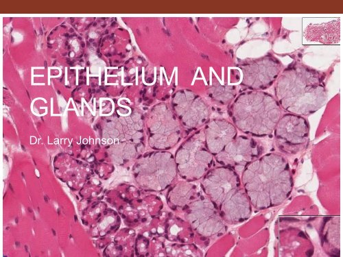
052<br />
<strong>EPITHELIUM</strong> <strong>AND</strong><br />
GL<strong>AND</strong>S<br />
Dr. Larry Johnson
Objectives<br />
• Identify every epithelium present in any tissue section.<br />
• Differentiate between mucus and serous secreting<br />
epithelia.<br />
• Identify single-celled glands, endocrine glands and the<br />
various types of exocrine glands.<br />
• Detail the structure of the sebaceous gland.<br />
• Identify the different types of sweat glands and distinguish<br />
the duct from the secretory region.<br />
• Identify myoepithelial cells and know their function.<br />
From: Douglas P. Dohrman and TAMHSC Faculty 2012 Structure and Function of Human Organ Systems, Histology<br />
Laboratory Manual
ORIGIN <strong>AND</strong> DISTRIBUTION OF<br />
<strong>EPITHELIUM</strong><br />
ECTODERM - EPIDERMIS OF SKIN <strong>AND</strong> <strong>EPITHELIUM</strong><br />
OF CORNEA TOGETHER COVERS THE ENTIRE<br />
SURFACE OF THE BODY; SEBACEOUS <strong>AND</strong><br />
MAMMARY GL<strong>AND</strong>S<br />
ECTODERM<br />
ENDODERM - ALIMENTARY TRACT,<br />
LIVER, PANCREAS, GASTRIC<br />
GL<strong>AND</strong>S, INTESTINAL GL<strong>AND</strong>S<br />
• ENDOCRINE GL<strong>AND</strong>S - LOSE<br />
CONNECTION WITH SURFACE<br />
MESODERM<br />
MESODERM<br />
• ENDOTHELIUM - LINING OF BLOOD VESSELS<br />
• MESOTHELIUM - LINING SEROUS CAVITIES<br />
ENDODERM
Characteristics of epithelium<br />
• Classification by # of layers<br />
• Simple = single layer<br />
• Stratified = multiple, stacked<br />
layers<br />
• Pseudostratified = appears to be<br />
multiple layers, but all cells<br />
touch basement membrane (all<br />
cells do not necessarily reach<br />
lumen)<br />
• Classification by shape<br />
• Squamous = flat<br />
• Cuboidal = square<br />
• Columnar = column
Laboratory Experience<br />
• Identify and characterize various types of epithelia<br />
• Recognize various glands and modes of secretion<br />
• Recognize gland associated structures/cells and differentiate glands<br />
from their ducts<br />
• Understand significance of cytological expression of epithelial cells<br />
with regard to function<br />
053<br />
Slide 53<br />
Slide 35<br />
Slide 93
Slide 33: Kidney (PAS/hematoxylin)<br />
Periodic acid – Schiff<br />
• Used to visualize: glycogen, glycoproteins, and glycosaminoglycans<br />
• Steps in PAS staining<br />
1. Periodic acid oxidizes 1,2-glycols to aldehydes<br />
2. Schiff’s reagent colors the aldehyde groups (pink to magenta)<br />
Brush border<br />
Basement<br />
membrane<br />
The brush border is composed of<br />
numerous microvilli which function<br />
to increase surface area for<br />
absorption of nutrient material and<br />
fluid.<br />
The structure on the brush border<br />
that stains with periodic-acid Schiff<br />
(PAS) stain is the glycocalyx.
EM 1 & 7<br />
Slide 33<br />
glycocalyx<br />
PAS<br />
staining<br />
Lipid in SER<br />
Observe various brush border - microvilli<br />
The brush border is composed of numerous microvilli of a<br />
uniform size projecting into a lumen. Each microvillus is<br />
composed of microfilaments (actin) surrounded by the cell<br />
membrane.
EM 2<br />
Stereocilia of the epididymis - long thin microvilli<br />
Stereocilia are very long, branched, non-motile microvilli of<br />
differing lengths that function in absorption from the lumen.
EM 5 & 6<br />
Observe cell junctions characteristic of epithelium
SPECIALIZATION OF EPITHELIA<br />
MAINTAIN EXTENSIVE<br />
CONTACTS AMONG CELLS<br />
STRUCTURALLY <strong>AND</strong><br />
FUNCTIONALLY POLARIZED<br />
JUNCTIONS<br />
ZONULA OCCLUDENS - TIGHT<br />
JUNCTION (BELT)<br />
ZONULA ADHERENS –<br />
ADHERING BELT<br />
DESMOSOME (MACULA<br />
ADHERENS) - SPOT ATTACHMENT<br />
GAP JUNCTIONS - COMMUNICATION
Terminal bars<br />
32409
Slide 32: Kidney (H&E)<br />
Simple squamous<br />
Simple cuboidal<br />
Simple squamous<br />
Simple cuboidal
Slide 75: Thyroid gland (endocrine)<br />
Simple cuboidal epithelium<br />
Basement membrane
SUMMARY OF TISSUE FEATURES OF<br />
<strong>EPITHELIUM</strong><br />
• AVASCULAR<br />
• EXTRANEOUS CELLS<br />
32409<br />
• REGENERATION<br />
• MIGRATION
SUMMARY OF TISSUE FEATURES OF<br />
<strong>EPITHELIUM</strong><br />
• AVASCULAR<br />
• EXTRANEOUS CELLS<br />
Slide 258<br />
• REGENERATION<br />
• MIGRATION<br />
• METAPLASIA<br />
• BASEMENT MEMBRANE
SECRETION – ACTIVE PROCESS<br />
CONSUMING ENERGY<br />
EXOCRINE GL<strong>AND</strong>S - DELIVER<br />
THEIR SECRETION INTO<br />
DUCTS OPENING INTO<br />
EXTERNAL OR INTERNAL<br />
SURFACE<br />
ENDOCRINE GL<strong>AND</strong>S -<br />
DUCTLESS, DELIVER THEIR<br />
SECRETIONS INTO THE LYMPH<br />
OR BLOOD STREAM<br />
PANCREAS<br />
has exocrine<br />
PANCREAS<br />
has endocrine
EXOCRINE GL<strong>AND</strong>S<br />
DUCT<br />
• SIMPLE - UNBRANCHED DUCT<br />
• COMPOUND - BRANCHED DUCT<br />
SECRETORY PORTION<br />
• TUBULAR<br />
• COILED TUBULAR<br />
• BRANCHED TUBULAR<br />
• ALVEOLAR<br />
• BRANCHED ACINAR<br />
• TUBULOACINAR<br />
• TUBULOALVEOLAR<br />
MUCUS VS SEROUS
TUBULAR<br />
COILED TUBULAR<br />
BRANCHED TUBULAR<br />
ALVEOLAR<br />
BRANCHED ACINAR<br />
TUBULOACINAR<br />
TUBULOALVEOLAR
ACINUS = FUNCTIONAL UNIT<br />
072<br />
Mucous - Light staining<br />
Cytoplasm and dark, flattened<br />
nucleus at base of cell<br />
MUCOUS<br />
SEROUS<br />
072<br />
Serous – dark red staining cytoplasm and<br />
lighter, spherical nucleus
Slide 072: Submandibular gland<br />
Simple columnar epithelia<br />
Stratified columnar epithelia
Slide 072: Submandibular gland<br />
Serous cells<br />
Mucous cells<br />
Serous demilune<br />
The submandibular gland is a mixed (seromucous) gland whose mode of<br />
secretion is merocrine secretion = exocytosis without loss of cellular components
MECHANISM FOR RELEASE OF<br />
SECRETORY PRODUCTS<br />
MEROCRINE SECRETION – EXOCYTOSIS W/O LOSS OF<br />
SURFACE MEMBRANE<br />
APOCRINE SECRETION – LOSS OF PART OF APICAL<br />
CYTOPLASM <strong>AND</strong> SOME PLASMA MEMBRANE<br />
HOLOCRINE SECRETION – RELEASE OF WHOLE cell<br />
CYTOCRINE SECRETION –<br />
MELANIN GRANULES<br />
TRANSFERRED FROM<br />
MELANOCYTE TO<br />
KERATINOCYTES
MEROCRINE<br />
APOCRINE
CYTOCRINE SECRETION - PASS MELANIN GRANULES<br />
FROM MELANOCYTES TO KERATINOCYTES
Alternative slide 250<br />
Slide 61: Terminal Ileum<br />
Brush border composed of<br />
microvilli<br />
Microvilli are fingerlike<br />
projections that greatly<br />
increase the surface<br />
area of certain cells to<br />
help increase<br />
absorption. Microvilli<br />
are non-motile and are<br />
composed of a core of<br />
thin microfilaments<br />
called actin.<br />
Goblet cell releasing contents<br />
Simple columnar epithelium
Alterative slide 242<br />
Slide 40: Trachea<br />
Goblet cell releasing<br />
contents<br />
Ciliated pseudostratified<br />
columnar epithelium with<br />
goblet cells<br />
Note the thick basement membrane of the respiratory<br />
pseudostratified columnar epithelium.<br />
The main function of<br />
cilia is to sweep or<br />
move fluids, cells, or<br />
particulate matter<br />
across cell surface<br />
in the lumen as to<br />
remove dust in the<br />
lungs. Microtubules<br />
of the axoneme are<br />
at the core of cilia<br />
and make them<br />
motile.
Slide 93: Epididymis<br />
Pseudostratified columnar epithelium with stereocilia<br />
Microvilli: small, non-motile projections composed of thin microfilaments<br />
Stereocilia: long, non-motile projections (branched microvilli) composed of<br />
thin microfilaments<br />
Cilia: larger, long, motile projections composed of thick microtubules
Slide 35: Urinary bladder<br />
Transitional epithelium<br />
Specialized “dome-shaped”<br />
cells<br />
Basal cells
Slide 34: Ureter<br />
Transitional<br />
epithelium
Stratified squamous epithelium: the<br />
protection epithelium<br />
• Keratinized stratified squamous<br />
(thick or thin)<br />
• Prevent dessication<br />
• Protect against abrasion<br />
• Prevent foreign invasion<br />
• Ex. Slide 29: Thick skin (ventral<br />
surface of finger)<br />
• Non-keratinized stratified<br />
squamous<br />
• Moist lubricated surface<br />
• Ex. Slide 52: Tongue<br />
Slide 029<br />
Slide 52
Slide 52 : Tongue<br />
Non-keratinized stratified<br />
squamous epithelium<br />
Mucous acini<br />
Serous acini<br />
Mucous cells stain light and serous cells stain dark.
Slide 53: Esophagus<br />
Non-keratinized stratified squamous epithelium
Slide 29: Thick skin (ventral surface of finger)<br />
Epidermis with<br />
keratinized stratified<br />
squamous epithelium<br />
Dermis<br />
Hypodermis<br />
The major function of this type of epithelium (thick skin) is for<br />
protection from mechanical stress, but it also prevents dehydration.
Slide 29 : Thick skin (ventral surface of<br />
finger)<br />
Keratinized stratified<br />
squamous epithelium<br />
Polyhedral cells<br />
Cuboidal cells of the stratum<br />
basale<br />
The cells in the stratum<br />
basale serve as stem<br />
cells for the epidermis,<br />
and their progeny<br />
differentiate as they move<br />
away from the base.
Slide 29 : Thick skin (ventral surface of<br />
finger)<br />
Duct of eccrine sweat<br />
gland with stratified<br />
cuboidal epithelium<br />
Eccrine sweat gland<br />
Myoepithelial cells<br />
Myoepithelial cells are eosinophilic because of the presence of a<br />
high density of contractile protein. These cells surround the gland<br />
like a net and expel glandular secretions upon contraction.
Slide 31: Thin skin (scalp)<br />
Sebaceous glands<br />
The mode of secretion<br />
used by sebaceous glands<br />
is holocrine secretion<br />
Keratinized, stratified<br />
squamous epithelium
Slide 66: Recto-anal junction<br />
Anus – stratified squamous<br />
Rectum - simple columnar epithelium
Slide 66 : Recto-anal junction<br />
Sebaceous glands<br />
Apocrine sweat gland<br />
Eccrine sweat gland
GL<strong>AND</strong>S OF EPIDERMAL ORIGIN<br />
SWEAT GL<strong>AND</strong>S<br />
• ECCRINE - COMMON<br />
SWEAT GL<strong>AND</strong> -<br />
LOCAL COOLING<br />
• APOCRINE AXILLARY<br />
REGION - FUNCTION<br />
IN ANIMALS,<br />
discharge in hair follicle
Epithelial tissues of the body<br />
• Use table as guide<br />
• Generalizations<br />
• Entire GI system from gastroesophageal<br />
junction to rectoanal<br />
junction is lined by<br />
simple columnar epithelium<br />
• Cilia is present in most<br />
respiratory passages
EPITHELIA ARE SPECIALIZED FOR<br />
FUNCTIONS<br />
ABSORPTION - INTESTINE<br />
SECRETION - PANCREAS<br />
TRANSPORT - EYE, ENDOTHELIUM IN VESSELS<br />
EXCRETION - KIDNEY<br />
PROTECTION – AGAINST<br />
MECHANICAL<br />
DAMAGE <strong>AND</strong><br />
DEHYDRATION<br />
SENSORY RECEPTION –<br />
PAIN TO AVOID<br />
INJURY, TASTE BUDS,<br />
OLFACTORY, ETC.<br />
CONTRACTION –<br />
MYO<strong>EPITHELIUM</strong>
SURFACE SPECIALIZATIONS OF<br />
EPITHELIA<br />
MICROVILLI - INTESTINE ABSORPTIVE CELL<br />
CILIA - RESPIRATORY <strong>EPITHELIUM</strong><br />
32409<br />
BASAL LAMINA – ALL<br />
<strong>EPITHELIUM</strong><br />
038b<br />
INTERCELLULAR CANALICULUS –<br />
HEPATOCYTE<br />
SECRETORY CANALICULUS –<br />
GASTRIC PARIETAL CELL<br />
FLAGELLA<br />
33
SURFACE SPECIALIZATIONS OF EPITHELIA<br />
INTERCELLULAR CANALICULUS –<br />
HEPATOCYTE
SURFACE SPECIALIZATIONS OF EPITHELIA<br />
SECRETORY CANALICULUS –<br />
GASTRIC PARIETAL CELL
Clinical Correlation<br />
Slide 38<br />
Edward C. Klatt, M.D.<br />
Mercer University School of Medicine<br />
Normal larynx with ciliated<br />
pseudostratified columnar epithelium<br />
Abnormal larynx with stratified<br />
squamous epithelium
Many illustrations in these VIBS Histology YouTube videos were modified<br />
from the following books and sources: Many thanks to original sources!<br />
• Bruce Alberts, et al. 1983. Molecular Biology of the Cell. Garland Publishing, Inc., New York, NY.<br />
• Bruce Alberts, et al. 1994. Molecular Biology of the Cell. Garland Publishing, Inc., New York, NY.<br />
• William J. Banks, 1981. Applied Veterinary Histology. Williams and Wilkins, Los Angeles, CA.<br />
• Hans Elias, et al. 1978. Histology and Human Microanatomy. John Wiley and Sons, New York, NY.<br />
• Don W. Fawcett. 1986. Bloom and Fawcett. A textbook of histology. W. B. Saunders Company, Philadelphia, PA.<br />
• Don W. Fawcett. 1994. Bloom and Fawcett. A textbook of histology. Chapman and Hall, New York, NY.<br />
• Arthur W. Ham and David H. Cormack. 1979. Histology. J. S. Lippincott Company, Philadelphia, PA.<br />
• Luis C. Junqueira, et al. 1983. Basic Histology. Lange Medical Publications, Los Altos, CA.<br />
• L. Carlos Junqueira, et al. 1995. Basic Histology. Appleton and Lange, Norwalk, CT.<br />
• L.L. Langley, et al. 1974. Dynamic Anatomy and Physiology. McGraw-Hill Book Company, New York, NY.<br />
• W.W. Tuttle and Byron A. Schottelius. 1969. Textbook of Physiology. The C. V. Mosby Company, St. Louis, MO.<br />
• Leon Weiss. 1977. Histology Cell and Tissue Biology. Elsevier Biomedical, New York, NY.<br />
• Leon Weiss and Roy O. Greep. 1977. Histology. McGraw-Hill Book Company, New York, NY.<br />
• Nature (http://www.nature.com), Vol. 414:88,2001.<br />
• A.L. Mescher 2013 Junqueira’s Basis Histology text and atlas, 13 th ed. McGraw<br />
• Douglas P. Dohrman and TAMHSC Faculty 2012 Structure and Function of Human Organ Systems, Histology Laboratory<br />
Manual - Slide selections were largely based on this manual for first year medical students at TAMHSC
The End!