ARTICLES undergo radiotherapy and chemotherapy for the treatment of cancer. Such treatments can have severely deleterious effects on the oocytes. By removing and freezing the oocytes, they are not exposed to the cancer treatments. After the patient has recovered from the cancer treatments and wants to start a family, the oocytes can be thawed and fertilized, and the embryos transferred back into her uterus. In the same way that cryopreservation can protect oocytes from cancer treatments, it could also be used to protect oocytes from natural loss and degeneration due to aging. Stachecki and Cohen (2004) have suggested that this may offer an approach to preserving fertility for women wishing to delay reproduction. Oocytes would be collected from young women and then cryopreserved until they are ready to begin their families. Although as yet largely experimental, the pregnancy rates from cryopreserved oocytes are improving. V. CONCLUSIONS There is a tendency for women in industrialized countries to delay having babies until their mid-thirties or later. There are important social and economic reasons for doing so, but it is imperative that women be aware that fertility decreases significantly with age, particularly after 35 years of age. From a purely biological perspective, the best approach to ensuring fertility is for women to have their babies before they have reached their mid-thirties, but for many women, this is not a desirable or even practical option. At any given age, assisted reproduction techniques may improve the chances of becoming pregnant, but cannot make up for the loss of fertility due to the effects of aging on the numbers and quality of oocytes. References Astolfi P, Zonta LA (1999) Risks of preterm delivery and association with maternal age, birth order, and fetal gender. Hum Reprod 14, 2891-4. Barritt JA, Cohen J, Brenner CA (2000) Mitochondrial DNA point mutation in human oocytes is associated with maternal age. Reprod Biomed Online 1, 96-100. Broekmans FJ, Klinkert ER (2004) Female age in ART: when to stop? Gynecol Obstet Invest 58, 225-34. Brzechffa PR, Daneshmand S, Buyalos RP (1998) Sequential clomiphene citrate and human menopausal gonadotrophin with intrauterine insemination: the effect of patient age on clinical outcome. Hum Reprod 13, 2110-4. Dunson DB, Colombo B, Baird DD (2002) Changes with age in the level and duration of fertility in the menstrual cycle. Hum Reprod 17, 1399-403. Ferrara I, Balet R, Grudzinskas JG (2002) Intrauterine insemination with frozen donor sperm. Pregnancy outcome in relation to age and ovarian stimulation regime. Hum Reprod 17, 2320-4. Fisch H (2005) ‘The Male Biological Clock.’ (Free Press: New York) Ford WC, North K, Taylor H, Farrow A, Hull MG, Golding J (2000) Increasing paternal age is associated with delayed conception in a large population of fertile couples: evidence for declining fecundity in older men. The ALSPAC Study Team (Avon Longitudinal Study of Pregnancy and Childhood). Hum Reprod 15, 1703-8. Gindoff PR, Jewelewicz R (1986) Reproductive potential in the older woman. Fertil Steril 46, 989-1001. Hassan MA, Killick SR (2004) Negative lifestyle is associated with a significant reduction in fecundity. Fertil Steril 81, 384-92. Jolly M, Sebire N, Harris J, Robinson S, Regan L (2000) The risks associated with pregnancy in women aged 35 years or older. Hum Reprod 15, 2433-7. Leridon H (2004) Can assisted reproduction technology compensate for the natural decline in fertility with age? A model assessment. Hum Reprod 19, 1548-53. Lim AS, Tsakok MF (1997) Age-related decline in fertility: a link to degenerative oocytes? Fertil Steril 68, 265-71. Mathews T, Hamilton B (2002) ‘Mean age of mother, 1970–2000.’ National Center for Health Statistics, Hyattsville, Maryland. Menken J, Trussell J, Larsen U (1986) Age and infertility. Science 233, 1389-94. Munné S, Sandalinas M, Escudero T, Velilla E, Walmsley R, Sadowy S, Cohen J, Sable D (2003) Improved implantation after preimplantation genetic diagnosis of aneuploidy. Reprod Biomed Online 7, 91-7. Pellestor F (2004) Âge maternel et anomalies chromosomiques dans les ovocytes humains. Med Sci (Paris) 20, 691-6. Piñón R (2002) ‘Biology of Human Reproduction.’ (University Science Books: Sausalito, CA, USA) Scheffer GJ, Broekmans FJ, Dorland M, Habbema JD, Looman CW, te Velde ER (1999) Antral follicle counts by transvaginal ultrasonography are related to age in women with proven natural fertility. Fertil Steril 72, 845-51. Simpson JL, Elias S (1994) Prenatal diagnosis of genetic disorders. In ‘Maternal-fetal Medicine : Principles and Practice’. (Eds RK Robert K. Creasy and R Resnik) pp. 61-87. (W.B. Saunders: Philadelphia) Stachecki J, Cohen J (2004) An overview of oocyte cryopreservation. Reprod BioMed. Online 9, 152–163. Tarin JJ, Gomez-Piquer V, Rausell F, Navarro S, Hermenegildo C, Cano A (2005) Delayed motherhood decreases life expectancy of mouse offspring. Biol Reprod 72, 1336-43. te Velde ER, Pearson PL (2002) The variability of female reproductive ageing. Hum Reprod Update 8, 141-54. U.S. Department of Health and Human Services – Centers for Disease Control and Prevention (2004) ‘2002 Assisted Reproductive Technology Success Rates: National Summary and Fertility Clinic Reports.’ Atlanta, GA, USA. U.S. National Center for Health Statistics (2003) Crude birth rates, fertility rates, and birth rates by age of mother, according to race and Hispanic origin: United States, selected years 1950-2002 .ftp://ftp.cdc.gov/pub/Health_Statistics/NCHS/ Publications/Health_US/hus04tables/Table003.xls Date of access, July 2005. Wiemer KE, Anderson AR, Kyslinger ML, Weikert ML (2002) Embryonic development and pregnancies following sequential culture in human tubal fluid and a modified simplex optimized medium containing amino acids. Reprod Biomed Online 5, 323-7. Wilding M, Fiorentino A, De Simone ML, Infante V, De Matteo L, Marino M, Dale B (2002) Energy substrates, mitochondrial membrane potential and human preimplantation embryo division. Reprod Biomed Online 5, 39-42. Page 22 – Fertility Genetics Magazine • Volume 2 • www.FertMag.com
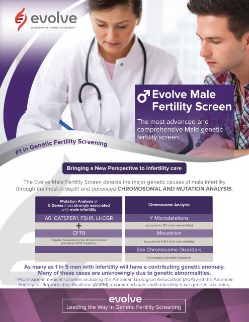
LEADERS IN GENETIC <strong>FERTILITY</strong> SCREENING TM #1 in Genetic Fertility Screening Evolve Male Fertility Screen The most advanced and comprehensive Male genetic fertility screen Bringing a New Perspective to infertility care The Evolve Male Fertility Screen detects the major genetic causes of male infertility through the most in depth and advanced CHROMOSOMAL AND MUTATION ANALYSIS. Mutation Analysis of 5 Genes most strongly associated with male infertility AR, CATSPER1, FSHR, LHCGR + CFTR (Targeted Screening for the 39 most common pan-ethnic CFTR mutations.) Chromosome Analysis Y Microdeletions (accounts for 15% of all male infertility) Mosaicism (accounts for 5-10% of all male infertility) Sex Chromosome Disorders (For example, Klinefelter Syndrome) As many as 1 in 5 men with infertility will have a contributing genetic anomaly. Many of these cases are unknowingly due to genetic abnormalities. * Professional medical societies including the American Urological Association (AUA) and the American Society for Reproductive Medicine (ASRM) recommend males with infertility have genetic screening. Leading the Way in Genetic Fertility Screening