Interpretation of Gram Stains for the Nonmicrobiologist
Interpretation of Gram Stains for the Nonmicrobiologist
Interpretation of Gram Stains for the Nonmicrobiologist

You also want an ePaper? Increase the reach of your titles
YUMPU automatically turns print PDFs into web optimized ePapers that Google loves.
interpretation [microbiology | generalist]<br />
<strong>Interpretation</strong> <strong>of</strong> <strong>Gram</strong> <strong>Stains</strong> <strong>for</strong> <strong>the</strong><br />
<strong>Nonmicrobiologist</strong><br />
Joan Barenfanger, MD, MMB, ABMM, and Cheryl A. Drake, SM(ASCP)<br />
From <strong>the</strong> Department <strong>of</strong> Laboratory Medicine, Memorial Medical Center, Springfield, IL<br />
� Guidelines <strong>for</strong> <strong>the</strong> interpretation <strong>of</strong><br />
<strong>Gram</strong> stains<br />
� Normal flora in respiratory secretions<br />
� Normal flora in <strong>the</strong> female genital tract<br />
� Presumptive identification <strong>of</strong><br />
microorganisms from <strong>Gram</strong> stain<br />
� Correlation <strong>of</strong> findings<br />
Laboratories everywhere are being<br />
asked to do more with less. To enable<br />
<strong>the</strong> laboratory to <strong>of</strong>fer increased services<br />
over an expanded period <strong>of</strong> time,<br />
many technologists with little experience<br />
in microbiology are now asked to<br />
per<strong>for</strong>m and read <strong>Gram</strong> stains. <strong>Interpretation</strong><br />
<strong>of</strong> <strong>Gram</strong> stains is notoriously difficult<br />
<strong>for</strong> nonmicrobiologists because<br />
such interpretation requires multiple<br />
observations and <strong>the</strong> judgment that<br />
comes with years <strong>of</strong> experience. This<br />
article <strong>of</strong>fers objective criteria <strong>for</strong> interpreting<br />
<strong>the</strong> most commonly<br />
encountered <strong>Gram</strong>-stained specimens.<br />
An adequate examination <strong>of</strong> a<br />
<strong>Gram</strong>-stained smear includes observing<br />
numerous representative fields. The<br />
fields containing neutrophils yield <strong>the</strong><br />
most useful in<strong>for</strong>mation. A minimum <strong>of</strong><br />
1 minute should be spent examining a<br />
smear; after that, judgment is needed.<br />
Obviously, a smear from <strong>the</strong> cerebrospinal<br />
fluid (CSF) with neutrophils deserves<br />
more time than an acellular<br />
smear. Similarly, more time should be<br />
spent on a specimen that was obtained<br />
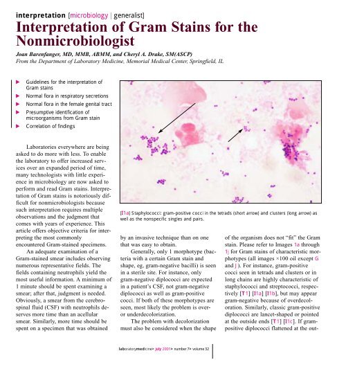
[I1a] Staphylococci: gram-positive cocci in <strong>the</strong> tetrads (short arrow) and clusters (long arrow) as<br />
well as <strong>the</strong> nonspecific singles and pairs.<br />
by an invasive technique than on one<br />
that was easy to obtain.<br />
Generally, only 1 morphotype (bacteria<br />
with a certain <strong>Gram</strong> stain and<br />
shape, eg, gram-negative bacilli) is seen<br />
in a sterile site. For instance, only<br />
gram-negative diplococci are expected<br />
in a patient’s CSF, not gram-negative<br />
diplococci as well as gram-positive<br />
cocci. If both <strong>of</strong> <strong>the</strong>se morphotypes are<br />
seen, most likely <strong>the</strong> problem is overor<br />
underdecolorization.<br />
The problem with decolorization<br />
must also be considered when <strong>the</strong> shape<br />
laboratorymedicine> july 2001> number 7> volume 32<br />
<strong>of</strong> <strong>the</strong> organism does not “fit” <strong>the</strong> <strong>Gram</strong><br />
stain. Please refer to Images 1a through<br />
1j <strong>for</strong> <strong>Gram</strong> stains <strong>of</strong> characteristic morphotypes<br />
(all images ×100 oil except G<br />
and J). For instance, gram-positive<br />
cocci seen in tetrads and clusters or in<br />
long chains are highly characteristic <strong>of</strong><br />
staphylococci and streptococci, respectively<br />
[T1] [I1a] [I1b], but may appear<br />
gram-negative because <strong>of</strong> overdecoloration.<br />
Similarly, classic gram-positive<br />
diplococci are lancet-shaped or pointed<br />
at <strong>the</strong> outside ends [T1] [I1c]. If grampositive<br />
diplococci flattened at <strong>the</strong> out-
Presumptive Identification Based on <strong>Gram</strong> Stain and Morphologic Features<br />
<strong>Gram</strong> Stain Findings<br />
<strong>Gram</strong>-positive cocci in clusters or tetrads<br />
<strong>Gram</strong>-positive cocci in long chains (>5 cocci)<br />
<strong>Gram</strong>-positive diplococci and chains;<br />
occasionally with a capsule<br />
<strong>Gram</strong>-positive cocci in singles, pairs, and short<br />
chains<br />
<strong>Gram</strong>-positive bacilli<br />
<strong>Gram</strong>-positive coccobacilli<br />
<strong>Gram</strong>-negative cocci<br />
<strong>Gram</strong>-negative coccobacilli<br />
<strong>Gram</strong>-negative diplococci<br />
<strong>Gram</strong>-negative rods (bacilli)<br />
CSF, cerebrospinal fluid; GI, gastrointestinal.<br />
©<br />
Presumptive Identification<br />
Staphylococci<br />
Streptococci (all groups, including<br />
Streptococcus viridans) and enterococci<br />
Streptococcus pneumoniae<br />
Staphylococci or streptococci<br />
Lactobacilli<br />
Diph<strong>the</strong>roids<br />
Clostridium organisms<br />
Listeria organisms<br />
—<br />
—<br />
Haemophilus influenzae,<br />
Gardnerella vaginalis<br />
Neisseria organisms, Moraxella organisms<br />
Pseudomonas aeruginosa, enterics including<br />
Escherichia coli, and anaerobes such as<br />
Bacteroides organisms<br />
laboratorymedicine> july 2001> number 7> volume 32<br />
Most Likely Site(s)/Comment<br />
�your lab focus�<br />
T1<br />
Any site including GI and respiratory tract, skin,<br />
blood, and urine<br />
Any site including GI and respiratory tract, skin,<br />
blood, and urine<br />
Any site, especially respiratory, blood, and CSF;<br />
<strong>the</strong> gram-positive diplococci differ from <strong>the</strong><br />
gram-negative diplococci in that <strong>the</strong> <strong>for</strong>mer are<br />
pointed at <strong>the</strong> ends, or lancet shaped, unlike<br />
<strong>the</strong> latter, which are flat or biscuit or kidneybean<br />
shaped<br />
Any site including GI and respiratory tract, skin,<br />
blood, and urine<br />
Lactobacilli are generally long, slender, large<br />
gram-positive bacilli but may be more variable<br />
or may be chains or spirals or short and<br />
coccobacillary <strong>for</strong>ms; <strong>the</strong>y are seen as normal<br />
flora in respiratory and genital sites but rarely<br />
cause infection<br />
Diph<strong>the</strong>roids are usually small, pleomorphic<br />
gram-positive bacilli <strong>of</strong>ten in club-shaped or<br />
angular arrangements (V and L shapes or<br />
"Chinese lettering"); <strong>the</strong>y are <strong>of</strong>ten seen as<br />
normal flora but rarely cause infection<br />
Clostridium organisms are large, box-carshaped<br />
bacilli that may contain spores and may<br />
decolorize easily, appearing to be gramnegative;<br />
<strong>the</strong> finding <strong>of</strong> gram-positive bacilli with<br />
spores in a wound without <strong>the</strong> presence <strong>of</strong><br />
neutrophils is highly suggestive <strong>of</strong> Clostridium<br />
organisms because <strong>the</strong>se organisms have<br />
enzymes that lyse WBCs, thus making <strong>the</strong><br />
smear appear acellular; Clostridium organisms<br />
may be present in wounds, especially with<br />
o<strong>the</strong>r organisms<br />
Listeria are generally small and regular grampositive<br />
bacilli that are seen mostly in CSF<br />
Rarely seen clinically<br />
Rarely seen clinically unless in <strong>the</strong> presence <strong>of</strong><br />
gram-negative diplococci, in which case <strong>the</strong><br />
gram-negative cocci should not be specifically<br />
reported<br />
H influenzae: respiratory tract or CSF<br />
G vaginalis: genital secretions<br />
Neisseria gonorrhoeae: genital and joint<br />
(sexually transmitted disease)<br />
Neisseria meningitidis: CSF and respiratory<br />
secretions<br />
Moraxella organisms: respiratory tract<br />
The gram-negative diplococci differ from <strong>the</strong><br />
gram-positive diplococci in that <strong>the</strong> <strong>for</strong>mer are<br />
flat or biscuit or kidney-bean shaped, unlike <strong>the</strong><br />
latter, which are pointed at <strong>the</strong> ends or lancet<br />
shaped<br />
Any site including GI and respiratory tract, skin,<br />
blood, and urine<br />
369
370<br />
�your lab focus�<br />
[I1b] Streptococci: gram-positive cocci in long (>5 cocci) chains. Note <strong>the</strong> intracellular location.<br />
[I1c] Streptococcus pneumoniae: gram-positive lancet-shaped (pointed ends) diplococci (long<br />
arrow). The capsule (short arrow) is visible as a halo around <strong>the</strong> bacteria, providing ano<strong>the</strong>r clue<br />
that this is S pneumoniae.<br />
side ends are seen, <strong>the</strong> smear might be<br />
underdecolorized because generally<br />
gram-negative diplococcci are kidneybean<br />
or biscuit shaped with flattened<br />
ends [T1] [I1e].<br />
<strong>Interpretation</strong> <strong>of</strong> <strong>Gram</strong> stains should<br />
be based only on areas <strong>of</strong> <strong>the</strong> smear that<br />
take up <strong>the</strong> <strong>Gram</strong> stain properly. Even in<br />
a well-per<strong>for</strong>med stain, a thick smear<br />
may contain areas that are underdecolorized<br />
or overdecolorized. Purple precipitate<br />
is <strong>of</strong>ten present in an area <strong>of</strong><br />
underdecolorization, and <strong>the</strong> nuclei <strong>of</strong><br />
neutrophils are purple. Examining an<br />
area <strong>of</strong> underdecolorization may result<br />
in <strong>the</strong> interpretation <strong>of</strong> gram-negative<br />
bacteria as gram-positive.<br />
The term “gram variable” refers to<br />
organisms that take up <strong>the</strong> positive<br />
(crystal violet) stain variably.<br />
Frequently, organisms such as Clostridium<br />
species will be gram-variable or<br />
even appear frankly gram-negative on<br />
smears made directly from patient spec-<br />
laboratorymedicine> july 2001> number 7> volume 32<br />
©<br />
imens [I1d]. Interestingly, when <strong>the</strong>se<br />
same organisms are grown in <strong>the</strong> laboratory<br />
and <strong>the</strong>n stained, <strong>the</strong>y are<br />
strongly gram-positive. Alternatively,<br />
<strong>the</strong>re are rare instances <strong>of</strong> classically<br />
gram-negative organisms such as<br />
Moraxella and Acinetobacter species<br />
that tend to retain <strong>the</strong> crystal violet<br />
stain and appear to be gram-positive.<br />
Use <strong>of</strong> <strong>the</strong> term gram-variable should<br />
be restricted to cases in which <strong>the</strong> laboratorian<br />
really cannot determine<br />
whe<strong>the</strong>r <strong>the</strong> organism is gram-positive<br />
or gram-negative.<br />
Occasionally, debris and artifacts<br />
mimic organisms. To avoid this problem,<br />
bacteria should not be reported<br />
automatically unless more than 1 is<br />
seen. For instance, if only 1 gram-positive<br />
coccus or even a pair <strong>of</strong> cocci is<br />
seen after examining numerous fields,<br />
one should consider: (1) not reporting<br />
this result, (2) obtaining consultation<br />
with ano<strong>the</strong>r technologist be<strong>for</strong>e reporting,<br />
or (3) reporting “possible” or<br />
“probable” bacteria.<br />
Normal Flora in Respiratory<br />
Secretions<br />
Unless clearly associated with neutrophils<br />
or seen intracellularly, <strong>the</strong> various<br />
organisms <strong>of</strong> normal flora need not<br />
necessarily be reported individually. In<br />
fact, doing so may be confusing and<br />
misleading to <strong>the</strong> clinician who may<br />
interpret <strong>the</strong>se organisms as potential<br />
pathogens simply because “<strong>the</strong> laboratory<br />
reported it.” However, if any single<br />
morphotype (even one that is considered<br />
normal flora) is closely associated<br />
with neutrophils, its presence should be<br />
reported, and if this morphotype is intracellular,<br />
its intracellular location<br />
should also be noted in <strong>the</strong> report.<br />
There are 2 main clues that an organism<br />
is truly intracellular and not<br />
merely lying on top <strong>of</strong> a neutrophil (RT<br />
Thompson, oral communication,<br />
Evanston Hospital, Evanston, IL). First,<br />
<strong>the</strong> organisms may be concentrated<br />
within a neutrophil because phagocytosis<br />
is an active process. Second, <strong>the</strong> organisms<br />
may be seen displacing<br />
something in <strong>the</strong> cytoplasm, usually <strong>the</strong><br />
nucleus or a vacuole. In addition, unless
Morphotypes Expected From Common Sites*<br />
Site<br />
CSF<br />
Joint<br />
Blood †<br />
Respiratory<br />
Wound/abscess<br />
Urine<br />
Genital<br />
CSF, cerebrospinal fluid.<br />
* In nonsterile sites, <strong>the</strong> common potential pathogens are in boldface.<br />
† Unlike <strong>the</strong> o<strong>the</strong>r specimen types, blood is not examined directly <strong>for</strong> organisms. This row describes what is to be expected from blood that has been cultured previously.<br />
©<br />
<strong>Gram</strong> Stain/Organism Expected<br />
<strong>Gram</strong>-negative diplococci (Neisseria<br />
organisms); gram-negative coccobacilli<br />
(Haemophilus organisms); gram-positive cocci<br />
(in long chains and diplococci, streptococci; in<br />
clusters and tetrads, staphylococci); grampositive<br />
rods (Listeria organisms)<br />
<strong>Gram</strong>-positive cocci (in tetrads or clusters,<br />
staphylococci; in long chains and diplococci,<br />
streptococci); gram-negative diplococci<br />
(Neisseria organisms); gram-negative<br />
coccobacilli (Haemophilus organisms); gramnegative<br />
rods (enterics such as Escherichia coli)<br />
<strong>Gram</strong>-positive cocci (especially in clusters and<br />
tetrads, staphylococci; in long chains,<br />
streptococci and enterococcci; diplococci,<br />
Streptococcus pneumoniae); gram-negative<br />
coccobacilli (Haemophilus); gram-negative rods<br />
(Pseudomonas aeruginosa, "enterics" including<br />
Escherichia coli, and anaerobes such as<br />
Bacteroides organisms); gram-positive bacilli<br />
(Clostridium or Listeria or diph<strong>the</strong>roids)<br />
<strong>Gram</strong>-positive cocci (especially diplococci,<br />
S pneumoniae; in chains, streptococci; in<br />
clusters and tetrads, staphylococci); gramnegative<br />
diplococci (Moraxella organisms);<br />
gram-negative coccobacilli (Haemophilus<br />
organisms); gram-negative rods<br />
(pseudomonads and enterics such as E coli);<br />
gram-positive bacilli (lactobacilli and<br />
diph<strong>the</strong>roids, almost never clinically significant);<br />
large gram-positive oval <strong>for</strong>ms with<br />
budding/hyphae (yeasts)<br />
<strong>Gram</strong>-positive cocci (especially in clusters and<br />
tetrads, staphylococci; in long chains,<br />
streptococci and enterococcci); gram-negative<br />
rods (Pseudomonas aeruginosa, enterics<br />
including E coli, and anaerobes such as<br />
Bacteroides organisms); gram-positive bacilli<br />
(Clostridium organisms and diph<strong>the</strong>roids)<br />
<strong>Gram</strong>-negative rods (pseudomonads and<br />
enterics such as E coli); gram-positive cocci<br />
(especially in long chains, streptococci and<br />
enterotococci; in clusters and tetrads,<br />
staphylococci); large gram-positive oval <strong>for</strong>ms<br />
with budding/hyphae (yeasts)<br />
<strong>Gram</strong>-negative diplococci (Neisseria organisms)<br />
gram-negative coccobacilli (Gardnerella<br />
organisms); gram-negative rods (enterics such<br />
as E coli and pseudomonads); gram-positive<br />
cocci (especially in long chains, enterococci<br />
and streptococci; in clusters and tetrads,<br />
staphylococci); gram-positive bacilli (lactobacilli,<br />
which almost never cause disease); large grampositive<br />
(or variable) oval <strong>for</strong>ms with<br />
budding/hyphae (yeasts)<br />
laboratorymedicine> july 2001> number 7> volume 32<br />
Comment<br />
�your lab focus�<br />
In a sterile site, expect only 1 morphotype;<br />
Staphylococcus epidermidis is generally<br />
expected if <strong>the</strong> CSF is from a shunt (here this<br />
organism is usually not a contaminant);<br />
streptococci and Listeria organisms are<br />
expected in infants and in elderly people<br />
In a sterile site, expect only 1 morphotype;<br />
staphylococci are <strong>the</strong> most common organisms<br />
in this site; Neisseria organisms would be<br />
expected only in a sexually active patient<br />
In a sterile site, expect only 1 morphotype<br />
Normal oral flora include: gram-positive cocci,<br />
gram-positive bacilli, gram-negative diplococci,<br />
gram-negative coccobacilli, gram-negative<br />
bacilli, and even yeasts; gram-negative bacilli<br />
are frequent causes <strong>of</strong> hospital-acquired<br />
pneumonia<br />
More than 1 morphotype may be present<br />
—<br />
T2<br />
Normal female flora include: gram-positive<br />
cocci, gram-positive bacilli, and gram-negative<br />
bacilli; Neisseria organisms would be expected<br />
in a sexually active or sexually abused patient;<br />
gram-negative diplococci are diagnostically<br />
significant <strong>for</strong> gonococcal infection only in a<br />
urethral smear from a man; women may have<br />
saprophytic gram-negative diplococci in <strong>the</strong><br />
genital tract; bacterial vaginosis<br />
characteristically has: (1) an absence <strong>of</strong><br />
inflammatory cells; (2) a decreased number <strong>of</strong><br />
gram-positive lactobacilli morphotypes; (3) an<br />
excess number <strong>of</strong> pleomorphic gram-negative<br />
bacilli, curved rods, and coccobacilli; and (4)<br />
clue cells<br />
371
372<br />
�your lab focus�<br />
[I1d] Clostridium species: gram-variable bacilli with spores (long arrows). Note <strong>the</strong> lack <strong>of</strong><br />
neutrophils, which were most likely lysed by <strong>the</strong> enzymes <strong>of</strong> <strong>the</strong> bacteria. The inset shows <strong>the</strong> <strong>Gram</strong><br />
stain <strong>of</strong> <strong>the</strong> same organism grown in <strong>the</strong> laboratory. Note that most <strong>of</strong> <strong>the</strong> bacilli are taking up <strong>the</strong><br />
<strong>Gram</strong> stain (short arrows), making <strong>the</strong>m easy to interpret as gram-positive bacilli.<br />
[I1e] Moraxella/Neisseria species: gram-negative diplococci with kidney-bean or biscuit-shaped<br />
ends (arrow). Note <strong>the</strong> intracellular location.<br />
<strong>the</strong> infection is polymicrobial, only 1<br />
morphotype will be seen intracellularly.<br />
The morphotypes <strong>of</strong> normal oral<br />
flora seen in respiratory specimens are<br />
gram-positive cocci, gram-positive bacilli,<br />
gram-negative diplococci, gram-negative<br />
coccobacilli, gram-negative bacilli, and<br />
even a very few yeasts [T2]. With <strong>the</strong> exceptions<br />
described in <strong>the</strong> following paragraph,<br />
<strong>the</strong>se organisms can be considered<br />
and reported as “bacteria consistent with<br />
normal oral flora.” Some specimens, notably<br />
sputum specimens, can contain<br />
anaerobes as part <strong>of</strong> normal flora. Caution<br />
should be used in reporting large<br />
gram-positive rods and/or fusi<strong>for</strong>m gramnegative<br />
rods. These may be included as<br />
part <strong>of</strong> <strong>the</strong> “bacteria consistent with normal<br />
oral flora.” All sputum specimens<br />
should be screened by <strong>Gram</strong> stain <strong>for</strong> acceptability<br />
be<strong>for</strong>e routine culturing. This<br />
process is well reviewed elsewhere. 1-4 As<br />
laboratorymedicine> july 2001> number 7> volume 32<br />
©<br />
part <strong>of</strong> this screen, a search <strong>for</strong><br />
neutrophils should be made on low power<br />
[I1j]. Generally, in a nonsterile specimen<br />
such as sputum, <strong>the</strong> only bacteria that are<br />
<strong>of</strong> interest are found closely associated<br />
with neutrophils.<br />
There is one exception to lumping<br />
<strong>the</strong> morphotypes <strong>of</strong> normal flora in sputum<br />
specimens ra<strong>the</strong>r than reporting <strong>the</strong>m<br />
individually. Sometimes organisms that<br />
are present in normal flora are also important<br />
causes <strong>of</strong> community-acquired<br />
pneumonia (eg, Streptococcus pneumoniae,<br />
Moraxella catarrhalis, and Haemophilus<br />
influenzae) or nosocomial<br />
pneumonia (eg, gram-negative bacilli).<br />
The specific morphotypes <strong>of</strong> normal flora<br />
should be reported: (1) if <strong>the</strong> bacteria are<br />
seen in moderate numbers and are associated<br />
with neutrophils; (2) if <strong>the</strong>y are seen<br />
intracellularly; or (3) if more than 2 to 3<br />
yeasts are seen, in which case <strong>the</strong>y should<br />
be specifically reported.<br />
For instance, morphotypes <strong>of</strong> M<br />
catarrhalis (gram-negative diplococci)<br />
are considered normal flora [T1] [T2]<br />
[I1e]. If <strong>the</strong>y predominate and are seen<br />
with neutrophils or (more significantly)<br />
are seen intracellularly within<br />
neutrophils, <strong>the</strong> report should specifically<br />
note <strong>the</strong> presence <strong>of</strong> <strong>the</strong> gramnegative<br />
diplococci, quantitate <strong>the</strong>m,<br />
and note <strong>the</strong>ir intracellular location.<br />
Noting in <strong>the</strong> report an intracellular location<br />
<strong>of</strong> an organism alerts <strong>the</strong> clinician<br />
that this organism is causing<br />
infection ra<strong>the</strong>r than simply colonizing<br />
<strong>the</strong> patient. [T2] indicates <strong>the</strong> common<br />
potential pathogens by specimen type.<br />
Normal Flora in <strong>the</strong> Female<br />
Genital Tract<br />
A wide variety <strong>of</strong> bacterial flora is<br />
found normally in <strong>the</strong> female genital<br />
tract, including gram-positive cocci in<br />
singles, pairs, chains, clusters, and<br />
tetrads, gram-positive bacilli that appear<br />
slender (lactobacilli), pleomorphic<br />
(diph<strong>the</strong>roids) or large box-car shaped<br />
(Clostridium organisms), gram-negative<br />
bacilli, gram-negative pleomorphic<br />
bacilli, gram-negative coccobacilli, and<br />
even a few yeasts. Although some<br />
saprophytic Neisseria species or organisms<br />
that look like Neisseria
gonorrhoeae on <strong>Gram</strong> stain and yeasts<br />
are occasionally seen in <strong>the</strong> genital<br />
tract, <strong>the</strong>y should be reported because<br />
<strong>the</strong>y share morphotypic characteristics<br />
with <strong>the</strong> common genital pathogens, N<br />
gonorrhoeae and Candida organisms. 5-8<br />
If extracellular organisms resembling<br />
<strong>the</strong> morphotypes <strong>of</strong> N gonorrhoeae are<br />
seen, <strong>the</strong> microscopist should continue<br />
examining <strong>the</strong> slide <strong>for</strong> intracellular<br />
gram-negative diplococci, which have<br />
more diagnostic significance.<br />
The predominance <strong>of</strong> slender grampositive<br />
bacilli (which are most likely<br />
lactobacilli) is normal. The <strong>Gram</strong> stain is<br />
an important component in making <strong>the</strong><br />
clinical diagnosis <strong>of</strong> bacterial vaginosis.<br />
The <strong>Gram</strong> stain in a patient with bacterial<br />
vaginosis has <strong>the</strong> following characteristics:<br />
(1) an absence <strong>of</strong> inflammatory<br />
cells; (2) a decreased number <strong>of</strong> grampositive<br />
lactobacilli morphotypes; (3) an<br />
excess number <strong>of</strong> pleomorphic gramnegative<br />
bacilli, curved rods, and coccobacilli;<br />
and (4) clue cells [I1g]. 9,10<br />
Clue cells are squamous epi<strong>the</strong>lial cells<br />
<strong>the</strong> borders <strong>of</strong> which are obscured by<br />
bacteria, classically by gram-negative<br />
coccobacilli, <strong>the</strong> morphotype characteristic<br />
<strong>of</strong> Gardnerella vaginalis [T1].<br />
Presumptive Identification<br />
Given a specific site and <strong>the</strong> presence<br />
<strong>of</strong> a characteristic organism, an<br />
educated guess can be made about its<br />
identity. Presumptive identification is<br />
not necessary or even expected in a report<br />
<strong>of</strong> a <strong>Gram</strong> stain. However, as part<br />
<strong>of</strong> self-imposed quality assurance, it is<br />
wise to consider presumptive identification<br />
when interpreting a <strong>Gram</strong> stain.<br />
While examining a smear from a specific<br />
site, <strong>the</strong> laboratorian should ask:<br />
“Is what I am seeing expected? Are<br />
<strong>the</strong>se gram-negative coccobacilli suggestive<br />
<strong>of</strong> H influenzae [T1] [I1f] expected<br />
from a wound?” According to<br />
[T1] and [T2], this is an unexpected<br />
finding. In that case, reexamining <strong>the</strong><br />
smear to verify <strong>the</strong> presence <strong>of</strong> gramnegative<br />
coccobacilli or consultation<br />
with ano<strong>the</strong>r technologist would<br />
improve confidence in <strong>the</strong> report and its<br />
quality, even if gram-negative<br />
coccobacilli are indeed present in <strong>the</strong><br />
wound. (In fact, wounds can be caused<br />
by o<strong>the</strong>r less common bacteria not covered<br />
in this article).<br />
As ano<strong>the</strong>r example, a sputum<br />
specimen with many lancet-shaped<br />
gram-positive diplococci with a<br />
surrounding capsule will most likely<br />
grow S pneumoniae [T1] [I1c]. Finding<br />
many neutrophils associated with <strong>the</strong>se<br />
bacteria makes <strong>the</strong> likelihood even<br />
©<br />
laboratorymedicine> july 2001> number 7> volume 32<br />
�your lab focus�<br />
[I1f] Haemophilus species: gram-negative coccobacilli. Some <strong>for</strong>ms appear coccoid (short arrows),<br />
o<strong>the</strong>rs more rod-like (long arrows).<br />
[I1g] Clue cell: Squamous epi<strong>the</strong>lial cell with borders (arrows) obscured by gram-negative<br />
coccobacilli, most likely Gardnerella species (×40). Note <strong>the</strong> predominance <strong>of</strong> gram-negative<br />
bacteria and <strong>the</strong> absence <strong>of</strong> both inflammatory cells and <strong>the</strong> morphotypes <strong>of</strong> normal flora, eg,<br />
gram-positive bacilli.<br />
higher. Similarly, <strong>the</strong> presence <strong>of</strong> gramnegative<br />
diplococci in a CSF specimen<br />
suggests Neisseria meningitidis, not N<br />
gonorrhoeae or M catarrhalis, all <strong>of</strong><br />
which may look identical to each o<strong>the</strong>r<br />
on <strong>Gram</strong> stain [T1] [I1e]. Neisseria<br />
gonorrhoeae would be expected in genital<br />
secretions (or even a joint fluid),<br />
and M catarrhalis would be expected in<br />
respiratory secretions [T1] [T2].<br />
373
374<br />
�your lab focus�<br />
[I1h] Enterobacteriaceae species and pseudomonads: gram-negative rods. Enterobacteriaceae<br />
organisms are <strong>of</strong>ten larger rods (long arrows); smaller rods (short arrows) are suggestive <strong>of</strong><br />
pseudomonads.<br />
[I1i] Yeasts: gram-positive and gram-negative (or gram-variable) large budding yeasts (short black<br />
arrow) and pseudohyphal <strong>for</strong>ms (long black arrow), which are mostly decolorized. Left inset, Note<br />
<strong>the</strong> size <strong>of</strong> <strong>the</strong> budding yeast (red arrow) compared with <strong>the</strong> gram-negative bacteria below. Right<br />
inset, A gram-positive extracellular yeast (long green arrow) and intracellular yeasts (short green<br />
arrow).<br />
If <strong>the</strong> gram-positive cocci are<br />
arranged in clusters or in tetrads,<br />
staphylococci are most likely present<br />
[T1] [I1a]. If long chains (>5 cocci) <strong>of</strong><br />
gram-positive cocci are present, streptococci<br />
are most likely [T1] [I1b]. Fur<strong>the</strong>r,<br />
if <strong>the</strong>re are pairs <strong>of</strong> cocci that are<br />
pointed at <strong>the</strong> outside ends, S pneumo-<br />
niae is likely [T1] [I1c]. Frequently,<br />
gram-positive cocci occur only in singles,<br />
pairs, and short chains, preventing<br />
reliable differentiation between staphylococci<br />
and streptococci (or enterococci).<br />
Although staphylococci are<br />
<strong>of</strong>ten larger than streptococci, this is<br />
not a characteristic reliable enough <strong>for</strong><br />
laboratorymedicine> july 2001> number 7> volume 32<br />
©<br />
differentiation. Similarly, although Enterobacteriaceae<br />
organsims are <strong>of</strong>ten<br />
larger than pseudomonads, this is not a<br />
characteristic sufficiently reliable <strong>for</strong><br />
differentiation [I1h].<br />
Although not seen commonly,<br />
Clostridium organisms in <strong>Gram</strong> stains<br />
are included here because <strong>of</strong> <strong>the</strong>ir clinical<br />
importance. If gram-positive (or<br />
gram-variable) bacilli contain spores,<br />
Clostridium or Bacillus species are<br />
likely [T1] [I1d]. Although Clostridium<br />
organisms are classically gram-positive<br />
bacilli that may or may not contain<br />
spores, in our experience <strong>the</strong>y <strong>of</strong>ten<br />
stain gram-negative in clinical specimens.<br />
Because some Clostridium organisms<br />
have enzymes that lyse <strong>the</strong><br />
host’s cells, <strong>the</strong>y are <strong>of</strong>ten seen in<br />
smears without neutrophils.<br />
Although <strong>the</strong> <strong>Gram</strong> stain is used <strong>for</strong><br />
detection and differentiation <strong>of</strong> bacteria,<br />
o<strong>the</strong>r microorganisms, most frequently<br />
yeasts and fungi, can be seen on a<br />
<strong>Gram</strong>-stained smear. Like Clostridium<br />
organisms, yeasts can appear gram-positive<br />
or gram-negative [I1i]. Yeasts are<br />
generally at least 10 to 20 times <strong>the</strong> size<br />
<strong>of</strong> bacteria [I1i], so differentiation from<br />
bacteria is not a problem. However,<br />
yeasts are <strong>the</strong> size <strong>of</strong> crystal violet precipitate,<br />
which is occasionally present.<br />
This large purple structure can even appear<br />
to be budding. Crystal violet precipitate<br />
can be differentiated from yeasts<br />
because (1) <strong>the</strong> precipitate may be present<br />
in an area with several o<strong>the</strong>r purple<br />
artifacts <strong>of</strong> various size and shape, (2)<br />
<strong>the</strong> precipitate has a homogenous deeppurple<br />
color while <strong>the</strong> yeast is <strong>of</strong>ten<br />
mottled, and (3) <strong>the</strong> precipitate may be<br />
perfectly round while Candida species,<br />
<strong>the</strong> yeast most commonly encountered,<br />
are generally oval.<br />
Correlation <strong>of</strong> Findings<br />
[T1] contains in<strong>for</strong>mation on which<br />
species <strong>the</strong> common morphotypes most<br />
likely represent, and [T2] contains in<strong>for</strong>mation<br />
on which site is expected <strong>for</strong><br />
a given morphotype. Correlating in<strong>for</strong>mation<br />
from both <strong>of</strong> <strong>the</strong>se tables will<br />
help <strong>the</strong> laboratorian better interpret <strong>the</strong><br />
<strong>Gram</strong>-stained smear. For instance, if<br />
gram-negative diplococci are seen in a
joint fluid specimen, <strong>the</strong> expected<br />
species represented by this finding is N<br />
gonorrhoeae. However, if this specimen<br />
were from a 70-year-old woman (who<br />
would be unlikely to have a sexually<br />
transmitted disease), one may want to<br />
reconsider. Are <strong>the</strong> bacteria really<br />
gram-negative diplococci, or are <strong>the</strong>y<br />
decolorized gram-positive cocci? A<br />
more extensive examination <strong>of</strong> <strong>the</strong> slide<br />
may reveal gram-positive cocci in clusters<br />
or tetrads, indicating <strong>the</strong> presence<br />
<strong>of</strong> Staphylococcus aureus, a more likely<br />
joint pathogen in an elderly patient.<br />
O<strong>the</strong>r sources <strong>of</strong> help in interpreting,<br />
and competency testing <strong>for</strong>, <strong>Gram</strong>stained<br />
specimens include both compact<br />
disks and color atlases. 11,12<br />
Summary<br />
If <strong>the</strong> laboratorian’s interpretation<br />
is not consistent with <strong>the</strong> guidelines<br />
presented in [T1] [T2], <strong>the</strong> interpretation<br />
should be reconsidered or consultation<br />
should be sought. Possibly over- or<br />
underdecolorization has occurred. Although<br />
<strong>the</strong>se guidelines give objective<br />
criteria <strong>for</strong> interpreting <strong>the</strong> most commonly<br />
encountered <strong>Gram</strong> stains, <strong>the</strong>re<br />
will be exceptions. The quality <strong>of</strong> <strong>the</strong><br />
reports and confidence in <strong>the</strong> reports<br />
will improve if consultation with ano<strong>the</strong>r<br />
technologist occurs be<strong>for</strong>e reporting<br />
such findings.<br />
Acknowledgment<br />
This article was part <strong>of</strong> a project<br />
supported by <strong>the</strong> Memorial Medical<br />
Foundation, Springfield, IL.<br />
1. Wilson ML. Clinically relevant, cost effective<br />
clinical microbiology. Am J Clin Pathol.<br />
1997;107:154-167.<br />
©<br />
2. Infections in <strong>the</strong> lower respiratory tract. In: Forbes<br />
BD, Sahm D, Weissfeld A. Bailey and Scott’s<br />
Diagnostic Microbiology. 10th ed. St Louis, MO:<br />
Mosby; 1998:318.<br />
3. Reisner BS, Woods GL, Thomson RB, et al.<br />
Specimen processing. In: Murray PR, ed. Manual<br />
<strong>of</strong> Clinical Microbiology. 7th ed. Washington DC:<br />
ASM Press; 1999:72.<br />
4. Introduction to microbiology, part I: <strong>the</strong> role <strong>of</strong> <strong>the</strong><br />
microbiology laboratory in <strong>the</strong> diagnosis <strong>of</strong><br />
infectious diseases: guidelines to practice and<br />
management. In: Koneman EW, Allen SD, Janda<br />
WM, et al, eds. Diagnostic Microbiology. 5th ed.<br />
New York, NY: Lippincott; 1997:83.<br />
5. Neisseria species and Moraxella catarrhalis. In<br />
Koneman EW, Allen SD, Janda WM, et al, eds.<br />
Diagnostic Microbiology. 5th ed. New York, NY:<br />
Lippincott; 1997:519.<br />
6. Neisseria and Moraxella catarrhalis. In: Forbes<br />
BD, Sahm D, Weissfeld A. Bailey and Scott’s<br />
Diagnostic Microbiology. 10th ed. St Louis, MO:<br />
Mosby; 1998:599.<br />
7. Knapp JS, Totten PA, Mulks MH, et al.<br />
Characterization <strong>of</strong> Neisseria cinerea, a<br />
nonpathogenic species isolated on Morton-Lewis<br />
medium selected <strong>for</strong> pathogenic Neisseria spp. J<br />
Clin Microbiol. 1984;19:63-67.<br />
8. Dossett JH, Appelbaum PC, Knapp JS, et al.<br />
Proctitis associated with Neisseria cinerea<br />
misidentified as Neisseria gonorrhoeae in a child.<br />
J Clin Microbiol. 1985;21:575-577.<br />
laboratorymedicine> july 2001> number 7> volume 32<br />
�your lab focus�<br />
[I1j] Left, A sputum specimen that should be rejected. Note <strong>the</strong> numerous squamous epi<strong>the</strong>lial cells<br />
(black arrows) and <strong>the</strong> absence <strong>of</strong> neutrophils. Right, Acceptable sputum specimen. Note <strong>the</strong><br />
numerous neutrophils (green arrow) and <strong>the</strong> absence <strong>of</strong> squamous epi<strong>the</strong>lial cells (×10).<br />
9. Nugent RP, Krohn MA, Hillier SL. Reliability <strong>of</strong><br />
diagnosing bacterial vaginosis is improved by a<br />
standardized method <strong>of</strong> <strong>Gram</strong> stain interpretation.<br />
J Clin Microbiol. 1991;29:297-301.<br />
10. Spiegel CA, Amsel R, Eschenbach DA, et al.<br />
Diagnosis <strong>of</strong> bacterial vaginosis by direct <strong>Gram</strong><br />
stain <strong>of</strong> vaginal fluid. J Clin Microbiol.<br />
1983;18:170-177.<br />
11. Marler LM, Siders JA, Allen SD. Direct Smear<br />
Atlas. New York, NY: Lippincott Williams and<br />
Wilkins; 1998.<br />
12. Cookson BT, Orkand AM, Curtis J, et al. <strong>Gram</strong>-<br />
Stain Tutor [CD-ROM, version 2.0]. Seattle, WA:<br />
University <strong>of</strong> Washington; 1999.<br />
Suggested Reading<br />
McClelland R. <strong>Gram</strong>’s stain: <strong>the</strong> key to<br />
microbiology. Med Lab Observer.<br />
2001;33:20-28.<br />
InterNetConnect<br />
Loyola University Medical Center’s <strong>Gram</strong><br />
stain page:<br />
<br />
375