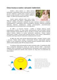
cistus laurifolius
The genus is one of the characteristic genera of the Mediterranean region, colonizing degraded areas (Attaguile et al., 2000). L. (cistaceae) is a common plant in Anatolia and is used against various ailments in traditional medicine. The plant leaves are used to treat rheumatic and related inflammatory diseases, externally as a bath or poultice to reduce pain in rheumatism, against fever in common cold or applied externally as a plaster on the dorsal part of the body in a line of the kidneys for urinary inflammations.
The genus is one of the characteristic genera of the Mediterranean region, colonizing degraded areas (Attaguile et al., 2000). L. (cistaceae) is a common plant in Anatolia and is used against various ailments in traditional medicine. The plant leaves are used to treat rheumatic and related inflammatory diseases, externally as a bath or poultice to reduce pain in rheumatism, against fever in common cold or applied externally as a plaster on the dorsal part of the body in a line of the kidneys for urinary inflammations.

Create successful ePaper yourself
Turn your PDF publications into a flip-book with our unique Google optimized e-Paper software.
Journal of Ethnopharmacology 103 (2006) 455–460<br />
Effect of<br />
L. leaf extracts and flavonoids on<br />
acetaminophen-induced hepatotoxicity in mice<br />
Esra Küpeli , Didem Deliorman Orhan, Erdem Yesilada<br />
Received 26 April 2005; received in revised form 15 August 2005; accepted 19 August 2005<br />
Available online 10 October 2005<br />
In this study, the effect of the flavonoids quercetin-3-methyl-ether (isorhamnetin) ( ), quercetin-3,7-dimethyl-ether ( ) and kaempferol-3,7-<br />
dimethyl-ether ( ) isolated from<br />
L. (cistaceae) leaves was assessed on lipid peroxidation (liver and plasma), cellular glutathione<br />
(GSH) level and plasma AST (aspartate aminotransferase), ALT (alanine aminotransferase) enzyme activities in acetaminophen-induced liver<br />
damage in mice. At 114 mg/kg oral dose quercetin-3,7-dimethyl-ether was shown to possess potent antioxidative activity.<br />
© 2005 Elsevier Ireland Ltd. All rights reserved.<br />
; Cistaceae; Flavonoid; Hepatotoxicity; Lipid peroxidation; Serum transaminase enzymes; Cellular GSH<br />
The genus is one of the characteristic genera of the<br />
Mediterranean region, colonizing degraded areas (Attaguile et<br />
al., 2000).<br />
L. (cistaceae) is a common plant<br />
in Anatolia and is used against various ailments in traditional<br />
medicine. The plant leaves are used to treat rheumatic and related<br />
inflammatory diseases, externally as a bath or poultice to reduce<br />
pain in rheumatism, against fever in common cold or applied<br />
externally as a plaster on the dorsal part of the body in a line of<br />
the kidneys for urinary inflammations (Yeşilada et al., 1997a).<br />
A tea prepared from the leaves is used as hypoglycaemic and the<br />
flowers and buds decoction is used for the treatment of peptic<br />
ulcers in Turkish folk medicine (Sezik et al., 1991; Yesilada et<br />
al., 1995). In previous phytochemical studies,<br />
have been reported to contain flavonoids including quercetin,<br />
kaempferol, apigenin, luteolin and theirs methyl–ethers, the<br />
coumarine scopoletin, diterpenoids, sesquiterpenoids, and sugars<br />
(Demetzos et al., 1989; Vogt et al., 1987; Wollenweber and<br />
Mann, 1984).<br />
Due to the widespread utilization of the plant as a traditional<br />
remedy, it is essential to investigate the potential effects of the<br />
crude drug on hepatic tissue for the evaluation of potential health<br />
Corresponding author. Fax: +90 312 223 50 18.<br />
esrak@gazi.edu.tr (E. Küpeli).<br />
risks to users as well as to disclose the possible antioxidant<br />
effect in the folkloric use. In the present study, the antioxidant<br />
effect of<br />
ethanol extract and fractions<br />
were investigated against acetaminophen-induced liver damage<br />
on subacute administration in mice. In order to assess the<br />
activity, some biochemical plasma and hepatic tissue parameters<br />
including malondialdehyde formation (MDA), transaminase<br />
levels in plasma; aspartate transferase (AST) and alanine transferase<br />
(ALT), and cellular glutathione (GSH) level in hepatic<br />
tissue were measured. Through bioassay-guided fractionation,<br />
the active antioxidant constituent(s) were isolated and identified<br />
by spectroscopy.<br />
NMR spectra were acquired on a JEOL instrument. 1 H NMR:<br />
500 MHz, 13 C NMR: 125 MHz, using TMS as internal standard.<br />
FAB-MS were obtained on a JEOL HX-110A instrument.<br />
The active compounds were isolated after extensive<br />
chromatography using Sephadex LH-20 (25–100 m, Lot<br />
124H0053, Sigma Chem. Co., column size 30 mm 500 mm).<br />
Silica gel (Kieselgel 230–400 mesh, Merck Art. No. 1.09385,<br />
column size 35 mm 750 mm) and reversed phase 18 (LiChroprep<br />
RP-18, Merck, column size 18.5 mm 352 mm) columns.<br />
0378-8741/$ – see front matter © 2005 Elsevier Ireland Ltd. All rights reserved.<br />
doi:10.1016/j.jep.2005.08.038
456<br />
Male Swiss albino mice (20–25 g) were purchased from the<br />
animal breeding laboratories of Refik Saydam Central Institute<br />
of Health (Ankara, Turkey). The animals left for 2 days for<br />
acclimatization to animal room conditions were maintained on<br />
standard pellet diet and water ad libitum. The food was withdrawn<br />
on the day before the experiment, but allowed free access<br />
of water. A minimum of six animals was used in each group.<br />
Throughout the experiments, animals were processed according<br />
to the suggested international ethical guidelines for the care of<br />
laboratory animals.<br />
L. leaves were collected from Bolu,<br />
Dörtdivan in May 2002 and was identified by Prof. Dr. M. Vural<br />
from the Department of Botany, Faculty of Science, Gazi University.<br />
A voucher specimen is deposited in the Herbarium of<br />
Faculty of Pharmacy, Gazi University (GUE-2300).<br />
Some 2 kg of powdered material was extracted three times<br />
with EtOH (10 L) by stirring in a 60 C water bath for 8 days<br />
each. The combined ethanol extract was evaporated to dryness<br />
under reduced pressure to yield ‘EtOH extract’ (315 g).<br />
The EtOH extract was then resuspanded in 1500 ml of MeOH<br />
and extracted with -hexane (7 500 ml). Combined hexane<br />
extract was evaporated under reduced pressure to yield<br />
‘Hexane fraction’ (64.8 g). MeOH was removed from the<br />
remaining solution and diluted with distilled H 2 O to 2000 ml<br />
and further fractionated by successive extractions with chloroform<br />
(7 500 ml), ethyl acetate (5 250 ml) and watersaturated<br />
-butanol (3 200 ml). The extracts as well as the<br />
remaining aqueous phase were evaporated to dryness under<br />
reduced pressure to yield the “CHCl 3 Fr.” (98.4 g), “EtOAc<br />
Fr.” (28.3 g), “BuOH Fr.” (29.7 g) and “R–H 2 O Fr.” (84.5 g),<br />
respectively.<br />
Four grams of the CHCl 3 fraction was permeated on a<br />
Sephadex LH-20 column using MeOH as eluent. Some 43<br />
fractions of 15 ml each were collected. Fractions were compared<br />
by TLC on silica gel using CHCl 3 /MeOH (8:2) and<br />
toluene/ether (1:1) as mobile systems and combined as follows:<br />
Fr.1–13 (1.23 g), Fr.14–19 (0.72 g), Fr.20–30 (0.91 g), Fr.31–43<br />
(1.12 g). Some additional 5.36 g of the CHCl 3 fraction were<br />
worked-up under identical conditions to obtain more material for<br />
further purification. Vacuum-chromatography of the flavonoidenriched<br />
fractions (silica gel; petroleum ether/EtOAc (1:1), (1:2)<br />
and EtOAc/MeOH (8:2) as solvent system) and reverse-phase<br />
(RP-18) chromatography using MeOH/H 2 O yielded three pure<br />
compounds: ( ) (1.23 g), ( ) (0.95 g), ( ) (0.82 g). The structure<br />
of compounds was elucidated by spectroscopic methods as<br />
quercetin-3-methyl-ether ( ), quercetin-3,7-dimethyl-ether ( )<br />
Scheme 1.<br />
and kaempferol-3,7-dimethyl-ether ( ) (Scheme 1). The spectral<br />
data are in agreement with the reported values (Barbera et<br />
al., 1986; Guerrero et al., 2002; Smolarz et al., 2003; Stevens et<br />
al., 1995).<br />
The extract, fractions and pure compounds were suspended<br />
in 0.5% CMC in distilled water prior to oral administration<br />
to experimental animals. Test groups of mice were orally<br />
treated with EtOH extract (500 mg/kg body weight), hexane<br />
(206 mg/kg), CHCl 3 (312 mg/kg), EtOAc (90 mg/kg), -BuOH<br />
(94 mg/kg) or R-H 2 O fractions (268 mg/kg) for 7 following<br />
days, once a day by gastric gavage. The control group<br />
(untreated) and acetaminophen group (positive control) were<br />
administered in 0.5% CMC suspension for the same period.<br />
The reference drug, ascorbic acid (Vitamin C) suspended in<br />
0.5% CMC was directly administered to animals at 100 mg/kg<br />
dose.<br />
Some 60 min after the administration of the last dose on<br />
seventh day, except the control group mice, each of the<br />
acetaminophen group and test group animals was challenged<br />
with the suspension of acetaminophen (800 mg/kg body weight)<br />
in 0.5% CMC to induce hepatic injury (Fairhurst et al., 1982).<br />
Four hours after acetaminophen administration, blood samples<br />
were withdrawn by cardiac puncture and then the mice were sacrificed<br />
by overdose of diethylether. Blood samples collected in<br />
heparinized tubes were centrifuged at 3000 (4 C) for 10 min<br />
to obtain plasma. Plasma samples were used to determine the<br />
lipid peroxide levels and the enzyme (AST, ALT) activity. Liver<br />
of each mouse was promptly removed and used to determine<br />
the tissue levels of malondialdehyde (MDA) and cellular glutathione<br />
(GSH).
457<br />
The methodology described by Kurtel et al. (1992) was<br />
used. Briefly, 1 ml of plasma was mixed with 2.0 ml of<br />
trichloroacetic acid (TCA; 15%, w/v)–thiobarbituric acid (TBA;<br />
0.375%)–0.25 N HCl and mixed throughly and centrifuged at<br />
10,000 for 5 min. The supernatant was mixed with 20 l<br />
of butyl hydroxy toluene (BHT; 0.02% in 95% EtOH, w/v) to<br />
prevent further oxidation and heated for 15 min in a boiling<br />
water bath. After cooling under running water, the flocculent<br />
precipitate was removed by centrifugation at 10,000 for<br />
5 min. The absorbance of the sample was measured at 532 nm<br />
against blank that contained all the reagents except plasma.<br />
1,1,3,3-Tetraethoxypropan was used as standard for the curve<br />
calibration.<br />
with no homogenate. Results were expressed as<br />
tissue.<br />
mol GSH/g<br />
Biocon standard kits and DAX-48 autoanalyzer were used to<br />
measure AST and ALT activities in plasma samples according<br />
to the method of Wilkinson et al. (1972).<br />
Animals employed in the experiments were observed during<br />
48 h and morbidity or mortality was recorded, if happens, for<br />
each group at the end of observation period.<br />
The method of Ohkawa et al. (1979) as modified by Jamall<br />
and Smith (1985) was used to determine lipid peroxidation in tissue<br />
samples. Mice were sacrified by an overdose of diethylether.<br />
The liver of each mouse was immediately excised and chilled in<br />
ice-cold 0.9% NaCl and then perfused via the portal vein with<br />
ice-cold 0.9% NaCl. After washing with 0.9% NaCl, 1.0 g of wet<br />
tissue was weighted exactly and homogenized in 9 ml of 0.25 M<br />
sucrose using a teflon homogenizer to obtain a 10% suspension.<br />
The cytosolic fraction was obtained by a two-step centrifugation<br />
first at 1000 for 10 min and then at 2000 for 30 min<br />
at 4 C. A volume of the homogenate (0.20 ml) was transferred<br />
to a vial and was mixed with 0.2 ml of a 8.1% (w/v) sodium<br />
dodecyl sulphate solution, 1.50 ml of a 20% acetic acid solution<br />
(adjusted to pH 3.5 with NaOH) and 1.50 ml of a 0.8% (w/v)<br />
solution of TBA and the final volume was adjusted to 4.0 ml<br />
with distilled water. Each vial was tightly capped and heated in<br />
boiling water bath for 60 min. The vials were then cooled under<br />
running water.<br />
Equal volumes of tissue blank or test sample and 10%<br />
TCA were transferred into a centrifuge tube and centrifuged at<br />
1000 for 10 min. The absorbance of the supernatant fraction<br />
was measured at 532 nm in a Beckman DU 650 spectrometer.<br />
Control experiment was processed using the same experimental<br />
procedure except the TBA solution was replaced with distilled<br />
water due to the peroxidative effect of acetaminophen on tissue.<br />
Livers of acetaminophen-treated mice were used as positive control<br />
and 1,1,3,3-tetraethoxypropan was used as standard for the<br />
curve calibration.<br />
Some 200 mg of liver was homogenized in 8.0 ml of 0.02 M<br />
EDTA in an ice bath. The homogenates were kept in the ice bath<br />
until used. Aliquots of 5.0 ml of the homogenates were mixed<br />
in 15.0 ml test tubes with 4.0 ml distilled water and 1.0 ml of<br />
50% trichloroacetic acid (TCA). The tubes were centrifuged for<br />
15 min at approximately 3000 . Some 2.0 ml of supernatant<br />
was mixed with 4.0 ml of 0.4 M Tris–buffer, pH 8.9, 0.1 ml Ellman’s<br />
reagent [5,5 -dithiobis-(2-nitro-benzoic acid)] (DTNB)<br />
added, and the sample shaken. The absorbance was read within<br />
5 min of the addition of DTNB at 412 nm against a reagent blank<br />
The data obtained were analyzed by one-way of variance<br />
(ANOVA) and Student–Newman–Keuls post hoc tests for the<br />
significant interrelation between the various groups using Instat<br />
computer software. < 0.05 was considered to be significant<br />
different from the control.<br />
In living systems, dietary antioxidants such as -tocopherol,<br />
ascorbic acid, carotenoids, as well as flavonoids and related<br />
phenolic compounds are suggested in protection from oxidative<br />
damage of tissues in the body and eventually for a healthy<br />
life (Haraguchi, 2001). Especially flavonoids have been shown<br />
to scavenge various reactive oxygen species and implicated as<br />
inhibitors of lipid peroxidation (Mora et al., 1990).<br />
Acetaminophen (paracetamol), a frequently used analgesic<br />
and antipyretic drug, is known to be hepatotoxic in high doses,<br />
which is primarily metabolized by sulfation and glucuronidation<br />
to unreactive metabolites, and then activated by the cytochrome<br />
P-450 system to produce liver injury. It is established that<br />
acetaminophen is bioactivated to a toxic electrophile, -acetyl-<br />
-benzoquinone imine (NAPQI), which binds covalently to tissue<br />
macromolecules, and probably also oxidizes lipids, or the<br />
critical sulphydryl groups (protein thiols) and alters the homeostasis<br />
of calcium (Lin et al., 1997). Post-mitochondrial supernatants<br />
isolated from livers of rats given a single large oral dose<br />
of acetaminophen (800 mg/kg) showed rapid rates of lipid peroxidation<br />
when incubated in vitro. Lipid peroxidation probably<br />
occurs simultaneously with the proposed covalent binding of<br />
the active metabolite of acetaminophen. Since the former process<br />
is known to cause severe and extensive membrane damage,<br />
it may be a very important factor in acetaminophen-induced liver<br />
necrosis (Fairhurst et al., 1982).<br />
As shown in Table 1, in the liver and plasma of<br />
acetaminophen-treated group, tissue and plasma lipid peroxidation<br />
levels (219.2 and 1172%) as evidenced by MDA determination<br />
increased significantly as compared to control group, however,<br />
the content of GSH in liver decreased (8.7%). Additionally,<br />
acetaminophen was found to cause several folds increases in<br />
plasma AST and ALT levels (106 and 71.6%). Ethanol (EtOH)<br />
extract of<br />
leaves administered in 500 mg/kg
458<br />
Table 1<br />
Effect of<br />
EtOH extract, subfractions and the isolated flavonoids on MDA and GSH levels against acetaminophen-induced liver damage in mice<br />
Materials Dose (mg/kg) Plasma MDA level Liver homogenate MDA level Tissue GSH<br />
Plasma (nmol/ml) % Change a Liver (nmol/g) % Change a mol/g % Change a<br />
Control (0.5% CMC) 1.8 0.1 118.2 15.4 106.1 8.1<br />
Acetaminophen b 800 11.4 2.1 *** +1172 377.8 29.6 *** +219.2 96.9 10.6 8.7<br />
Ascorbic acid c 100 4.6 0.2 *** 59.5 183.7 17.9 ** 51.4 126.3 11.5 +30.3<br />
EtOH extract c 500 5.8 0.9 *** 49.3 194.7 19.3 *** 48.5 135.6 7.9 ** +39.9<br />
Hexane Fr. c 206 7.9 1.5 * 31.1 296.1 24.1 * 21.6 117.3 8.6 +21<br />
CHCl 3 Fr. c 312 6.7 0.8 *** 41.3 260.8 16.7 ** 31.0 153.4 6.1 *** +58.3<br />
EtOAc Fr. c 90 7.0 1.0 * 38.4 278.8 14.4 ** 26.2 117.8 7.2 +21.6<br />
-BuOH Fr. c 94 8.7 2.2 23.4 307.8 21.6 18.5 128 8.4 * +32.1<br />
R-H 2 O Fr. c 268 8.9 1.9 21.3 303.2 18.8 19.8 106 4.4 +9.4<br />
Quercetin-3-methyl-ether c 147 6.8 0.6 * 40.3 316.6 12.3 * 16.2 109.7 13.1 +13.2<br />
Quercetin-3,7-dimethyl-ether c 114 6.1 0.4 *** 46.8 300.8 14.8 ** 20.5 115.5 6.3 +19.2<br />
Kaempferol-3,7-dimethyl-ether c 98 7.1 0.5 * 37.5 355.15 14.3 6.0 104.5 9.5 +7.8<br />
Results are presented as mean S.E.M.<br />
a (+) Represents percentage of increase and ( ) decrease when compared to either vehicle or acetaminophen.<br />
b Compared to vehicle control (0.5% CMC).<br />
c Compared to acetaminophen as hepatotoxin.<br />
* Change to significance from control or acetaminophen; < 0.05.<br />
** Change to significance from control or acetaminophen; < 0.01.<br />
*** Change to significance from control or acetaminophen; < 0.001.<br />
dose as well as ascorbic acid, as reference compound, showed<br />
a significant effect at plasma AST, ALT, MDA and liver tissue<br />
MDA, GSH levels (Tables 1 and 2). Besides, we observed that<br />
EtOH extract was more effective than ascorbic acid in plasma<br />
AST levels.<br />
The active EtOH extract was fractionated through successive<br />
solvent-solvent extractions and five fractions namely Hexane<br />
Fr., CHCl 3 Fr., EtOAc Fr., -BuOH Fr. and R-H 2 O Fr. were<br />
obtained.<br />
As shown in Table 1, CHCl 3 Fr. (41.3% for plasma and 31%<br />
for tissue) and, in a lesser degree, EtOAc Fr. (38.4% for plasma<br />
and 26.2% for tissue) possessed a potent activity against both<br />
plasma and tissue MDA levels. Of the five fractions tested, the<br />
CHCl 3 Fr. (58.3%) appeared to be the most effective in increasing<br />
the hepatic GSH levels, as compared to the acetaminophen<br />
group. Additionally, only CHCl 3 (21.5%), EtOAc (10.1%) and<br />
-BuOH (19.6%) fractions produced a decrease in plasma ALT<br />
levels while all of the fractions caused reduction in plasma AST<br />
Table 2<br />
Effect of<br />
in mice<br />
EtOH extract, subfractions and the isolated flavonoids on plasma transaminase enzyme levels against acetaminophen-induced liver damage<br />
Materials Dose (mg/kg) ALT AST<br />
IU (L) % Change a IU (L) % Change a<br />
Control (0.5% CMC) 3474 16 1683 57<br />
Acetaminophen b 800 5960 251 *** +71.6 3468 219 *** +106.0<br />
Ascorbic acid c 100 5864 188 1.6 2108 115 *** 39.2<br />
EtOH extract c 500 4692 113 *** 21.3 2319 119 *** 33.1<br />
Hexane Fr. c 206 6452 114 +8.3 2660 167 * 23.3<br />
CHCl 3 Fr. c 312 4680 115 *** 21.5 2576 235 * 25.7<br />
EtOAc Fr. c 90 5360 240 * 10.1 2194.4 161 ** 36.7<br />
-BuOH Fr. c 94 4792 138 *** 19.6 2796 258 * 19.4<br />
R-H 2 O Fr. c 268 6180 139 +3.7 2656 300 * 23.4<br />
Quercetin-3-methyl-ether c 147 4704 124 ** 21.1 2289.6 121 ** 34.0<br />
Quercetin-3,7-dimethyl-ether c 114 4932 147 *** 17.3 3120 167 10.0<br />
Kaempferol-3,7-dimethyl-ether c 98 4904 124 *** 17.7 2288 150 ** 34.0<br />
Results are presented as mean S.E.M.<br />
a (+) Represents percentage of increase and ( ) decrease when compared to either vehicle or acetaminophen.<br />
b Compared to vehicle control (0.5% CMC).<br />
c Compared to acetaminophen as hepatotoxin.<br />
* Change to significance from control or acetaminophen; < 0.05.<br />
** Change to significance from control or acetaminophen; < 0.01<br />
*** Change to significance from control or acetaminophen; < 0.001
459<br />
levels (19.4–36.7%). The results showed that pretreatment with<br />
CHCl 3 Fr. (in ALT) and EtOAc Fr. (in AST) represented the most<br />
significant alleviation of the plasma enzyme activity induced by<br />
acetaminophen. AST can be generally found in the liver, cardiac<br />
muscle, skeletal muscle, kidneys, brain, pancreas, lungs,<br />
leukocytes, and erythrocytes, whereas, ALT is present in highest<br />
concentration in liver (Rej, 1978). Therefore, the CHCl 3<br />
Fr. was considered to be more effective than the EtOAc Fr. on<br />
acetaminophen-induced liver damage.<br />
According to the biochemical test results, the following<br />
experiments were directed to CHCl 3 Fr. On TLC analysis,<br />
CHCl 3 Fr. was found to be rich in flavonoids. Through successive<br />
column chromatographies three main flavonoids were<br />
isolated, and their structures were elucidated as quercetin-3-<br />
methyl-ether (isorhamnetin) ( ), quercetin-3,7-dimethyl-ether<br />
( ), kaempferol-3,7-dimethyl-ether ( ) by spectral techniques.<br />
Effect of the isolated flavonoid aglycones on acetaminopheninduced<br />
liver damage was also studied using the same biochemical<br />
methods on plasma and liver tissue.<br />
Mice treated with flavonoid significantly reduced<br />
acetaminophen-induced increases in plasma (46.8%) and liver<br />
(20.5%) MDA production compared with acetaminophentreated<br />
mice. On the other hand, flavonoid also caused reduce<br />
of acetaminophen-induced increase in plasma (40.3%) and liver<br />
(16.2%) MDA production. According to these results, flavonoid<br />
is considered to have more inhibitory effect than that of<br />
flavonoid on acetaminophen-induced lipid peroxidation.<br />
Glutathione (GSH) plays an essential role in the detoxification<br />
of acetaminophen and protects hepatocytes by uniting with<br />
reactive metabolites of acetaminophen. Thus, it prevents them<br />
from binding covalently to liver proteins. Intracellular decrease<br />
of the reducted GSH exposes the cell to the destructive effects of<br />
the oxidative stress (Lauterburg and Velez, 1988). In other word,<br />
the hepatic GSH is non-protein reserve of the liver and responsible<br />
in reducing the NAPQI-induced liver damage (Ischiropolus<br />
et al., 1992). The GSH activities showed alleviation of 13.2, 19.2<br />
and 7.8%, respectively, in liver of mice, received flavonoids ,<br />
and , compared with the acetaminophen group.<br />
The rise in serum AST and ALT levels has been attributed<br />
to the damaged structural integrity of the liver (Chenoweth<br />
and Hake, 1962), because these are cytoplasmic in location<br />
and are released into circulation after cellular damage (Sallie<br />
et al., 1991). Pretreatment of mice with flavonoid , and<br />
noticeably decreased the ALT activities (21.1, 17.3 and 17.7%)<br />
compared with the acetaminophen group. Flavonoids (34%)<br />
and 3 (34%) provided a significant reduction in AST activities,<br />
whereas flavonoid (10%) exhibited an insignificant reduction.<br />
The reversal of alleviation of plasma enzyme activity in<br />
acetaminophen-induced hepatic damage by these flavonoids<br />
could explain the prevention of leakage of the intracellular<br />
enzymes by its membrane stabilizing activity (Thabrew et al.,<br />
1987).<br />
While quercetin-3,7-dimethyl-ether ( ) displayed identical<br />
activity than the CHCl 3 fraction in MDA levels, it did not show<br />
any comparable effect with CHCl 3 fraction in tissue GSH and<br />
MDA levels. As to ALT levels, all three flavonoids had equal<br />
activity than the CHCl 3 fraction, whereas quercetin-3-methylether<br />
( ) and kaempferol-3,7-dimethyl-ether ( ) were less active<br />
than the CHCl 3 fraction.The isolated flavonoids did not induce<br />
any apparent acute toxicity during the 48 h observation period.<br />
As shown in Tables 1 and 2, flavonoid was the most active<br />
compound, as can be expected due to the presence of -<br />
catechol group (3 ,4 -OH) in the B-ring, which is known to be<br />
an important substituent pattern in the antioxidant activity of<br />
flavonoids. Although the same functional group also exists in<br />
flavonoid , the effect was somewhat lesser, probably due to the<br />
weaker lipophilic nature of the compound.<br />
Previously, Dok-Go et al. (2003) studied the neuroprotective<br />
effects of three antioxidant flavonoids, including quercetin,<br />
quercetin 3-methyl-ether ( ), and 1-dihydroquercetin isolated<br />
from - var. and the results indicated<br />
that quercetin 3-methyl-ether was the most potent and suggested<br />
as a promising neuroprotectant. It is evident that due to the neuroprotective<br />
activity of this flavonoid, it would be beneficial for<br />
the prevention and treatment of oxidative stress-induced neurological<br />
disorders (Dok-Go et al., 2003).<br />
On the other hand, several studies have reported that quercetin<br />
3-methyl-ether ( ) displayed a remarkable activity against a<br />
wide range of human picornaviruses, platelet aggregation and<br />
histamine-induced tracheal relaxation in vitro (Ko et al., 1999;<br />
Lin et al., 1995; Vanden Berghe et al., 1986). Moreover,<br />
quercetin 3-methyl-ether isolated from<br />
has<br />
been reported to be effective for inhibiting the aldose reductase<br />
activity. Therefore, this compound could be used to treat<br />
the complications associated with diabetes (Enomoto et al.,<br />
2004). Yeşilada et al. (1997a) reported that the methanolic<br />
extract and liposoluble fractions (hexane and<br />
chloroform) were active on IL-1 when assayed high concentrations.<br />
In a previous study, the polysaccharide mixture<br />
obtained from the flowers and flower buds of<br />
was found to be active against pylorus ligation-, absolute<br />
ethanol-, indomethacin-, indomethacin plus HCl/EtOH-induced<br />
gastric and cysteamine-induced duodenal lesions (Yeşilada<br />
et al., 1997b). The CHCl 3 extract and the precipitate obtained<br />
from<br />
leaves showed significant analgesic<br />
activity on the tail flick assay (Ark et al., 2004).<br />
The in vivo antioxidant effect of the flavonoid aglycones isolated<br />
from , quercetin-3,7-dimethyl-ether ( )<br />
and kaempferol-3,7-dimethyl-ether ( ) is reported for the first<br />
time.<br />
According to the results of the present study, the extracts<br />
and isolated flavonoids from<br />
possess a potent<br />
antioxidant activity. Since oxidation is known to be involved in<br />
the pathogenesis of many diseases in which treatment with<br />
is claimed to be effective, further studies should<br />
be carried out on the active compound(s) using other in vivo<br />
and in vitro antioxidant models in order to assess the role and<br />
elucidate the action mechanism.<br />
The authors are extremely thankful to Dr. Emi Okuyama of<br />
Graduate School of Pharmacy Science, Chiba University, Chiba,<br />
Japan, for NMR and MS measurements.
460<br />
Ark, M., Üstün, O., Yeşilada, E., 2004. Analgesic activity of<br />
in mice. Pharmaceutical Biology 42, 176–178.<br />
Attaguile, G., Russo, A., Campisi, A., Savoca, F., Acquaviva, R., Ragusa,<br />
N., Vanella, A., 2000. Antioxidant activity and protective effect on DNA<br />
cleavage of extracts from L. and L.<br />
Cell Biology and Toxicology 16, 83–90.<br />
Barbera, O., Marco, J.A., Sanz, J.F., Parareda, J.S., 1986. 3-Methoxyflavones<br />
and coumarins from . Phytochemistry 25, 2357–<br />
2360.<br />
Chenoweth, M., Hake, C., 1962. The smaller halogenated aliphatic hydrocarbons.<br />
Annual Reviews of Pharmacology 2, 363–398.<br />
Demetzos, C., Homatidou, V., Loukis, A.E., Philianos, S.M., 1989. The essential<br />
oil of<br />
: comparison with five other species of the genus<br />
. Planta Medica 55, 633–634.<br />
Dok-Go, H., Lee, K.H., Kim, H.J., Lee, E.H., Lee, J., Song, Y.S., Lee, Y.H.,<br />
Jin, C., Lee, Y.S., Cho, J., 2003. Neuroprotective effects of antioxidative<br />
flavonoids, quercetin, (1)-dihydroquercetin and quercetin 3-methyl-ether,<br />
isolated from - var. . Brain Research 965,<br />
130–136.<br />
Enomoto, S., Okada, Y., Guvenc, A., Erdurak, C.S., Coskun, M., Okuyama,<br />
T., 2004. Inhibitory effect of traditional Turkish folk medicines on aldose<br />
reductase (AR) and haematological activity, and on AR inhibitory activity<br />
of quercetin-3- -methyl-ether isolated from<br />
L. Biological<br />
and Pharmaceutical Bulletin 27, 1140–1143.<br />
Fairhurst, S., Barber, D.J., Clark, B., Horton, A.A., 1982. Studies on<br />
paracetamol-induced lipid peroxidation. Toxicology 23, 249–259.<br />
Guerrero, M.F., Puebla, P., Carro, R., Martin, M.L., Roman, L.S., 2002.<br />
Quercetin 3,7-dimethyl-ether: a vasorelaxant flavonoid isolated from<br />
Schlecht. Journal of Pharmacy and Pharmacology 54,<br />
1373–1378.<br />
Haraguchi, H., 2001. Antioxidative plant constituents. In: Tringali, C. (Ed.),<br />
Bioactive Compounds from Natural Sources. Taylor and Francis Group,<br />
London (Chapter 9).<br />
Ischiropolus, H., Zhu, L., Chen, J., Tsai, M., Martin, J.C., Smith, C.D.,<br />
Beckman, J.S., 1992. Peroxynitrite-mediated tyrosine nitration catalyzed<br />
by superoxide dismutase. Archives of Biochemistry and Biophysics 298,<br />
431–437.<br />
Jamall, I.S., Smith, J.C., 1985. Effects of cadmium on glutathione peroxidase,<br />
superoxide dismutase, and lipid peroxidation in the rat heart: a<br />
possible mechanism of cadmium cardiotoxicity. Toxicology and Applied<br />
Pharmacology 80, 33–42.<br />
Ko, W.C., Kuo, S.W., Sheu, J.R., Lin, C.H., Tzeng, S.H., Chen, C.M., 1999.<br />
Relaxant effects of quercetin-methyl-ether derivatives in isolated guinea<br />
pig trachea and their structure-activity relationships. Planta Medica 5,<br />
273–275.<br />
Kurtel, H., Granger, D.N., Tso, P., Grisham, M.B., 1992. Vulnerability of<br />
intestinal interstitial fluid oxidant stress. American Journal of Physiology<br />
268, 573–575.<br />
Lauterburg, B.H., Velez, M.E., 1988. Glutathione deficiency in alcoholics:<br />
risk factor for acetaminophen hepatotoxicity. Gut 29, 1153–1157.<br />
Lin, C.N., Lu, C.M., Lin, H., Ko, F.N., Teng, C.M., 1995. Novel antiplatelet<br />
naphthalene from . Journal of Natural Product 58,<br />
1934–1940.<br />
Lin, C.C., Shieh, D.E., Yen, M.H., 1997. Hepatoprotective effect of the fractions<br />
of Ban-zhi-lian on experimental liver injuries in rats. Journal of<br />
Ethnopharmacology 56, 193–200.<br />
Mora, A., Paya, M., Rios, J.L., Alcaraz, M.J., 1990. Structure-activity relationships<br />
of polymethoxyflavones and other flavonoids as inhibitors of<br />
non-enzymic lipid peroxidation. Biochemistry and Pharmacology 40,<br />
793–797.<br />
Ohkawa, H., Ohishi, N., Yagi, K., 1979. Assay for lipid peroxides in animal<br />
tissues by thiobarbituric acid reaction. Analytical Biochemistry 95,<br />
351–358.<br />
Rej, R., 1978. Aspartate aminotransferase activity and isoenzyme proportions<br />
in human liver tissues. Clinical Chemistry 24, 1971.<br />
Sallie, R., Tredger, J., William, R., 1991. Drugs and the liver. Biopharmaceuticals<br />
Drug Disposition 12, 251–259.<br />
Sedlak, J., Lindsay, R.H., 1968. Estimation of total protein-band and nonprotein<br />
sulfhydryl group in tissue with Ellman’s reagent. Analytical Biochemistry<br />
25, 192–205.<br />
Sezik, E., Tabata, M., Yesilada, E., Honda, G., Goto, K., Ikeshiro, Y., 1991.<br />
Traditional medicine in Turkey I. Folk medicine in Northeast Anatolia.<br />
Journal of Ethnopharmacology 35, 191–196.<br />
Smolarz, H.D., Surdacka, A., Rolinski, J., 2003. Influence of ethyl acetate<br />
extract and quercetin-3-methyl-ether from<br />
on activation<br />
lymphocytes from peripheral blood of healthy in vitro. Phytotherapy<br />
Research 17, 744–747.<br />
Stevens, J.F., Hart, H.T., Wollenweber, E., 1995. The systematic and evolutionary<br />
significance of exudate flavonoids in . Phytochemistry<br />
39, 805–813.<br />
Thabrew, M., Joice, P., Rajatissa, W., 1987. A comparative study of the<br />
efficacy of and in the treatment of<br />
liver dysfunction. Planta Medica 53, 239–241.<br />
Vanden Berghe, D.A., Vlietinck, A.J., Van Hoof, L., 1986. Plant products as<br />
potential antiviral agents. Bulletin De L’Institut Pasteur 84, 101–147.<br />
Vogt, T., Prosch, P., Gülz, P.G., 1987. Epicuticular flavonoid aglycones in the<br />
genus , Cistaceae. Journal of Plant Physiology 131, 25–36.<br />
Wilkinson, J.H., Baron, D.N., Moss, D.W., Wolter, P.G., 1972. Standardization<br />
of clinical enzyme assays: reference method for aspartate and alanine<br />
transaminases. Journal of Clinical Pathology 25, 940.<br />
Wollenweber, E., Mann, K., 1984. Flavonoid aglycones in the leaf resin of<br />
some species. Zeitschrift für Naturforschung 39c, 303–306.<br />
Yesilada, E., Honda, G., Sezik, E., Tabata, M., Fujita, T., Tanaka, T., Takeda,<br />
Y., Takaishi, Y., 1995. Traditional medicine in Turkey V. Folk medicine<br />
in inner Taurus mountains. Journal of Ethnopharmacology 46, 133–152.<br />
Yeşilada, E., Ustun, O., Sezik, E., Takaishi, Y., Ono, Y., Honda, G., 1997a.<br />
Inhibitory effects of Turkish folk remedies on inflammatory cytokines:<br />
interleukin-1 , interleukin-1 and tumor necrosis factor . Journal of<br />
Ethnopharmacology 58, 59–73.<br />
Yeşilada, E., Gürbüz, İ., Ergun, E., 1997b. Effects of L.<br />
flowers on gastric and duodenal lesion. Journal of Ethnopharmacology<br />
55, 201–211.