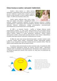
cistus laurifolius
The genus is one of the characteristic genera of the Mediterranean region, colonizing degraded areas (Attaguile et al., 2000). L. (cistaceae) is a common plant in Anatolia and is used against various ailments in traditional medicine. The plant leaves are used to treat rheumatic and related inflammatory diseases, externally as a bath or poultice to reduce pain in rheumatism, against fever in common cold or applied externally as a plaster on the dorsal part of the body in a line of the kidneys for urinary inflammations.
The genus is one of the characteristic genera of the Mediterranean region, colonizing degraded areas (Attaguile et al., 2000). L. (cistaceae) is a common plant in Anatolia and is used against various ailments in traditional medicine. The plant leaves are used to treat rheumatic and related inflammatory diseases, externally as a bath or poultice to reduce pain in rheumatism, against fever in common cold or applied externally as a plaster on the dorsal part of the body in a line of the kidneys for urinary inflammations.

You also want an ePaper? Increase the reach of your titles
YUMPU automatically turns print PDFs into web optimized ePapers that Google loves.
457<br />
The methodology described by Kurtel et al. (1992) was<br />
used. Briefly, 1 ml of plasma was mixed with 2.0 ml of<br />
trichloroacetic acid (TCA; 15%, w/v)–thiobarbituric acid (TBA;<br />
0.375%)–0.25 N HCl and mixed throughly and centrifuged at<br />
10,000 for 5 min. The supernatant was mixed with 20 l<br />
of butyl hydroxy toluene (BHT; 0.02% in 95% EtOH, w/v) to<br />
prevent further oxidation and heated for 15 min in a boiling<br />
water bath. After cooling under running water, the flocculent<br />
precipitate was removed by centrifugation at 10,000 for<br />
5 min. The absorbance of the sample was measured at 532 nm<br />
against blank that contained all the reagents except plasma.<br />
1,1,3,3-Tetraethoxypropan was used as standard for the curve<br />
calibration.<br />
with no homogenate. Results were expressed as<br />
tissue.<br />
mol GSH/g<br />
Biocon standard kits and DAX-48 autoanalyzer were used to<br />
measure AST and ALT activities in plasma samples according<br />
to the method of Wilkinson et al. (1972).<br />
Animals employed in the experiments were observed during<br />
48 h and morbidity or mortality was recorded, if happens, for<br />
each group at the end of observation period.<br />
The method of Ohkawa et al. (1979) as modified by Jamall<br />
and Smith (1985) was used to determine lipid peroxidation in tissue<br />
samples. Mice were sacrified by an overdose of diethylether.<br />
The liver of each mouse was immediately excised and chilled in<br />
ice-cold 0.9% NaCl and then perfused via the portal vein with<br />
ice-cold 0.9% NaCl. After washing with 0.9% NaCl, 1.0 g of wet<br />
tissue was weighted exactly and homogenized in 9 ml of 0.25 M<br />
sucrose using a teflon homogenizer to obtain a 10% suspension.<br />
The cytosolic fraction was obtained by a two-step centrifugation<br />
first at 1000 for 10 min and then at 2000 for 30 min<br />
at 4 C. A volume of the homogenate (0.20 ml) was transferred<br />
to a vial and was mixed with 0.2 ml of a 8.1% (w/v) sodium<br />
dodecyl sulphate solution, 1.50 ml of a 20% acetic acid solution<br />
(adjusted to pH 3.5 with NaOH) and 1.50 ml of a 0.8% (w/v)<br />
solution of TBA and the final volume was adjusted to 4.0 ml<br />
with distilled water. Each vial was tightly capped and heated in<br />
boiling water bath for 60 min. The vials were then cooled under<br />
running water.<br />
Equal volumes of tissue blank or test sample and 10%<br />
TCA were transferred into a centrifuge tube and centrifuged at<br />
1000 for 10 min. The absorbance of the supernatant fraction<br />
was measured at 532 nm in a Beckman DU 650 spectrometer.<br />
Control experiment was processed using the same experimental<br />
procedure except the TBA solution was replaced with distilled<br />
water due to the peroxidative effect of acetaminophen on tissue.<br />
Livers of acetaminophen-treated mice were used as positive control<br />
and 1,1,3,3-tetraethoxypropan was used as standard for the<br />
curve calibration.<br />
Some 200 mg of liver was homogenized in 8.0 ml of 0.02 M<br />
EDTA in an ice bath. The homogenates were kept in the ice bath<br />
until used. Aliquots of 5.0 ml of the homogenates were mixed<br />
in 15.0 ml test tubes with 4.0 ml distilled water and 1.0 ml of<br />
50% trichloroacetic acid (TCA). The tubes were centrifuged for<br />
15 min at approximately 3000 . Some 2.0 ml of supernatant<br />
was mixed with 4.0 ml of 0.4 M Tris–buffer, pH 8.9, 0.1 ml Ellman’s<br />
reagent [5,5 -dithiobis-(2-nitro-benzoic acid)] (DTNB)<br />
added, and the sample shaken. The absorbance was read within<br />
5 min of the addition of DTNB at 412 nm against a reagent blank<br />
The data obtained were analyzed by one-way of variance<br />
(ANOVA) and Student–Newman–Keuls post hoc tests for the<br />
significant interrelation between the various groups using Instat<br />
computer software. < 0.05 was considered to be significant<br />
different from the control.<br />
In living systems, dietary antioxidants such as -tocopherol,<br />
ascorbic acid, carotenoids, as well as flavonoids and related<br />
phenolic compounds are suggested in protection from oxidative<br />
damage of tissues in the body and eventually for a healthy<br />
life (Haraguchi, 2001). Especially flavonoids have been shown<br />
to scavenge various reactive oxygen species and implicated as<br />
inhibitors of lipid peroxidation (Mora et al., 1990).<br />
Acetaminophen (paracetamol), a frequently used analgesic<br />
and antipyretic drug, is known to be hepatotoxic in high doses,<br />
which is primarily metabolized by sulfation and glucuronidation<br />
to unreactive metabolites, and then activated by the cytochrome<br />
P-450 system to produce liver injury. It is established that<br />
acetaminophen is bioactivated to a toxic electrophile, -acetyl-<br />
-benzoquinone imine (NAPQI), which binds covalently to tissue<br />
macromolecules, and probably also oxidizes lipids, or the<br />
critical sulphydryl groups (protein thiols) and alters the homeostasis<br />
of calcium (Lin et al., 1997). Post-mitochondrial supernatants<br />
isolated from livers of rats given a single large oral dose<br />
of acetaminophen (800 mg/kg) showed rapid rates of lipid peroxidation<br />
when incubated in vitro. Lipid peroxidation probably<br />
occurs simultaneously with the proposed covalent binding of<br />
the active metabolite of acetaminophen. Since the former process<br />
is known to cause severe and extensive membrane damage,<br />
it may be a very important factor in acetaminophen-induced liver<br />
necrosis (Fairhurst et al., 1982).<br />
As shown in Table 1, in the liver and plasma of<br />
acetaminophen-treated group, tissue and plasma lipid peroxidation<br />
levels (219.2 and 1172%) as evidenced by MDA determination<br />
increased significantly as compared to control group, however,<br />
the content of GSH in liver decreased (8.7%). Additionally,<br />
acetaminophen was found to cause several folds increases in<br />
plasma AST and ALT levels (106 and 71.6%). Ethanol (EtOH)<br />
extract of<br />
leaves administered in 500 mg/kg