

Podosomes at a Glance - Journal of Cell Science - The Company of ...
Podosomes at a Glance - Journal of Cell Science - The Company of ...
Podosomes at a Glance - Journal of Cell Science - The Company of ...

You also want an ePaper? Increase the reach of your titles
YUMPU automatically turns print PDFs into web optimized ePapers that Google loves.
<strong>Journal</strong> <strong>of</strong> <strong>Cell</strong> <strong>Science</strong><br />
<strong>Cell</strong> <strong>Science</strong> <strong>at</strong> a <strong>Glance</strong> 2079<br />
<strong>Podosomes</strong> <strong>at</strong> a glance<br />
Stefan Linder* and Petra Kopp<br />
Institut für Prophylaxe und Epidemiologie der<br />
Kreislaufkrankheiten, Ludwig-Maximilians-<br />
Universität, Pettenk<strong>of</strong>erstr. 9, 80336 München,<br />
Germany<br />
*Author for correspondence (e-mail:<br />
stefan.linder@med.uni-muenchen.de)<br />
<strong>Journal</strong> <strong>of</strong> <strong>Cell</strong> <strong>Science</strong> 118, 2079-2082<br />
Published by <strong>The</strong> <strong>Company</strong> <strong>of</strong> Biologists 2005<br />
doi:10.1242/jcs.02390<br />
<strong>Cell</strong>s can contact the extracellular m<strong>at</strong>rix<br />
(ECM) through a variety <strong>of</strong> specialized<br />
structures. <strong>The</strong>se cell-m<strong>at</strong>rix contacts<br />
contain membrane proteins such as<br />
integrins, which bind both m<strong>at</strong>rix<br />
components and cellular proteins, thus<br />
bridging the substr<strong>at</strong>um and the<br />
cytoskeleton (reviewed in Adams,<br />
2001). M<strong>at</strong>rix contacts provide the<br />
physical linkage th<strong>at</strong> enables cells to<br />
Overlay Talin F-actin<br />
Structure<br />
Functions<br />
10 µm<br />
Adhesion<br />
Close contact to substr<strong>at</strong>um<br />
Enrichment <strong>of</strong> adhesion-medi<strong>at</strong>ing integrins<br />
Form<strong>at</strong>ion <strong>at</strong> substr<strong>at</strong>e-<strong>at</strong>tached cell side<br />
adhere to and migr<strong>at</strong>e on (or through) the<br />
m<strong>at</strong>rix. Moreover, they can also act as<br />
localized ‘relay st<strong>at</strong>ions’ for important<br />
signaling p<strong>at</strong>hways. Well-known cellm<strong>at</strong>rix<br />
contacts include focal complexes<br />
and focal adhesions, whereas others,<br />
such as fibrillar adhesions, podosomes<br />
and invadopodia are just beginning to<br />
<strong>at</strong>tract widespread <strong>at</strong>tention.<br />
Focal complexes have a dash-like<br />
appearance, a length <strong>of</strong> ~0.5 µm and<br />
constitute some <strong>of</strong> the earliest m<strong>at</strong>rix<br />
contacts formed by a protruding cell.<br />
<strong>The</strong>y are integrin-based and enriched in<br />
talin, paxillin and vinculin (Zamir and<br />
Geiger, 2001). Focal complexes can<br />
m<strong>at</strong>ure into focal adhesions through the<br />
applic<strong>at</strong>ion <strong>of</strong> force – for example, during<br />
the retraction <strong>of</strong> cellular protrusions. This<br />
transform<strong>at</strong>ion is accompanied by an<br />
increase in size, the form<strong>at</strong>ion <strong>of</strong> actin<br />
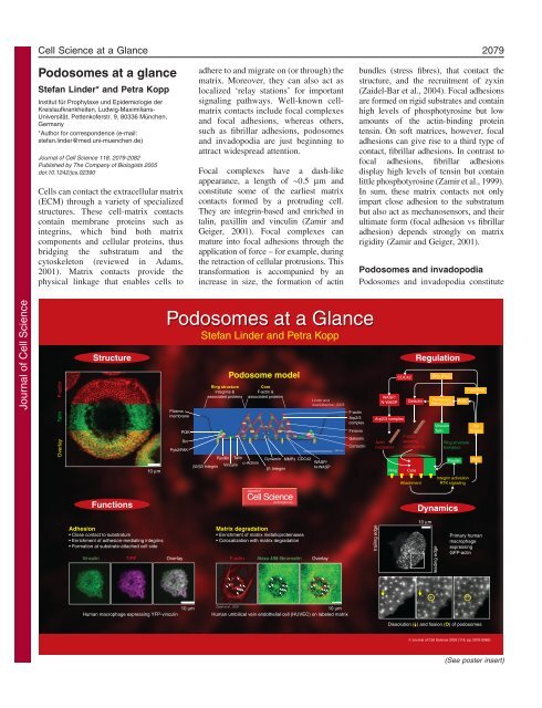
<strong>Podosomes</strong> <strong>at</strong> a <strong>Glance</strong><br />
Stefan Linder and Petra Kopp<br />
Plasma<br />
membrane<br />
Vinculin TIRF Overlay<br />
PI3K<br />
Src<br />
10 µm<br />
Human macrophage expressing YFP-vinculin<br />
Ring structure<br />
Integrins &<br />
associ<strong>at</strong>ed proteins<br />
Podosome model<br />
Core<br />
F-actin &<br />
associ<strong>at</strong>ed proteins<br />
Pyk2/FAK<br />
Paxillin Talin<br />
Dynamin MMPs CDC42<br />
WASP/<br />
Vinculin<br />
α-Actinin<br />
β2/β3 Integrin<br />
β1 Integrin<br />
N-WASP<br />
jcs.biologists.org<br />
M<strong>at</strong>rix degrad<strong>at</strong>ion<br />
Enrichment <strong>of</strong> m<strong>at</strong>rix metalloproteinases<br />
Colocaliz<strong>at</strong>ion with m<strong>at</strong>rix degrad<strong>at</strong>ion<br />
F-actin Alexa 488-fibronectin Overlay<br />
Linder and<br />
Aepfelbacher, 2003<br />
F-actin<br />
Arp2/3<br />
complex<br />
Fimbrin<br />
Gelsolin<br />
Cortactin<br />
Osiak et al., 2005<br />
10 µm<br />
Human umbilical vein endothelial cell (HUVEC) on labeled m<strong>at</strong>rix<br />
bundles (stress fibres), th<strong>at</strong> contact the<br />
structure, and the recruitment <strong>of</strong> zyxin<br />
(Zaidel-Bar et al., 2004). Focal adhesions<br />
are formed on rigid substr<strong>at</strong>es and contain<br />
high levels <strong>of</strong> phosphotyrosine but low<br />
amounts <strong>of</strong> the actin-binding protein<br />
tensin. On s<strong>of</strong>t m<strong>at</strong>rices, however, focal<br />
adhesions can give rise to a third type <strong>of</strong><br />
contact, fibrillar adhesions. In contrast to<br />
focal adhesions, fibrillar adhesions<br />
display high levels <strong>of</strong> tensin but contain<br />
little phosphotyrosine (Zamir et al., 1999).<br />
In sum, these m<strong>at</strong>rix contacts not only<br />
impart close adhesion to the substr<strong>at</strong>um<br />
but also act as mechanosensors, and their<br />
ultim<strong>at</strong>e form (focal adhesion vs fibrillar<br />
adhesion) depends strongly on m<strong>at</strong>rix<br />
rigidity (Zamir and Geiger, 2001).<br />
<strong>Podosomes</strong> and invadopodia<br />
<strong>Podosomes</strong> and invadopodia constitute<br />
trailing edge<br />
WASP/<br />
N-WASP<br />
Arp2/3 complex<br />
Actin<br />
nucle<strong>at</strong>ion<br />
CDC42<br />
F-actin<br />
severing<br />
+uncapping<br />
Ring Core<br />
Attachment<br />
Regul<strong>at</strong>ion<br />
Gelsolin PI3K<br />
Dynamics<br />
10 µm<br />
Rho (Rac)<br />
PtdIns(3,4)P2 PtdIns(3,4,5)P3 Vinculin<br />
Talin<br />
leading edge<br />
Ring structure<br />
form<strong>at</strong>ion<br />
Paxillin<br />
Integrin activ<strong>at</strong>ion<br />
RTK signaling<br />
Pyk2/FAK<br />
Src<br />
kinase<br />
PKC<br />
Primary human<br />
macrophage<br />
expressing<br />
GFP-actin<br />
Dissolution ( ) and fission (O) <strong>of</strong> podosomes<br />
© <strong>Journal</strong> <strong>of</strong> <strong>Cell</strong> <strong>Science</strong> 2005 (118, pp. 2079-2082)<br />
(See poster insert)
<strong>Journal</strong> <strong>of</strong> <strong>Cell</strong> <strong>Science</strong><br />
2080<br />
<strong>Journal</strong> <strong>of</strong> <strong>Cell</strong> <strong>Science</strong> 118 (10)<br />
two forms <strong>of</strong> dot-like m<strong>at</strong>rix contact th<strong>at</strong><br />
differ from those described above<br />
structurally and functionally (reviewed<br />
in Linder and Aepfelbacher, 2003;<br />
Buccione et al., 2004). Structurally, their<br />
most distinguishing fe<strong>at</strong>ure is their twopart<br />
architecture: they have a core <strong>of</strong> Factin<br />
and actin-associ<strong>at</strong>ed proteins th<strong>at</strong> is<br />
surrounded by a ring structure consisting<br />
<strong>of</strong> plaque proteins such as talin or<br />
vinculin. This actin-rich core is not<br />
present in other cell-m<strong>at</strong>rix contacts.<br />
Consequently, actin regul<strong>at</strong>ory p<strong>at</strong>hways<br />
exert a major additional influence on<br />
podosome-type contacts. Functionally,<br />
the ability <strong>of</strong> podosomes and<br />
invadopodia to engage in m<strong>at</strong>rix<br />
degrad<strong>at</strong>ion clearly sets them apart from<br />
other cell-m<strong>at</strong>rix contacts.<br />
<strong>Podosomes</strong> are typically formed in cells<br />
<strong>of</strong> the monocytic lineage, such as<br />
macrophages (Lehto et al., 1982; Linder<br />
et al., 1999), osteoclasts (Marchisio et<br />
al., 1984) or (imm<strong>at</strong>ure) dendritic cells<br />
(Burns et al., 2001). However, they can<br />
also be found or induced in a variety <strong>of</strong><br />
other cell types, including smooth<br />
muscle cells (Gimona et al., 2003) and<br />
endothelial cells (Moreau et al., 2003;<br />
Osiak et al., 2005).<br />
By contrast, invadopodia are found in<br />
fibroblasts transformed with viral<br />
oncogenes encoding protein tyrosine<br />
kinases (Tarone et al., 1985) and in some<br />
malignant cell types (Buccione et al.,<br />
2004). Indeed, the first observ<strong>at</strong>ion <strong>of</strong><br />
podosome-type adhesions 25 years ago<br />
described such virally induced<br />
invadopodia (David-Pfeuty and Singer,<br />
1980).<br />
<strong>Podosomes</strong> and invadopodia differ in<br />
their respective sizes and numbers.<br />
Typically, a cell forms dozens <strong>of</strong><br />
podosomes, but there are only a few<br />
invadopodia per cell. However, wh<strong>at</strong><br />
invadopodia lack in numbers, they make<br />
up for in size: <strong>Podosomes</strong> have a<br />
diameter <strong>of</strong> 0.5-1 µm and a depth <strong>of</strong> 0.2-<br />
0.4 µm, whereas invadopodia can<br />
correspond to an array <strong>of</strong> membrane<br />
invagin<strong>at</strong>ions th<strong>at</strong> have a diameter <strong>of</strong> ~8<br />
µm and also can form root-like<br />
extensions into the m<strong>at</strong>rix th<strong>at</strong> are<br />
several micrometers deep (Buccione et<br />
al., 2004; McNiven et al., 2004).<br />
Despite these differences, basic<br />
similarities between the two structures,<br />
both in composition and architecture, are<br />
apparent. Indeed, it has been proposed<br />
th<strong>at</strong> invadopodia might develop from<br />
podosomal precursors (Linder and<br />
Aepfelbacher, 2003; Buccione et al.,<br />
2004). It is therefore an <strong>at</strong>tractive<br />
specul<strong>at</strong>ion th<strong>at</strong> podosomes and<br />
invadopodia represent a continuum <strong>of</strong><br />
specialized m<strong>at</strong>rix contacts comparable<br />
to the succession <strong>of</strong> focal complexes,<br />
focal adhesions and fibrillar adhesions<br />
described above.<br />
<strong>The</strong> growing list <strong>of</strong> podosomecontaining<br />
cells may point to a<br />
widespread ability <strong>of</strong> cells to form this<br />
type <strong>of</strong> actin-rich structure. Furthermore,<br />
the fact th<strong>at</strong> podosomes are also formed<br />
on s<strong>of</strong>t substr<strong>at</strong>es, such as endothelial<br />
monolayers (Linder and Aepfelbacher,<br />
2003), adds to the notion th<strong>at</strong> podosomes<br />
are physiological structures th<strong>at</strong> are also<br />
formed in the context <strong>of</strong> tissues.<br />
Functions<br />
<strong>The</strong> main functions <strong>of</strong> podosomes<br />
appear to be adhesion and m<strong>at</strong>rix<br />
degrad<strong>at</strong>ion. Moreover, podosomes have<br />
also been implic<strong>at</strong>ed in cell migr<strong>at</strong>ion<br />
and invasion. However, much <strong>of</strong> the<br />
evidence for these functions is still<br />
circumstantial and needs to be rigorously<br />
tested.<br />
It is very possible th<strong>at</strong> podosomes have<br />
a role in adhesion, because they establish<br />
close contact to the substr<strong>at</strong>um, which<br />
can be shown by total internal reflection<br />
(TIRF) microscopy, are enriched in<br />
adhesion-medi<strong>at</strong>ing integrins (Linder<br />
and Aepfelbacher, 2003) and form only<br />
<strong>at</strong> the substr<strong>at</strong>e-<strong>at</strong>tached cell side.<br />
<strong>The</strong> aptly named invadopodia were<br />
defined through their ability to perform<br />
m<strong>at</strong>rix degrad<strong>at</strong>ion (Buccione et al.,<br />
2004; McNiven et al., 2004). For<br />
podosomes, the case has been less clear<br />
cut: podosomes in osteoclasts are<br />
enriched in m<strong>at</strong>rix metalloproteases<br />
(S<strong>at</strong>o et al., 1997; Delaissé et al., 2000),<br />
but it was not shown until recently th<strong>at</strong>,<br />
in various cell systems, podosomes<br />
overlap regions <strong>of</strong> m<strong>at</strong>rix degrad<strong>at</strong>ion<br />
(Burgstaller and Gimona, 2005; Osiak et<br />
al., 2005). It is therefore very possible<br />
th<strong>at</strong> podosomes, like invadopodia, have<br />
an inherent ability to lyse the ECM.<br />
<strong>Podosomes</strong> may also play an accessory<br />
role in cell migr<strong>at</strong>ion. <strong>The</strong>y might help to<br />
establish localized anchorage, thus<br />
stabilizing sites <strong>of</strong> cell protrusion and<br />
ultim<strong>at</strong>ely enabling productive directional<br />
movement. <strong>The</strong> observ<strong>at</strong>ion th<strong>at</strong><br />
podosomes are recruited to sites <strong>of</strong> cell<br />
protrusion, especially to the leading edge,<br />
appears to be in line with such a concept.<br />
Podosome-type adhesions are mainly<br />
formed in cells th<strong>at</strong> have to cross tissue<br />
boundaries such as monocytes,<br />
imm<strong>at</strong>ure dendritic cells or some types<br />
<strong>of</strong> cancer cell (invadopodia in this case).<br />
Podosome-localized m<strong>at</strong>rix degrad<strong>at</strong>ion<br />
<strong>at</strong> the leading edge may therefore also<br />
confer invasive potential to cells.<br />
Finally, podosomes are a prominent part<br />
<strong>of</strong> the actin cytoskeleton in osteoclasts,<br />
where they form continuous belts <strong>at</strong> the<br />
cell periphery (Pfaff and Jurdic, 1999).<br />
<strong>The</strong>ir probable roles in adhesion and<br />
m<strong>at</strong>rix degrad<strong>at</strong>ion suggest an important<br />
role in bone homeostasis. Moreover, their<br />
fusion was believed to form the boneremodeling<br />
organelle <strong>of</strong> osteoclasts, the<br />
so-called sealing zone, a tightly adherent<br />
structure surrounding the space into<br />
which lytic enzymes and protons are<br />
secreted (Lakkakorpi and Väänänen,<br />
1996). However, recent d<strong>at</strong>a suggest th<strong>at</strong><br />
both types <strong>of</strong> organelle are formed<br />
independently (Saltel et al., 2004).<br />
Furthermore, on mineralized bone, their<br />
physiological substr<strong>at</strong>e, osteoclasts form<br />
only sealing zones, but not podosomes<br />
(Saltel et al., 2004). <strong>The</strong> proposed role<br />
for podosomes – as opposed to sealing<br />
zones – in bone homeostasis is therefore<br />
again open for deb<strong>at</strong>e.<br />
Structure and components<br />
<strong>Podosomes</strong> comprise a core <strong>of</strong> F-actin<br />
and actin-associ<strong>at</strong>ed proteins embedded<br />
in a ring structure <strong>of</strong> integrins and<br />
integrin-associ<strong>at</strong>ed proteins (see poster,<br />
middle and right). Ring and core are<br />
probably linked by bridging molecules<br />
such as α-actinin, and the whole structure<br />
is surrounded by a cloud <strong>of</strong> mostly<br />
monomeric actin molecules (Destaing et<br />
al., 2003). Many podosome components<br />
show a distinct localiz<strong>at</strong>ion to either the<br />
core or ring structure. Typical core<br />
components are F-actin, actin regul<strong>at</strong>ors<br />
such as members <strong>of</strong> the Wiskott-Aldrich<br />
Syndrome protein (WASP) family, the
<strong>Journal</strong> <strong>of</strong> <strong>Cell</strong> <strong>Science</strong><br />
<strong>Cell</strong> <strong>Science</strong> <strong>at</strong> a <strong>Glance</strong> 2081<br />
Arp2/3 complex, gelsolin and cortactin,<br />
whereas adhesion medi<strong>at</strong>ors such as<br />
paxillin, vinculin or talin, and kinases<br />
such as PI3K or Pyk2/FAK preferentially<br />
associ<strong>at</strong>e with the ring structure (Linder<br />
and Aepfelbacher, 2003; Buccione et al.,<br />
2004).<br />
<strong>Podosomes</strong> are anchored in the<br />
extracellular m<strong>at</strong>rix through integrins.<br />
<strong>The</strong>se are distributed over the whole<br />
outer surface <strong>of</strong> the podosome, but show<br />
an isotype-specific localiz<strong>at</strong>ion: β1<br />
integrins localize mostly to the core,<br />
whereas β2 and β3 integrins localize to<br />
the ring.<br />
Regul<strong>at</strong>ion<br />
<strong>Podosomes</strong> are influenced by a variety <strong>of</strong><br />
cellular signaling p<strong>at</strong>hways. Major<br />
modes <strong>of</strong> podosome regul<strong>at</strong>ion include<br />
signaling by Rho family GTPases, actin<br />
regul<strong>at</strong>ory p<strong>at</strong>hways, protein tyrosine<br />
phosphoryl<strong>at</strong>ion, and the influence <strong>of</strong> the<br />
microtubule system.<br />
Rho GTPases<br />
<strong>The</strong> RhoGTPases RhoA, Rac1 and<br />
CDC42 have all been shown to regul<strong>at</strong>e<br />
podosome turnover in various cell types.<br />
<strong>The</strong>ir influence on podosomes is<br />
undisputed; however, their particular<br />
mode <strong>of</strong> action may depend on the cell<br />
type. For example, both dominant active<br />
and inactive mutants <strong>of</strong> these GTPases<br />
can interfere with podosome form<strong>at</strong>ion<br />
or localiz<strong>at</strong>ion in dendritic cells (Burns<br />
et al., 2001), while expression <strong>of</strong><br />
dominant active CDC42 leads to<br />
podosome form<strong>at</strong>ion in aortic<br />
endothelial cells (Moreau et al., 2003).<br />
In any case, podosome turnover appears<br />
to need subcellular fine tuning <strong>of</strong> the<br />
GTP/GDP cycles <strong>of</strong> these cellular master<br />
switches (Linder and Aepfelbacher,<br />
2003). Accordingly, active Rho has been<br />
localized to invadopodia in transformed<br />
fibroblasts (Berdeaux et al., 2004).<br />
Actin regul<strong>at</strong>ion<br />
<strong>The</strong> most prominent fe<strong>at</strong>ure <strong>of</strong><br />
podosomes is their F-actin-rich core,<br />
which is also necessary for the stability<br />
<strong>of</strong> the whole structure (Lehto et al.,<br />
1982). Members <strong>of</strong> the WASP family, as<br />
well as actin-nucle<strong>at</strong>ing Arp2/3 complex<br />
are both strongly enriched in the<br />
podosome core. Absence <strong>of</strong> these<br />
components, either induced artificially<br />
(Linder et al., 2000a) or in disease<br />
(Linder et al., 1999; Burns et al., 2001)<br />
(reviewed in Calle et al., 2004), results<br />
in disruption <strong>of</strong> podosomes. Another<br />
prominent podosome component<br />
involved in actin regul<strong>at</strong>ion is gelsolin. It<br />
probably contributes to actin turnover<br />
through its ability to both sever and cap<br />
actin filaments (Chellaiah et al., 1998).<br />
Tyrosine phosphoryl<strong>at</strong>ion<br />
It was noted early on th<strong>at</strong> podosome-type<br />
adhesions can be induced by<br />
transform<strong>at</strong>ion <strong>of</strong> cells with viruses<br />
whose oncogenes encode tyrosine<br />
kinases such as v-Src (Tarone et al.,<br />
1985; Marchisio et al., 1987). Indeed,<br />
one <strong>of</strong> the best ways to visualize<br />
podosomes is by staining phosphoryl<strong>at</strong>ed<br />
tyrosine (Linder and Apfelbacher, 2003),<br />
which is highly enriched in some types <strong>of</strong><br />
adhesive structure. Not surprisingly,<br />
cellular tyrosine kinases such as Src and<br />
Csk play major roles in podosome<br />
regul<strong>at</strong>ion (Howell and Cooper, 1994),<br />
and tools for podosome manipul<strong>at</strong>ion<br />
include Src kinase inhibitors, which<br />
disrupt podosomes (Linder et al., 2000b),<br />
and vanad<strong>at</strong>e, a phosphotyrosine<br />
phosph<strong>at</strong>ase inhibitor, which is able to<br />
induce podosome form<strong>at</strong>ion (Marchisio<br />
et al., 1988).<br />
Microtubules<br />
Microtubules are closely associ<strong>at</strong>ed with<br />
podosomes. Moreover, they have been<br />
shown to stabilize podosome p<strong>at</strong>terns<br />
such as the marginal belts in osteoclasts<br />
(Babb et al., 1997), to influence the<br />
fusion and fission r<strong>at</strong>es <strong>of</strong> podosome<br />
precursors in murine macrophages<br />
(Evans et al., 2003) and to regul<strong>at</strong>e<br />
podosome form<strong>at</strong>ion in human<br />
macrophages (Linder et al., 2000b).<br />
A model for podosome form<strong>at</strong>ion<br />
Analysis <strong>of</strong> podosome-inducing p<strong>at</strong>hways<br />
has been performed in a variety <strong>of</strong><br />
cell types. From these d<strong>at</strong>a we can<br />
propose the following simplified model<br />
for podosome form<strong>at</strong>ion: <strong>The</strong> key signal<br />
for initi<strong>at</strong>ion is <strong>at</strong>tachment <strong>of</strong> the cell to<br />
the substr<strong>at</strong>e, because podosomes are<br />
only observed in adherent cells. This<br />
leads to clustering and activ<strong>at</strong>ion <strong>of</strong><br />
integrins and signaling by receptor<br />
tyrosine kinases. One <strong>of</strong> the most<br />
upstream signals is probably PKC<br />
activity. This is underscored by the fact<br />
th<strong>at</strong> podosome form<strong>at</strong>ion can be induced<br />
by PKC-activ<strong>at</strong>ing agents, such as<br />
phorbol esters (Gimona et al., 2003).<br />
An important subsequent switch is<br />
activ<strong>at</strong>ion <strong>of</strong> Src. Downstream, a variety<br />
<strong>of</strong> p<strong>at</strong>hways are initi<strong>at</strong>ed, most notably<br />
PI3K signaling (Chellaiah et al., 1998),<br />
which leads to the form<strong>at</strong>ion <strong>of</strong> the<br />
phosph<strong>at</strong>idylinositols PtdIns(3,4)P2 and<br />
PtdIns(3,4,5)P3, and activ<strong>at</strong>ion <strong>of</strong> focal<br />
adhesion kinase (FAK) or its<br />
haem<strong>at</strong>opoietic rel<strong>at</strong>ive Pyk2. 3�<br />
phosphoinositide signaling is also<br />
influenced by the RhoGTPases Rho and<br />
(probably) Rac (Chellaiah et al., 2001;<br />
Sechi and Wehland, 2000).<br />
CDC42, another RhoGTPase, is crucial<br />
for the local initi<strong>at</strong>ion <strong>of</strong> actin filament<br />
form<strong>at</strong>ion, which gives rise to the<br />
form<strong>at</strong>ion <strong>of</strong> the actin-rich core<br />
structure. This is achieved by releasing<br />
the autoinhibition <strong>of</strong> N-WASP or its<br />
rel<strong>at</strong>ive WASP, which in turn activ<strong>at</strong>es<br />
actin-nucle<strong>at</strong>ing Arp2/3 complex<br />
(Linder et al., 1999; Linder et al., 2000a;<br />
Burns et al., 2001). Turnover <strong>of</strong> corelocalized<br />
actin filaments is probably<br />
facilit<strong>at</strong>ed by gelsolin (Chellaiah et al.,<br />
1998) and the GTPase dynamin<br />
(McNiven et al., 2004; Buccione et al.,<br />
2004). Additionally, dynamin may also<br />
play a role in podosome-localized m<strong>at</strong>rix<br />
degrad<strong>at</strong>ion by facilit<strong>at</strong>ing m<strong>at</strong>rix<br />
metalloprotease release.<br />
It is unclear how core- and ringform<strong>at</strong>ion<br />
are coordin<strong>at</strong>ed. However,<br />
similarly to core molecules, central<br />
components <strong>of</strong> the ring structure, such as<br />
talin and vinculin, are also influenced by<br />
phosphoinositides (Sechi and Wehland,<br />
2000), and activ<strong>at</strong>ion <strong>of</strong> paxillin through<br />
integrin-rel<strong>at</strong>ed signaling (Pfaff and<br />
Jurdic, 2001) may constitute one <strong>of</strong> the<br />
earliest signals in ring form<strong>at</strong>ion.<br />
Dynamics<br />
<strong>Podosomes</strong> are highly dynamic<br />
organelles with a half-life <strong>of</strong> 2-12<br />
minutes. Moreover, their inner dynamic<br />
is even faster: F-actin in the core turnes<br />
over 2-3 times during the life span <strong>of</strong> a<br />
podosome (Destaing et al., 2003).<br />
Individual podosomes are motile within
<strong>Journal</strong> <strong>of</strong> <strong>Cell</strong> <strong>Science</strong><br />
2082<br />
<strong>Journal</strong> <strong>of</strong> <strong>Cell</strong> <strong>Science</strong> 118 (10)<br />
a certain radius, but movement <strong>of</strong> larger<br />
podosome groups is achieved through<br />
assembly <strong>at</strong> the front and disassembly <strong>at</strong><br />
the rear. This is especially visible in<br />
migr<strong>at</strong>ory cells where podosomes are<br />
recruited to the leading edge.<br />
<strong>Podosomes</strong> can assemble de novo or by<br />
branching <strong>of</strong>f precursor clusters, which<br />
undergo constant fusion and fission<br />
(Evans et al., 2003) (see poster, lower<br />
right). ‘Regular’ podosomes are mostly<br />
found in the inner regions <strong>of</strong> the cell,<br />
while the larger precursor clusters<br />
localize to the cell periphery or the<br />
leading lamella <strong>of</strong> migr<strong>at</strong>ing cells.<br />
Disease<br />
<strong>Podosomes</strong> are assembled from<br />
molecules th<strong>at</strong> have multiple functions<br />
in the cell, such as actin and Src kinases.<br />
Inhibitors <strong>of</strong> podosome assembly<br />
therefore also have pr<strong>of</strong>ound effects<br />
on other parts <strong>of</strong> the cytoskeleton.<br />
Similarly, the absence <strong>of</strong> podosomes in<br />
diseases based on defects in a podosome<br />
component(s) may only constitute a side<br />
effect. <strong>The</strong> podosome aficionado should<br />
therefore take care to ascertain whether<br />
a lack <strong>of</strong> podosomes causes or is simply<br />
symptom<strong>at</strong>ic <strong>of</strong> a particular phenotype.<br />
Podosome-associ<strong>at</strong>ed diseases include<br />
Wiskott-Aldrich Syndrome (WAS) and<br />
chronic myeloid leukemia (CML).<br />
WAS-p<strong>at</strong>ient macrophages (Linder et<br />
al., 1999) and dendritic cells from CML<br />
p<strong>at</strong>ients (Dong et al., 2003) both display<br />
pronounced defects in podosome<br />
form<strong>at</strong>ion and chemotaxis.<br />
We thank Bettina Ebbing for help with TIRF<br />
microscopy, Alexander Bershadsky for the gift <strong>of</strong><br />
YFP-vinculin, Peter C. Weber and Jürgen<br />
Heesemann for continuous support and Barbara<br />
Böhlig for expert technical assistance. Work from<br />
our lab is supported by the Deutsche<br />
Forschungsgemeinschaft (GRK 438, SFB 413), the<br />
Friedrich Baur Stiftung and the August Lenz<br />
Stiftung. We apologize to all whose work was not<br />
mentioned owing to space limit<strong>at</strong>ions.<br />
References<br />
Adams, J. C. (2001). <strong>Cell</strong>-m<strong>at</strong>rix contact structures.<br />
<strong>Cell</strong>. Mol. Life Sci. 58, 371-392.<br />
Babb, S. G., M<strong>at</strong>sudaira, P., S<strong>at</strong>o, M., Correia, I.<br />
and Lim, S. S. (1997). Fimbrin in podosomes <strong>of</strong><br />
monocyte-derived osteoclasts. <strong>Cell</strong> Motil.<br />
Cytoskeleton 37, 308-325.<br />
Berdeaux, R. L., Diaz, B., Kim, L. and Martin, G.<br />
S. (2004). Active Rho is localized to podosomes<br />
induced by oncogenic Src and is required for their<br />
assembly and function. J. <strong>Cell</strong> Biol. 166, 317-323.<br />
Buccione, R., Orth, J. D. and McNiven, M. A.<br />
(2004). Foot and mouth: podosomes, invadopodia and<br />
circular dorsal ruffles. N<strong>at</strong>. Rev. Mol.<strong>Cell</strong> Biol. 5, 647-<br />
657.<br />
Burgstaller, G. and Gimona, M. (2005). Podosomemedi<strong>at</strong>ed<br />
m<strong>at</strong>rix resorption and cell motility in<br />
vascular smooth muscle cells. Am. J. Physiol. Heart<br />
Circ. Physiol. (epub Feb. 4)<br />
Burns, S., Thrasher, A. J., Blundell, M. P.,<br />
Machesky, L. and Jones, G. E. (2001). Configur<strong>at</strong>ion<br />
<strong>of</strong> human dendritic cell cytoskeleton by Rho GTPases,<br />
the WAS protein, and differenti<strong>at</strong>ion. Blood 98, 1142-<br />
1149.<br />
Calle, Y., Chou, H. C., Thrasher, A. J. and Jones,<br />
G. E. (2004). Wiskott-Aldrich syndrome protein and<br />
the cytoskeletal dynamics <strong>of</strong> dendritic cells. J. P<strong>at</strong>hol.<br />
204, 460-469.<br />
Chellaiah, M., Fitzgerald, C., Alvarez, U. and<br />
Hruska, K. (1998). c-Src is required for stimul<strong>at</strong>ion<br />
<strong>of</strong> gelsolin-associ<strong>at</strong>ed phosph<strong>at</strong>idylinositol 3-kinase.<br />
J. Biol. Chem. 273, 11908-11916.<br />
Chellaiah, M. A., Biswas, R. S., Yuen, D., Alvarez,<br />
U. M. and Hruska, K. A. (2001).<br />
Phosph<strong>at</strong>idylinositol 3,4,5-trisphosph<strong>at</strong>e directs<br />
associ<strong>at</strong>ion <strong>of</strong> Src homology 2-containing signaling<br />
proteins with gelsolin. J. Biol. Chem. 276, 47434-<br />
47444.<br />
David-Pfeuty, T. and Singer, S. J. (1980). Altered<br />
distributions <strong>of</strong> the cytoskeletal proteins vinculin and<br />
alpha-actinin in cultured fibroblasts transformed by<br />
Rous sarcoma virus. Proc. N<strong>at</strong>l. Acad. Sci. USA 77,<br />
6687-6691.<br />
Delaissé, J. M., Engsig, M. T., Everts, V., del<br />
Carmen Ovejero, M., Ferreras, M., Lund, L., Vu,<br />
T. H., Werb, Z., Winding, B., Lochter, A. et al.<br />
(2000). Proteinases in bone resorption: obvious and<br />
less obvious roles. Clin. Chim. Acta 291, 223-234.<br />
Destaing, O., Saltel, F., Geminard, J. C., Jurdic, P.<br />
and Bard, F. (2003). <strong>Podosomes</strong> display actin<br />
turnover and dynamic self-organiz<strong>at</strong>ion in osteoclasts<br />
expressing actin-green fluorescent protein. Mol. Biol.<br />
<strong>Cell</strong> 14, 407-416.<br />
Dong, R., Cwynarski, K., Entwistle, A., Marelli-<br />
Berg, F., Dazzi, F., Simpson, E., Goldman, J. M.,<br />
Melo, J. V., Lechler, R. I., Bellantuono, I. et al.<br />
(2003). Dendritic cells from CML p<strong>at</strong>ients have<br />
altered actin organiz<strong>at</strong>ion, reduced antigen processing,<br />
and impaired migr<strong>at</strong>ion. Blood 101, 3560-3567.<br />
Evans, J.G., Correia, I., Krasavina, O., W<strong>at</strong>son, N.<br />
and M<strong>at</strong>sudaira, P. (2003). Macrophage podosomes<br />
assemble <strong>at</strong> the leading lamella by growth and<br />
fragment<strong>at</strong>ion. J. <strong>Cell</strong> Biol. 161, 697-705.<br />
Gimona, M., Kaverina, I., Resch, G. P., Vignal, E.<br />
and Burgstaller, G. (2003). Calponin repe<strong>at</strong>s regul<strong>at</strong>e<br />
actin filament stability and form<strong>at</strong>ion <strong>of</strong> podosomes in<br />
smooth muscle cells. Mol. Biol. <strong>Cell</strong> 14, 2482-2491.<br />
Howell, B. W. and Cooper, J. A. (1994). Csk<br />
suppression <strong>of</strong> Src involves movement <strong>of</strong> Csk to sites<br />
<strong>of</strong> Src activity. Mol. <strong>Cell</strong> Biol. 14, 5402-5411.<br />
Lakkakorpi, P. T. and Väänänen, H. K. (1996).<br />
Cytoskeletal changes in osteoclasts during the<br />
resorption cycle. Microsc. Res. Tech. 33, 171-181.<br />
Lehto, V. P., Hovi, T., Vartio, T., Badley, R. A. and<br />
Virtanen, I. (1982). Reorganiz<strong>at</strong>ion <strong>of</strong> cytoskeletal<br />
and contractile elements during transition <strong>of</strong> human<br />
monocytes into adherent macrophages. Lab. Invest.<br />
47, 391-399.<br />
Linder, S. and Aepfelbacher, M. (2003).<br />
<strong>Podosomes</strong>: adhesion hot-spots <strong>of</strong> invasive cells.<br />
Trends <strong>Cell</strong> Biol. 13, 376-385.<br />
Linder, S., Nelson, D., Weiss, M. and Aepfelbacher,<br />
M. (1999). Wiskott-Aldrich syndrome protein<br />
regul<strong>at</strong>es podosomes in primary human macrophages.<br />
Proc. N<strong>at</strong>l. Acad. Sci. USA 96, 9648-9653.<br />
Linder, S., Higgs, H., Hufner, K., Schwarz, K.,<br />
Pannicke, U. and Aepfelbacher, M. (2000a). <strong>The</strong><br />
polariz<strong>at</strong>ion defect <strong>of</strong> Wiskott-Aldrich syndrome<br />
macrophages is linked to dislocaliz<strong>at</strong>ion <strong>of</strong> the Arp2/3<br />
complex. J. Immunol. 165, 221-225.<br />
Linder, S., Hufner, K., Wintergerst, U. and<br />
Aepfelbacher, M. (2000b). Microtubule-dependent<br />
form<strong>at</strong>ion <strong>of</strong> podosomal adhesion structures in<br />
primary human macrophages. J. <strong>Cell</strong> Sci. 113, 4165-<br />
4176.<br />
Marchisio, P. C., Cirillo, D., Naldini, L.,<br />
Primavera, M. V., Teti, A. and Zambonin-Zallone,<br />
A. (1984). <strong>Cell</strong>-substr<strong>at</strong>um interaction <strong>of</strong> cultured<br />
avian osteoclasts is medi<strong>at</strong>ed by specific adhesion<br />
structures. J. <strong>Cell</strong> Biol. 99, 1696-1705.<br />
Marchisio, P. C., Cirillo, D., Teti, A., Zambonin-<br />
Zallone, A. and Tarone, G. (1987). Rous sarcoma<br />
virus-transformed fibroblasts and cells <strong>of</strong> monocytic<br />
origin display a peculiar dot-like organiz<strong>at</strong>ion <strong>of</strong><br />
cytoskeletal proteins involved in micr<strong>of</strong>ilamentmembrane<br />
interactions. Exp. <strong>Cell</strong> Res. 169, 202-214.<br />
Marchisio, P. C., D’Urso, N., Comoglio, P. M.,<br />
Giancotti, F. G. and Tarone, G. (1988). Vanad<strong>at</strong>etre<strong>at</strong>ed<br />
baby hamster kidney fibroblasts show<br />
cytoskeleton and adhesion p<strong>at</strong>terns similar to their<br />
Rous sarcoma virus-transformed counterparts. J. <strong>Cell</strong><br />
Biochem. 37, 151-159.<br />
McNiven, M. A., Baldassarre, M. and Buccione, R.<br />
(2004). <strong>The</strong> role <strong>of</strong> dynamin in the assembly and<br />
function <strong>of</strong> podosomes and invadopodia. Front.<br />
Biosci. 9, 1944-1953.<br />
Moreau, V., T<strong>at</strong>in, F., Varon, C. and Genot, E.<br />
(2003). Actin can reorganize into podosomes in aortic<br />
endothelial cells, a process controlled by Cdc42 and<br />
RhoA. Mol. <strong>Cell</strong> Biol. 23, 6809-6822.<br />
Osiak, A.-E., Zenner, G. and Linder, S. (2005).<br />
Subconfluent endothelial cells form podosomes<br />
downstream <strong>of</strong> cytokine and RhoGTPase signaling.<br />
Exp. <strong>Cell</strong> Res., in press<br />
Pfaff, M. and Jurdic, P. (2001). <strong>Podosomes</strong> in<br />
osteoclast-like cells: structural analysis and<br />
cooper<strong>at</strong>ive roles <strong>of</strong> paxillin, proline-rich tyrosine<br />
kinase 2 (Pyk2) and integrin alphaVbeta3. J. <strong>Cell</strong> Sci.<br />
114, 2775-2786.<br />
Saltel, F., Destaing, O., Bard, F., Eichert, D. and<br />
Jurdic, P. (2004). Ap<strong>at</strong>ite-medi<strong>at</strong>ed actin dynamics in<br />
resorbing osteoclasts. Mol. Biol. <strong>Cell</strong> 15, 5231-5241.<br />
S<strong>at</strong>o, T., del Carmen Ovejero, M., Hou, P.,<br />
Heegaard, A. M., Kumegawa, M., Foged, N. T. and<br />
Delaisse, J. M. (1997). Identific<strong>at</strong>ion <strong>of</strong> the<br />
membrane-type m<strong>at</strong>rix metalloproteinase MT1-MMP<br />
in osteoclasts. J. <strong>Cell</strong> Sci. 110, 589-596.<br />
Sechi, A. S. and Wehland, J. (2000). <strong>The</strong> actin<br />
cytoskeleton and plasma membrane connection:<br />
PtdIns(4,5)P(2) influences cytoskeletal protein activity<br />
<strong>at</strong> the plasma membrane. J. <strong>Cell</strong> Sci. 113, 3685-3695.<br />
Tarone, G., Cirillo, D., Giancotti, F. G., Comoglio,<br />
P. M. and Marchisio, P. C. (1985). Rous sarcoma<br />
virus-transformed fibroblasts adhere primarily <strong>at</strong><br />
discrete protrusions <strong>of</strong> the ventral membrane called<br />
podosomes. Exp. <strong>Cell</strong> Res. 159, 141-157.<br />
Zaidel-Bar, R., Cohen, M., Addadi, L. and Geiger,<br />
B. (2004). Hierarchical assembly <strong>of</strong> cell-m<strong>at</strong>rix<br />
adhesion complexes Biochem. Soc. Trans. 32, 416-<br />
420.<br />
Zamir, E. and Geiger, B. (2001). Molecular<br />
complexity and dynamics <strong>of</strong> cell-m<strong>at</strong>rix adhesions. J.<br />
<strong>Cell</strong> Sci. 114, 3583-3590.<br />
Zamir, E., K<strong>at</strong>z, B. Z., Aota, S., Yamada, K. M.,<br />
Geiger, B. and Kam, Z. (1999). Molecular diversity<br />
<strong>of</strong> cell-m<strong>at</strong>rix adhesions. J. <strong>Cell</strong> Sci. 112, 1655-1669.<br />
<strong>Cell</strong> <strong>Science</strong> <strong>at</strong> a <strong>Glance</strong> on the Web<br />
Electronic copies <strong>of</strong> the poster insert are<br />
available in the online version <strong>of</strong> this article<br />
<strong>at</strong> jcs.biologists.org. <strong>The</strong> JPEG images can<br />
be downloaded for printing or used as<br />
slides.


