RESEARCH REPORT - Peter MacCallum Cancer Centre
RESEARCH REPORT - Peter MacCallum Cancer Centre
RESEARCH REPORT - Peter MacCallum Cancer Centre

Create successful ePaper yourself
Turn your PDF publications into a flip-book with our unique Google optimized e-Paper software.
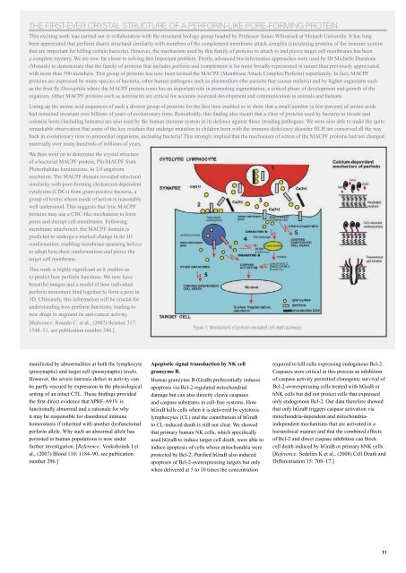
the first-ever CrYstaL struCture of a perforin-Like pore-forMinG protein<br />
This exciting work was carried out in collaboration with the structural biology group headed by Professor James Whisstock at Monash University. It has long<br />
been appreciated that perforin shares structural similarity with members of the complement membrane attack complex (circulating proteins of the immune system<br />
that are important for killing certain bacteria). However, the mechanism used by this family of proteins to attach to and pierce target cell membranes has been<br />
a complete mystery. We are now far closer to solving this important problem. Firstly, advanced bio-informatics approaches were used by Dr Michelle Dunstone<br />
(Monash) to demonstrate that the family of proteins that includes perforin and complement is far more broadly represented in nature than previously appreciated,<br />
with more than 500 members. This group of proteins has now been termed the MACPF (Membrane Attack Complex/Perforin) superfamily. In fact, MACPF<br />
proteins are expressed by many species of bacteria, other human pathogens such as plasmodium (the parasite that causes malaria) and by higher organisms such<br />
as the fruit fly Drosophila where the MACPF protein torso has an important role in promoting segmentation, a critical phase of development and growth of the<br />
organism. Other MACPF proteins such as astrotactin are critical for accurate neuronal development and communication in animals and humans.<br />
Lining up the amino acid sequences of such a diverse group of proteins for the first time enabled us to show that a small number (a few percent) of amino acids<br />
had remained invariant over billions of years of evolutionary time. Remarkably, this finding also meant that a class of proteins used by bacteria to invade and<br />
colonise hosts (including humans) are also used by the human immune system in its defence against those invading pathogens. We were also able to make the quite<br />
remarkable observation that some of the key residues that undergo mutation in children born with the immune-deficiency disorder HLH are conserved all the way<br />
back in evolutionary time to primordial organisms, including bacteria! This strongly implied that the mechanism of action of the MACPF proteins had not changed<br />
materially over many hundreds of millions of years.<br />
We then went on to determine the crystal structure<br />
of a bacterial MACPF protein, Plu-MACPF from<br />
Photorhabdus luminescens, to 2.0 angstrom<br />
resolution. The MACPF domain revealed structural<br />
similarity with pore-forming cholesterol-dependent<br />
cytolysins (CDCs) from gram-positive bacteria, a<br />
group of toxins whose mode of action is reasonably<br />
well understood. This suggests that lytic MACPF<br />
proteins may use a CDC-like mechanism to form<br />
pores and disrupt cell membranes. Following<br />
membrane attachment, the MACPF domain is<br />
predicted to undergo a marked change in its 3D<br />
conformation, enabling membrane spanning helices<br />
to adopt beta sheet conformations and pierce the<br />
target cell membrane.<br />
This work is highly significant as it enables us<br />
to predict how perforin functions. We now have<br />
beautiful images and a model of how individual<br />
perforin monomers bind together to form a pore in<br />
3D. Ultimately, this information will be crucial for<br />
understanding how perforin functions, leading to<br />
new drugs to augment its anti-cancer activity.<br />
[Reference: Rosado C. et al., (2007) Science 317:<br />
Figure 1: Mechanisms of perforin-mediated cell death pathways.<br />
1548–51, see publication number 240.]<br />
manifested by abnormalities at both the lymphocyte<br />
(presynaptic) and target cell (postsynaptic) levels.<br />
However, the severe intrinsic defect in activity can<br />
be partly rescued by expression in the physiological<br />
setting of an intact CTL. These findings provided<br />
the first direct evidence that hPRF-A91V is<br />
functionally abnormal and a rationale for why<br />
it may be responsible for disordered immune<br />
homeostasis if inherited with another dysfunctional<br />
perforin allele. Why such an abnormal allele has<br />
persisted in human populations is now under<br />
further investigation. [Reference: Voskoboinik I et<br />
al., (2007) Blood 110: 1184–90, see publication<br />
number 296.]<br />
Apoptotic signal transduction by NK cell<br />
granzyme B.<br />
Human granzyme B (GraB) preferentially induces<br />
apoptosis via Bcl-2-regulated mitochondrial<br />
damage but can also directly cleave caspases<br />
and caspase substrates in cell-free systems. How<br />
hGraB kills cells when it is delivered by cytotoxic<br />
lymphocytes (CL) and the contribution of hGraB<br />
to CL-induced death is still not clear. We showed<br />
that primary human NK cells, which specifically<br />
used hGraB to induce target cell death, were able to<br />
induce apoptosis of cells whose mitochondria were<br />
protected by Bcl-2. Purified hGraB also induced<br />
apoptosis of Bcl-2-overexpressing targets but only<br />
when delivered at 5 to 10 times the concentration<br />
required to kill cells expressing endogenous Bcl-2.<br />
Caspases were critical in this process as inhibition<br />
of caspase activity permitted clonogenic survival of<br />
Bcl-2-overexpressing cells treated with hGraB or<br />
hNK cells but did not protect cells that expressed<br />
only endogenous Bcl-2. Our data therefore showed<br />
that only hGraB triggers caspase activation via<br />
mitochondria-dependent and mitochondriaindependent<br />
mechanisms that are activated in a<br />
hierarchical manner and that the combined effects<br />
of Bcl-2 and direct caspase inhibition can block<br />
cell death induced by hGraB or primary hNK cells.<br />
[Reference: Sedelies K et al., (2008) Cell Death and<br />
Differentiation 15: 708–17.]<br />
11