RESEARCH REPORT - Peter MacCallum Cancer Centre
RESEARCH REPORT - Peter MacCallum Cancer Centre
RESEARCH REPORT - Peter MacCallum Cancer Centre

Create successful ePaper yourself
Turn your PDF publications into a flip-book with our unique Google optimized e-Paper software.
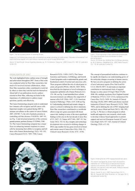
Figure 1: The role of polarity proteins in controlling cell fate.<br />
Images of a T cell dividing (grey arrows) while still attached to an antigen-presenting cell (white arrows). Polarisation of the attached T cell<br />
means that the two daughter cells inherit different molecules and so go on to adopt different fates.<br />
[Source: Dr Sarah Russell, Immune Signalling Laboratory, pp. 18–19. Reference: Chang et al., (2007) Science 315: 1687–91; publication<br />
number 28.]<br />
highlights of 2007<br />
The work highlighted below outlines areas of strength<br />
and achievement throughout 2007. Some of this work<br />
was conducted solely by <strong>Peter</strong> Mac researchers, but<br />
much of it involved external collaboration, where<br />
<strong>Peter</strong> Mac researchers either contributed to work led<br />
by others or where they took the lead role themselves.<br />
About half of our publications involve authors<br />
external to <strong>Peter</strong> Mac, reflecting our belief in the<br />
importance of collaboration to address major<br />
questions, quickly and effectively.<br />
The <strong>Cancer</strong> Immunology program seeks to understand<br />
how immune cells recognise and kill cancer cells.<br />
Important insights were gained during 2007 in<br />
immune surveillance in the control of cancer (Nature,<br />
2007, 450 903–7), the role of polarity proteins in<br />
controlling cell fate (Science 315(5819): 1687–91;<br />
see Fig. 1) and structural properties of the cytotoxic<br />
granule protein, perforin (Science 317(5844): 1548–<br />
51). The <strong>Cancer</strong> Immunology program has a major<br />
focus on enhancing immune responses to cancer<br />
cells by increasing their ability to recognise and kill<br />
these cells (Nature Biotechnology 25(2): 192–193;<br />
<strong>Cancer</strong> Research 67(15): 7495–7504; <strong>Cancer</strong><br />
Research 67(23): 11428–11437). The <strong>Cancer</strong><br />
Genomics and Genetics, Cell Biology, and Growth<br />
Control programs seek to understand the genetic and<br />
biochemical control of normal and cancerous cells.<br />
These programs defined the role of critical genes in<br />
colon cell growth (PNAS, 104(10): 3829–3834),<br />
described the development of novel techniques to<br />
manipulate epithelial cells (Nature Protocols 2(1):<br />
178–186, see Fig. 2) and identified how cellular<br />
microenvironment can influence the outgrowth of<br />
tumour cells (Cell 129(6): 1097–1110; American<br />
Journal of Pathology 170(6): 2135–2148 see Fig.<br />
4). Understanding inherited and somatic change in<br />
cancer is critical to defining key driver mutations.<br />
<strong>Peter</strong> Mac researchers contributed to a worldwide<br />
effort to map new genes associated with breast<br />
cancer risk, leading to some of the most significant<br />
findings in this area in the last decade (<strong>Cancer</strong> Res<br />
67(7): 3027–35; Nature 447(7148): 1087–93; Nat<br />
Genet 39(3): 352–8). Key insights and technology<br />
development also occurred in defining changes in<br />
DNA copy number for key genes involved in breast<br />
and ovarian cancer (<strong>Cancer</strong> Res 67(6): 2544 –51;<br />
Clinical <strong>Cancer</strong> Research 13(16): 4731– 4739).<br />
Figure 2. Tissue regeneration of early differentiating keratinocytes.<br />
This image demonstrates tissue reconstitution six weeks<br />
after transplantation.<br />
[Source: Dr Pritinder Kaur, Epithelial Stem Cell Biology Laboratory,<br />
pp. 58–9. Reference: Gangatirkar et al., (2007) Nature Protocols<br />
2(1): 178 –86; publication number 93.]<br />
The concept of personalised medicine continues to<br />
be rapidly developed as our understanding grows of<br />
the molecular changes occurring in human cancers.<br />
We have an active program in defining the action<br />
of novel anticancer agents (Proc Natl Acad Sci<br />
U S A 104(19): 8071–6) and made an important<br />
contribution to international trials of targeted<br />
therapy in colon cancer (N Engl J Med 357(20):<br />
2040–48), multiple myeloma (New England Journal<br />
of Medicine 357(21): 2123–2132), melanoma (Clin<br />
<strong>Cancer</strong> Res 13(12): 3630–36; Journal of Clinical<br />
Oncology 25(28): 4493–4494) and chronic myeloid<br />
leukaemia (Clinical <strong>Cancer</strong> Research 13(23): 7080–<br />
7085), and in defining the role of PET imaging head<br />
and neck cancer (Head and Neck 29(11): 986–995;<br />
see Fig. 3). We have a vibrant program in allied<br />
health and supportive care research, which seeks<br />
to develop evidence-based approaches to patient<br />
support and survival (European Journal of <strong>Cancer</strong><br />
Care (Engl) 16(5): 417– 423; Journal of Clinical<br />
Oncology 25(9): e13–14).<br />
Figure 3. Defining the role of PET imaging head and neck cancer.<br />
This research investigated the clinical impact and prognostic<br />
stratification of F-18 FDG PET/CT in head and neck mucosal<br />
squamous cell carcinoma. PET/CT indentified uptake at the<br />
previously ‘not suspicious’ neck node (arrow), leading to improved<br />
identification of disease state.<br />
[Source: Connell CA, et al., (2007) Head and Neck, 29 (11),<br />
986–95. Copyright 2007 Wiley Periodicals, Inc. See pages 102–3,<br />
and publication number 40.]<br />
3