Management of choledochal cyst with portal ... - APAMED Central
Management of choledochal cyst with portal ... - APAMED Central
Management of choledochal cyst with portal ... - APAMED Central

You also want an ePaper? Increase the reach of your titles
YUMPU automatically turns print PDFs into web optimized ePapers that Google loves.
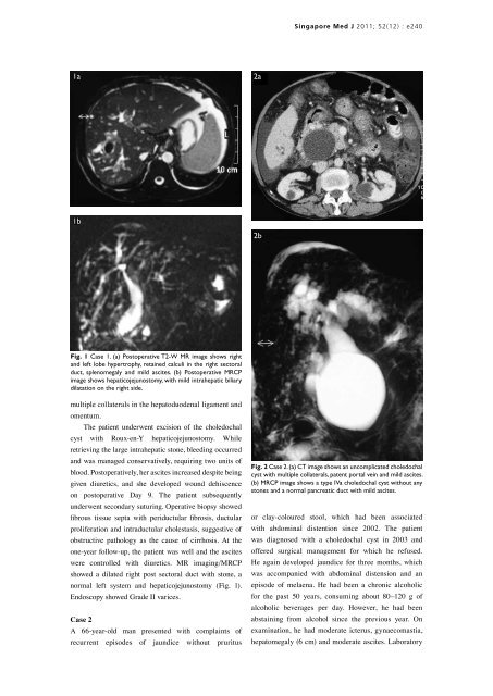
1a<br />
1b<br />
Fig. 1 Case 1. (a) Postoperative T2-W MR image shows right<br />
and left lobe hypertrophy, retained calculi in the right sectoral<br />
duct, splenomegaly and mild ascites. (b) Postoperative MRCP<br />
image shows hepaticojejunostomy, <strong>with</strong> mild intrahepatic biliary<br />
dilatation on the right side.<br />
multiple collaterals in the hepatoduodenal ligament and<br />
omentum.<br />
The patient underwent excision <strong>of</strong> the <strong>choledochal</strong><br />
<strong>cyst</strong> <strong>with</strong> Roux-en-Y hepaticojejunostomy. While<br />
retrieving the large intrahepatic stone, bleeding occurred<br />
and was managed conservatively, requiring two units <strong>of</strong><br />
blood. Postoperatively, her ascites increased despite being<br />
given diuretics, and she developed wound dehiscence<br />
on postoperative Day 9. The patient subsequently<br />
underwent secondary suturing. Operative biopsy showed<br />
fibrous tissue septa <strong>with</strong> periductular fibrosis, ductular<br />
proliferation and intraductular cholestasis, suggestive <strong>of</strong><br />
obstructive pathology as the cause <strong>of</strong> cirrhosis. At the<br />
one-year follow-up, the patient was well and the ascites<br />
were controlled <strong>with</strong> diuretics. MR imaging/MRCP<br />
showed a dilated right post sectoral duct <strong>with</strong> stone, a<br />
normal left system and hepaticojejunostomy (Fig. 1).<br />
Endoscopy showed Grade II varices.<br />
Case 2<br />
A 66-year-old man presented <strong>with</strong> complaints <strong>of</strong><br />
recurrent episodes <strong>of</strong> jaundice <strong>with</strong>out pruritus<br />
2a<br />
2b<br />
Singapore Med J 2011; 52(12) : e240<br />
Fig. 2 Case 2. (a) CT image shows an uncomplicated <strong>choledochal</strong><br />
<strong>cyst</strong> <strong>with</strong> multiple collaterals, patent <strong>portal</strong> vein and mild ascites.<br />
(b) MRCP image shows a type IVa <strong>choledochal</strong> <strong>cyst</strong> <strong>with</strong>out any<br />
stones and a normal pancreatic duct <strong>with</strong> mild ascites.<br />
or clay-coloured stool, which had been associated<br />
<strong>with</strong> abdominal distention since 2002. The patient<br />
was diagnosed <strong>with</strong> a <strong>choledochal</strong> <strong>cyst</strong> in 2003 and<br />
<strong>of</strong>fered surgical management for which he refused.<br />
He again developed jaundice for three months, which<br />
was accompanied <strong>with</strong> abdominal distension and an<br />
episode <strong>of</strong> melaena. He had been a chronic alcoholic<br />
for the past 50 years, consuming about 80–120 g <strong>of</strong><br />
alcoholic beverages per day. However, he had been<br />
abstaining from alcohol since the previous year. On<br />
examination, he had moderate icterus, gynaecomastia,<br />
hepatomegaly (6 cm) and moderate ascites. Laboratory