Cell~vizioTM
Cell~vizioTM
Cell~vizioTM
Create successful ePaper yourself
Turn your PDF publications into a flip-book with our unique Google optimized e-Paper software.
Microscopic<br />
Resolution<br />
Macroscopic<br />
Resolution<br />
Cell~vizio TM<br />
Taking the Microscope into the Animal<br />
Invasive<br />
Intra-vital<br />
Imaging Systems<br />
Wide-field<br />
microscopy<br />
Two-photon<br />
Cell~vizio TM<br />
Functionalities Overview<br />
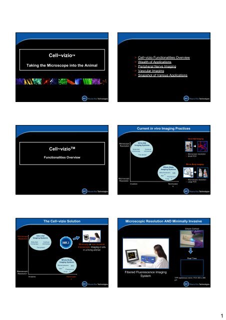
The Cell~vizio Solution<br />
Confocal<br />
microscopy<br />
Whole Body<br />
Imaging Systems<br />
Bioluminescenc MRI<br />
e<br />
PET<br />
Fluorescenc<br />
X-ray e<br />
Noninvasiv<br />
e<br />
Minimally or non invasive<br />
microscopic imaging in situ<br />
in a living animal<br />
Microscopic<br />
Resolution<br />
Macroscopic<br />
Resolution<br />
Invasive<br />
> Cell~vizio Functionalities Overview<br />
> Wealth of Applications<br />
> Peripheral Nerve Imaging<br />
> Vascular Imaging<br />
> Snapshot of Various Applications<br />
Wide-field<br />
microscopy<br />
Current in vivo Imaging Practices<br />
Intra-vital<br />
Imaging Systems<br />
Two-photon<br />
Confocal<br />
microscopy<br />
Whole Body<br />
Imaging Systems<br />
Bioluminescenc MRI<br />
e<br />
PET<br />
Fluorescenc<br />
X-ray e<br />
Noninvasiv<br />
e<br />
Intra-vital Imaging<br />
> Microscopic resolution<br />
> Small FOV<br />
Whole Body Imaging<br />
> Macroscopic resolution<br />
> Large FOV<br />
Microscopic Resolution AND Minimally Invasive<br />
Fibered Fluorescence Imaging<br />
System<br />
Simple Contact<br />
Real Time<br />
QuickTime?and a<br />
YUV420 codec decompressor<br />
are needed to see this picture.<br />
YFP saphenous nerve. FOV 400 x 280<br />
µm<br />
1
Laser Scanning Unit<br />
• 488 nm coherent source<br />
• High-speed scanning system<br />
• Ultra-sensitive detector<br />
Cell~vizio’s Three Components<br />
ProFlex TM flexible microprobe<br />
• Tens of thousands of fiber optics<br />
• Specific high-precision connector<br />
• Custom miniature objective<br />
ImageCell TM software<br />
• Real time control<br />
• On-the-fly image processing<br />
• Quantitative capabilities<br />
Image Construction<br />
QuickTime?and a<br />
YUV420 codec decompressor<br />
are needed to see this picture.<br />
• 30 000 fibers<br />
• Point-by-point injection<br />
• Point-by-point detection<br />
• Optical section generated<br />
• Images formed at 12<br />
frames/second<br />
Fibered ProFlex to Access Virtually Anywhere<br />
> Proprietary connector<br />
> Micro-precision fiber to<br />
laser interface<br />
> Optimal laser injection<br />
> Automatically recognized<br />
by the LSU (i-button)<br />
> Bundle of tens of<br />
thousands of fiber<br />
optics<br />
> Monolithic bundle<br />
construction<br />
> Flexible and robust<br />
> Wide range of distal tips<br />
for various uses and<br />
applications<br />
> With or without optics<br />
> From 1.8 mm to 300 µm<br />
Laser Scanning Unit<br />
ProFlex<br />
ImageCell<br />
Laser Scanning Unit<br />
ProFlex<br />
ImageCell<br />
Miniaturized ProFlex Microprobes<br />
> Direct contact imaging by gliding ProFlex along tissue<br />
QuickTime?and a<br />
YUV420 codec decompressor<br />
are needed to see this picture.<br />
2
Miniaturized ProFlex Microprobes<br />
> Direct contact imaging by gliding ProFlex along tissue<br />
> Smallest diameter of 300 µm for penetration into soft tissue with minimal<br />
disturbance<br />
Simple Acquisition<br />
Controls<br />
QuickTime?and a<br />
YUV420 codec decompressor<br />
are needed to see this picture.<br />
Laser Scanning Unit<br />
ProFlex<br />
ImageCell<br />
Acquisition Tools<br />
Laser Power<br />
Control<br />
Movie<br />
Thumbnails<br />
LookUp Table<br />
Control<br />
> Maximum diameter of 1.8<br />
mm for confocal optical<br />
slicing with working<br />
distances between 20 and<br />
100 µm<br />
Confocal Optical Slicing<br />
Courtesy of Elisabeth Laemmel and Eric Vicaut, LEM, Paris, France<br />
z<br />
Quantificatio<br />
n<br />
ImageCell Versatile Software<br />
QuickTime?and a<br />
YUV420 codec decompressor<br />
are needed to see this picture.<br />
Calibration process for<br />
experimental repeatability<br />
QuickTime?and a<br />
YUV420 codec decompressor<br />
are needed to see this picture.<br />
Different horizontal planes visible<br />
with large vessel clearly above<br />
smaller one<br />
FOV 160x120 µm<br />
Controls system for proper<br />
image acquisition, treatment<br />
and viewing<br />
Analysis and Processing Tools<br />
Display<br />
Enhancing<br />
Comprehensive ROI<br />
Management<br />
5 Place Jules Janssen<br />
Enables post-acquisition<br />
image browsing and<br />
analysis<br />
Editing and<br />
Exporting<br />
3
External<br />
External<br />
Why Use the Cell~vizio TM?<br />
Versatile Use and Applicability<br />
Minimally<br />
Invasive<br />
Versatile Use and Applicability<br />
Minimally<br />
Invasive<br />
Endoscopic<br />
Endoscopic<br />
QuickTime?and a<br />
YUV420 codec decompressor<br />
are needed to see this picture.<br />
Colonic crypts<br />
> Anyone can use the Cell~vizio, no particular training<br />
needed<br />
> Turn-key system requires no alignment or adjustments<br />
> Acquiring an image is as simple as placing the ProFlex<br />
in direct contact with a fluorescent organ<br />
><br />
Immediate and Intuitive Use<br />
Hand-held ProFlex can be<br />
glided along fluorescent<br />
structures over long<br />
distances<br />
External<br />
QuickTime?and a<br />
YUV420 codec decompressor<br />
are needed to see this picture.<br />
Cornea epithelium<br />
External<br />
400 µm<br />
QuickTime?and a<br />
YUV420 codec decompressor<br />
are needed to see this picture.<br />
Versatile Use and Applicability<br />
Minimally<br />
Invasive<br />
Versatile Use and Applicability<br />
GFP Neurons in the<br />
striatum<br />
Minimally<br />
Invasive<br />
QuickTime?and a<br />
YUV420 codec decompressor<br />
are needed to see this picture.<br />
Isolated sensory fiber. FOV 400 x<br />
280µm<br />
GFP tumor cells<br />
Endoscopic<br />
Endoscopic<br />
4
Longitudinal Studies<br />
TABLETOP FLUORESCENCE MICROSCOPY<br />
• Sacrificed animal<br />
• Explanted and fixed nerve<br />
• One mouse per<br />
measurement<br />
• 50 minutes per measurement<br />
FIBERED CONFOCAL FLUORESCENCE MICROSCOPY<br />
• Live, anesthetized animal<br />
• In vivo and in situ imaging<br />
• Repeated measurement<br />
on the same mouse<br />
• 5 minutes per<br />
measurement<br />
Actual Case Study: Peripheral<br />
Nerve Imaging After Crush<br />
Injury<br />
> Three times fewer animals for three time-point study<br />
> 1/10 of time for each measurement<br />
> More relevant data with all measurements on same animal<br />
Morphometry and Signal Quantification<br />
> Basis for accurate and reliable<br />
morphometry and quantification<br />
> Integrated, real-time analytical tools<br />
> Quantification and comparison<br />
between sequences<br />
> Precise in-depth localization of<br />
objects thanks to confocality<br />
> Linear response over a large<br />
detection range<br />
Courtesy of E. Laemmel and E. Vicaut, Microcirculation Lab’, Hospital F. Widal, Paris, France<br />
Wealth of Applications<br />
Real Time Dynamic Sequences<br />
> Dynamic events captured at 12 frames per second<br />
> Stable and fluid sequences even with a hand-held<br />
ProFlex<br />
> Real time made possible by on-the-fly image<br />
processing<br />
QuickTime?and a<br />
YUV420 codec decompressor<br />
are needed to see this picture.<br />
Erythrocytes, stained ex vivo with FITC,<br />
going through a capillary. FOV 160 x 120<br />
µm<br />
Courtesy of Elisabeth Laemmel and Eric Vicaut, LEM, Paris, France<br />
Deep<br />
Brain<br />
Corne<br />
a<br />
Heart<br />
> Cell~vizio Functionalities Overview<br />
> Wealth of Applications<br />
> Peripheral Nerve Imaging<br />
> Vascular Imaging<br />
> Snapshot of Various Applications<br />
Ear<br />
Mesentery<br />
A World of Possibilities<br />
Lymph<br />
Node<br />
Liver<br />
Muscle<br />
Kidney<br />
Colon<br />
Bladder<br />
NMJ<br />
5
Excised tissue<br />
imaged with a<br />
fluorescence<br />
microscope<br />
In situ images<br />
acquired with the<br />
Cell~vizio<br />
Cancer Research<br />
Endoscopic In Vivo Histology of Rat Bladder<br />
Superficial<br />
prismatic<br />
cells<br />
Control cells<br />
Basal<br />
cells<br />
Published work:<br />
M.A. D'Hallewin, S. El Khatib, A. Leroux, L. Bezdetnaya, F. Guillemin<br />
“Endoscopic Confocal Flurorescence Microscopy of Normal and Tumor Bearing<br />
Rat Bladder” (2005)Journal of Urology - In press (August 2005)<br />
Peripheral Neuropathies<br />
Tumor<br />
cells<br />
Scale bar: 100 µm<br />
Subcutaneous<br />
Tumor<br />
Tumor Vasculature and Angiogenesis<br />
Applications:<br />
> Vasculature Characterization<br />
• Functional capillary density<br />
• Vessel diameter<br />
• Permeability<br />
QuickTime?and a<br />
YUV420 codec decompressor<br />
are needed to see this picture.<br />
Courtesy of Descartes Image, O. Clément, Necker School of Medicine, Paris, France<br />
Scale bar: 100 µm<br />
> Angiogenesis<br />
• Endothelial cells recruitment<br />
• Anti-angiogenic therapies<br />
Observation of Aberrant Crypt Foci (ACF)<br />
Applications:<br />
> Colorectal cancer study<br />
> Aberrant Crypt Foci and adenoma characterization<br />
> Orthotopic tumor model observation<br />
> In vivo histology<br />
> Cytotoxic treatment monitoring<br />
Courtesy of Danijela Vignejevic, Sylvie Robine, Daniel Louvard, Institut Curie, Paris,<br />
France<br />
QuickTime?and a<br />
YUV420 codec decompressor<br />
are needed to see this picture.<br />
Scale bar: 100 µm<br />
Image all Parts of Neuron in vivo and in situ<br />
QuickTime?and a<br />
TIFF (LZW) decompressor<br />
are needed to see this picture.<br />
QuickTime?and a<br />
TIFF (LZW) decompressor<br />
are needed to see this picture.<br />
QuickTime?and a<br />
TIFF (LZW) decompressor<br />
are needed to see this picture.<br />
6
Vascularization<br />
Inject, Incise & Image<br />
Imaging is as easy as “1 2 3”<br />
1. Inject your dye IV<br />
2. Incise at the desired site<br />
3. Put ProFlex in contact with<br />
tissue<br />
Courtesy of Pr Vicaut, Microcirculation Lab, Hopital Fernand Widal, Paris, France<br />
QuickTime?and a<br />
YUV420 codec decompressor<br />
are needed to see this picture.<br />
Red blood cells with arrival of<br />
plasmatic dye<br />
FOV 400 x 280 µm<br />
Cell-endothelium interaction<br />
QuickTime?and a<br />
YUV420 codec decompressor<br />
are needed to see this picture.<br />
Rolling and adhering<br />
leukocytes<br />
Applications:<br />
> Leukocyte rolling<br />
>Recruitment of endothelial cells in neo-angiogenesis<br />
> Platelet aggregation<br />
> Atheromateous plaque formation<br />
Courtesy of Elisabeth Laemmel and Eric Vicaut, LEM, Paris, France<br />
Imaging from Vascular Networks to Single Cells<br />
QuickTime?and a<br />
YUV420 codec decompressor<br />
are needed to see this picture.<br />
Capillaries<br />
QuickTime?and a<br />
YUV420 codec decompressor<br />
are needed to see this picture.<br />
Cells in the<br />
blood stream<br />
Organ vascularization<br />
QuickTime?and a<br />
YUV420 codec decompressor<br />
are needed to see this picture.<br />
Vessels<br />
Courtesy of Ac Sinica, Taipei, Taiwan; CEA, Orsay, France; Hopital Fernand Widal, Paris, France<br />
QuickTime?and a<br />
YUV420 codec decompressor<br />
are needed to see this picture.<br />
Quantification of Vascular Parameters<br />
With ImageCell, measure:<br />
> Vessel diameter<br />
> Perfused capillary<br />
density<br />
> Permeability<br />
> Vasodynamics<br />
Published work:<br />
Laemmel, M. Genet, G. Le Goualher, A. Perchant, J.F. Le Gargasson, E. Vicaut<br />
« Fibered confocal fluoresence microscopy (Cell~vizio ェ) facilitates extended imaging in the<br />
field of microcirculation » (2004) Journal of Vascular Research 41(5):400-411<br />
Other Applications<br />
8
Snapshot of Other Applications<br />
Ophthalmology Gene Expression<br />
Kidney<br />
Damaged cells of the cornea<br />
epithelium as revealed by topical<br />
application of fluorescein.<br />
YFP transfected muscular fibers<br />
from the mouse skeletal muscle.<br />
Pharmacokinetics Immune Responses<br />
Detection of a drug candidate at the<br />
membrane of liver hepatocytes after<br />
IV injection.<br />
CSFE labeled T cells detected in<br />
the inguinal lymph node after<br />
injection in the foot-pad.<br />
Kidney glomerulus in a beta-actin-<br />
GFP mouse.<br />
Courtesy of Rothschild Foundation,<br />
CEA-SHFJ, Animage (France),<br />
Stanford University (USA), CMU-<br />
Geneva Medical Research Center<br />
(Switzerland).<br />
FOV 400 x 280 µm<br />
> Cell~vizio Functionalities Overview<br />
> Wealth of Applications<br />
> Peripheral Nerve Imaging<br />
> Vascular Imaging<br />
> Snapshot of Various Applications<br />
Apoptotic bodies in a cornea graft as<br />
revealed by topical application of<br />
YoYo-1.<br />
Cells in a GFP-rice specimen.<br />
Snapshot of Other Applications<br />
Apoptosis In vivo Heart<br />
Pancreas<br />
Plants<br />
Cardiomyocytes in a ß-actin-GFP<br />
mouse.<br />
Excretion<br />
Excretion of small MW dextran in the<br />
kidney afetr IP injection.<br />
Glandular organization of the mouse<br />
pancreas as revealed by a Rhodamine<br />
123 topical staining.<br />
Courtesy of Rotschild Foundation,<br />
INRA Montpellier (France),<br />
Stanford University (USA), CMU-<br />
Geneva Medical Research Center<br />
(Switzerland), Hebrew University of<br />
Jerusalem (Israel) .<br />
FOV 400 x 280 µm<br />
Applying the Cell~vizio<br />
to<br />
Research in Peripheral Neuropathies<br />
Image all Parts of Neuron in vivo and in situ … Detect Immune Response and Vascularization<br />
QuickTime?and a<br />
TIFF (LZW) decompressor<br />
are needed to see this picture.<br />
QuickTime?and a<br />
TIFF (LZW) decompressor<br />
are needed to see this picture.<br />
QuickTime?and a<br />
TIFF (LZW) decompressor<br />
are needed to see this picture.<br />
> Local recruitment and<br />
infiltration of immune cells<br />
Capillaries after I.V. injection of FITC-<br />
Albumin<br />
FOV 400 x 280 µm<br />
Courtesy of C. Combadière, Cellular Immunology, Pitié-Salpétrière, and<br />
E. Laemmel, Microcirculation Lab’, Hospital F. Widal, Paris<br />
GFP T cells after adoptive transfer<br />
FOV 400 x 280 µm<br />
> Micro-architecture of the<br />
local vascular bed<br />
9
… For Wealth of Potential Applications<br />
> Peripheral nerve injuries<br />
> Diabetic neuropathies<br />
> Inflammatory demyelinating<br />
diseases<br />
> Drug-related neuropathies<br />
> Drug discovery for neurotrophic or<br />
neuroprotective molecules<br />
Easy Animal Preparation and Image Acquisition<br />
> Access to e.g. the saphenous<br />
nerve is only a small incision in<br />
the skin away<br />
> A simple contact with the<br />
fluorescent organ is enough to<br />
get real-time images<br />
> The hand-held ProFlex can be<br />
glided along fluorescent<br />
structures over long distances<br />
Courtesy of Igor Charvet and Paolo Meda, CMU, Geneva, Switzerland<br />
400 µm<br />
QuickTime?and a<br />
YUV420 codec decompressor<br />
are needed to see this picture.<br />
YFP saphenous nerve. FOV 400 x 280<br />
µm<br />
Monitoring Peripheral Nerve<br />
Injuries and Regeneration<br />
Follow Up of Whole Nerve’s Condition…<br />
QuickTime?and a<br />
YUV420 codec decompressor<br />
are needed to see this picture.<br />
D0, H0: Intact Saphenous<br />
Nerve<br />
… with a single fiber resolution<br />
QuickTime?and a<br />
YUV420 codec decompressor<br />
are needed to see this picture.<br />
D1 post crush: Degenerated<br />
Nerve<br />
D0, H1: Crushed<br />
Nerve<br />
D4 post crush: Regenerated<br />
Nerve<br />
Courtesy of Igor Charvet and Paolo Meda, Medical Research Center, Geneva, Switzerland<br />
QuickTime?and a<br />
YUV420 codec decompressor<br />
are needed to see this picture.<br />
QuickTime?and a<br />
YUV420 codec decompressor<br />
are needed to see this picture.<br />
Daily Quantification of Nerve Regrowth In vivo Screening of Drug Candidates<br />
Cell~vizio vs. Conventional Fluorescence Microscope<br />
Length of outgrowth (mm)<br />
9<br />
8<br />
7<br />
6<br />
5<br />
4<br />
3<br />
2<br />
Reliable measurements of nerve outgrowth with Cell~vizio imagin<br />
Courtesy of Igor Charvet and Paolo Meda, CMU, Geneva, Switzerland<br />
3 4 5<br />
Days after crush<br />
QuickTime?and a<br />
TIFF (LZW) decompressor<br />
are needed to see this picture.<br />
> Evaluate in vivo neurotrophic molecules and their<br />
effect on nerve repair after a trauma<br />
> Study neuroprotection in vivo and address drug- or<br />
disease-related neuropathies<br />
> Quicker and more relevant evaluation of<br />
neurotrophic/ neuroprotectant drug candidates<br />
Thy1-YFP mouse FOV 400 x 280 µm<br />
10
In vivo Detection of Nerve Endings<br />
QuickTime?and a<br />
YUV420 codec decompressor<br />
are needed to see this picture.<br />
Dendritic endings of a YFP nerve<br />
FOV 400 x 280 µm<br />
Courtesy of Igor Charvet and Paolo Meda, CMU, Geneva, Switzerland<br />
QuickTime?and a<br />
YUV420 codec decompressor<br />
are needed to see this picture.<br />
Motor endings of a YFP nerve<br />
FOV 400 x 280 µm<br />
> Cell~vizio Functionalities Overview<br />
> Wealth of Applications<br />
> Peripheral Nerve Imaging<br />
> Vascular Imaging<br />
> Snapshot of Various Applications<br />
Imaging from Vascular Networks to Single Cells<br />
QuickTime?and a<br />
YUV420 codec decompressor<br />
are needed to see this picture.<br />
Capillaries<br />
QuickTime?and a<br />
YUV420 codec decompressor<br />
are needed to see this picture.<br />
Cells in the<br />
blood stream<br />
Organ vascularization<br />
Vessels<br />
QuickTime?and a<br />
YUV420 codec decompressor<br />
are needed to see this picture.<br />
Courtesy of Ac Sinica, Taipei, Taiwan; CEA, Orsay, France; Hopital Fernand Widal, Paris, France<br />
QuickTime?and a<br />
YUV420 codec decompressor<br />
are needed to see this picture.<br />
Cell~vizio Benefits in Peripheral Nerve Studies<br />
> One instrument for all aspects of a pathology<br />
> Immediate and intuitive (real-time imaging, quick animal preparation)<br />
> Cost- and time- saving<br />
Example: to screen 100 candidate neurotrophic<br />
molecules on a 3 day follow-up<br />
300 mice<br />
250 hours<br />
100 mice<br />
25 hours<br />
> Longitudinal studies thanks to organ preservation and minimally<br />
invasive access to the organ<br />
Applying the Cell~vizio<br />
to<br />
Research on Vascularization<br />
Organ Vascularization within ProFlex Reach!<br />
Organ-Specific Patterns Vessels & Capillaries<br />
Liver<br />
Tongue<br />
Kidney<br />
Lung<br />
Courtesy of Ac. Sinica; CEA; Chang Gung University; Hopital Fernand Widal; Pasteur Institute.<br />
Mesentery Muscle<br />
Brain<br />
Ear<br />
12
Macromolecular leakage<br />
5, 11 and 25 min after<br />
histamine suffusion<br />
Extravasation<br />
Courtesy of Pr Vicaut, Microcirculation Lab, Hopital Fernand Widal, Paris, France<br />
QuickTime?and a<br />
YUV420 codec decompressor<br />
are needed to see this picture.<br />
Cell-Wall Interactions<br />
Leukocytes stained with<br />
Rhodamine6G, FITC-Albumin in the<br />
plasma<br />
FOV 160 x 120 µm<br />
Courtesy of Hopital Fernand Widal, Paris, France; UniMaas, Maastricht, Netherlands<br />
Extravasation<br />
1400<br />
1200<br />
1000<br />
800<br />
600<br />
400<br />
200<br />
0<br />
0 5 10 15 20 25<br />
Leukocytes stained with<br />
Rhodamine6G<br />
FOV 400 x 280 µm<br />
Time (mn)<br />
QuickTime?and a<br />
YUV420 codec decompressor<br />
are needed to see this picture.<br />
Observe, characterize and quantify the cell-wall interactions with minimal animal<br />
preparation!<br />
Meeting the Requirements for<br />
Vascularization Imaging<br />
Application Examples<br />
Tumoral Angiogenesis Made Visible<br />
> Minimal incision required to access subcutaneous tumors<br />
> High molecular weight dye to avoid leakage<br />
> The protocol can be applied for ischemia studies<br />
Courtesy of Anne-Carole Duconseille and Olivier Clément, Descartes Image, Small Animal<br />
Imaging Facility, Université Paris V, Paris, France<br />
> Access<br />
QuickTime?and a<br />
YUV420 codec decompressor<br />
are needed to see this picture.<br />
Injection of FITC-Dextran 500 kDa in a<br />
mouse with a subcutaneous PC3 tumor<br />
Requirements<br />
> Dynamic Events Recording<br />
> Study Vascular Development<br />
14
Cell~vizio Facilitates Access<br />
Conventional Lens:<br />
> X, Y & Z movement only<br />
> Diameter many mm’s<br />
• Major surgical disturbance<br />
• Difficult access<br />
• Constraints on experiments<br />
ProFlex:<br />
> X, Y, Z, theta & phi movement<br />
> Diameter < 1 mm<br />
• Minimal surgical disturbance<br />
• Easy access<br />
• Access to remote locations (ex.<br />
Kidneys)<br />
• New access possibilities (ex. Deep<br />
brain)<br />
> Cell~vizio Functionalities Overview<br />
> Wealth of Applications<br />
> Peripheral Nerve Imaging<br />
> Vascular Imaging<br />
> Snapshot of Various Applications<br />
Glomerulus of the Kidney<br />
Kidney glomerulus in a beta-actin-GFP mouse imaged with the ProFlex S-1500 in<br />
direct contact with the intact kidney. FOV 400 x 280 µm<br />
Courtesy of Christopher H. Contag and Tim Doyle, Stanford,<br />
CA<br />
Answer to Requirements<br />
> Dynamic Events: Fast and Slow Events<br />
Recording<br />
• Flowing red blood cells, rolling leukocytes => Real time recording<br />
• Vasoconstriction studies => Time lapse recording<br />
> Study Vascular Development<br />
• Longitudinal studies => Minimal invasiveness<br />
• Statistical analyses => Screening possibilities<br />
• Quantification needs => ImageCell tools<br />
Snapshot of Existing Applications<br />
with the Cell~vizio<br />
Tumor Capillaries<br />
QuickTime?and a<br />
YUV420 codec decompressor<br />
are needed to see this picture.<br />
Capillaries of a mouse subcutaneous prostate tumor stained by IV injection of FITC-<br />
Albumin. Circulating blood cells appear in negative contrast in the movie. Imaged<br />
using a ProFlex S-1500. FOV 400 x 300 µm<br />
Courtesy of Nathalie Faye, Laure Fournier and Olivier Clément, LRI, Faculté Necker,-EM<br />
Paris<br />
15
Dual Plane of Axial Resolution<br />
Microvasculature of mouse mesentery, showing two different planes of different<br />
microcirculation architecture. ProFlex S-1500. FOV 400 x 280 µm<br />
Courtesy of Mauna Kea Technologies, Paris, France<br />
Erythrocytes Circulating Through a Capillary<br />
QuickTime?and a<br />
YUV420 codec decompressor<br />
are needed to see this picture.<br />
Erythrocytes going through a capillary. Erythrocytes are stained ex-vivo with FITC<br />
while blood plasma is stained by FITC-Albumin. Imaged with a ProFlex HD-1800/80,<br />
2.5 fixed on a probe holder. FOV: 160 x 120 µm<br />
Courtesy of Elizabeth Laemmel, Eric Vicaut, Microcirculation Lab, Hopital Fernand Widal,<br />
Paris<br />
Colonic Mucosa<br />
Mouse colonic crypts stained with both Syto 13 (nuclear staining) and cresyl<br />
violet (cytoplasmic staning) ProFlex S-1500. FOV 400 x 280 µm<br />
Courtesy of Igor Charvet, Centre Medical Universitaire, Geneva,<br />
Switzerland<br />
Rolling Leukocytes<br />
QuickTime?and a<br />
YUV420 codec decompressor<br />
are needed to see this picture.<br />
Leukocytes rolling on a vessels wall. Leukocytes are stained with Rhodamine 6G<br />
while blood plasma is stained by FITC-Albumin. Imaged with a ProFlex HD-1800/80,<br />
2.5 fixed on a probe holder. FOV: 160 x 120 µm<br />
Courtesy of Elizabeth Laemmel, Eric Vicaut, Microcirculation Lab, Hopital Fernand Widal,<br />
Paris<br />
Circulating Erythrocytes<br />
QuickTime?and a<br />
YUV420 codec decompressor<br />
are needed to see this picture.<br />
Circulation of erythrocytes in blood vessels. Erythrocytes are stained ex-vivo with<br />
FITC while blood plasma is stained by FITC-Albumin. Imaged with a ProFlex HD-<br />
1800/80, 2.5 fixed on a probe holder. FOV: 160 x 120 µm<br />
Courtesy of Elizabeth Laemmel, Eric Vicaut, Microcirculation Lab, Hopital Fernand Widal,<br />
Paris<br />
In vivo detection of Aberrant Crypt Foci<br />
QuickTime?and a<br />
YUV420 codec decompressor<br />
are needed to see this picture.<br />
Movie acquired on a mouse treated with the carcinogen AOM. The colonoscopy<br />
was performed after instillation of acriflavine. ACF AOM-induced are clearly<br />
visible at the end of the movie. ProFlex S-1500. FOV 400 x 280 µm<br />
Courtesy of Danijela Vignjevic, Sylvie Robine, Daniel Louvard, Institut Curie, Paris France<br />
16
Orthotopic Visualization of a Colonic Tumor<br />
Visualization of a colonic AOM-induced tumor in a mouse. The colonoscopy was<br />
performed after instillation of acriflavine. ProFlex S-1500. FOV 400 x 280 µm<br />
Courtesy of Danijela Vignjevic, Sylvie Robine, Daniel Louvard, Institut Curie, Paris France<br />
Cornea Apoptosis<br />
Apoptotic bodies in a rejected cornea graft, marked with Yoyo1, imaged with a<br />
ProFlex S-1500 placed in direct contact with the cornea, with a totally non-invasive<br />
external access. FOV 400 x 280 µm<br />
Courtesy of Francine Behart-Cohen, U450, INSERM, Paris France<br />
Endoscopic Access to Bladder Umbrella Cells<br />
Umbrella cells of healthy urothelial mucosa after instillation of rhodamine 123<br />
(mitochondria marker), with cell nuclei appearing dark. Imaged with a ProFlex S-<br />
0650 in view of optical biopsies for early cancer detection. FOV 400 x 280 µm<br />
Text<br />
Courtesy of Samy Elkatib and M.A. D'Hallewin, Centre Alexis Vautrin, Nancy, France<br />
Cremaster Microvasculature<br />
Image showing mouse cremaster vessels stained with FITC-Albumin, injected<br />
intravenously, with vessels as small as 5 µm. Hand-held ProFlex S-1500.<br />
FOV 400 x 280 µm<br />
Courtesy of Elisabeth Laemmel and Eric Vicaut, LEM, Paris, France<br />
Endoscopic Access to Bladder Basal Cells<br />
QuickTime?and a<br />
YUV420 codec decompressor<br />
are needed to see this picture.<br />
Basal cells of healthy urothelial mucosa after instillation of rhodamine 123<br />
(mitochondria marker), with cell nuclei appearing dark. Imaged with a ProFlex S-<br />
0650 in view of optical biopsies for early cancer detection. FOV 400 x 280 µm<br />
Courtesy of Samy Elkhatib and M.A. D'Hallewin, Centre Alexis Vautrin, Nancy, France<br />
Endoscopic Access to Bladder Tumor Cells<br />
QuickTime?and a<br />
YUV420 codec decompressor<br />
are needed to see this picture.<br />
In vivo visualization of a tumor after instillation of rhodamine 123 (mitochondria<br />
marker), with cell nuclei appearing dark. Imaged with a ProFlex S-0650 in view of<br />
optical biopsies for early cancer detection. FOV 400 x 280 µm<br />
Courtesy of Samy Elkatib and M.A. D'Hallewin, Centre Alexis Vautrin, Nancy, France<br />
17
Homing T cells<br />
CFSE labeled T cells detected in the inguinal lymph node after injection in the<br />
foot-pad. FOV 400 x 280 µm<br />
Courtesy of Hamida Hammad and Bart Lambrecht, Erasmus MC, Rotterdam.<br />
> Cell~vizio Functionalities Overview<br />
> Wealth of Applications<br />
> Peripheral Nerve Imaging<br />
> Vascular Imaging<br />
> Snapshot of Various Applications<br />
18


