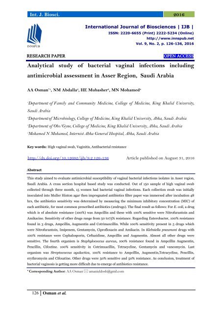
Analytical study of bacterial vaginal infections including antimicrobial assessment in Asser Region, Saudi Arabia
This study aimed to evaluate antimicrobial susceptibility of vaginal bacterial infections isolates in Asser region, Saudi Arabia. A cross section hospital based study was conducted. Out of 150 sample of high vaginal swab collected through three month, 15 women had bacterial vaginal infections. Each collection swab was initially inoculated into Muller Hinton agar then impregnated antibiotics filter paper was immersed after incubation 48 hrs, the antibiotics sensitivity was determined by measuring the minimum inhibitory concentration (MIC) of each antibiotic, for most common prescribed antibiotics (20drugs). The final result as follows: For E. coli, a drug which is of absolute resistance (100%) was Ampcillin and these with 100% sensitive were Nitrofurantoin and Amikacine. Sensitivity of other drugs range from 50 to75% resistance. Regarding Enterobacter, 100% resistance found in 3 drugs, Ampcillin, Augmentin and Cotrimaxcillin. While 100% sensitivity present in 5 drugs which were Nitrofurantoin, Imipenem, Gentamycin, Ciprofloxacin and Amikacin. In Klebsiella pneumoni drugs with 100% resistance were Cephalosporin, Ceftazidime, Ampcillin and Augmentin. Almost all other drugs were sensitive. The fourth organism is Staphylococcus aureus, 100% resistance found in Ampcillin Augmentin, Pencillin, Cifoxtine. 100% sensitivity in Cotrimxacillin, Tetracycline, Gentamycin and vancomycin. Last organism was Streptococcus agalactica, 100% resistance to Ampcillin, Augmentin, Tetracycline, Pencillin, erythromycin and Cifoxatine. Other drugs were 50% sensitive and 50% resistance. As conclusion, treatment of bacterial vaginosis is getting more difficult due to emerge of antibiotics resistance.
This study aimed to evaluate antimicrobial susceptibility of vaginal bacterial infections isolates in Asser region, Saudi Arabia. A cross section hospital based study was conducted. Out of 150 sample of high vaginal swab collected through three month, 15 women had bacterial vaginal infections. Each collection swab was initially inoculated into Muller Hinton agar then impregnated antibiotics filter paper was immersed after incubation 48
hrs, the antibiotics sensitivity was determined by measuring the minimum inhibitory concentration (MIC) of each antibiotic, for most common prescribed antibiotics (20drugs). The final result as follows: For E. coli, a drug which is of absolute resistance (100%) was Ampcillin and these with 100% sensitive were Nitrofurantoin and Amikacine. Sensitivity of other drugs range from 50 to75% resistance. Regarding Enterobacter, 100% resistance found in 3 drugs, Ampcillin, Augmentin and Cotrimaxcillin. While 100% sensitivity present in 5 drugs which were Nitrofurantoin, Imipenem, Gentamycin, Ciprofloxacin and Amikacin. In Klebsiella pneumoni drugs with 100% resistance were Cephalosporin, Ceftazidime, Ampcillin and Augmentin. Almost all other drugs were sensitive. The fourth organism is Staphylococcus aureus, 100% resistance found in Ampcillin Augmentin, Pencillin, Cifoxtine. 100% sensitivity in Cotrimxacillin, Tetracycline, Gentamycin and vancomycin. Last organism was Streptococcus agalactica, 100% resistance to Ampcillin, Augmentin, Tetracycline, Pencillin, erythromycin and Cifoxatine. Other drugs were 50% sensitive and 50% resistance. As conclusion, treatment of bacterial vaginosis is getting more difficult due to emerge of antibiotics resistance.
You also want an ePaper? Increase the reach of your titles
YUMPU automatically turns print PDFs into web optimized ePapers that Google loves.
Int. J. Biosci. 2016<br />
International Journal <strong>of</strong> Biosciences | IJB |<br />
ISSN: 2220-6655 (Pr<strong>in</strong>t) 2222-5234 (Onl<strong>in</strong>e)<br />
http://www.<strong>in</strong>nspub.net<br />
Vol. 9, No. 2, p. 126-136, 2016<br />
RESEARCH PAPER<br />
OPEN ACCESS<br />
<strong>Analytical</strong> <strong>study</strong> <strong>of</strong> <strong>bacterial</strong> <strong>vag<strong>in</strong>al</strong> <strong><strong>in</strong>fections</strong> <strong><strong>in</strong>clud<strong>in</strong>g</strong><br />
<strong>antimicrobial</strong> <strong>assessment</strong> <strong>in</strong> <strong>Asser</strong> <strong>Region</strong>, <strong>Saudi</strong> <strong>Arabia</strong><br />
AA Osman *1 , NM Abdalla 2 , HE Mubasher 3 , MN Mohamed 4<br />
1<br />
Department <strong>of</strong> Family and Community Medic<strong>in</strong>e, College <strong>of</strong> Medic<strong>in</strong>e, K<strong>in</strong>g Khalid University,<br />
<strong>Saudi</strong> <strong>Arabia</strong><br />
2<br />
Department <strong>of</strong> Microbiology, College <strong>of</strong> Medic<strong>in</strong>e, K<strong>in</strong>g Khalid University, Abha, <strong>Saudi</strong> <strong>Arabia</strong><br />
3<br />
Department <strong>of</strong> Obs/Gyne, College <strong>of</strong> Medic<strong>in</strong>e, K<strong>in</strong>g Khalid University, Abha, <strong>Saudi</strong> <strong>Arabia</strong><br />
4<br />
Mohamed N Mohamed, Internist Abha General Hospital, Abha, <strong>Saudi</strong> <strong>Arabia</strong><br />
Key words: High <strong>vag<strong>in</strong>al</strong> swab, Vag<strong>in</strong>itis, Anti<strong>bacterial</strong> resistance<br />
http://dx.doi.org/10.12692/ijb/9.2.126-136 Article published on August 31, 2016<br />
Abstract<br />
This <strong>study</strong> aimed to evaluate <strong>antimicrobial</strong> susceptibility <strong>of</strong> <strong>vag<strong>in</strong>al</strong> <strong>bacterial</strong> <strong><strong>in</strong>fections</strong> isolates <strong>in</strong> <strong>Asser</strong> region,<br />
<strong>Saudi</strong> <strong>Arabia</strong>. A cross section hospital based <strong>study</strong> was conducted. Out <strong>of</strong> 150 sample <strong>of</strong> high <strong>vag<strong>in</strong>al</strong> swab<br />
collected through three month, 15 women had <strong>bacterial</strong> <strong>vag<strong>in</strong>al</strong> <strong><strong>in</strong>fections</strong>. Each collection swab was <strong>in</strong>itially<br />
<strong>in</strong>oculated <strong>in</strong>to Muller H<strong>in</strong>ton agar then impregnated antibiotics filter paper was immersed after <strong>in</strong>cubation 48<br />
hrs, the antibiotics sensitivity was determ<strong>in</strong>ed by measur<strong>in</strong>g the m<strong>in</strong>imum <strong>in</strong>hibitory concentration (MIC) <strong>of</strong><br />
each antibiotic, for most common prescribed antibiotics (20drugs). The f<strong>in</strong>al result as follows: For E. coli, a drug<br />
which is <strong>of</strong> absolute resistance (100%) was Ampcill<strong>in</strong> and these with 100% sensitive were Nitr<strong>of</strong>uranto<strong>in</strong> and<br />
Amikac<strong>in</strong>e. Sensitivity <strong>of</strong> other drugs range from 50 to75% resistance. Regard<strong>in</strong>g Enterobacter, 100% resistance<br />
found <strong>in</strong> 3 drugs, Ampcill<strong>in</strong>, Augment<strong>in</strong> and Cotrimaxcill<strong>in</strong>. While 100% sensitivity present <strong>in</strong> 5 drugs which<br />
were Nitr<strong>of</strong>uranto<strong>in</strong>, Imipenem, Gentamyc<strong>in</strong>, Cipr<strong>of</strong>loxac<strong>in</strong> and Amikac<strong>in</strong>. In Klebsiella pneumoni drugs with<br />
100% resistance were Cephalospor<strong>in</strong>, Ceftazidime, Ampcill<strong>in</strong> and Augment<strong>in</strong>. Almost all other drugs were<br />
sensitive. The fourth organism is Staphylococcus aureus, 100% resistance found <strong>in</strong> Ampcill<strong>in</strong> Augment<strong>in</strong>,<br />
Pencill<strong>in</strong>, Cifoxt<strong>in</strong>e. 100% sensitivity <strong>in</strong> Cotrimxacill<strong>in</strong>, Tetracycl<strong>in</strong>e, Gentamyc<strong>in</strong> and vancomyc<strong>in</strong>. Last<br />
organism was Streptococcus agalactica, 100% resistance to Ampcill<strong>in</strong>, Augment<strong>in</strong>,Tetracycl<strong>in</strong>e, Pencill<strong>in</strong>,<br />
erythromyc<strong>in</strong> and Cifoxat<strong>in</strong>e. Other drugs were 50% sensitive and 50% resistance. As conclusion, treatment <strong>of</strong><br />
<strong>bacterial</strong> vag<strong>in</strong>osis is gett<strong>in</strong>g more difficult due to emerge <strong>of</strong> antibiotics resistance.<br />
* Correspond<strong>in</strong>g Author: AA Osman amanieldosh@gmil.com<br />
126 Osman et al.
Int. J. Biosci. 2016<br />
Introduction<br />
Vag<strong>in</strong>itis means <strong>in</strong>flammation <strong>of</strong> the vag<strong>in</strong>a. In most<br />
cases it is due to a fungal <strong>in</strong>fection. The patient<br />
typically has a discharge, itch<strong>in</strong>g, burn<strong>in</strong>g, and<br />
possibly pa<strong>in</strong>. It is frequently l<strong>in</strong>ked to an irritation or<br />
<strong>in</strong>fection <strong>of</strong> the vulva. Vag<strong>in</strong>itis is a very common<br />
condition. It is especially common <strong>in</strong> women<br />
with diabetes. (B. Lunenfeld, 2004).<br />
Vag<strong>in</strong>a is a muscular canal from the cervix to the<br />
outside <strong>of</strong> the body. It has an average length <strong>of</strong> about<br />
six to seven <strong>in</strong>ches. The walls <strong>of</strong> the vag<strong>in</strong>a are l<strong>in</strong>ed<br />
with mucus membrane People frequently refer to the<br />
vag<strong>in</strong>a when really they mean the vulva or female<br />
genitals, strictly speak<strong>in</strong>g the vag<strong>in</strong>a is a specific<br />
<strong>in</strong>ternal structure. The only part <strong>of</strong> the vag<strong>in</strong>a that<br />
can be normally viewed from the outside (without any<br />
<strong>in</strong>struments or carry<strong>in</strong>g out a pelvic exam<strong>in</strong>ation) is<br />
the <strong>vag<strong>in</strong>al</strong> open<strong>in</strong>g. The rest <strong>of</strong> the areas are parts <strong>of</strong><br />
the vulva, which <strong>in</strong>clude the labia majora, mons<br />
pubis, labia m<strong>in</strong>ora, clitoris, bulb <strong>of</strong> the vestibule,<br />
vestibule <strong>of</strong> the vag<strong>in</strong>a, etc.<br />
There are several types <strong>of</strong> vag<strong>in</strong>itis. The most<br />
common are; Atrophic vag<strong>in</strong>itis (or senile vag<strong>in</strong>itis) -<br />
the endothelium, the l<strong>in</strong><strong>in</strong>g <strong>of</strong> the vag<strong>in</strong>a, gets th<strong>in</strong>ner<br />
when estrogen levels go down dur<strong>in</strong>g the menopause.<br />
This makes the l<strong>in</strong><strong>in</strong>g more susceptible to irritation<br />
and <strong>in</strong>flammation. (FR. Ochsendorf, 2006). Bacterial<br />
vag<strong>in</strong>osis caused by overgrowth <strong>of</strong> normal bacteria <strong>in</strong><br />
the vag<strong>in</strong>a. Patients usually have less <strong>of</strong> the normal<br />
<strong>vag<strong>in</strong>al</strong> bacteria called lactobacilli. Trichomoniasis<br />
sometimes referred to as trich. It is a sexually<br />
transmitted s<strong>in</strong>gle-celled protozoan parasite<br />
Trichonomas <strong>vag<strong>in</strong>al</strong>is. It may <strong>in</strong>fect other parts <strong>of</strong><br />
the urogenital tract, <strong><strong>in</strong>clud<strong>in</strong>g</strong> the urethra (where<br />
ur<strong>in</strong>e comes out <strong>of</strong>) as well as the vag<strong>in</strong>a. Candida<br />
albicans this yeast-like fungal organism is what<br />
causes thrush. It exists <strong>in</strong> small amounts <strong>in</strong> the gut<br />
and is normally kept <strong>in</strong> check by bacteria. Cl<strong>in</strong>ically<br />
the hallmark symptoms <strong>of</strong> vag<strong>in</strong>itis <strong>in</strong>clude itch<strong>in</strong>g,<br />
burn<strong>in</strong>g and a discharge. Irritation <strong>of</strong> the genital area,<br />
<strong>vag<strong>in</strong>al</strong> discharge, <strong>in</strong>flammation redness, swell<strong>in</strong>g <strong>of</strong><br />
the labia majora, labia m<strong>in</strong>ora and per<strong>in</strong>eal area;<br />
ma<strong>in</strong>ly because <strong>of</strong> the presence <strong>of</strong> extra immune cells.<br />
Dysuria-pa<strong>in</strong> or discomfort when ur<strong>in</strong>at<strong>in</strong>g.<br />
Dyspareunia - pa<strong>in</strong>ful sexual <strong>in</strong>tercourse and foul<br />
<strong>vag<strong>in</strong>al</strong> odor. Aetiologically; Vulvovag<strong>in</strong>itis<br />
<strong>in</strong>flammation <strong>of</strong> the vag<strong>in</strong>a and vulva - can affect all<br />
women <strong>of</strong> all ages from every socioeconomic and<br />
ethnic backgrounds. Infectious vag<strong>in</strong>itis makes up<br />
90% <strong>of</strong> all cases <strong>in</strong> post-pubescent females. Infectious<br />
vag<strong>in</strong>itis <strong>in</strong>cludes candidiasis, <strong>bacterial</strong> vag<strong>in</strong>osis and<br />
trichmonisasis. Less commonly vag<strong>in</strong>itis may also be<br />
caused by gonnrrhea, Chlamydia, mycoplasma,<br />
herpes, campylobacter, some parasites and poor<br />
hygiene. Young girls, before they reach puberty, may<br />
also develop vag<strong>in</strong>itis, but the cause is <strong>of</strong>ten different<br />
from those for older females. While Streptococcus<br />
spp causes <strong>bacterial</strong> vag<strong>in</strong>osis <strong>in</strong> pre-pubescent girls,<br />
for post-pubescent females it is Gardnerella (both are<br />
types <strong>of</strong> bacteria). Improper hygiene <strong>in</strong> pre-pubescent<br />
girls can transfer bacteria and/or other irritants to the<br />
<strong>vag<strong>in</strong>al</strong> area from the anal region. Pre-pubescent girls<br />
do not usually get yeast <strong>in</strong>fection because their pH<br />
balance is different from older women's.<br />
An allergic reaction can cause vag<strong>in</strong>itis. For example,<br />
some women may be allergic to condoms,<br />
spermicides, certa<strong>in</strong> soaps and perfumes, douches,<br />
topical medications, lubricants, and even semen.<br />
Irritation from a tampon can cause vag<strong>in</strong>itis <strong>in</strong> some<br />
women. (Abdalla, 2011). The diagnosis <strong>of</strong> vag<strong>in</strong>itis<br />
require physical exam<strong>in</strong>ation and medical history. A<br />
sample <strong>of</strong> discharge may be taken to try to determ<strong>in</strong>e<br />
the cause <strong>of</strong> the <strong>in</strong>flammation. Vag<strong>in</strong>itis is diagnosed<br />
by check<strong>in</strong>g <strong>vag<strong>in</strong>al</strong> fluid appearance, <strong>vag<strong>in</strong>al</strong> pH<br />
levels, the presence <strong>of</strong> volatile am<strong>in</strong>es (the gas that<br />
causes a bad smell) and the microscopic detection <strong>of</strong><br />
specific cells. The common Pathogenic agent which<br />
might lead to female <strong>in</strong>fertility was shown <strong>in</strong> table 1.<br />
The type <strong>of</strong> treatment recommended depends on the<br />
cause <strong>of</strong> the <strong>in</strong>fection, and may <strong>in</strong>clude topical<br />
(applied onto the sk<strong>in</strong>) or oral antibiotics or<br />
anti<strong>bacterial</strong> creams. Antifungals eg. (Candida<br />
albicans), antiviral eg. (Herpes simplex) and<br />
antiparasitic eg. (Entrobious vermicularis and<br />
Tichomonas <strong>vag<strong>in</strong>al</strong>is). Cortisone cream may be<br />
prescribed if irritation symptoms are severe.<br />
127 Osman et al.
Int. J. Biosci. 2016<br />
An antihistam<strong>in</strong>e may be given if the doctor<br />
determ<strong>in</strong>es that the <strong>in</strong>flammation has been caused by<br />
an allergic reaction. If the vag<strong>in</strong>itis was caused by low<br />
estrogen levels, a topical estrogen cream may be<br />
recommended.<br />
Table 1. Disease Pathogenic agent which might lead<br />
to female <strong>in</strong>fertility.<br />
Bacteria Viruses Protozoa Yeasts<br />
Gonorrhoea<br />
Neisseria<br />
gonorrhoea<br />
Chlamydia<strong>in</strong>fection<br />
Chlamydia<br />
trachomatis<br />
serotype (D-K)<br />
Urethritis<br />
to)<br />
Ureaplasma<br />
urealyticum<br />
Syphilis<br />
Treponema<br />
pallidum<br />
HSV<br />
(due<br />
Chancroid caused Adenovirus<br />
by Haemophilus Infertility<br />
ducrey<br />
Lymphogranulo<br />
ma<br />
venereum by<br />
Chlamydia<br />
trachomatis<br />
(L1-L3)<br />
Granuloma<br />
<strong>in</strong>gu<strong>in</strong>aly<br />
AIDS HIV Urethritis<br />
Mononucleo due<br />
sis<br />
CMV<br />
to<br />
Tricho-<br />
Asymptomatmonas<br />
ic<br />
<strong>in</strong>fection<br />
Asymptomat<br />
ic<br />
<strong>in</strong>fection<br />
HPV<br />
Asymptomat<br />
ic<br />
<strong>in</strong>fection<br />
<strong>vag<strong>in</strong>al</strong>is<br />
Balanitis,<br />
urethritis<br />
due<br />
to<br />
Candida<br />
albicans<br />
Sometimes treatment is needed to restore <strong>vag<strong>in</strong>al</strong><br />
flora balance, which may have been altered after<br />
treatment for an <strong>in</strong>fection. Vag<strong>in</strong>al flora refers to a<br />
balance <strong>of</strong> bacteria <strong>in</strong> the vag<strong>in</strong>a that has significant<br />
implications for a woman's overall health.<br />
The prevention <strong>of</strong> vag<strong>in</strong>itis can be met by good<br />
hygiene - keep <strong>vag<strong>in</strong>al</strong> area clean. Use a mild soap<br />
(without irritants). Avoid douch<strong>in</strong>g and irritat<strong>in</strong>g<br />
agents - many are present <strong>in</strong> hygiene sprays, soaps,<br />
and other fem<strong>in</strong><strong>in</strong>e products. Avoid wip<strong>in</strong>g from your<br />
bottom to your vag<strong>in</strong>a (do it the other way<br />
round). Wear loose cloth<strong>in</strong>g. Practice safe sex.<br />
Materials and methods<br />
Study design<br />
<strong>Analytical</strong> cross sectional hospital based <strong>study</strong>.<br />
Study area<br />
Abha General Hospital, Abha city, Aseer, Western <strong>of</strong> KSA.<br />
Study Samples<br />
Samples <strong>of</strong> high <strong>vag<strong>in</strong>al</strong> swap (HVS) sent by<br />
gynecology department <strong>in</strong> the period from February<br />
to April 2016 was reviewed. Samples <strong>of</strong> HVS with<br />
<strong>bacterial</strong> results from the total samples were selected.<br />
Antimicrobial resistant test was conducted to all<br />
samples with <strong>bacterial</strong> <strong>in</strong>fection.<br />
High Vag<strong>in</strong>al Swab (HVS)<br />
High Vag<strong>in</strong>al Swab (HVS) is a technique used<br />
<strong>in</strong> Obstetrics and Gynaecology to obta<strong>in</strong> a sample<br />
<strong>of</strong> discharge from the vag<strong>in</strong>a. This is then sent<br />
for culture and sensitivity. Samples should be<br />
transported to laboratory immediately. If this is not<br />
possible the sample should be stored at room<br />
temperature and must reach the laboratory with<strong>in</strong> 48<br />
hours <strong>of</strong> collection. It is commonly used to test for the<br />
presence <strong>of</strong> candidiasis <strong>in</strong>fection, <strong>bacterial</strong> vag<strong>in</strong>osis<br />
and trichomonas <strong>vag<strong>in</strong>al</strong>is. (Niger, 2014)<br />
Antimicrobial Sensitivity Tests (Kirby- Pauer<br />
Diffusion Disk)<br />
Vag<strong>in</strong>al swabs collected from all 150 new cases<br />
presented with <strong>vag<strong>in</strong>al</strong> secretions will be collected.<br />
Direct microscopy us<strong>in</strong>g gram sta<strong>in</strong> and cultured <strong>in</strong><br />
Blood Agar to assess the hemolysis. Subcultured on<br />
blood agar and/or nutrient and Muller Hunter agar,<br />
<strong>in</strong>cubated at 35°C for 18 to 20 h, these cultures will be<br />
put <strong>in</strong> a bactech system and/or suncultured <strong>in</strong><br />
nutrient agar to which antibiotic laden filter papers<br />
will be applied then kept for 48 hrs <strong>in</strong> <strong>in</strong>cubator at<br />
36C o and then by measur<strong>in</strong>g the clear zone around<br />
each antibiotic disc (MIC) the sensitivity and<br />
resistance will be assessed.<br />
Exclusion criteria<br />
All results <strong>of</strong> HVS with non hemolytic gram positive<br />
bacteria such as Staphylococus epidermidis and<br />
saprophyticus.<br />
128 Osman et al.
Int. J. Biosci. 2016<br />
Non hemolytic Streptocccal spp., <strong>in</strong>tracellular<br />
bacteria such as Chylamdia spp., other bacteria<br />
Mucobacterium tuberculosis and cell wall deficient<br />
bacteria, Mycoplasma and Ureaplasma, anaerobics<br />
such as Bcteroides fragilis and Provetella sp. Gram<br />
negative cocci such as Hemophilus <strong>in</strong>fluenza and<br />
Nisseria gonorrheoa. Parasitic such as Trichomonas<br />
<strong>vag<strong>in</strong>al</strong>is, fungal eg. Candida albicans, viral<br />
<strong><strong>in</strong>fections</strong> and normal commensal such as<br />
Lactobacillus spp.<br />
Results and discussion<br />
A total <strong>of</strong> 150 samples <strong>of</strong> HVS conducted dur<strong>in</strong>g three<br />
month <strong>in</strong> gynecology department, Abha general<br />
hospital. Among them there were 15 cases <strong>of</strong> <strong>bacterial</strong><br />
<strong>in</strong>fection, which represent 10% <strong>of</strong> the total sample.<br />
Five pathogenic organism were isolated, they were, E.<br />
coli, Enterobacter sp., Klebsiella pneumonia,<br />
Staphylococcus aureus and Streptococcus agalactica.<br />
Antimicrobials resistance pr<strong>of</strong>ile was done to these<br />
organisms by us<strong>in</strong>g 20 commonest antibiotics as<br />
shown <strong>in</strong> table 2.<br />
Table 2. Five pathogenic groups isolated by HVS aga<strong>in</strong>st 20 common used <strong>antimicrobial</strong>s resistance pr<strong>of</strong>ile.<br />
Anti-<br />
Entero-<br />
Staphylo-<br />
Strepto-<br />
Klebsiella<br />
Microbials<br />
E. coli bacter<br />
coccus<br />
coccus<br />
pneumoniae<br />
Groups<br />
spp.<br />
aureus agalactica<br />
Penicil<strong>in</strong> R 50% Not done Not done R 100% R 100%<br />
Methicill<strong>in</strong> R 50% Not done Not done R 66% R 50%<br />
Erythromyc<strong>in</strong>3 Not done Not done Not done R 66& R 100%<br />
Cifoxt<strong>in</strong>e R 25% R 100% S 100% R 100% R 100%<br />
Tetracycl<strong>in</strong>3 R 50% S 50% S 100% S 100% R 100%<br />
Vancomyc<strong>in</strong> Not done Not done Not done S 100% R 50%<br />
Fucid<strong>in</strong>3 R 50% Not done Not done R 66% R 50%<br />
Augmant<strong>in</strong> R 50% R 100% R 100% R 66% R 100%<br />
Ampicil<strong>in</strong> R 100% R 100% R 100% R 100% R 100%<br />
Amikac<strong>in</strong>3 S 100% S 100% S 100% R 33% R 50%<br />
Cefaclor Not done Not done Not done R 66% R 50%<br />
Cipr<strong>of</strong>loxac<strong>in</strong>4 R 50% S 100% S 100% S 66% R 50%<br />
Gentamyc<strong>in</strong>3 R 50% S 100% S 100% S 100% R 50%<br />
Carb<strong>in</strong>cil<strong>in</strong>3 S 50% Not done Not done R 33% R 50%<br />
Cotrimxacill<strong>in</strong>5 75% R 100% S 75% S 100% R 50%<br />
Imipenem R 50% S 100% S 100% S 66% R 50%<br />
Ceftazidime R 75% R 50% R 100% S 66% R 50%<br />
Cephalospor<strong>in</strong>e S 75% R 50% R 100% S 33% S 50%<br />
Nitr<strong>of</strong>uranto<strong>in</strong>4 S 100% S 100% S 100% S 66% S 50%<br />
Rifamp<strong>in</strong>4 Not done Not done Not done S 33% S 50%<br />
S= sensitive R=resistance<br />
For E. coli drugs absolute resistance 100% was<br />
Ampcill<strong>in</strong> and these 100% sensitive were<br />
Nitr<strong>of</strong>uranto<strong>in</strong> and Amikac<strong>in</strong>e. Sensitivity <strong>of</strong> other<br />
drugs range from 50 to75% resistance.<br />
Regard<strong>in</strong>g Enterobacter, 100% resistancy found <strong>in</strong><br />
Ampcill<strong>in</strong>, Augment<strong>in</strong> and Cotrimaxcill<strong>in</strong>. While<br />
100% sensitivity present <strong>in</strong> 4 drugs which were<br />
Nitr<strong>of</strong>uranto<strong>in</strong>, Imipenem, Gentamyc<strong>in</strong>,<br />
Cipr<strong>of</strong>loxac<strong>in</strong> and Amikac<strong>in</strong>.<br />
129 Osman et al.
Int. J. Biosci. 2016<br />
In Klebsiella pneumoni drugs with 100% resistance<br />
were Cephalospor<strong>in</strong>, Ceftazidime, Ampcill<strong>in</strong> and<br />
Augment<strong>in</strong>. Almost all other drugs were sensitive.<br />
The fourth organism is Staphylococcus aureus, 100%<br />
resistance found <strong>in</strong> Ampcill<strong>in</strong> Augment<strong>in</strong>, Pencill<strong>in</strong>,<br />
Cifoxt<strong>in</strong>e. 100% sensitivity <strong>in</strong> Cotrimxacill<strong>in</strong>,<br />
Tetracycl<strong>in</strong>e, Gentamyc<strong>in</strong> and vancomyc<strong>in</strong>.<br />
Last organism was Streptococcus agalactica, 100%<br />
resistance to Ampcill<strong>in</strong>, augment<strong>in</strong>, tetracycl<strong>in</strong>e,<br />
pencill<strong>in</strong>, erythromyc<strong>in</strong> and Cifoxat<strong>in</strong>e. Other drugs<br />
were 50% sensitive and 50% resistance.<br />
In table 3, Antimicrobial resistance was done aga<strong>in</strong>st<br />
the five pathogenic organism isolated (E. coli,<br />
Enterobacter, Klebsielpneumone, Staphylococcus<br />
aureus and Streptococcus agalactica) the<br />
<strong>antimicrobial</strong> used has different mode <strong>of</strong> action. The<br />
first group is Cell wall <strong>in</strong>hibitor; this group <strong>in</strong>cludes<br />
around 10 commonly used <strong>antimicrobial</strong>s drugs. Over<br />
this entire group the most resistance organism was<br />
Streptococcus agalactica and E. coli, while the least<br />
resistance is Staphylococcus aureus.<br />
Table 4 showed the second group used was that work<br />
<strong>in</strong> Prote<strong>in</strong> Synthesis <strong>in</strong>hibitors. There were 6 common<br />
used drugs, the most resistance organism<br />
Streptococcus agalactica, and the most sensitive<br />
organism to this group was Klebsiela pneumoni.<br />
The third <strong>antimicrobial</strong> group is Inhibitors <strong>of</strong> nucleic<br />
acids synthesis. It <strong>in</strong>cludes common 3 drugs, It looks<br />
all organism were sensitive to this group, as shown <strong>in</strong><br />
table 5.<br />
The fourth group is antimetabolite (antifolate) <strong>in</strong>hibitor<br />
group. The most sensitive organism was Staphylococcus<br />
aureus while Enterobacter sp. was the most resistant<br />
one to this group as shown <strong>in</strong> table 6.<br />
Table 3. Five pathogenic groups isolated by HVS aga<strong>in</strong>st 10 common used <strong>antimicrobial</strong>s <strong>of</strong> Cell wall <strong>in</strong>hibitors group.<br />
Enterobacter Klebsiella Staphylo-coccus Streptococcus<br />
Anti-Microbiials Groups E. coli<br />
spp. pneumoniae aureus agalactica<br />
Penicil<strong>in</strong> R 50% Not done Not done R 100% R 100%<br />
Methicill<strong>in</strong> R 50% Not done Not done R 66% R 50%<br />
Cifoxt<strong>in</strong>e R 25% R 100% S 100% R 100% R 100%<br />
Vancomyc<strong>in</strong> Not done Not done Not done S 100% R 50%<br />
Augmant<strong>in</strong> R 50% R 100% R 100% R 66% R 100%<br />
Ampicil<strong>in</strong> R 100% R 100% R 100% R 100% R 100%<br />
Cefaclor Not done Not done Not done R 66% R 50%<br />
Imipenem R 50% S 100% S 100% S 66% R 50%<br />
Ceftazidime R 75% R 50% R 100% S 66% R 50%<br />
Cephalospor<strong>in</strong>e S 75% R 50% R 100% S 33% S 50%<br />
S= sensitive R=resistance<br />
Table 4. Five pathogenic groups isolated by HVS aga<strong>in</strong>st 6 common used <strong>antimicrobial</strong>s resistance pr<strong>of</strong>ile<br />
Prote<strong>in</strong> Synthesis <strong>in</strong>hibitors groups (S 50 & S 30).<br />
Entero-bacter Klebsiella Staphylococcus Streptococcus<br />
Anti-Microbiials Groups E. coli<br />
spp. pneumoniae aureus agalactica<br />
Erythromyc<strong>in</strong> S 50 Not done Not done Not done R 66& R 100%<br />
Tetracycl<strong>in</strong> S 30 R 50% S 50% S 100% S 100% R 100%<br />
Fucid<strong>in</strong> S 50 R 50% Not done Not done R 66% R 50%<br />
Amikac<strong>in</strong> S 30 S 100% S 100% S 100% R 33% R 50%<br />
Gentamyc<strong>in</strong> S 30 R 50% S 100% S 100% S 100% R 50%<br />
Carb<strong>in</strong>cil<strong>in</strong> S 50 S 50% Not done Not done R 33% R 50%<br />
S= sensitive R=resistance<br />
130 Osman et al.
Int. J. Biosci. 2016<br />
Table 5.Five pathogenic groups isolated by HVS aga<strong>in</strong>st 3 common used <strong>antimicrobial</strong>s resistance pr<strong>of</strong>ile,<br />
Inhibitors <strong>of</strong> nucleic acids synthesis group.<br />
Anti-<br />
Microbiials<br />
Groups<br />
E. coli<br />
Enterobacter<br />
spp.<br />
Klebsiella<br />
pneumoniae<br />
Staphylococcus<br />
aureus<br />
Streptococcus<br />
agalactica<br />
Cipr<strong>of</strong>loxac<strong>in</strong> R 50% S 100% S 100% S 66% R 50%<br />
Nitr<strong>of</strong>uranto<strong>in</strong> S 100% S 100% S 100% S 66% S 50%<br />
Rifamp<strong>in</strong> Not done Not done Not done S 33% S 50%<br />
S= sensitive R=resistance<br />
Table 6. Resistance pr<strong>of</strong>ile for the Five pathogenic organism isolated by HVS aga<strong>in</strong>st one common used<br />
<strong>antimicrobial</strong> drug from Antimetabolite (antifolate ) <strong>in</strong>hibitor group.<br />
Anti-Microbiials<br />
Entero-bacter Klebsiella Staphylo-coccus Strepto-coccus<br />
E. coli<br />
Groups<br />
spp.<br />
pneumoniae aureus<br />
agalactica<br />
Cotrimxacill<strong>in</strong>5 R75% R 100% S 75% S 100% R 50%<br />
S= sensitive R=resistance<br />
In Western countries STD-<strong><strong>in</strong>fections</strong> are <strong>of</strong> m<strong>in</strong>or<br />
relevance. In other regions, i.e. Africa or South East<br />
Asia, the situation appears to be different. Chronic<br />
<strong><strong>in</strong>fections</strong> (gonorrhoea) can cause urethral strictures<br />
and epididymoorchitis.<br />
Chlamydia trachomatis and Neisseria gonorrhoea<br />
can be transmitted to the female partner and cause<br />
pelvic <strong>in</strong>flammatory disease with tubal obstruction.<br />
Ureaplasma urealyticum may impair spermatozoa<br />
(motility, DNA condensation). Trichomonas<br />
<strong>vag<strong>in</strong>al</strong>is has, if any, only m<strong>in</strong>or <strong>in</strong>fluence on male<br />
fertility. The relevance <strong>of</strong> viral <strong><strong>in</strong>fections</strong> (HPV, HSV)<br />
for male <strong>in</strong>fertility is not resolved. Any STD <strong>in</strong>creases<br />
the chances <strong>of</strong> transmission <strong>of</strong> the human<br />
immunodeficiency virus (HIV). The HIV <strong>in</strong>fection is<br />
associated with <strong>in</strong>fectious semen and the risk <strong>of</strong> virus<br />
transmission. Semen quality deteriorates with the<br />
progression <strong>of</strong> immunodeficiency. Special counsell<strong>in</strong>g<br />
<strong>of</strong> serodiscordant couples is needed. STDs should be<br />
treated early and adequately to prevent late sequelae<br />
for both men and women (Dohle, 2003).<br />
Gonorrhoea, however, may impair male fertility.<br />
Gonorrhoeic urethritis was associated with urethral<br />
strictures (Zhou, 2004). Few cases had urithritis was<br />
identified by ur<strong>in</strong>e analysis. This may be expla<strong>in</strong>ed by<br />
large differences <strong>in</strong> the prevalence <strong>of</strong> STDs <strong>in</strong><br />
different regions <strong>of</strong> the world. Exclud<strong>in</strong>g human<br />
immunodeficiency virus (HIV) <strong><strong>in</strong>fections</strong>,<br />
the prevalence ranges from 4 per million <strong>in</strong> Western<br />
Europe to 32 <strong>in</strong> Sub-Saharan Africa and 48 <strong>in</strong> South<br />
East Asia with annual <strong>in</strong>cidences <strong>of</strong> 17, 69 and 151 per<br />
million respectively.<br />
Epidemiological data propose an association <strong>of</strong> a past<br />
C. trachomatis <strong>in</strong>fection and subfertility both <strong>in</strong> men<br />
and women (Kar<strong>in</strong>en, 2004; Eley, 2005). It is<br />
accepted that C. trachomatis impairs female fertility<br />
by caus<strong>in</strong>g tubal obstruction (Eggert-Kruse,1990).<br />
The exact role <strong>of</strong> Mycoplasmae, i.e. M. hom<strong>in</strong>is, U.<br />
urealyticum and M. genitalium has still to be<br />
elucidated. It was shown that M. genitalium can<br />
attach to spermatozoa and thus can be transported to<br />
the female genital tract (Sakar, 2008).<br />
Ureaplasma urealyticum may cause <strong>in</strong>fertility via<br />
deleterious effects on sperm chromat<strong>in</strong> and DNA,<br />
lead<strong>in</strong>g to impairment <strong>of</strong> embryo development<br />
(Gorkemli, 2006). In the group <strong>of</strong> <strong>in</strong>fertile patients<br />
with PCR positive for U. urealyticum, the volume,<br />
count and morphology <strong>of</strong> semen samples were lower<br />
than <strong>in</strong> the <strong>in</strong>fertile patients with PCR negative<br />
results. Trichomonas <strong>vag<strong>in</strong>al</strong>is was more <strong>of</strong>ten found<br />
<strong>in</strong> <strong>in</strong>fertile women than <strong>in</strong> fertile controls. A large<br />
women population <strong>of</strong> the world is suffer<strong>in</strong>g from a<br />
<strong>vag<strong>in</strong>al</strong> <strong>in</strong>fection commonly known as <strong>bacterial</strong><br />
vag<strong>in</strong>osis. The disease is associated with the decrease<br />
<strong>in</strong> the lactobacilli count <strong>in</strong> the vag<strong>in</strong>a. Till date, there<br />
is a lack <strong>of</strong> full pro<strong>of</strong> treatment modalities for the cure<br />
<strong>of</strong> the disease.<br />
131 Osman et al.
Int. J. Biosci. 2016<br />
The treatment <strong>in</strong>cludes the use <strong>of</strong> <strong>antimicrobial</strong>s<br />
and/or acidify<strong>in</strong>g agents and probiotics, either<br />
separately or <strong>in</strong> comb<strong>in</strong>ation (Nikhil, 2011).<br />
The vag<strong>in</strong>a it harbors a number <strong>of</strong> microorganisms<br />
and Lactobacillusis the predom<strong>in</strong>ant species as<br />
normal flora. Gram positive non hemolytics such as<br />
Staphylococus epidermidis and saprophyticus (sk<strong>in</strong><br />
normal flora). Fungal <strong>in</strong>fection (Candida albicans)<br />
parasitic (Trichomonas <strong>vag<strong>in</strong>al</strong>is) viral (HIV) were<br />
not <strong>in</strong>cluded <strong>in</strong> the <strong>study</strong>.<br />
Glycogen, an analogue <strong>of</strong> starch found <strong>in</strong> animals, is<br />
the ma<strong>in</strong> source <strong>of</strong> nutrients for the microbial flora<br />
resid<strong>in</strong>g <strong>in</strong> the lumen <strong>of</strong> the vag<strong>in</strong>a. The metabolism<br />
<strong>of</strong> glycogen <strong>in</strong> the <strong>vag<strong>in</strong>al</strong> system is mediated by the<br />
estrogen hormone via estrogen receptors located <strong>in</strong><br />
the epithelial cells cover<strong>in</strong>g the <strong>vag<strong>in</strong>al</strong> lumen (Owen,<br />
1975). The quantity <strong>of</strong> the mucus, estrogen level<br />
<strong>in</strong>creases. The normal pH <strong>of</strong> the vag<strong>in</strong>a 3.5-4.5 6<br />
<strong>vag<strong>in</strong>al</strong> micr<strong>of</strong>lora. In general, it is regarded that<br />
lactobacilli species is the predom<strong>in</strong>ant micr<strong>of</strong>lora<br />
responsible for ma<strong>in</strong>ta<strong>in</strong><strong>in</strong>g the pH <strong>of</strong> the <strong>vag<strong>in</strong>al</strong><br />
lumen (Eschenbach, 2000).<br />
The vag<strong>in</strong>a has been attributed to the regular contact<br />
<strong>of</strong> the external <strong>vag<strong>in</strong>al</strong> structures with the ur<strong>in</strong>e.<br />
(Hawthorn, 1991; Otero, 2007) gram negative such as<br />
E. coli and Klebsilla spp. undergo physicochemical<br />
<strong>in</strong>teraction with the <strong>vag<strong>in</strong>al</strong> epithelia, which helps <strong>in</strong><br />
the colonization <strong>of</strong> these pathogens as well as the<br />
lactobacilli lead<strong>in</strong>g to bi<strong>of</strong>ilm formation with<strong>in</strong> the<br />
mucosal and the epithelial layer <strong>of</strong> the vag<strong>in</strong>a<br />
(Busscher, 1987). The bi<strong>of</strong>ilm consists <strong>of</strong> the <strong>bacterial</strong><br />
cell layer(s) and the secretory components from the<br />
vag<strong>in</strong>a (Bibel, 1987).<br />
Vag<strong>in</strong>al secretions, especially mucus known as, postmenopausal<br />
atrophic vulvo-vag<strong>in</strong>itis. This results <strong>in</strong><br />
the decrease <strong>in</strong> the lubrication <strong>of</strong> the vag<strong>in</strong>a caus<strong>in</strong>g<br />
discomfort dur<strong>in</strong>g coitus (Kamarashev, 1997). In<br />
addition, there is a decrease <strong>in</strong> the thickness <strong>of</strong> the<br />
epithelial layer thereby <strong>in</strong>creas<strong>in</strong>g the susceptibility <strong>of</strong><br />
the <strong>vag<strong>in</strong>al</strong> tissue toward <strong>in</strong>fection and associated<br />
irritation (Farage, 2006).<br />
In normal healthy women, there is an <strong>in</strong>crease <strong>in</strong> the<br />
dynamic nature <strong>of</strong> the micr<strong>of</strong>lora with an <strong>in</strong>crease <strong>in</strong><br />
the hydration <strong>of</strong> the epithelial layer (Fre<strong>in</strong>kel, 1969).<br />
This results <strong>in</strong> the decreased permeability <strong>of</strong> the<br />
mucosal layer for the pathogenic organisms.<br />
The metabolic products secreted by the microbes may<br />
<strong>in</strong>fluence the availability <strong>of</strong> the nutrients (Elias,<br />
2007). Fatty acids have shown <strong>antimicrobial</strong> activity<br />
(aga<strong>in</strong>st Streptococcus pyogenes, Staphylococcus<br />
aureus, and sk<strong>in</strong> micrococci) and may help <strong>in</strong> f<strong>in</strong>e<br />
tun<strong>in</strong>g the composition <strong>of</strong> the microbial flora (Kabara,<br />
1972). They failed to produce substantial activity<br />
aga<strong>in</strong>st gram-negative bacteria. (Ouattara B, 1997)<br />
Apart from this, some peptides have also shown<br />
<strong>antimicrobial</strong> activity aga<strong>in</strong>st various pathogenic<br />
bacteria, fungi, viruses, and protozoa (Zasl<strong>of</strong>f 2002).<br />
A large number <strong>of</strong> transient organisms cont<strong>in</strong>uously<br />
migrate from the exogenous source (e.g., anus and<br />
urethra).<br />
Among the Lactobacillus species, Lactobacillus<br />
acidophilus was considered to be the dom<strong>in</strong>ant<br />
microbe present <strong>in</strong> the <strong>vag<strong>in</strong>al</strong> micr<strong>of</strong>lora. (Lachlak N<br />
1996, Vásquez A, 2002) The molecular identification<br />
techniques have the ability to specify the species<br />
easily <strong>in</strong> case <strong>of</strong> diverse micr<strong>of</strong>lora.<br />
In general, the lactobacilli are replaced with the<br />
<strong>in</strong>creased population <strong>of</strong> pathogenic gram-negative<br />
anaerobic bacteria such as E. coli, Gardnerella<br />
<strong>vag<strong>in</strong>al</strong>is, Mycoplasma hom<strong>in</strong>is, and Mycoplasma<br />
curtisii (Hillier S.2005, Hill GB. 1993). This condition<br />
may lead to several complications, which <strong>in</strong>clude<br />
cont<strong>in</strong>uous <strong>vag<strong>in</strong>al</strong> discharge, high HIV risk, malodor<br />
(fishy smell), stomach pa<strong>in</strong>, abortion, <strong>in</strong>fertility,<br />
preterm birth, chorioamnionitis, and ur<strong>in</strong>ary tract<br />
<strong>in</strong>fection. (Darwish A, 2007) researchers have<br />
associated BV with various factors <strong><strong>in</strong>clud<strong>in</strong>g</strong> <strong>vag<strong>in</strong>al</strong><br />
douch<strong>in</strong>g by the use <strong>of</strong> scented soaps or perfumed<br />
bubble bath and antiseptics dur<strong>in</strong>g bath, (MA.<br />
Kleban<strong>of</strong>f 2010) multiple sex partners and/or a new<br />
sex partner, smok<strong>in</strong>g (Ryckman KK, 2009) and use <strong>of</strong><br />
contraceptives (e.g., spermacides) which may <strong>in</strong>crease<br />
the probability <strong>of</strong> the <strong>in</strong>fection <strong>in</strong> a women.<br />
132 Osman et al.
Int. J. Biosci. 2016<br />
The presence <strong>of</strong> a strong fishy smell <strong>in</strong>dicates that the<br />
patient is suffer<strong>in</strong>g from BV. The microscopic<br />
exam<strong>in</strong>ation <strong>of</strong> the <strong>vag<strong>in</strong>al</strong> smear, which is analyzed<br />
for the presence <strong>of</strong> bacteria, white blood cells and clue<br />
cells, and the presence <strong>of</strong> clue cells, <strong>in</strong>dicates BV. The<br />
gram sta<strong>in</strong><strong>in</strong>g method, <strong>in</strong>troduced by Dunkelberg <strong>in</strong><br />
the year 1965, is also a simple method for the<br />
diagnosis <strong>of</strong> BV (Dunkelberg WE, 1965). the method<br />
helps <strong>in</strong> the confirmation <strong>of</strong> the presence <strong>of</strong> grampositive<br />
and gram-negative bacteria <strong>in</strong> the <strong>vag<strong>in</strong>al</strong><br />
discharge. Recently, a scor<strong>in</strong>g system (on a scale <strong>of</strong><br />
10) based on the presence <strong>of</strong> large gram-positive rods,<br />
small gram-negative or variable rods, and small<br />
curved gram-negative to variable rods have been<br />
reported. If the score lies <strong>in</strong> between 7 and 10, then<br />
the patient is suffer<strong>in</strong>g from BV (M. Joesoef, 1999).<br />
A lot <strong>of</strong> <strong>antimicrobial</strong> agents (e.g., ampicill<strong>in</strong>,<br />
penicill<strong>in</strong>, and metronidazole) have been used <strong>in</strong> the<br />
treatment <strong>of</strong> <strong>bacterial</strong> vag<strong>in</strong>itis. (Spiegel CG,1991)<br />
Metronidazole have evolved as a drug <strong>of</strong> choice for<br />
the treatment <strong>of</strong> BV and is the widely prescribed drug.<br />
It is a nitroimidazole derivative hav<strong>in</strong>g activity<br />
aga<strong>in</strong>st anaerobic microbes and protozoans.<br />
Adm<strong>in</strong>istration <strong>of</strong> the drug <strong>in</strong>clude gels and<br />
suppositories. (Sobel JD, 2006) Metronidazole and<br />
t<strong>in</strong>idazole (a chemical analogue <strong>of</strong> metronidazole) are<br />
preferred for the treatment <strong>of</strong> BV as aga<strong>in</strong>st<br />
ampicill<strong>in</strong>. T<strong>in</strong>idazole has a better pharmacok<strong>in</strong>etics<br />
and longer half-life than metronidazole and its<br />
recommendation for the treatment <strong>of</strong> BV is on the<br />
rise. (Dickey LJ, 2009) The use <strong>of</strong> ampicill<strong>in</strong> is<br />
avoided due to the emergence <strong>of</strong> ampicill<strong>in</strong>-resistant<br />
bacteria <strong>in</strong> patients with BV. It also <strong>in</strong>hibits the<br />
growth <strong>of</strong> lactobacilli. (Spiegel CG, 1991) Reports on<br />
cl<strong>in</strong>damyc<strong>in</strong> have suggested that it can be used <strong>in</strong> the<br />
treatment <strong>of</strong> BV and may be adm<strong>in</strong>istered either<br />
orally or locally. (Mc Gregor JA, 1994). Development<br />
<strong>of</strong> <strong>antimicrobial</strong> resistance mechanism with<strong>in</strong> the<br />
microbes. (RH. Beigi, 2004). Such as B lactamase,<br />
MRSA, Mc A gene, Bi<strong>of</strong>ilm and Absence <strong>of</strong> Enzymes<br />
such as RNA polymerase <strong>in</strong> Rifampic<strong>in</strong> Resistance.<br />
Hence, the researchers and cl<strong>in</strong>icians are look<strong>in</strong>g for<br />
alternative methods for the treatment <strong>of</strong> BV.<br />
The absence <strong>of</strong> Lactobacillus from the vag<strong>in</strong>a is the<br />
specific feature <strong>of</strong> BV. (SL. Hillier,1993). Lactic acidproduc<strong>in</strong>g<br />
bacteria, as probiotic. Various<br />
commercially available Lactobacillus-based products<br />
for the treatment <strong>of</strong> BV <strong>in</strong>clude yoghurt, acidophilus<br />
milk, and available Lactobacillus powder and tablets.<br />
(Hughes VL, 1990)<br />
Vag<strong>in</strong>al douch<strong>in</strong>g is the treatment helped <strong>in</strong><br />
improv<strong>in</strong>g the conditions <strong>of</strong> the patients. (M.<br />
Tasdemir,1996) , the <strong>vag<strong>in</strong>al</strong> pH was with<strong>in</strong> the<br />
normal range and the presence<br />
<strong>of</strong> Lactobacillus with<strong>in</strong> the lumen was also observed.<br />
(Neri A,1993)<br />
The use <strong>of</strong> probiotics for the treatment <strong>of</strong> BV has<br />
provided a ray <strong>of</strong> hope by natural and nontoxic<br />
treatment modality. (Dover SE, 2008) Apart from the<br />
above, the probiotics may <strong>of</strong>fer cost-effective<br />
treatment <strong>of</strong> BV.<br />
Conclusion<br />
People can help tackle resistance by: hand wash<strong>in</strong>g,<br />
and avoid<strong>in</strong>g close contact with sick people to prevent<br />
transmission <strong>of</strong> <strong>bacterial</strong> <strong><strong>in</strong>fections</strong> and viral<br />
<strong><strong>in</strong>fections</strong> such as <strong>in</strong>fluenza or rotavirus, and us<strong>in</strong>g<br />
condoms to prevent the transmission <strong>of</strong> sexuallytransmitted<br />
<strong><strong>in</strong>fections</strong>; gett<strong>in</strong>g vacc<strong>in</strong>ated, and<br />
keep<strong>in</strong>g vacc<strong>in</strong>ations up to date; us<strong>in</strong>g <strong>antimicrobial</strong><br />
drugs only when they are prescribed by a certified<br />
health pr<strong>of</strong>essional; complet<strong>in</strong>g the full treatment<br />
course (which <strong>in</strong> the case <strong>of</strong> antiviral drugs may<br />
require life-long treatment), even if they feel better;<br />
never shar<strong>in</strong>g <strong>antimicrobial</strong> drugs with others or<br />
us<strong>in</strong>g leftover prescriptions.<br />
Health workers and pharmacists can help tackle<br />
resistance by: enhanc<strong>in</strong>g <strong>in</strong>fection prevention and<br />
control <strong>in</strong> hospitals and cl<strong>in</strong>ics; only prescrib<strong>in</strong>g and<br />
dispens<strong>in</strong>g antibiotics when they are truly needed;<br />
prescrib<strong>in</strong>g and dispens<strong>in</strong>g the right <strong>antimicrobial</strong><br />
drugs to treat the illness. Policymakers can help<br />
tackle resistance by: improv<strong>in</strong>g monitor<strong>in</strong>g around<br />
the extent and causes <strong>of</strong> resistance; strengthen<strong>in</strong>g<br />
<strong>in</strong>fection control and prevention;<br />
133 Osman et al.
Int. J. Biosci. 2016<br />
regulat<strong>in</strong>g and promot<strong>in</strong>g appropriate use <strong>of</strong><br />
medic<strong>in</strong>es; mak<strong>in</strong>g <strong>in</strong>formation widely available on<br />
the impact <strong>of</strong> <strong>antimicrobial</strong> resistance and how the<br />
public and health pr<strong>of</strong>essionals can play their part;<br />
reward<strong>in</strong>g <strong>in</strong>novation and development <strong>of</strong> new<br />
treatment options and other tools. Policymakers,<br />
scientists and <strong>in</strong>dustry can help tackle resistance by:<br />
foster<strong>in</strong>g <strong>in</strong>novation and research and development <strong>of</strong><br />
new vacc<strong>in</strong>es, diagnostics, <strong>in</strong>fection treatment options<br />
and other tools.<br />
Acknowledgement<br />
We confer our gratitude to the laboratory staff <strong>of</strong><br />
Assir Central Hospital. Our s<strong>in</strong>cere thanks are to the<br />
staff Department <strong>of</strong> Obstetrics and gynecology, K<strong>in</strong>g<br />
Khalid University, <strong>Saudi</strong> <strong>Arabia</strong> for their support.<br />
References<br />
Abdalla NM. 2011. Sudanese male fertility and<br />
<strong><strong>in</strong>fections</strong>. Journal <strong>of</strong> scientica-parasitologica:<br />
September 12(3), 123-129.<br />
www.scientia.zooparaz.net/2011_12_03/sp2011-123-<br />
129-Abdalla.pdf<br />
Aroutcheva A, Gariti D, Simon M, Shott S,<br />
Faro J, Simoes JA, et al. 2001. Defense factors <strong>of</strong><br />
<strong>vag<strong>in</strong>al</strong> lactobacilli. American Journal <strong>of</strong> Obstetrics<br />
and Gynecology 185, 375–9.<br />
DOI: 10.1067/mob.2001.115867<br />
Beigi RH, Aust<strong>in</strong> MN, Meyn LA, Krohn MA,<br />
Hillier SL. 2004. Antimicrobial resistance<br />
associated with the treatment <strong>of</strong> <strong>bacterial</strong><br />
vag<strong>in</strong>osis. American Journal <strong>of</strong> Obstetrics and<br />
Gynecology 191, 1124–9.<br />
DOI: 10.1016/j.ajog.2004.05.033<br />
Bibel DJ, Aly R, Lahti L, Sh<strong>in</strong>efield HR,<br />
Maibach HI. 1987. Microbial adherence to vulvar<br />
epithelial cells. Journal <strong>of</strong> Medical Microbiology 23,<br />
75–82.<br />
DOI:10.1099/00222615-23-1-75<br />
Busscher HJ, Weerkamp AH. 1987. Specific and<br />
non-specific <strong>in</strong>teractions <strong>in</strong> <strong>bacterial</strong> adhesion to solid<br />
substrata. Journal <strong>of</strong> FEMS Microbiology Letters 46,<br />
165–73.<br />
Castelo-Branco C, Cancelo MJ, Villero J,<br />
Nohales F, Juliá MD. 2005 Management <strong>of</strong> postmenopausal<br />
<strong>vag<strong>in</strong>al</strong> atrophy and atrophic<br />
vag<strong>in</strong>itis. Maturitas Journal 52(Suppl 1): S46–52.<br />
Darwish A, Elnshar EM, Hamadeh SM,<br />
Makarem MH. 2007. Treatment options for<br />
<strong>bacterial</strong> vag<strong>in</strong>osis <strong>in</strong> patients at high risk <strong>of</strong> preterm<br />
labor and premature rupture <strong>of</strong> membranes. Journal<br />
<strong>of</strong> Obstetrics and Gynecology Reserch 33, 781–7.<br />
DOI: 10.1111/j.1447-0756.2007.00656.x<br />
Dickey LJ, Nailor MD, Sobel JD. 2009.<br />
Guidel<strong>in</strong>es for the treatment <strong>of</strong> <strong>bacterial</strong> vag<strong>in</strong>osis:<br />
Focus on t<strong>in</strong>idazole. Journal <strong>of</strong> Therapeutics<br />
and Cl<strong>in</strong>ical Risk Management 5, 485–9.<br />
PMCID:PMC2710380<br />
Dohle GR. 2003. Inflammatory-associated<br />
obstructions <strong>of</strong> the male genital tract, Andrologia<br />
Journal. 35, 321-324. PMID:14535864<br />
Dover SE, Aroutcheva AA, Faro S, Chik<strong>in</strong>das<br />
ML. 2008. Natural <strong>antimicrobial</strong>s and their role <strong>in</strong><br />
<strong>vag<strong>in</strong>al</strong> health: A short review. International Journal<br />
<strong>of</strong> Probiotics Prebiotics 3, 219-30.<br />
Dunkelberg WE, Jr. 1965. Diagnosis <strong>of</strong><br />
hemophilus <strong>vag<strong>in</strong>al</strong>is vag<strong>in</strong>itis by gram-sta<strong>in</strong>ed<br />
smears. American Journal <strong>of</strong> Obstetric and<br />
Gynecology 91, 998.<br />
Eggert-Kruse W, Gerhard I, Naher H, Tilgen<br />
W, Runnebaum B. 1990. Chlamydial <strong>in</strong>fection – a<br />
female and/or male <strong>in</strong>fertility factor? Fertility and<br />
sterility Journal 53, 1037-1043.<br />
Eley A, Pacey AA, Galdiero M, Galdiero M,<br />
Galdiero F. 2005. Can Chlamydia trachomatis<br />
directly damage your sperm? Lancet Infectious<br />
Disease 5, 53-57.<br />
Elias P. 2007. The sk<strong>in</strong> barrier as an <strong>in</strong>nate immune<br />
element. Sem<strong>in</strong>ars <strong>in</strong> Immunopathology 29, 3–14.<br />
PMID: 17621950<br />
134 Osman et al.
Int. J. Biosci. 2016<br />
Eschenbach DA, Thw<strong>in</strong> SS, Patton DL, Hooton<br />
TM, Stapleton AE, Agnew K, et al. 2000.<br />
Influence <strong>of</strong> the normal menstrual cycle on <strong>vag<strong>in</strong>al</strong><br />
tissue, discharge, and micr<strong>of</strong>lora. Cl<strong>in</strong>ical and<br />
Infectious Diseases Journal 30, 901–7.<br />
Farage M, Maibach H. 2006. Lifetime changes <strong>in</strong><br />
the vulva and vag<strong>in</strong>a. Archive <strong>of</strong> Gynecology and<br />
Obstetric 273, 195–202.<br />
Fre<strong>in</strong>kel RK, Shen Y. 1969. The orig<strong>in</strong> <strong>of</strong> free fatty<br />
acids <strong>in</strong> sebum. II. Assay <strong>of</strong> the lipases <strong>of</strong> the<br />
cutaneous bacteria and effects <strong>of</strong> pH. Journal <strong>of</strong><br />
Investigative Dermatology 53, 422–7.<br />
Gorkemli H, Cicek MN, Akyurek C, Celik C.<br />
2006. Approach to <strong>in</strong>fertile patient. In: Cicek MN,<br />
Akyurek C, Celik C. (Eds.), Gynecology and obstetrics.<br />
Chapter 29. Ankara, Turkey: Gunes Kitabevi pp. 1081.<br />
Hawthorn LA, Bruce AW, Reid G. 1991. Ability<br />
<strong>of</strong> uropathogens to b<strong>in</strong>d to Tamm Horsfall prote<strong>in</strong>coated<br />
renal tubular cells. Urological Research<br />
Journal 19, 301–4.<br />
Hill GB. 1993. The microbiology <strong>of</strong> <strong>bacterial</strong><br />
vag<strong>in</strong>osis. American Journal <strong>of</strong> Obstetric and<br />
Gynecology 169, 450-4.<br />
Kabara JJ, Swieczkowski DM, Conley AJ,<br />
Truant JP. 1972. Fatty acids and derivatives as<br />
<strong>antimicrobial</strong> agents..Antimicrobial Agents and<br />
Chemotherapy Journal 2, 23–8.<br />
Kamarashev JA, Vassileva SG. 1997.<br />
Dermatologic diseases <strong>of</strong> the vulva. Cl<strong>in</strong>ics <strong>in</strong><br />
Dermatology 15, 53–65.<br />
Kar<strong>in</strong>en L, Pouta A, Hartika<strong>in</strong>en AL, Bloigu A,<br />
Paldanius M, Le<strong>in</strong>onen M, Saikku P, Jarvel<strong>in</strong><br />
MR. 2004. Association between Chlamydia<br />
trachomatis antibodies and subfertility <strong>in</strong> the<br />
Northern F<strong>in</strong>land Birth Cohort 1966 (NFBC1966), at<br />
the age <strong>of</strong> 31 years. Epidemiology and Infection.<br />
Journal 132, 977-984.<br />
Kleban<strong>of</strong>f MA, Nansel TR, Brotman RM,<br />
Zhang J, Yu KF, Schwebke JR, et al. 2010.<br />
Personal hygienic behaviors and <strong>bacterial</strong><br />
vag<strong>in</strong>osis. Sexual Transmitted Disease 37, 94–9.<br />
Lachlak N, Ageron E, Zampatti O, Michel G,<br />
Grimont PA. 1996. Composition <strong>of</strong> the<br />
Lactobacillus acidophilus complex isolated from<br />
<strong>vag<strong>in</strong>al</strong> flora. New Microbiology Journal 19, 123–32.<br />
Hillier S. 2005. The complexity <strong>of</strong> microbial<br />
diversity <strong>in</strong> <strong>bacterial</strong> vag<strong>in</strong>osis. The New England<br />
Journal <strong>of</strong> Medic<strong>in</strong>e 353, 1886–7.<br />
Hillier SL, Krohn MA, Rabe LK, Kleban<strong>of</strong>f SJ,<br />
Eschenbach DA. 1993. The normal <strong>vag<strong>in</strong>al</strong> flora,<br />
H2O2-produc<strong>in</strong>g lactobacilli, and <strong>bacterial</strong> vag<strong>in</strong>osis<br />
<strong>in</strong> pregnant women. Cl<strong>in</strong>ical and Infectious Disease<br />
Journal 16(Suppl 4), S273–81.<br />
Hughes VL, Hillier SL. 1990. Microbiologic<br />
characteristics <strong>of</strong> Lactobacillus products used for<br />
colonization <strong>of</strong> the vag<strong>in</strong>a. Obstetric and Gynecology<br />
Journal 75, 244–8.<br />
Joesoef M, Schmid G, Hillier S. 1999. Bacterial<br />
vag<strong>in</strong>osis: Review <strong>of</strong> treatment options and potential<br />
cl<strong>in</strong>ical <strong>in</strong>dications for therapy. Cl<strong>in</strong>ical Infectous<br />
Disease Journal 28(Suppl 1), S57–65.<br />
Lunenfeld B, Van Steirteghem A. 2004.<br />
Foundation Bertarelli. Infertility <strong>in</strong> the third<br />
millennium: implications for the <strong>in</strong>dividual, family<br />
and society: condensed meet<strong>in</strong>g report from the<br />
Bertarelli Foundation's second global conference.<br />
Human Reproduction. Update 10(4), 317-326.<br />
McGregor JA, French JI, Jones W, Milligan K,<br />
McK<strong>in</strong>ney PJ, Patterson E, et al. 1994. Bacterial<br />
vag<strong>in</strong>osis is associated with prematurity and <strong>vag<strong>in</strong>al</strong><br />
fluid muc<strong>in</strong>ase and sialidase: Results <strong>of</strong> a controlled<br />
trial <strong>of</strong> topical cl<strong>in</strong>damyc<strong>in</strong> cream. Amrican Journal<br />
<strong>of</strong> Obstetric and Gynecology 170, 1048–59.<br />
Neri A, Sabah G, Samra Z. 1993. Bacterial<br />
vag<strong>in</strong>osis <strong>in</strong> pregnancy treated with yoghurt.<br />
Acta Obstetricia et Gynecologica Scand<strong>in</strong>avica 72, 17–9.<br />
135 Osman et al.
Int. J. Biosci. 2016<br />
Niger J. 2014. Cl<strong>in</strong>ical Practice. Mar-Apr; 17(2),<br />
154-8. doi: 10.4103/1119-3077.127424.<br />
Nikhil Kumar, Beauty Behera, Sai S<br />
Sagiri, Kunal Pal, Sirsendu S Ray, Saroj Roy.<br />
2011. Bacterial vag<strong>in</strong>osis: Etiology and modalities <strong>of</strong><br />
treatment—A brief note. Journal <strong>of</strong> Pharmacy and<br />
Bioallied Sciences. Oct-Dec 3(4), 496–503.<br />
DOI:10.4103/0975-7406.90102.<br />
PMCID: PMC3249696<br />
Ochsendorf FR, Schill WB, Comhaire FH,<br />
Hargreave TB. 2006. Urethritis, sexually<br />
transmitted diseases (STD), acquired immunodeficiency<br />
syndrome (AIDS). In Berl<strong>in</strong> –Heidelberg –<br />
New York, Andrology for the cl<strong>in</strong>ician. Spr<strong>in</strong>ger pp.<br />
125-130.<br />
Otero CM, Nader-Macías EM. 2007.<br />
Lactobacillus adhesion to epithelial cells from bov<strong>in</strong>e<br />
vag<strong>in</strong>a. Communicat<strong>in</strong>g Current Research and<br />
Educational Topics and Trends <strong>in</strong> Applied<br />
Microbiology 2, 749–57.<br />
Ouattara B, Simard RE, Holley RA, Piette GJ,<br />
Bég<strong>in</strong> A. 1997. Anti<strong>bacterial</strong> activity <strong>of</strong> selected fatty<br />
acids and essential oils aga<strong>in</strong>st six meat spoilage<br />
organisms. International Journal <strong>of</strong> Food<br />
Microbiology 37, 155–62.<br />
Owen JA, Jr. 1975. Physiology <strong>of</strong> the menstrual<br />
cycle. Amrican Journal <strong>of</strong> Cl<strong>in</strong>ical Nutrition 28, 333–<br />
8.<br />
Ryckman KK, Simhan HN, Krohn MA,<br />
Williams SM. 2009. Predict<strong>in</strong>g risk <strong>of</strong> <strong>bacterial</strong><br />
vag<strong>in</strong>osis: The role <strong>of</strong> race, smok<strong>in</strong>g and<br />
corticotrop<strong>in</strong>-releas<strong>in</strong>g hormone-related genes.<br />
Molecular Human Reproduction 15, 131–7.<br />
Sakar MN, Gul T, Atay AE, Celik Y. 2008.<br />
Comparison <strong>of</strong> hysterosal p<strong>in</strong>gography and<br />
laparoscopy <strong>in</strong> the evaluation <strong>of</strong> <strong>in</strong>fertile Women,<br />
<strong>Saudi</strong> Medical Journal 29(9), 1315-1318.<br />
Sobel JD, Ferris D, Schwebke J, Nyirjesy P,<br />
Wiesenfeld HC, Peipert J, et al. 2006.<br />
Suppressive anti<strong>bacterial</strong> therapy with 0.75%<br />
metronidazole <strong>vag<strong>in</strong>al</strong> gel to prevent recurrent<br />
<strong>bacterial</strong> vag<strong>in</strong>osis. Amarican Journal Obstetric and<br />
Gynecology 194, 1283–9.36.<br />
Spiegel CG. 1991. Bacterial vag<strong>in</strong>osis. Cl<strong>in</strong>ical<br />
Microbiology Reviews Journal 4, 485–502.<br />
PMCID:PMC358214<br />
Tasdemir M, Tasdemir I, Tasdemir S,<br />
Tavukcuoglu S. 1996. Alternative treatment for<br />
<strong>bacterial</strong> vag<strong>in</strong>osis <strong>in</strong> pregnant patients; restoration<br />
<strong>of</strong> <strong>vag<strong>in</strong>al</strong> acidity and flora. Archive <strong>of</strong> AIDS Research<br />
Journal 10, 239–41.<br />
Vásquez A, Jakobsson T, Ahrné S, Forsum U,<br />
Mol<strong>in</strong> G. 2002. Vag<strong>in</strong>al Lactobacillus flora <strong>of</strong><br />
healthy Swedish women. Journal <strong>of</strong> Cl<strong>in</strong>ical<br />
Microbiology 40, 2746–9. PMCID:PMC120688<br />
Zasl<strong>of</strong>f M. 2002. Antimicrobial peptides <strong>of</strong><br />
multicellular organisms. Nature Journal 415, 389–<br />
95.<br />
DOI:10.1038/415389a<br />
Zhou X, Bent SJ, Schneider MG, Davis CC,<br />
Islam MR, Forney LJ. 2004. Characterization <strong>of</strong><br />
<strong>vag<strong>in</strong>al</strong> microbial communities <strong>in</strong> adult healthy<br />
women us<strong>in</strong>g cultivation-<strong>in</strong>dependent methods.<br />
Microbiology Journals 150, 2565–73.<br />
136 Osman et al.


