FRCEM FINAL SBA eBook 1(Preview)
FRCEM FINAL SBA book is designed to present concise, easy-to-read, practical information on the diagnosis and treatment of a wide spectrum of conditions that present to the emergency department. The chapters emphasize the immediate management of life-threatening problems & then present the evaluation and treatment of specific disorders. In keeping with the curriculum spanning ST 1- ST 6 I have strived to provide the reader with a broad-based text written in a clear and point form manner. My goal was to not only provide practising emergency physicians quick access to accurate and useful information that will aid in their everyday practice of emergency medicine but also mainly to help trainees throughout their 6-year training in Emergency Medicine. Because this text focuses on the practical aspects of emergency care, there is little discussion of disease processes' basic science or pathophysiology. This is the only book of its kind available which covers the entire curriculum and helps prepare for the FINAL Clinical SAQ. The book will be useful to all practitioners of Emergency Medicine, including Physicians, Residents, Medical Students as well as Physician Extenders.
FRCEM FINAL SBA book is designed to present concise, easy-to-read, practical information on the diagnosis and treatment of a wide spectrum of conditions that present to the emergency department.
The chapters emphasize the immediate management of life-threatening problems & then present the evaluation and treatment of specific disorders.
In keeping with the curriculum spanning ST 1- ST 6 I have strived to provide the reader with a broad-based text written in a clear and point form manner.
My goal was to not only provide practising emergency physicians quick access to accurate and useful information that will aid in their everyday practice of emergency medicine but also mainly to help trainees throughout their 6-year training in Emergency Medicine.
Because this text focuses on the practical aspects of emergency care, there is little discussion of disease processes' basic science or pathophysiology.
This is the only book of its kind available which covers the entire curriculum and helps prepare for the FINAL Clinical SAQ.
The book will be useful to all practitioners of Emergency Medicine, including Physicians, Residents, Medical Students as well as Physician Extenders.

You also want an ePaper? Increase the reach of your titles
YUMPU automatically turns print PDFs into web optimized ePapers that Google loves.
40 <strong>FRCEM</strong> Final<br />
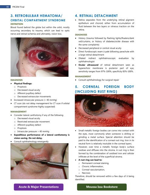
3. RETROBULBAR HEMATOMA/<br />
ORBITAL COMPARTMENT SYNDROME<br />
DEFINITION<br />
Blood found behind the globe but within the orbit, mostly<br />
occurring secondary to trauma, which can lead to optic<br />
nerve and retinal ischemia and ultimately, vision loss.<br />
DIAGNOSIS<br />
• Physical findings:<br />
o Proptosis<br />
o Decreased visual acuity<br />
o Afferent pupillary defect<br />
o Decreased extraocular movements<br />
• Increased intraocular pressure (> 40 mmHg)<br />
• CT scan (do not delay management for CT scan if orbital<br />
compartment syndrome highly suspected)<br />
MANAGEMENT<br />
• Consider lateral canthotomy if any of the following<br />
o Decreased visual acuity<br />
o Restricted extraocular movement<br />
o Afferent pupillary defect<br />
o Proptosis<br />
o Intraocular pressure > 40 mmHg<br />
• Expeditious performance of a lateral canthotomy is<br />
vision saving. Do not delay.<br />
• Consult ophthalmology emergently<br />
4. RETINAL DETACHMENT<br />
• Retina separates from the underlying retinal pigment<br />
epithelium and choroid, either from accumulation of<br />
fluid between the two layers or vitreous traction on the<br />
retina.<br />
DIAGNOSIS<br />
• History (trauma followed by flashing lights/floaters/dark<br />
veil/curtains, or history of diabetes/sickle disease with<br />
the same complaints)<br />
• Decreased peripheral or central visual acuity<br />
• Direct fundoscopic exam à pale billowing parachute with<br />
a large retinal detachment<br />
• Dilated indirect ophthalmoscopic evaluation by<br />
ophthalmologist<br />
• Ocular ultrasound of retinal detachment seen as<br />
hyperechoic membrane is posterior part of eye,<br />
sensitivity ranges from 97%-100%, specificity 83%-100%.<br />
MANAGEMENT<br />
• Consult ophthalmology for surgical repair<br />
5. CORNEAL FOREIGN BODY<br />
(INCLUDING RUST RING)<br />
• Small metallic foreign bodies can come into contact with<br />
the eyes, most commonly when someone is drilling or<br />
grinding a metal surface. Special attention should be<br />
paid to the identification of a corneal rust ring. Iron in its<br />
neutral form is relatively insoluble in the corneal layers.<br />
• However, over time a metallic foreign body’s surface<br />
oxidises and diffuses into the stroma. A rust ring is then<br />
formed by the combination of oxidised iron and cellular<br />
infiltrate at the level of the superficial stroma.<br />
• A rust ring can lead to:<br />
o Permanent corneal staining,<br />
o Chronic inflammation,<br />
o Corneal vascularisation,<br />
o Necrosis<br />
Therefore, should be removed within a few days of it being<br />
identified.<br />
Acute & Major Presentations<br />
Moussa Issa Bookstore