YSM Issue 92.1

Create successful ePaper yourself
Turn your PDF publications into a flip-book with our unique Google optimized e-Paper software.
Yale Scientific<br />
Established in 1894<br />
THE NATION’S OLDEST COLLEGE SCIENCE PUBLICATION<br />
MARCH 2019 VOL. 92 NO. 1 | $6.99<br />
12<br />
PREYING ON<br />
POLLUTANTS<br />
EVADING THE<br />
IMMUNE SYSTEM15<br />
SEQUENCING<br />
HALF A CELL18<br />
LEARNING TO<br />
REMEMBER20<br />
ESSENTIAL<br />
ERRORS22
INDIUM AND<br />
GALLIUM<br />
COMPOUNDS<br />
AND METALS<br />
49<br />
In<br />
114.818<br />
Indium Corporation’s online store allows<br />
you to browse our selection of metals,<br />
compounds, and solder products.<br />
Our ecommerce site:<br />
• Is easy to use<br />
• Accepts credit cards<br />
• Offers a wide variety<br />
of products<br />
Compounds<br />
31<br />
Ga<br />
69.723<br />
EZ-Pour ®<br />
Gallium<br />
Trichloride<br />
Contact our technical engineers today:<br />
techsupport@indium.com<br />
Learn more:<br />
www.indium.com/YALE<br />
From One Engineer<br />
To Another<br />
®<br />
www.indium.com<br />
askus@indium.com<br />
ASIA • CHINA • EUROPE • USA<br />
©2016 Indium Corporation
TABLE OF CONTENTS<br />
VOL. 92 ISSUE NO. 1<br />
More articles available online at www.yalescientific.org.<br />
12<br />
15<br />
18<br />
20<br />
22<br />
PREYING ON POLLUTANTS<br />
COVER ARTICLE: A collaboration between researchers at Yale and Peking University has led to a nanocoagulant that mimics<br />
the sea anemone Actinia. It can remove multiple types of contaminants simultaneously and outperforms conventional<br />
coagulants in multiple classes, thereby offering a cost-effective solution to water purification challenges.<br />
EVADING THE IMMUNE SYSTEM<br />
With the discovery of the immunological role of fibrinogen-like protein 1 (FGL-1) and its potential to aid the growth of cancerous<br />
cells, Yale researchers have solved a mystery that has baffled cancer immunologists for years.<br />
SEQUENCING HALF A CELL<br />
Researchers at Yale have developed a method to sequence both mRNA and microRNA in a single cell, leading to new insights into<br />
non-genetic variability at the single-cell level.<br />
LEARNING TO REMEMBER<br />
When do we start remembering? A new study conducted at the Yale School of Medicine probes this question and reveals<br />
the developmental timeline of memory formation.<br />
ESSENTIAL ERRORS<br />
The accurate translation of genetic material into proteins is essential to life. For parasitic bacteria with mutated genomes,<br />
however, errors in translation are important for their life cycles and could be therapeutic targets, Yale researchers report.<br />
www.yalescientific.org<br />
March 2019<br />
Yale Scientific Magazine<br />
3
CAN<br />
WE ENGI-<br />
NEER SPICY<br />
TOMATOES?<br />
By Serena Thaw-Poon<br />
Chili peppers, often conjuring both tears of pain and<br />
pleasure, serve an important role in the culinary exploits of<br />
many cultures. Less well known, however, are the medicinal<br />
properties of the compound responsible for the distinct heat<br />
of chilis. Peppers make a molecule called capsaicin, which has<br />
anti-inflammatory and weight-loss properties. But what if other<br />
plants could be modified to produce capsaicin as well? Could we<br />
make spicy tomatoes? Augustin Zsögön and his team have recently<br />
discovered genes in tomatoes that encode for capsaicin, suggesting<br />
that the “spicy tomato” might become a reality. Not only<br />
would chefs reap the benefits of a culinary marvel, but scientists<br />
would also have an easier time producing capsaicin to research as<br />
tomatoes are easier to breed and cultivate than typical Capsicum<br />
plants. The process of genetically engineering tomatoes, however,<br />
presents certain challenges. Many cellular components combine<br />
forces to make the process possible. “Enzymes that are acting in<br />
the mitochondrion, cytosol, and chloroplast all need to work<br />
together in sequence,” Zsögön said. Consequently, the production<br />
of capsaicin involves the manipulation of multiple<br />
genes and an understanding of how each gene affects the<br />
overall pathway. Zsögön makes it clear that any commercial<br />
opportunities would be unintentional, though<br />
welcome, consequences of his team’s research. “We<br />
don’t really care about the money, we just really<br />
like the ideas and get carried away,” he said,<br />
laughing. Whether for monetary or gastronomical<br />
purposes, spicy tomatoes<br />
are sure to appease everyone’s<br />
appetite.<br />
Q&A<br />
.<br />
CAN TETRIS<br />
HELP TREAT<br />
PTSD?<br />
By Nadean Alnajjar<br />
Tetris is the best-selling videogame of all time, but<br />
what if this simple computer game could relieve symptoms<br />
of an even more prevalent psychiatric disorder? Professor<br />
Henrik Kessler and Aram Kehyayan from Ruhr-Universität<br />
Bochum in Germany studied possible links between<br />
Tetris and post-traumatic stress disorder (PTSD), an anxiety<br />
disorder induced by stressful, recurrent memories called<br />
intrusions. “Our study is the first worldwide study to apply this<br />
Tetris paradigm in real patients with complex PTSD,” Kessler<br />
said. His study involved twenty patients undergoing a six-toeight-week<br />
period of regular inpatient treatment. The treatment<br />
involved playing Tetris after writing about an intrusion. Sixteen<br />
of the twenty patients responded well to the treatment–reporting<br />
a sixty-four percent decrease in intrusions on average. Further<br />
studies with more patients and better controls are now under way<br />
to replicate these results. PTSD intrusions use visuospatial regions<br />
of the brain, which are also activated while playing Tetris.<br />
“You supposedly need the same working memory resources<br />
and capacity that an intrusion would require,” Kessler said.<br />
“Tetris could lock the processing of intrusions.” Another<br />
theory proposes that if you reactivate an old memory, it<br />
becomes vulnerable, and interference through Tetris can<br />
be performed to weaken the memory. While professional<br />
treatment is available for PTSD, few people<br />
utilize it. “The implications of this research are<br />
huge because this would mean that people<br />
who had traumatic experiences could<br />
mend their intrusions by themselves<br />
without needing professional<br />
help,” Kessler said.<br />
4 Yale Scientific Magazine March 2019 www.yalescientific.org
SPIRIT OF CREATIVITY<br />
Great scientific discoveries begin with a pinch of curiosity and a dash of creativity. Our<br />
world faces complex problems which require scientists to work together to compose outof-the-box<br />
solutions. This issue features the work of incredible teams of investigators who<br />
have pushed the limits of human understanding of life in the universe (pg. 28), the immune<br />
system (pg. 15), memory formation (pg. 18), and Earth’s old supercontinents (pg. 34).<br />
Employing unique approaches to solve intricate problems, researchers have grown<br />
artificial brains from stem cells to better understand neurological disorders (pg. 10),<br />
developed a method to sequence two types of RNA in the same cell to learn how they<br />
regulate each other (pg. 22), and designed microscopes mounted on top of the heads<br />
of mice to collect real-time data on hundreds of thousands of neurons (pg. 35).<br />
As the human population explodes, and clean, drinkable water becomes less abundant,<br />
scientists are looking to nature for solutions. In our cover article, researchers<br />
have designed a water purification technology inspired from the feeding process of<br />
a sea anemone known as the “flower of the sea” (pg. 12). Based on the principles<br />
of coagulation, this system could lead to a more<br />
THE<br />
cost-effective approach to making cleaner water.<br />
Researchers have also sequenced the genome<br />
of a giant tortoise named Lonesome George,<br />
whose death at age 100 marked the last of his<br />
kind (pg. 8). Not only has Lonesome George’s<br />
venerable story inspired new conservation efforts<br />
in the Galapagos, but his genome might<br />
EDITORalso<br />
reveal the secrets to living a long life.<br />
All of us want to live long, full lives, but equally<br />
important is ensuring our quality of life doesn’t<br />
decline as we age. One study explains how a<br />
promising new compound restores memories and<br />
neural connections in the brains of mice afflicted<br />
with Alzheimer’s disease (pg. 10).<br />
With a new year comes a new masthead. Each<br />
IN-CHIEF<br />
year we grow as a community of writers, artists,<br />
and designers. We are excited to continue the<br />
work of our predecessors in celebrating compelling,<br />
new research being done at Yale and<br />
around the world. The stories we have chosen<br />
to share this issue have the potential to lead to<br />
a brighter future for our planet, our health, and<br />
SPEAKS<br />
our well-being. One of our goals is to build a<br />
conversation surrounding this research so that,<br />
one day, these visions become reality.<br />
William Burns, Editor-in-Chief<br />
ABOUT THE ART<br />
The cover art for this issue is inspired by the<br />
incredible breakthrough in water purification<br />
technology, modeling the molecular “tentacle”<br />
nano-coagulants of a novel system after<br />
a beautiful organism: the sea anemone. It’s<br />
an elegant and inspiring solution, reminding<br />
us that we need not look farther than natural<br />
forms to develop solutions.<br />
Ivory Fu, Arts Editor<br />
MASTHEAD<br />
MARCH 2019 VOL. 92 NO. 1<br />
EDITORIAL BOARD<br />
Editor-in-Chief<br />
Managing Editors<br />
News Editor<br />
Features Editor<br />
Articles Editor<br />
Online Editors<br />
Copy Editors<br />
Scope Editors<br />
PRODUCTION & DESIGN<br />
Production Manager<br />
Layout Editor<br />
Art Editor<br />
Photography Editor<br />
Webmaster<br />
BUSINESS<br />
Publisher<br />
Operations Manager<br />
Subscriptions Manager<br />
Advertising Managers<br />
OUTREACH<br />
Synapse Presidents<br />
Synapse Vice President<br />
Social Media Coordinator<br />
Outreach Coordinators<br />
STAFF<br />
Sophia Sanchez-Maes<br />
Ameera Billings<br />
Rami Rajjoub<br />
Viola Lee<br />
Miriam Ross<br />
James Han<br />
Ashwin Chetty<br />
Megan He<br />
Isaac Wendler<br />
Sami Elrazky<br />
ADVISORY BOARD<br />
Priyamvada Natarajan<br />
Sandy Chang<br />
Kurt Zilm, Chair<br />
Fred Volkmar<br />
Stanley Eisenstat<br />
James Duncan<br />
Stephen Stearns<br />
Jakub Szefer<br />
Werner Wolf<br />
John Wettlaufer<br />
William Summers<br />
Scott Strobel<br />
Robert Bazell<br />
Craig Crews<br />
Ayaska Fernando<br />
Robert Cordova<br />
Serena Thaw-Poon<br />
Nadean Alnajjar<br />
Katie Schlick<br />
Khue Mai Tran<br />
Tiffany Liao<br />
Grace Chen<br />
Brett Jennings<br />
Alison Ho<br />
Michael Adeyi<br />
Makayla Conley<br />
William Burns<br />
Conor Johnson<br />
Sunnie Liu<br />
Anna Sun<br />
Lukas Corey<br />
Marcus Sak<br />
James Han<br />
Lauren Kim<br />
Isabella Li<br />
Xiaoying Zheng<br />
Kelly Farley<br />
Georgia Woscoboinik<br />
Mafalda Von Alvensleben<br />
Maria Lee<br />
Ivory Fu<br />
Kate Kelly<br />
Matt Tu<br />
Richard Li<br />
Alexandra Brocato<br />
Sebastian Tsai<br />
Tony Leche<br />
Annie Yang<br />
Leslie Sim<br />
Lisa Wu<br />
Hannah Ro<br />
Chelsea Wang<br />
Oscar Garcia<br />
Katherine Dai<br />
Ellie Gabriel<br />
Britt Bistis<br />
Elissa Martin<br />
Anusha Bishop<br />
Antalique Tran<br />
Sandra Li<br />
Adrian Bebenek<br />
Andrea Ouyang<br />
Christie Yu<br />
Carli Roush<br />
Astronomy<br />
Biological and Biomedical Sciences<br />
Chemistry<br />
Child Study Center<br />
Computer Science<br />
Diagnostic Radiology<br />
Ecology & Evolutionary Biology<br />
Electrical Engineering<br />
Emeritus<br />
Geology & Geophysics<br />
History of Science, Medicine, & Public Health<br />
Molecular Biophysics & Biochemistry<br />
Molecular, Cellular, & Developmental Biology<br />
Molecular, Cellular, & Developmental Biology<br />
Undergraduate Admissions<br />
Yale Science & Engineering Association<br />
The Yale Scientific Magazine (<strong>YSM</strong>) is published four times a year by<br />
Yale Scientific Publications, Inc. Third class postage paid in New Haven,<br />
CT 06520. Non-profit postage permit number 01106 paid for May 19,<br />
1927 under the act of August 1912. ISN:0091-287. We reserve the right<br />
to edit any submissions, solicited or unsolicited, for publication. This<br />
magazine is published by Yale College students, and Yale University<br />
is not responsible for its contents. Perspectives expressed by authors<br />
do not necessarily reflect the opinions of <strong>YSM</strong>. We retain the right to<br />
reprint contributions, both text and graphics, in future issues as well as<br />
a non-exclusive right to reproduce these in electronic form. The <strong>YSM</strong><br />
welcomes comments and feedback. Letters to the editor should be under<br />
200 words and should include the author’s name and contact information.<br />
We reserve the right to edit letters before publication. Please send<br />
questions and comments to ysm@yale.edu. Special thanks to Yale STC.
SELECTING<br />
FOR CANCER<br />
Using evolution to explain the<br />
spread of cancer<br />
BY AMEERA BILLINGS<br />
In 2018, the<br />
International Agency for<br />
Research on Cancer estimated<br />
seventeen million new cancer diagnoses<br />
and 9.5 million cancer-related deaths worldwide.<br />
Though the numbers are grim, researchers have made<br />
great strides in explaining what causes and promotes cancer<br />
through an unexpected lens: evolution.<br />
The Townsend Lab at the Yale School of Public Health has<br />
developed a model to predict the evolutionary progression of<br />
cancerous cells. Now, we can quantify the relative importance<br />
of different gene mutations in stimulating cancer growth–the<br />
so-called ‘cancer effect size’ of a gene. By applying evolutionary<br />
principles, scientists can help analyze what mutations are driven<br />
by natural selection in a cancer lineage. “Selection is what drives<br />
[cancer] once you get a mutation…[causing cells to] replicate<br />
more,” explained Professor Jeffrey Townsend.<br />
Townsend and his team were able to calculate the mutation<br />
rate of cancer cells using tumor DNA sequencing. While some<br />
mutations may actually lead to cancer, others may have just<br />
happened to occur in those tumors. “If we see more [mutations]<br />
than expected by random mutation, then…it was more likely<br />
to lead to a tumor when you had those mutations in that gene,”<br />
Townsend said.<br />
What does this mean for the future of cancer medicine? “The<br />
cure for various cancers will be therapies that manage to corner<br />
all of the evolutionary avenues of an evolving tumor and make<br />
it impossible for it to find a new way to grow,” Townsend said.<br />
Being able to determine the cancer effect size of gene mutations<br />
will provide insight into the development of cancer cells, and<br />
thus, improve treatments for cancer patients in the future.<br />
Paleontology has long been regarded as the domain of dinosaur<br />
bones, teeth, and shells. During fossilization, these hard,<br />
organic scaffolds behave like a rough cast, providing structure<br />
for the assembly of minerals that will endure after the organics<br />
degrade. In this paradigm, soft tissues degrade too quickly<br />
for fossilization timescales, but perplexingly, structures which<br />
morphologically resemble them are common.<br />
A team led by Yale graduate student Jasmina Wiemann investigated<br />
the nature of these structures using a sample of vertebrate<br />
fossils from the Peabody Museum of Natural History<br />
collection. The researchers removed hard material from the<br />
fossils in a process called decalcification to isolate potential<br />
organic matter. Curiously, all the organic samples were brown<br />
in color, suggesting they had a common chemical composition.<br />
To explore this further, the team took fresh tissues rich<br />
in protein and subjected them to accelerated conditions for<br />
fossilization. Remarkably, the transformed tissues exhibited<br />
a brown color and composition reminiscent of the Peabody<br />
specimens. These findings suggest that the organic proteins<br />
in the Peabody specimens survived and metamorphized into<br />
a more stable, discolored polymer, visible to the unaided eye<br />
as the dark tinge on some fossils. The study shows that not<br />
only are organic structures, like cells and even organelles, preserved,<br />
but also that their preservation is surprisingly common<br />
in fossils formed under oxidative conditions.<br />
Scientists often use DNA testing to determine how living<br />
species are related, but DNA, which degrades easily, does not<br />
survive long in fossils. For long-extinct species, preserved<br />
proteins could be the next widely-used bio-informed<br />
molecule to more accurately<br />
reconstruct the tree of life.<br />
DINOSAUR<br />
SECRETS<br />
Preserved proteins reveal a<br />
more accurate tree of life<br />
BY SOPHIA S´ANCHEZ-MAES<br />
6 Yale Scientific Magazine March 2019
We are all familiar with the sensation of pain, but have<br />
you ever wondered why some people experience pain more<br />
vividly than others? For years, perplexed researchers have<br />
documented individual differences in pain tolerance to no<br />
avail. In a paper recently published in the Journal of Neuroscience,<br />
Malgorzata Mis, a postdoctoral associate in the<br />
Department of Neurology at Yale University, working with<br />
a team of researchers from the Yale School of Medicine<br />
and Veterans Affairs Connecticut Healthcare System, has<br />
taken the first steps towards identifying the genes involved<br />
in pain perception.<br />
The researchers analyzed a mother and son with inherited<br />
erythromelalgia, a disease caused by a mutation<br />
within a gene that kindles pain and burning sensations<br />
when expressed in the nervous system. Despite having<br />
the same mutation, the mother and son reported different<br />
levels of pains in a previous study. “The son reported<br />
over one-hundred nightly awakenings due to his mutation<br />
within a fifteen-day period, while the mother reported<br />
only one awakening,” Mis said.<br />
To further study this disparity, the research team grew<br />
sensory neurons derived from the mother’s and son’s<br />
stem cells in a laboratory dish. Using whole exome sequencing<br />
coupled with computer-aided dynamic clamp,<br />
a method that merges the visualization of live neurons<br />
with computational models, the researchers were able to<br />
identify a variation in the mother’s potassium channel<br />
encoded by the KCNQ2 gene, which ultimately led to<br />
her resilience to pain. The team is optimistic about<br />
implementing these new genetic methods<br />
that will guide them toward better<br />
treatments for pain.<br />
RETHINKING<br />
PAIN<br />
Uncovering the genes for<br />
pain resilience<br />
BY RAMI RAJJOUB<br />
THE BRAIN<br />
ON A DISH<br />
Organoids in the Age of<br />
Big Data<br />
BY VIOLA KYOUNG A LEE<br />
Yale neurobiologists<br />
are growing brain organoids to<br />
better understand the causes of neurological<br />
disorders. Brain organoids are artificial<br />
organs which are grown by differentiating pluripotent<br />
stem cells into neuronal tissues.<br />
Researchers in professor Flora Vaccarino’s lab at the Yale School of<br />
Medicine recently verified brain organoids’ ability to closely model fetal<br />
brain development. This project analyzed large-scale gene expression<br />
data from organoids’ RNA transcripts on Yale’s supercomputing clusters.<br />
“An extensive verification of the ability of brain organoids to serve<br />
as a model for the human brain has not been done before,” first author<br />
Anahita Amiri remarks. For years, professor Vaccarino has grown thousands<br />
of organoids to observe the correlation between gene expression<br />
and time-dependent cell differentiation. These brain organoids have<br />
many advantages over traditional neurobiological techniques. “[We<br />
have] the ability to observe the development of human neurons in a living<br />
system, in a time-dependent manner, and in 3D,” Vaccarino said.<br />
With the fidelity of their model system confirmed, the lab can grow<br />
hundreds of organoids and get massive amounts of data, including enhancer<br />
networks–which control which genes will be transcribed at each<br />
specific stage of brain development–and potential interactions with<br />
their target genes.<br />
Vaccarino and her lab are currently using organoids grown from the<br />
cells of patients with Autism Spectrum Disorder (ASD) to gain insights<br />
into how specific enhancers might contribute to ASD. “Once we can<br />
identify ASD biomarkers, the next steps could include developing therapies<br />
to target genes and enhancers involved through gene therapy or<br />
drugs,” Vaccarino said. With her brain organoid research, we are closer<br />
to understanding the mechanisms and causes of some neurodevelopmental<br />
diseases.<br />
March 2019<br />
Yale Scientific Magazine<br />
7
NEWS<br />
genetics<br />
LONESOME<br />
GEORGE’S<br />
LEGACY<br />
Genetic markers<br />
of longevity<br />
in giant tortoises<br />
BY MIRIAM ROSS<br />
IMAGE COURTESY OF FLICKR<br />
“Gone but not forgotten;” an epithet that applies to Lonesome George,<br />
the giant tortoise, and his newly-extinct species. In 2012, Lonesome<br />
George died as the last member of Chelonoidis abingdonii, at the ripe<br />
old age of 100. Galapagos tortoises are among the longest-lived vertebrate<br />
organisms, and researchers have long been curious about which<br />
specific factors contribute to their long lives. In Lonesome George’s<br />
case, the main research to examine tortoise longevity began after his<br />
death. “We were at the point in which it would have been very useful for<br />
us to have a whole genome, [to give] better information of how polymorphisms<br />
of different species are distributed,” said Adalgisa Caccone,<br />
a senior research scientist in Yale’s Ecology and Evolutionary Biology<br />
Department and director of the Center for Genetic Analysis of Biodiversity.<br />
After Lonesome George’s death in 2012, Caccone worked with<br />
colleagues at the Galapagos National Park Service, the Galapagos Conservancy,<br />
and the University of Oviedo to sequence his entire genome<br />
from a blood sample. With the full code of Lonesome George’s genome,<br />
the research team could begin making cross-species comparisons with<br />
smaller DNA sequences coded from several related tortoise species.<br />
In particular, Caccone and her research colleagues wanted to investigate<br />
variants linked to longevity in other species, which affected<br />
functions such as DNA repair, inflammation, and cancer development.<br />
The team had previously identified nine major “hallmarks<br />
of aging” in humans, and found variants affecting six of these “hallmarks”<br />
in the elderly tortoise’s genome–variants which are not found<br />
in the DNA of shorter-lived tortoise species. Of particular interest<br />
were a group of enzyme variants that seemed to offer protection<br />
against the sequence of protein activation which causes Parkinson’s<br />
disease. However, interpreting the meaning of variant changes within<br />
DNA is difficult. “It’s one thing to produce a genome, [and another]<br />
to annotate it, [and to] to know the function of a particular gene,”<br />
Caccone said. Traits are controlled by multiple genes which interact<br />
in multifaceted ways, and isolating the effect of a single variant can<br />
prove especially challenging. Although genome analysis reveals many<br />
variants, there is no easy way to map their functions; what is found<br />
are potential associations. And even in forming these associations,<br />
it is not clear from one genome alone whether a change is associated<br />
with longevity more broadly or effects in an individual organism.<br />
The first step towards identifying a variant’s function comes through<br />
cross-species comparisons. The research team had partial DNA samples<br />
of multiple individuals from related species of long-lived tortoises,<br />
which they compared to the full genome of Lonesome George. “Using<br />
this comparative approach, we can identify the hallmarks among all<br />
these genes, [and] eventually carry out functional studies on other organisms...to<br />
see if the [variants] have any associations with disease [or]<br />
longevity,” Caccone explained. Additionally, cross-species comparisons<br />
can allow the researchers to rule out the importance of changes<br />
which are not shared between species to longevity. The variants most<br />
likely to have functional significance should be those disproportionately<br />
present within the genomes of both Lonesome George and other<br />
Giant Galapagos tortoise species, all of which are long-lived.<br />
In addition to studying longevity, Lonesome George’s genome could<br />
also help with tortoise conservation. An effort is already underway to<br />
bring back extinct Galapagos tortoise species. “The Lonesome George<br />
genome is helping us is to identify molecular markers that are diagnostic<br />
for each species,” Caccone said. In previous research using DNA<br />
analysis, she discovered tortoises living on Galapagos islands that were<br />
actually genetic hybrids between the local species and an extinct one.<br />
The hybrids arose when early mariners brought tortoises with them<br />
for meat, and occasionally dropped them at random islands if the ship’s<br />
hold was too full. Caccone is helping the Galapagos National Park to<br />
start a breeding program that crosses these hybrids with each other to<br />
produce offspring more closely related to the original extinct species,<br />
the first breeding program of this type. The eventual goal is to release<br />
the tortoises’ offspring back onto the islands of their species’ origins.<br />
The sequencing of Lonesome George’s genome has given rise to<br />
many exciting potential associations, but the search for which variants<br />
actively contributed to his long life has just begun. With a group of<br />
potential associations and the sequences of other species, the secret to<br />
tortoises’ longevity will become clearer over time. Lonesome George<br />
may have died as the last member of his species, but discoveries from<br />
his genes can help us both to understand his long life, and perhaps<br />
even bring another extinct species back to their home island.<br />
8 Yale Scientific Magazine March 2019 www.yalescientific.org
cell biology<br />
NEWS<br />
HOPS<br />
INTO<br />
THE CELL<br />
IMAGE COURTESY OF WIKIMEDIA COMMONS<br />
When we take a drug, it flows through our bloodstream before<br />
entering the cells in our bodies–where the action happens. Most<br />
drugs that are currently used are small organic molecules that<br />
perform specific functions according to their chemical composition<br />
and shape. However, there is also substantial interest in using<br />
larger proteins and peptides as therapies for diseases. Unlike<br />
most small organic molecules, which are small enough to cross<br />
cell membranes, larger proteins or enzymes–molecular machines<br />
which have many more functions than small organic molecules–<br />
are tougher to shuttle across membranes inside cells. In a recent<br />
study published in the Proceedings of the National Academy of<br />
Sciences, a team led by Yale professor Alanna Schepartz uncovered<br />
new pathways and mechanisms that help proteins enter cells.<br />
Schepartz focused on two classes of proteins–cell-penetrating peptides<br />
(CPPs) and cell-permeant miniature proteins (CPMPs)–and the<br />
mechanism they use to enter the cell. In the study, Schepartz hoped to<br />
shed light on some of the unknown mechanisms employed by CPPs<br />
and CPMPs to enter cells. Both CPPs and CPMPs use what is called an<br />
endocytic pathway, taking advantage of the built-in cellular machinery<br />
for bringing larger molecules into the cell. During endocytosis, the cellular<br />
membrane surrounds the target molecule, pinches off, and creates<br />
an endosome inside the cell. This process is like the surface of a<br />
bubble pinching off inwards to become a new bubble inside the original<br />
bubble. Though scientists knew CPPs and CPMPs used the endocytic<br />
pathway, they were still unsure how CPPs and CPMPs are able to<br />
escape the endosome and enter the cellular fluid to fulfill their roles.<br />
Schepartz first tested the hypothesis that CPPs and CPMPs<br />
simply rupture the endosome to escape. The researchers used<br />
fluorescent tags to look for signs of endosome damage in the<br />
cell and locate ruptured endosomes. They discovered that the<br />
endosomes in cells that had taken in CPPs and CPMPs were relatively<br />
unharmed. The researchers then tested for signs of leakage<br />
from the endosome, which came back negative. Based on<br />
these results, the researchers were certain that endosomes are<br />
not harmed when the CPPs and CPMPs are released into cells.<br />
The team then decided to look for genes that could be regulating<br />
Uncovering<br />
how proteins<br />
enter cells<br />
BY JAMES HAN<br />
this release of proteins into the cell. They implemented a technique using<br />
small interfering RNAs (siRNAs), which allow researchers to “turn<br />
off” certain genes and observe the effects. After testing genes across<br />
the genome, Schepartz found that if VPS39, a gene that codes for a<br />
piece of the homotypic fusion and protein-sorting (HOPS) complex,<br />
was turned off, CPPs and CPMPs could not leave the endosome and<br />
enter the cell. They compared HOPS to similar complexes, but only<br />
turning off HOPS prevented proteins from leaving the endosome.<br />
Schepartz and her team then decided to look at the effects of<br />
CPPs and CPMPs on the function of HOPS. Using a fluorescence<br />
technique that looks for the products of HOPS’s reactions,<br />
Schepartz found that HOPS remained active even with delivery<br />
of CPPs and CPMPs. Finally, using fluorescent microscopy, the<br />
researchers found that HOPS functions by acting as a guide for<br />
CPPs and CPMPs, directing them to a class of endosome, called<br />
Lamp1, which has factors which facilitate the escape of proteins.<br />
Though the confirmation of HOPS as a key player in cell entry<br />
for proteins is a major breakthrough, further research is needed<br />
to understand the final steps of entry into the cell–particularly,<br />
how CPMPs escape from Lamp1 endosomes. Nevertheless,<br />
Schepartz’s research has great therapeutic potential and furthers<br />
our understanding of movement into and out of cells. As researchers<br />
learn more about these mechanisms, intricate molecular<br />
machines may replace small organic chemicals in medicine.<br />
SCHEPARTZ’S RESEARCH HAS GREAT<br />
THERAPEUTIC POTENTIAL AND<br />
FURTHERS OUR UNDERSTANDING OF<br />
MOVEMENT INTO AND OUT OF CELLS.<br />
www.yalescientific.org<br />
March 2019<br />
Yale Scientific Magazine<br />
9
NEWS<br />
neuroscience<br />
PIONEERING<br />
ALZHEIMER’S<br />
TREATMENT<br />
A compound that<br />
can reverse neural<br />
connection loss<br />
BY MIRIAM ROSS<br />
Alzheimer’s disease (AD), which currently affects more that 5.7<br />
million people in the US, is a neurodegenerative disease that greatly<br />
impairs memory. Despite the vast field of research dedicated toward<br />
tackling Alzheimer’s, there is still no cure. Erik Gunther, along with<br />
a team of Yale researchers in Professor Stephen Strittmatter’s lab, has<br />
discovered a promising drug to reverse the progression of AD. After<br />
screening over 60,000 compounds at the Yale Small Molecule<br />
Discovery Center, they developed a new compound that, instead of<br />
simply treating AD symptoms like currently approved drugs, can reverse<br />
neural connection loss and memory defects in mice.<br />
To better unravel the mechanism of the drug, it is important to<br />
understand AD development. AD results from an accumulation of<br />
β-amyloid peptides, which are small proteins present only in low concentrations<br />
in non-Alzheimer’s patients. In AD patients, β-amyloid<br />
peptides clump together to form β-amyloid oligomers, which then<br />
further combine to form β-amyloid plaques that look like “rocks in the<br />
brain,” according to Strittmatter. β-amyloid oligomers are problematic<br />
because they interact with neurons to set off a cascade of molecular<br />
events that damages synapses–the connections between neurons–and<br />
thus lead to memory impairment. Ten years ago, Strittmatter demonstrated<br />
that β-amyloid peptide binds strongly to cellular prion protein<br />
(PrP), a protein on the surface of neurons, so he began to investigate<br />
how to block this damaging interaction from occurring.<br />
Strittmatter’s lab initially screened over 12,000 compounds, hoping<br />
to find one that would inhibit the β-amyloid oligomer interaction<br />
with PrP. They first identified the antibiotic cefixime as a potential<br />
candidate, which, unusually, was only effective after it had degraded–since<br />
most drugs are effective before they start to degrade. If cefixime<br />
had not degraded by the time the research team screened it,<br />
Strittmatter believes, “We might still be searching [for a compound].”<br />
Other antibiotics with analogous structures to cefixime’s yielded similar<br />
results. After further testing, the team switched their focus toward<br />
the degraded product of the antibiotic ceftazidime because it was<br />
more effective than the degraded product of cefixime. Through further<br />
analysis, the research team learned that ceftazidime decomposes<br />
and reacts with itself to create a different polymer, referred to as<br />
IMAGE COURTESY OF FLICKR<br />
Compound Z, that inhibits β-amyloid oligomer interaction with PrP.<br />
Although the researchers knew Compound Z has inhibitory activity,<br />
they were uncertain whether it binds to PrP. Using antibodies constructed<br />
to bind to specific regions of PrP, they found that Compound<br />
Z binds to two regions of PrP that interact with the β-amyloid oligomer,<br />
preventing any interaction between β-amyloid oligomers and PrP.<br />
Once the researchers elucidated this pathway, they tested Compound<br />
Z’s effect on neurons. Even in a petri dish, neurons exposed to β-amyloid<br />
oligomers have damaged synapses, but Strittmatter’s lab found that Compound<br />
Z reduces β-amyloid oligomer binding to neurons by over eighty<br />
percent and reduces the loss of dendritic spines–an essential component<br />
of neuron signaling–by eight-fold. With these promising results, they administered<br />
Compound Z to mice engineered with Alzheimer’s-like disease.<br />
The researchers knew they had discovered a potential treatment for<br />
AD when the mice recovered learning and memory function.<br />
This compound, however, was unable to bypass the brain’s filtration<br />
system, the blood-brain barrier, rendering it difficult to<br />
use as a potential therapeutic. Strittmatter’s lab needed to find a<br />
compound that could cross the blood-brain barrier. They tested<br />
over 56,000 molecules until they landed on a synthetic compound<br />
poly(4-styrenesulfonnic acid-co-maleic acid), abbreviated as PSC-<br />
MA. Similar to Compound Z, PSCMA has inhibitory properties,<br />
but unlike Compound Z it can be administered orally and thus is<br />
considerably more practical as a potential drug.<br />
Though the mice in this study were able to complete memory tasks<br />
with the remaining β-amyloid oligomers in the brain, the results will<br />
not necessarily translate in human subjects. “[The mice models] are<br />
not a complete picture of human AD.” Strittmatter acknowledged.<br />
There are numerous steps before human trials can begin, but the<br />
researchers’ main goal now is to find the ideal balance between maintaining<br />
the drug’s inhibitory activity while optimizing the drug’s access<br />
to the brain. Strittmatter hopes to eventually test whether a drug<br />
from his lab might prove effective in treating AD in humans. “My<br />
optimism is high because I really believe we have a molecular basis<br />
for its effectiveness, and while these animal models are not perfect,<br />
they are the best predictors we have at this time,” he said.<br />
10 Yale Scientific Magazine March 2019 www.yalescientific.org
ecology<br />
NEWS<br />
SOURCES<br />
AND<br />
SINKS<br />
IMAGE COURTESY OF WIKIMEDIA COMMONS<br />
The global carbon cycle is an essential aspect of life on Earth,<br />
and there has been extensive scientific research concerning<br />
how the movement and exchange of carbon compounds can<br />
help manage atmospheric carbon dioxide (CO2) concentrations<br />
and predict climate change. Contemporary carbon cycle<br />
models largely look at carbon that is involved in live plant biomass<br />
and organic matter and the relationship between plants<br />
across landscapes and the atmosphere. Yet, these models ignore<br />
the effects of animals in higher trophic levels of ecosystems.<br />
An interdisciplinary team of researchers led by professor<br />
Oswald Schmitz at Yale sought to dispel the notion that animals<br />
have little or no impact on the carbon cycle. The researchers<br />
argue that wild animal populations are key players<br />
in shaping how ecosystems mitigate climate change and<br />
should be considered when making carbon measurements.<br />
Their paper, published in Science, found that the direct and<br />
indirect effects of animals can control the magnitude of carbon<br />
exchange in various global reservoirs. This can consequently<br />
influence carbon turnover rates, or the time it takes<br />
for carbon fixed in a plant by photosynthesis to be released<br />
back into the atmosphere.<br />
The animals in question are a diverse group spread across<br />
varying locations, including both large and small herbivores,<br />
carnivores, vertebrates, and invertebrates. Herbivores consume<br />
plant biomass and assimilate carbon into their own<br />
animal biomass after digestion, releasing additional carbon<br />
through defecation and respiration. With less plant biomass<br />
comes a decrease in photosynthesis and an increase in respiration,<br />
which reduces the chemical energy created by an ecosystem.<br />
Additionally, the heavy trampling of soil and sediment by<br />
larger animals can change surface temperatures that increase<br />
either carbon retention or release.<br />
These animals can even have conflicting positive and negative<br />
effects on the carbon cycle. For example, in some cases,<br />
grazing and browsing herbivores such as muskox and geese<br />
enhance carbon uptake and storage from the atmosphere into<br />
How animals<br />
shape the<br />
carbon cycle<br />
BY MEGAN HE<br />
organic compounds by twenty to twenty-five percent. In other<br />
cases, these grazing animals cause a fifteen to seventy percent decrease<br />
in CO2 uptake. The predators of these herbivores, on the<br />
other hand, can reverse these effects. In grassland ecosystems,<br />
grasshoppers cause a seventeen percent reduction in CO2 uptake,<br />
but the spiders that forage on these grasshoppers “reverse” the effect<br />
by increasing CO2 uptake by forty-six percent. In freshwater,<br />
stickleback fish that feed on macroinvertebrate stonefly insects,<br />
increase algal CO2 uptake, which in turn decreases concentrations<br />
of dissolved inorganic carbon in the water, enhancing carbon<br />
capture by around ninety percent.<br />
Other underlooked factors in ecosystem models are animal<br />
movement and migration. “Since large animals roam widely<br />
across landscapes, being able to monitor their movements and<br />
quantify the amount of material that is moved along with them<br />
is crucial to understanding their impact,” Schmitz said. Schmitz<br />
outlined a one instrument–named LIDAR–able to provide 3D<br />
portraits of habitat structure, but these methods still are limited<br />
in their ability to quantify the impacts of animals. On-the-ground<br />
sampling is often coupled with remote sensing to produce a more<br />
comprehensive analysis of carbon distribution. For example, the<br />
impact of African elephant populations on densities of woody<br />
vegetation can be monitored through both methods.<br />
An interesting challenge faced by the team was the pushback<br />
from skeptical reviewers, who were not convinced of the<br />
importance of incorporating animals into these models. Ultimately,<br />
however, Schmitz thinks that the data speaks for itself.<br />
“In some cases, you can triple how much carbon you take up if<br />
you protect these animals,” Schmitz said.<br />
What about the role of humans? “We have to protect the<br />
food chains–the animals and plants–that drive carbon dynamics.<br />
Nature provides all these services for our survival benefit<br />
and for other species, so we have to reimagine ourselves as being<br />
part of nature,” Schmitz said. At the end of the day, wildlife<br />
conservation proves to be an essential strategy for creating<br />
carbon storage capacity, and that task is up to the people.<br />
www.yalescientific.org<br />
March 2019<br />
Yale Scientific Magazine<br />
11
cell biology<br />
FOCUS<br />
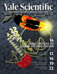
The sea anemone Actinia is a deceptive organism, in<br />
both name and appearance. For one, it is known as the<br />
“flower of the sea” for its colorful ring of tentacles. It also<br />
adopts a sessile lifestyle, preferring to lie and wait for food<br />
rather than go looking for it. When an unassuming plankton<br />
or fish passes by and stimulates its tentacles, the marine<br />
predator stuns it, abruptly retracts its tentacles, and<br />
gracefully encapsulates its catch.<br />
In a classic example of biomimetics–the imitation of<br />
natural systems for the purpose of solving human problems–a<br />
team of researchers from Yale and Peking University<br />
has designed a coagulant that can capture contaminants<br />
in water via a shape eversion process reminiscent<br />
of Actinia’s feeding process. Due to its unique structure,<br />
the innovative technology can trap many different types<br />
of contaminants in a single step. This versatile feature can<br />
potentially enable tremendous increases in water purification<br />
efficiency.<br />
Water and Energy: A Modern Compromise<br />
This innovation comes at a crucial time, as it promises<br />
to revolutionize the way we treat water. One in nine people<br />
worldwide do not have access to potable water. Water scarcity<br />
is a multifaceted problem without a clear solution, and cleaning<br />
up water with modern water purification systems has a<br />
high energetic and economic cost. Water and wastewater facilities<br />
are responsible for approximately thirty-five percent<br />
of a typical US municipal energy budget. Today, in order to<br />
supply consumers with clean water, facilities release over forty-five<br />
million tons of greenhouse gases annually–an output<br />
equivalent to that of more than six million cars.<br />
Current water treatment systems feature an extensive<br />
number of steps to ensure complete purification, because<br />
each step cannot remove all contaminants at once. Water<br />
contains many types of contaminants, each with drastically<br />
different chemical properties–think bacteria,<br />
harmful chemicals, and organic matter.<br />
One popular purification method is coagulation,<br />
in which positively charged<br />
coagulants, when added into water,<br />
bind to negatively charged<br />
dissolved organic carbon<br />
(DOC) and carbon-based<br />
remains<br />
of organisms. The<br />
added coagulants<br />
and<br />
organic<br />
mat-<br />
by HANNAH RO<br />
art by ANUSHA BISHOP<br />
PREYING<br />
ON POLLUTANTS<br />
technology imitates life<br />
to generate cleaner water<br />
www.yalescientific.org<br />
March 2019<br />
Yale Scientific Magazine<br />
12
FOCUS<br />
cell biology<br />
ter clump together into larger particles, then settle by gravity<br />
to the bottom of the basin so they can be more easily removed<br />
from the water. Another common step, sedimentation, allows<br />
heavier particles to settle, and a further chlorination step kills<br />
remaining microorganisms. Each of these methods requires<br />
specific technologies and tools, and the entire package together<br />
accounts for the high energy costs incurred today.<br />
The newly developed technology developed at Yale and Peking<br />
University, aptly named Actinia-like micellar nanocoagulant<br />
(AMC), streamlines water purification in two ways:<br />
one, it decreases the number of steps; two, it maximizes energy<br />
efficiency. If AMC enters the water purification industry,<br />
it could well eliminate multiple types of contaminants in one<br />
step, an optimal solution to lowering both the time and cost<br />
of cleaning water.<br />
Form Follows Function<br />
Last month, the collaborative project published results<br />
demonstrating that AMC could capture a spectrum of different<br />
contaminants in a single step. Conventional coagulants,<br />
such as aluminum sulfate, can typically only remove bulky<br />
organic matter. AMC inherits its special properties from Actinia.<br />
Like the sea anemone, the coagulant features a flexible<br />
outer shell that can be everted–turned outward/inside-out–<br />
to reveal an inner core, which has contrasting chemical properties.<br />
The positively charged outer shell binds to DOCs and<br />
other organic substances, while the negatively charged inner<br />
core contains organic micelles that capture smaller micropollutants.<br />
Notably, AMC can also extract nitrates, an excess<br />
of which can be harmful to humans.<br />
Both inner and outer surfaces can be accessed<br />
by altering the pH–a scale<br />
from zero to fourteen that<br />
corresponds to a<br />
solution’s acidity–of<br />
the surrounding solution. When stored in acidic conditions<br />
with a pH lower than four, the coagulant maintains its stable<br />
form, with the inorganic shell completely shielding the<br />
organic core. Raising the pH around AMC is akin to stimulating<br />
the tentacles of the Actinia–in wastewater above pH<br />
four, AMC changes its configuration to reveal the organic<br />
core. “Shape eversion of the coagulant adds additional functionality.<br />
Coagulants neutralize the charges on the organic<br />
matter and cause them to clump together so they can settle.<br />
But when the core is exposed, it absorbs additional contaminants<br />
that other coagulants would not be able to,” said Ryan<br />
DuChanois, a graduate student in the Elimelech group, who<br />
worked on the project.<br />
Another unique characteristic of the AMC is its ability<br />
to self-assemble. Much like the phospholipid bilayers that<br />
make up cell membranes, the AMC contains both hydrophobic<br />
and hydrophilic elements that rearrange in an aqueous<br />
solution. During the synthesis of AMC, hydrophobic<br />
carbon chains gather inwards, driven away from water, to<br />
form the organic core, and hydrophilic ionic complexes,<br />
comprising ammonium, silicon, and aluminum, form the<br />
outer shell. The researchers closely regulated the ratios of<br />
the reactants to achieve the product’s desired shape. Thanks<br />
to AMC’s optimized and stable build, it does not aggregate<br />
in solution and instead maintains its size and shape even after<br />
one year of storage.<br />
Having synthesized the particles, the researchers had<br />
to perform characterization to confirm and optimize its<br />
structure. However, one significant challenge of this project<br />
was the nanoscale of the coagulant; specialized equipment<br />
was required to observe the structure and function<br />
of the miniscule AMC particles. Researchers confirmed<br />
the spherical shape of AMC using transmission electron<br />
microscope (TEM) images and approximated its size with<br />
dynamic light scattering (DLS) measurements. For both<br />
types of technology, a beam of electrons, or light, is transmitted<br />
through the molecule to map out its structure. The<br />
caveat is that these methods only work when the molecule<br />
being observed is stationary. This is not the natural<br />
working state of the AMC. As such, the<br />
researchers had to turn to other technologies<br />
to learn about AMC’s mobile<br />
functionality.<br />
To observe how<br />
AMC self-as-<br />
sem-<br />
THE AMC RESEMBLES THE STRUCTURE OF SEA ANEMONE, WITH AN OUTER SHELL AND AN INNER CORE THAT TARGET
cell biology<br />
FOCUS<br />
bles, researchers conducted molecular dynamics<br />
simulations. These computational<br />
simulations, among other visualization<br />
techniques, predict and explain how a coagulant<br />
will behave when exposed to different<br />
types of wastewater. “[Simulations] show<br />
why the shell is removing certain contaminants<br />
and why the core is removing certain<br />
[other] contaminants. You can pinpoint the<br />
exact mechanisms, like electrostatic and hydrophobic<br />
interactions,” DuChanois said.<br />
Comparing Coagulants<br />
Once the structure and function of AMC<br />
were confirmed and better understood, researchers<br />
progressed to the field research<br />
portion of the project. Jar tests–in which<br />
treatment parameters, such as dosage, mixing<br />
rate, and aeration time, are altered to determine<br />
how a coagulant will behave with<br />
specific contaminants–were used to simulate<br />
full-scale water purification processes.<br />
These were performed to compare the<br />
efficiency of AMC and conventional coagulants.<br />
Efficiency was determined by two<br />
factors: the resulting contaminant concentration,<br />
as well as turbidity, a quantitative<br />
measurement of a liquid’s murkiness.<br />
AMC edged out other commercially<br />
used coagulants tested in the study–including<br />
a polymer called polyDADMAC,<br />
aluminum sulfate, and iron(III) chloride–<br />
with an average efficiency of over ninety<br />
percent. While all of the coagulants exhibited<br />
similar efficiency in removing turbidity,<br />
AMC was more successful at lowering<br />
DOC, phosphorous, and nitrate concentrations.<br />
Furthermore, while traditional coagulants<br />
demonstrated negligible removal of<br />
nitrate, AMC’s nitrate removal efficiency<br />
exceeded ninety percent.<br />
Other key contaminants the researchers<br />
considered were organic micropollutants<br />
and certain pharmaceuticals.<br />
Commonly, water is contaminated by<br />
micropollutants from residue left behind<br />
by personal care products, hormones,<br />
and pesticides. Because these particles<br />
are nonbiodegradable, they persist in<br />
wastewater treatment discharges<br />
and enter the environment.<br />
Conventional coagulants<br />
perform<br />
poorly<br />
in this category. Instead, the existing methods<br />
of removing micropollutants are ozonation<br />
and ultrasound. These approaches are energy-expensive,<br />
time-consuming, and work for<br />
only their specific class of contaminants.<br />
In this study, the conventional coagulants<br />
demonstrated removal efficiencies between<br />
zero and sixty percent. AMC outshone all of<br />
them, achieving removal efficiencies of over<br />
ninety percent for all tested micropollutants.<br />
This makes AMC all the more promising as a<br />
future water purification technique because<br />
it offers an entire functional package. “The<br />
real benefit is that if you can remove many<br />
different types of contaminants in one step<br />
instead of using many steps, you can simplify<br />
the process, use less money, and take up<br />
less land. Overall, the process becomes more<br />
efficient,” DuChanois said.<br />
The Big Picture<br />
Ultimately, this project demonstrates<br />
how adaptations which species develop<br />
to face problems in nature can inspire<br />
technological innovations to solve human<br />
problems. Encouraged by the success of<br />
AMC, the researchers hope to continue<br />
applying their novel ideas to other areas<br />
of water treatment and materials science.<br />
“Coagulation has been in use for centuries<br />
and nothing has really changed. You<br />
would think at this point in time, after<br />
thousands of people have thought about<br />
this, that we would have perfected the water<br />
treatment process. But there’s always<br />
opportunity to innovate,” DuChanois<br />
said. Perhaps more innovations, building<br />
on the success of the AMC, will lead to effective<br />
solutions to the abiding challenge<br />
of energy-efficient water purification.<br />
ABOUT THE AUTHOR<br />
IMAGE COURTESY OF RYAN DUCHANOIS<br />
HANNAH RO<br />
HANNAH RO is a first-year in Trumbull from Orange County, California. In addition to writing for the<br />
Yale Scientific, she volunteers with Synapse, researches in the Bergwitz lab, and plans events for Korean<br />
American Students at Yale.<br />
THE AUTHOR WOULD LIKE TO THANK Ryan DuChanois for his time and enthusiasm to share their<br />
research.<br />
FURTHER READING<br />
Jinwei Liu, Shihan Cheng, Na Cao, Chunxiang Geng, Chen He, Quan Shi, Chunming Xu, Jinren Ni, Ryan<br />
M. DuChanois, Menachem Elimelech, Huazhang Zhao. Actinia-like multifunctional nanocoagulant for singlestep<br />
removal of water contaminants. Nature Nanotechnology, 2018; DOI: 10.1038/s41565-018-0307-8<br />
T TARGETS DIFFERENT TYPES OF CONTAMINANTS. IMAGE COURTESY OF RYAN DUCHANOIS<br />
www.yalescientific.org<br />
March 2019<br />
Yale Scientific Magazine<br />
14
THE DEFENSES CANCERS RAISE AGAINST<br />
US, AND HOW WE DEFEAT THEM<br />
RESCUING<br />
the<br />
IMMUNE<br />
SYSTEM<br />
BY SAMI ELRAZKY<br />
ART BY ELISSA MARTIN
FOCUS<br />
medicine<br />
The process of drug development is enigmatic<br />
to most of us. Several groups, labs,<br />
and industries contribute considerable<br />
time and resources to producing the medicines<br />
which keep us healthy. The key to a<br />
drug’s success is the interplay between industry,<br />
which ultimately produces the drug,<br />
and academic research, where the “next big<br />
drug” is usually discovered. Research can<br />
be a slow and uncertain process–years pass<br />
seamlessly between major discoveries. In<br />
contrast, the companies that translate these<br />
discoveries into the clinic follow a strict,<br />
fast-moving timeline.<br />
In rapidly expanding fields of science like<br />
cancer immunotherapy, the disconnect between<br />
academic research and industry can<br />
sometimes lead to problems. A recent study<br />
in the Chen group at the Yale School of Medicine<br />
has overturned our traditional understanding<br />
of a certain immunological mechanism–brining<br />
under scrutiny the use of<br />
certain drugs which have been used by pharmaceutical<br />
industry for years. In the study,<br />
the researchers characterized and examined<br />
the interaction between fibrinogen-like protein<br />
1 (FGL-1) and the LAG-3 receptor on<br />
immune T cells. The results of this study<br />
could lead to the development of better treatments<br />
in the field of cancer immunotherapy.<br />
The immune system<br />
The human immune system is an intricate<br />
machine because of the amazing variety<br />
of threats it has to recognize and deal with,<br />
from bacteria, to viruses, to even fungi. Its<br />
operation is centered around innate immunity,<br />
which involves recognizing interactions<br />
between molecular sensors attached<br />
to immune cells and common types of molecules<br />
on foreign cells. But some invaders,<br />
such as viruses, cannot be recognized in this<br />
way. In response, every non-immune human<br />
cell has evolved antibodies and the major<br />
histocompatibility complex ligands class<br />
I or II (MHC I/II), which are attached to the<br />
cell surface and serve as a diagnostic tool for<br />
immune T cells.<br />
The MHC ligand samples protein fragments<br />
from inside the cell and presents it<br />
for inspection by the T cells, which roam<br />
around like policemen. When a virus invades<br />
and forces the cell to produce abnormal<br />
proteins, these proteins will appear on<br />
the MHC-I, and the T cell responsible for<br />
identifying it will be activated. In many cases,<br />
the T cell will kill the infected cell and<br />
stop widespread infection.<br />
In a pinch, this process also allows T cells<br />
to detect and kill cancer cells. Cancer cells<br />
originate from accumulated random mutations<br />
that endow them with an abnormally<br />
high proliferation rate, allowing these cancer<br />
cells to grow unchecked in the body.<br />
These mutations often result in abnormal<br />
protein production, which are sampled and<br />
presented on the MHC-I ligand on the cell’s<br />
surface. T cells can recognize this abnormality<br />
and act as necessary.<br />
I GOT TO THINKING,<br />
MAYBE MHC-II ISN’T<br />
THE MAJOR LIGAND.<br />
MAYBE THE LIGAND<br />
IS SOMEWHERE ELSE.<br />
What is cancer immunology?<br />
This study was supervised by Lieping<br />
Chen, United Technologies Corporation<br />
professor in cancer research and professor<br />
of Immunobiology, Dermatology, and Medicine<br />
at Yale. He had previously revolutionized<br />
cancer treatment with his discovery<br />
and characterization of the PD-L1 ligand, a<br />
cell-death pathway that cancer cells hijack to<br />
prevent an immune response. His research<br />
group works to use the human body’s natural<br />
immune system to fight cancer. To do so,<br />
the team faces a number of challenges. Cancer<br />
cells proliferate rapidly and can thereby<br />
rapidly evolve defenses against T cells. One<br />
such defense is misdirection–when binding<br />
between an abnormal MHC-I ligand and a<br />
T cell receptor sends off an alarm signal, the<br />
cancer cell can induce other interactions that<br />
cancel out that signal, or even kill the T cell.<br />
For example, a cancer cell under attack<br />
by the immune system can evolve the PD-<br />
L1 ligand. The next time a T cell binds to<br />
the MHC-I, the cancer cell will attach its<br />
PD-L1 ligand to the PD-1 receptor on the<br />
T cell. This binding sends a signal to the T<br />
cell to both stop attacking and to self-ter-<br />
16 Yale Scientific Magazine March 2019 www.yalescientific.org
medicine<br />
FOCUS<br />
minate through apoptosis. The PD-L1 ligand<br />
is, in essence, the cancer cell’s “get<br />
out of jail free” card.<br />
To mitigate the cancer cell’s defenses, cancer<br />
immunologists can introduce antibodies<br />
that block the T cell’s PD-1 receptor. The<br />
T cell regains the ability to bind to the MHC<br />
ligand and sound the alarm. This, of course,<br />
prompts the cancer cell line to either find another<br />
way to suppress immune cells or die<br />
out. What results is a game of cat-and-mouse<br />
between the cancer cell and the T cell. Clearly,<br />
an understanding of the various interactions<br />
between cancer cells and immune system receptors,<br />
as well as the cellular responses they<br />
induce, is critical to controlling cancer.<br />
Signals of an issue<br />
IMAGE COURTSEY OF FLICKR<br />
Another receptor that caught the eye of<br />
the Chen group was LAG-3 on T cells. LAG-<br />
3, like PD-1, is an inhibitory receptor on T<br />
cells. That is, if a suitable molecule binds in<br />
a complementary manner to LAG-3, the T<br />
cell loses its ability to attack foreign and cancerous<br />
cells. Discovered before PD-1, the<br />
MHC-II ligand–the version of MHC ligand<br />
on specialized immune cells–was commonly<br />
thought to be the complementary ligand to<br />
the LAG-3 receptor for decades. As a result,<br />
numerous pharmaceutical companies produced<br />
drugs to block LAG-3 from binding<br />
to MHC-II. The rationale was that introducing<br />
molecules that crowd out the MHC-II<br />
ligands will allow LAG-3 will be unbound,<br />
allowing T cells to remain active.<br />
However, evidence had been mounting<br />
that MHC-II is not the major ligand that<br />
pairs with LAG-3. A number of research<br />
studies demonstrated that other kinds of<br />
molecules that prevent LAG-3-mediated inactivation<br />
without blocking MHC-II were<br />
effective at amplifying T-cell responses and<br />
managing tumors, indicating that the MHC-<br />
II/LAG-3 interaction may not be as important<br />
as researchers thought.<br />
Unfortunately, the majority of LAG-3 targeting<br />
drugs pushed to clinical trials are still<br />
MHC-II blockers. Jun Wang, an associate<br />
research scientist in the Chen lab and first<br />
author on the paper, is acutely aware of this<br />
issue. “This phenomenon has been in this<br />
field for more than fifteen years…people<br />
know the issue is there, but they don’t know<br />
the biology and so they continue to produce<br />
MHC-II blockers,” Wang said. It was this<br />
widespread issue that drew Wang to investigate.<br />
“I got to thinking, ‘maybe MHC-II isn’t<br />
the major ligand. Maybe the ligand is somewhere<br />
else,” Wang said.<br />
Dispelling an old misunderstanding<br />
Wang began experimenting by growing T<br />
cells and cancer cells together in a medium<br />
lacking serum, a slurry of the different proteins<br />
and fluids typically used to help cells<br />
remain healthy in laboratory dishes. In this<br />
case, using MHC-II blocking antibodies induced<br />
no therapeutic response whatsoever,<br />
whereas use of the same antibodies on<br />
cells grown on regular serum medium had<br />
ABOUT THE AUTHOR<br />
a small effect on cancer cell growth. This<br />
experiment indicated that the antibodies<br />
weren’t blocking MHC-II, but rather something<br />
in the serum that was responsible for<br />
binding to LAG-3.<br />
To find the molecule responsible in the serum,<br />
the team scanned the human genome<br />
for proteins that would bind to LAG-3. Using<br />
what is termed a GSRA assay, they found<br />
that fibrinogen-like protein 1 (FGL-1), a<br />
protein normally produced in the liver, had<br />
a very high binding affinity for LAG-3. “We<br />
generate antibodies as therapeutic drugs because<br />
they have very high affinity for receptors…but<br />
[the FGL-1/LAG-3 interaction]<br />
has a very high affinity, just like on the antibody<br />
level,” Wang said.<br />
Further experimentation confirmed the<br />
importance of this interaction towards cancer<br />
research. A biophysical study found that<br />
FGL-1 binds to the area of LAG-3 that is<br />
blocked by the non-MHC-II blockers often<br />
used in laboratory studies, finally explaining<br />
the effectiveness of these blockers<br />
in years of studies. Furthermore, knocking<br />
out the FGL-1 gene reduced T cell deactivation<br />
at a comparable level to LAG-3, further<br />
strengthening the connection between<br />
the two. Finally, just like PD-L1, the team<br />
found that FGL-1 was greatly upregulated<br />
in humans with solid tumor cancers, indicating<br />
that the protein acts as a defensive<br />
measure used by cancer cells against T cells<br />
and could serve as a helpful biomarker for<br />
cancer immunotherapy.<br />
This research demonstrates that FGL-<br />
1 is the complementary ligand to LAG-3,<br />
meaning that the numerous drugs produced<br />
to target MHC-II are relying on biological<br />
assumptions proven to be false.<br />
Chen hopes that his team’s research will<br />
lead to the creation of new treatments<br />
which take advantage of this newly discovered<br />
FGL-1/LAG-3 interaction.<br />
SAMI ELRAZKY<br />
SAMI ELRAZKY is a first-year prospective Molecular, Cellular and Developmental Biology major<br />
in Saybrook College. Hailing from Tacoma, Washington, he also writes for the Yale Journal of<br />
Medicine and Law.<br />
THE AUTHOR WOULD LIKE TO THANK Dr. Jun Wang for his extensive explanation of the work, and<br />
Dr. Lieping Chen for his insights into the world of cancer immunology.<br />
FURTHER READING<br />
Wang, Jun, Miguel F. Sanmamed, Ila Datar, Tina Tianjiao Su, Lan Ji, Jingwei Sun, Ling Chen et al.<br />
“Fibrinogen-like Protein 1 Is a Major Immune Inhibitory Ligand of LAG-3.” Cell 176, no. 1-2 (2019):<br />
334-347.<br />
www.yalescientific.org<br />
March 2019<br />
Yale Scientific Magazine<br />
17
FOCUS<br />
neuroscience<br />
Can you remember when you started to remember?<br />
At what point in time, and by what<br />
mechanism, does our brain gain the ability to<br />
form memories? A recently published study<br />
conducted by Usman Farooq and George<br />
Dragoi, assistant professor of psychiatry and<br />
neuroscience at the Yale School of Medicine,<br />
has probed the developmental timeline of<br />
memory formation and extracted key information<br />
about this process. This study aimed to uncover<br />
the mechanism through which animals<br />
like us develop the ability to form memories<br />
that link our past experiences with our present<br />
and future selves.<br />
Untangling<br />
the formation<br />
ofmemories<br />
LEARNING<br />
to<br />
REMEMBER<br />
B Y I S A A C W E N D L E R<br />
Conventional views of memory development<br />
The scientific community has long<br />
known that memories are established,<br />
stored, and maintained in the brain. Broadly<br />
speaking, there are three types of memory:<br />
sensory, which lasts only a few seconds;<br />
working, which lasts from a few seconds<br />
to a minute; and long-term, which can last<br />
for years. Different parts of the brain, such<br />
as the hippocampus or amygdala, serve<br />
as mediators for these different types of<br />
memory. These brain regions house the<br />
networks and ‘bundles’ of interconnected<br />
neurons whose interplay forms the physiological<br />
foundation of memory.<br />
Moreover, it is known that newborns<br />
are not able to form long-lasting memories<br />
immediately post-birth; for their first<br />
weeks to months, they are consciously<br />
in the present, after which they gain the<br />
ability to form memories. Although much<br />
is known about the anatomical and neurological<br />
support of new memory formation<br />
in adulthood, scientists know far less<br />
about the exact mechanisms that prompt<br />
the genesis of memory in early animal development.<br />
The present study fills this gap<br />
in knowledge, looking specifically at the<br />
neurological changes during the development<br />
of episodic memory–a type of longterm<br />
memory dealing with first-hand experiences–in<br />
rats.<br />
Stages of memory formation<br />
Farooq and Dragoi wanted to study the<br />
developmental timeline of memory formation<br />
in real time, so they monitored the<br />
electrophysiological activity of different<br />
“neuronal ensembles”–bundles of functionally<br />
connected neurons–in the hippocampi<br />
of rats over the third and fourth<br />
weeks after birth. They chose to monitor<br />
the hippocampi because they are known<br />
to be critical for the formation of new<br />
memories in adulthood. These rats were<br />
placed onto a linear track and allowed to<br />
move freely while their neuronal activity<br />
was recorded and decoded to display<br />
spatial information. “Particular neurons<br />
in the hippocampus ‘code’ or ‘fire’ for discrete<br />
locations; together, a population of<br />
these so-called ‘place cells’ can represent a<br />
whole arena composed of many locations,”<br />
Farooq said. In other words, each instantaneous<br />
location along the track was rep-<br />
18 Yale Scientific Magazine March 2019 www.yalescientific.org
neuroscience<br />
FOCUS<br />
IMAGE COURTESY OF FLIKR<br />
Much of the structure and function of the<br />
animal brain is conserved across different<br />
species; this allows the use of rats as model<br />
organisms for investigating phenomena that<br />
affect even humans<br />
resented by a specific pattern of neuronal<br />
activity. Furthermore, the neural activity<br />
of the rats was recorded during periods of<br />
sleep and “awake rest,” where the rats were<br />
awake but resting on the track.<br />
The goal of the study was to see how<br />
the activity of these neuronal ensembles<br />
changes over time, as the rats gain the ability<br />
to remember their previous experiences<br />
on the track. This experimental design allowed<br />
for direct investigation of the development<br />
of episodic memory.<br />
The researchers observed that episodic<br />
memory development occurs in three separate<br />
stages. In the first stage, around two<br />
weeks after birth, rats displayed neural<br />
activity characterized only by discrete, instantaneous<br />
locations represented by single<br />
neurons, lacking any pattern of activity<br />
characteristic of memory. “These young animals<br />
are completely ‘stuck’ in the present<br />
and cannot form memories,” Farooq said.<br />
In the second stage, around the middle of<br />
week three, the rats began to display signals<br />
that functionally connected different neurons<br />
to one another. Although these signals<br />
were not yet characteristic of full-fledged<br />
memories, the neural patterns of the rats at<br />
this stage gradually started to organize into<br />
sequential motifs that “preplay” the patterns<br />
expressed during memory encoding,<br />
a kind of memory storage practice intrinsic<br />
to the adult animal brain. Interestingly,<br />
these preconfigured patterns were expressed<br />
during sleep, independently of any<br />
external stimuli.<br />
By the onset of stage three–about three<br />
and a half weeks after birth–the rats had<br />
developed an ability to modify their preconfigured<br />
neural network of activity in<br />
response to a new experience. With this<br />
in place, the rats were now able to develop<br />
episodic memories. These memories were<br />
characterized by the strengthening and manipulation<br />
of the preexisting sequential patterns.<br />
Crucially, the rats in this final stage<br />
had developed the ability to use “replay”,<br />
wherein the preconfigured neural connections<br />
are used to relate different neurons–<br />
and thus, different locations on the track–in<br />
a sequential, long-lasting temporal manner.<br />
In other words, these rats had developed<br />
the ability to form memories.<br />
Learning about memory<br />
“Prior to our study, there was essentially a<br />
gap in our understanding of how and when<br />
these sequential patterns develop,” Dragoi<br />
said. Having observed these three stages of<br />
memory formation, the researchers were<br />
able to identify two interdependent prerequisites<br />
for memory development. First, the<br />
preconfigured patterns of sequential neural<br />
activity must form; next, the external<br />
world must impose some changes to these<br />
neural patterns of the brain. These steps<br />
must occur in that order, otherwise the<br />
brain has an insufficient foundation upon<br />
which to build a memory.<br />
These distinct stages of memory development<br />
might have evolutionary roots. In<br />
the same time it takes for a newborn experimental<br />
rat to progress through the developmental<br />
steps in the confines of the linear<br />
track, a wild newborn would leave its nest<br />
to explore the surrounding environment.<br />
The emergence of an episodic memory is<br />
ABOUT THE AUTHOR<br />
almost certainly essential for these animals<br />
to navigate an unforgiving external environment<br />
and stay alive.<br />
The results of this study provide interesting<br />
insights into the animal mind. They bring us<br />
closer to elucidating the system by which the<br />
brain makes and stores memories. “This is an<br />
important question which has attracted the<br />
attention of philosophers for centuries. Now<br />
we’re able to directly test some of their hypotheses,”<br />
Farooq said.<br />
Experimental challenges and future plans<br />
Aside from valuable scientific contributions<br />
made by this study, it is also an archetypal<br />
developmental study. A tremendous<br />
amount of time and preparation was necessary<br />
to lay the groundwork and design the<br />
right approach to the problem. “For this<br />
developmental study, it took close to four<br />
years from the start of the experiments until<br />
its publication,” Dragoi explained.<br />
As for the future, Dragoi and Farooq are<br />
looking to pursue a few different routes.<br />
One possible effort might focus on dissecting<br />
the factors, mechanisms, and neuronal<br />
circuits underlying the developmental processes<br />
investigated in this study. Another<br />
potential direction involves studying the<br />
development of neural patterns in animal<br />
models of neurodevelopmental disorders,<br />
potentially allowing comparisons to the<br />
current findings, to elucidate any deficits in<br />
the developmental system associated with<br />
the disorders.<br />
In any case, this study has managed to<br />
provide some answers to the fundamental<br />
question of memory formation. This understanding<br />
will prove crucial to support<br />
further exploration of the miracle that is the<br />
animal brain.<br />
A R T B Y S U N N I E L I U<br />
Isaac Wendler<br />
Isaac Wendler is a junior in Berkeley College, majoring in Chemistry and MCDB. When he’s not<br />
putting in hours on his chemistry research project, he can be found at Payne Whitney playing<br />
pick-up basketball or representing the Thundercoqs in intramurals.<br />
THE AUTHOR WOULD LIKE TO THANK Dr. George Dragoi and Usman Farooq for their time and<br />
enthusiasm about their research.<br />
FURTHER READING<br />
Farooq, U., and G. Dragoi. "Emergence of preconfigured and plastic time-compressed sequences in<br />
early postnatal development." Science 363, no. 6423 (2019): 168-173.<br />
www.yalescientific.org<br />
March 2019<br />
Yale Scientific Magazine<br />
19
FOCUS<br />
cell biology<br />
E S S E<br />
In the average adult human, half a pound<br />
of proteins is turned over daily–destroyed<br />
and rebuilt from scratch. Among the many<br />
biological processes that occupy an organism<br />
throughout its lifetime, protein production<br />
ranks among the most intensive. Most forms of<br />
life therefore depend on accurate protein production.<br />
For instance, in humans, single base<br />
pair mutations in DNA blueprints, leading to<br />
a change in one amino acid building block, are<br />
known to cause diseases ranging from sickle<br />
cell anemia to cancer. However, a recent study<br />
at Yale has found an exception. Researchers in<br />
professor Dieter Soll’s group, working in collaboration<br />
with Antonia van den Elzen from<br />
professor Carson Thoreen’s group, examined<br />
the genomes–the complete set of genetic material<br />
in an organism–of thousands of bacterial<br />
species and found that parasitic bacteria with<br />
reduced genomes lack protein proofreading<br />
organisms do not face the same evolutionary<br />
pressures as free-living organisms. They do<br />
not have to be the best in their habitat to survive<br />
and reproduce. They merely need to be<br />
good enough.<br />
As a result, parasites can tolerate mutations<br />
and genomic deletions that weaken their survival<br />
ability. As free-living organisms transfer<br />
to parasitic lifestyles, they lose small pieces of<br />
their DNA over time due to the imperfect nature<br />
of DNA replication. Unlike in free-living<br />
organisms, these small losses of functionality<br />
are not important in the relatively noncompetitive<br />
host environment, so the bacteria survive<br />
and continue replicating, eventually losing up<br />
to ninety-five percent of their DNA. This process,<br />
known as “erosive evolution” or “Muller’s<br />
Ratchet,” explains why the genomes of parasitic<br />
organisms are much smaller than those of the<br />
free-living organisms from which they evolved.<br />
This erosion of the genome has consequences<br />
for the cellular machinery. During<br />
this reduction of genetic code, certain features<br />
typically thought to be essential for<br />
life can be lost, such as the ability of the<br />
protein production machinery to proofread<br />
the amino acid sequence being generated.<br />
After studying 10,423 genomes of 2,277<br />
bacterial species, Melnikov and his colleagues<br />
determined that parasitic bacteria<br />
with reduced genomes lose mechanisms responsible<br />
for protein quality control. “Parasitic<br />
life eradicates one important property<br />
of cells: accurate processing of genetic information,”<br />
Melkinov said.<br />
According to their research, an unusually<br />
large percentage of the parasitic bacteria<br />
studied had mutations in their protein<br />
synthesis quality control system. One<br />
such system identified was the aaRSs editing<br />
site of tRNA, where messenger RNAs<br />
coming from the organism's DNA are<br />
converted to protein. Seventeen percent<br />
of the bacteria had multiple mutations in<br />
one synthetase, an enzyme that links two<br />
amino acids together, and five percent had<br />
multiple mutations in several synthetases.<br />
Mutations were more common in species<br />
with reduced genomes. For instance, only<br />
6.3 percent of species with genome sizes<br />
greater than three million base pairs had<br />
editing-site mutations, while 83.8 percent<br />
of species with genome sizes less than one<br />
million base pairs had percent editing-site<br />
mutations. These mutations indicate that<br />
these species do not correct erroneous amino<br />
acids incorporated into the produced<br />
protein.<br />
ADVANTAGEOUS ERROR-<br />
mechanisms. How is it that these bacteria survive–and<br />
even thrive–with chunks of faulty<br />
protein production machinery?<br />
Parasites only have to be “good enough”<br />
The parasitic lifestyle may provide an answer.<br />
Organisms can be divided into two major<br />
lifestyles: free-living or parasitic. Free-living<br />
organisms live in open environments–air,<br />
soil, water, or ground–while parasitic organisms<br />
live inside other organisms. “These<br />
lifestyles are fundamentally different because<br />
of the difference in competition,” said Sergey<br />
Melnikov, a postdoctoral associate who led<br />
the project. While a free-living organism<br />
competes with others for environmental resources,<br />
a parasitic organism does not; the<br />
resources available inside its host are plentiful.<br />
Without intense competition, parasitic<br />
Loss of quality control<br />
Surviving and thriving with mutations<br />
Although their amino acid sequences may<br />
be incorrect, these parasites somehow still<br />
survive. To explain this, Melnikov uses an<br />
analogy between headphone cords and proteins.<br />
“Think of headphones. It is very easy for<br />
them to form knots and fold incorrectly, but<br />
only one form of the untangled headphones<br />
cord is useful to you,” Melnikov said. Just like<br />
headphone cords, proteins can misfold into<br />
incorrect structures that prevent them from<br />
executing their intended function.<br />
To offset the increased possibility of protein<br />
misfolding, parasites have elevated amounts<br />
of chaperone proteins. These chaperone proteins<br />
help guide the folding of the protein into<br />
its biologically active form. Consequently,<br />
parasitic proteins are still functional despite<br />
defects in their sequence. Moreover, protein<br />
E R R<br />
20 Yale Scientific Magazine March 2019 www.yalescientific.org
cell biology<br />
FOCUS<br />
N T I A L<br />
IMAGE COURTESY OF FLICKR<br />
Helicobacter pylori, a common bacterium that can<br />
lead to stomach ulcers and eventually stomach<br />
cancer. Its protein production mechanism is<br />
characteristic of a parasite, making it a potential<br />
target for amino acid therapy.<br />
clumping, a common and catastrophic consequence<br />
of misfolding, is less likely.<br />
All of the errors in protein synthesis proofreading<br />
actually help the bacteria survive the<br />
host’s immune defenses. This is because the<br />
errors result in incredible protein diversity.<br />
Leveraging the errors<br />
of the parasite could be suppressed without<br />
affecting the growth of the host.<br />
The reliance of parasites on chaperone proteins<br />
can also be targeted with small molecules<br />
that inactivate chaperones. Although<br />
this therapeutic approach could impact the<br />
chaperone proteins of the host as well, it likely<br />
will affect the parasites much more than it<br />
will the host, meaning that even a low dose<br />
could disrupt the growth of parasites without<br />
significantly impacting host growth and survival.<br />
Potential targets for these approaches<br />
include the parasites responsible for ulcers<br />
(Helicobacteri pylori), sexually transmitted<br />
diseases (Ureaplasma parvum), and Lyme disease<br />
(Borrelia afzelii).<br />
Studying genomes of over 10,000 bacteria<br />
has shown that a parasitic lifestyle leads to<br />
the degeneration of the genome, severely diminishing<br />
quality control systems in protein<br />
PRONE DNA TRANSLATION<br />
While typical proteins are constructed from<br />
twenty stock amino acids, these bacterial<br />
proteins were found to have over a hundred<br />
additional amino acid variants. The erroneous<br />
use of these unnatural amino acids<br />
would typically be prevented by the tRNA<br />
proofreading system, which is mutated in<br />
these bacteria. The result is that every “copy”<br />
of a protein produced is slightly different<br />
from the next.<br />
The constant flux in protein structure allows<br />
the parasite to evade the host immune<br />
system. As soon as the host develops immunity–that<br />
is, learns to recognize and destroy<br />
certain parasite proteins–the parasite changes<br />
its identity. These organisms have evolved<br />
to perfect their role as parasites.<br />
Researchers are devising novel approaches<br />
to fight parasite infection that exploit precisely<br />
this property. “What we are doing here<br />
is developing a treatment not based on what<br />
the parasite has, but based on what the parasite<br />
does not have: the ability to proofread<br />
amino acids when building proteins,” Melnikov<br />
said.<br />
Traditionally, therapeutics target a feature<br />
that is present in a specific parasite, but not<br />
in the host, in order to selectively destroy the<br />
specific parasite. But this knowledge of faulty<br />
protein synthesis can now be exploited to create<br />
a therapeutic that targets parasites in general.<br />
By increasing the availability of unnatural<br />
amino acids, the protein synthesis of the parasite<br />
could be disrupted without disrupting<br />
the protein synthesis of the host, whose proofreading<br />
mechanisms would prevent the use of<br />
these erroneous amino acids. If the amino acid<br />
sequence is sufficiently disrupted so that the<br />
proteins can no longer properly fold and perform<br />
functions necessary for life, the growth<br />
ABOUT THE AUTHOR<br />
synthesis. These errors are a parasite’s greatest<br />
strength, as its ever-changing composition<br />
ensures it avoids the host’s immune response,<br />
but they also present a key weakness to be exploited<br />
by future therapeutic approaches.<br />
KELLY FARLEY<br />
KELLY FARLEY is a first-year in Morse College interested in science communication. She is the Yale<br />
Scientific Magazine's Special Sections Editor, teaches a bilingual 3rd grade science class through<br />
Demos, and leads dance classes for children at Yale-New Haven Hospital through Peristalsis.<br />
THE AUTHOR WOULD LIKE TO THANK Dr. Sergey Melnikov for his time, knowledge, and enthusiasm<br />
while explaining his work.<br />
FURTHER READING<br />
Melnikov, Sergey et al. 2018. “Loss of protein synthesis quality control in host-restricted organisms.”<br />
Proc Natl Acad Sci USA.<br />
A R T B Y S U N N I E L I U<br />
O R S21<br />
www.yalescientific.org<br />
March 2019<br />
Yale Scientific Magazine
FOCUS<br />
molecular biology<br />
CONQUER<br />
DIVIDE &<br />
Sequencing Two<br />
Types of RNA in<br />
a Single Cell<br />
BY MATT TU<br />
ART BY SUNNIE LIU
molecular biology<br />
FOCUS<br />
Imagine a police officer chasing two suspects<br />
down a narrow alleyway. He almost<br />
catches up to them, when suddenly, the<br />
alleyway splits into two paths going in opposite<br />
directions. One of the suspects takes<br />
the left path while the accomplice takes<br />
the right path. No matter which choice he<br />
makes, one convict will go behind bars,<br />
and the other will remain at large.<br />
This might seem a far-fetched scenario<br />
for scientists, but they in fact face a similar<br />
dilemma when genetically sequencing<br />
a single cell. Conventionally, much like the<br />
one policeman, researchers can sequence<br />
either the messenger RNA (mRNA) or<br />
micro RNA (microRNA) of a single cell,<br />
but never both simultaneously. Therefore,<br />
it remains poorly understood how both<br />
types of RNA affect and regulate each<br />
other within a single cell. Recently, a collaborative<br />
study involving the Lu and Fan<br />
labs at Yale has led to a method that allows<br />
both types of RNA from the same cell to<br />
be sequenced. This method could prove<br />
useful in a variety of fields, ranging from<br />
epigenetics to cancer research.<br />
A look inside the protein factory<br />
RNA is generally known to serve as a<br />
template and regulator in protein synthesis,<br />
but the true complexity of the specific<br />
roles played by various types of RNAs<br />
and how they interact with each other is<br />
less well-known. Each RNA type is like a<br />
worker with a unique job on an assembly<br />
line, and each of these workers coordinates<br />
with every other component in the overall<br />
production pipeline. The three main types<br />
are large messenger RNAs (mRNA), the<br />
intermediate between DNA and protein,<br />
transfer RNAs (tRNA), and ribosomal<br />
RNAs (rRNA), both of which are involved<br />
in the translation of mRNA into functional<br />
proteins. Less commonly known is microRNA,<br />
a small RNA composed of only<br />
twenty-two nucleotides that inhibits gene<br />
expression by attaching itself to specific<br />
strands of mRNA, physically blocking<br />
translation. MicroRNA, alongside a myriad<br />
of other RNA types, is crucial in gene<br />
expression regulation. For instance, abnormal<br />
activities of some microRNAs have<br />
been linked to the development of cancer<br />
and the onset of Alzheimer’s disease.<br />
Divide and conquer<br />
As a result, many researchers are interested<br />
in better understanding microRNA<br />
and other types of small RNA, especially<br />
at the single-cell level. For Jun Lu, associate<br />
professor of Genetics and co-principal<br />
investigator of the study, studying microRNA’s<br />
role in gene expression can help<br />
explain slight variations in functionally<br />
similar cells. “Looking at many genes in a<br />
single cell at a time has allowed us to detect<br />
variations of gene expression in cells<br />
of the same tissue…We are interested in<br />
understanding microRNA in single cells<br />
and their relationship to this variability,”<br />
Lu said.<br />
The only way to exactly determine the<br />
effects of microRNA on a cell’s genetic<br />
variability would be to also simultaneously<br />
sequence its messenger RNA, since the two<br />
regulate each other. Although it has long<br />
been possible to sequence both large RNA<br />
and small RNA in a group of cells, no previous<br />
methodology has proven successful<br />
at sequencing both large and small RNA<br />
types in a single cell. Directly combining<br />
current approaches does not work. “Just<br />
like how certain cooking ingredients cannot<br />
be added together, any technique to sequence<br />
large RNA, such as mRNA, cannot<br />
be combined with techniques to sequence<br />
small RNA like microRNA,” Lu explained.<br />
Therefore, in the first part of their<br />
study, Lu and his collaborator Rong Fan,<br />
associate professor of Biomedical Engineering,<br />
worked together to develop their<br />
own methodology that could co-sequence<br />
both types of RNA. “If we could somehow<br />
split the single cell in half, then we could<br />
sequence the microRNA and mRNA separately,”<br />
Lu said. In other words, what if<br />
the police officer in the beginning of this<br />
story had a partner to catch the other suspect?<br />
Initially, the team thought about<br />
engineering an extremely precise tool to<br />
cut the cell in half like a microscopic cake<br />
cutter. However, they eventually realized<br />
that this approach was nearly impossible<br />
to implement in practice, due to physical<br />
constraints and the inevitability of damaging<br />
the genetic material inside the cell.<br />
Instead, they decided to divide the cell using<br />
an approach called half-cell genomics.<br />
In this method, the membranes of single<br />
PHOTOGRAPHY BY KATE KELLY<br />
Professor Jun Lu holds a component of the<br />
laboratory robotics system the team used in<br />
their methodology.<br />
cells were first broken. The resulting lysate<br />
was split into two “half-cell lysates” where<br />
one was sequenced for its microRNA while<br />
the other was sequenced for its mRNA. To<br />
account for the smaller amount of genetic<br />
material in a half cell compared to a group<br />
of cells, the researchers chose sensitive sequencing<br />
techniques.<br />
Perfecting the “cell-splitting” methodology<br />
was difficult. For instance, when the<br />
researchers realized that each “half-cell”<br />
didn’t have equal amounts of microRNA,<br />
they had to tinker with freezing and heating<br />
processes in the cell lysis protocol to<br />
produce lysates that had equal amounts of<br />
mRNA and microRNA. With the kinks sorted<br />
out, the technique proved to be remarkably<br />
successful–even to the surprise of the<br />
researchers. Out of twenty trials with single<br />
cells, nineteen of them passed the quality<br />
check for both microRNA and mRNAs.<br />
Insights from the new method<br />
In addition to verifying their methodology<br />
on single cell RNA samples, the researchers<br />
also analyzed the resulting sequencing<br />
data to determine the degree to<br />
which variations in microRNA expression<br />
related to mRNA expression in a single<br />
cell. As predicted, the variation of abundantly<br />
expressed microRNAs was significantly<br />
anti-correlated with the mRNAs<br />
that those microRNAs targeted, suggesting<br />
that differences in cell-specific microRNA<br />
expression alone can lead to non-genetic<br />
variation between cells. Furthermore, the<br />
researchers found evidence suggesting that<br />
some large RNAs could also regulate small<br />
www.yalescientific.org<br />
March 2019<br />
Yale Scientific Magazine<br />
23
FOCUS<br />
molecular biology<br />
LU AND FAN BELIEVE<br />
THAT THEIR "HALF-<br />
CELL GENOMICS"<br />
METHOD CAN SERVE<br />
AS A POWERFUL<br />
MOLECULAR TOOL<br />
TO UNDERSTAND<br />
GENOMICS ON A<br />
CELL-BY-CELL BASIS.<br />
of thousands and even millions. To further<br />
advance this technology and broaden its<br />
impact, designs of higher throughput is the<br />
direction to go.” Chen said. Another foreseeable<br />
direction is to automate and enhance<br />
the microRNA sequencing process<br />
using tools like microfluidics.<br />
A large part of this project’s success was<br />
attributed to the close collaboration between<br />
the Lu and Fan labs, and a shared<br />
interest in developing single-cell techniques<br />
to study microRNA. “It was natural<br />
for us. We had expertise on the molecular<br />
aspects of small RNA research, while Dr.<br />
Fan had expertise in developing single-cell<br />
technologies,” Lu said. Zhuo Chen, a graduate<br />
student in the Fan lab who was part of<br />
the project team, spoke to the contrasting<br />
strengths of the collaborators. “Engineers<br />
are more results-driven and are always trying<br />
to build things that work. Biologists<br />
are more detail-oriented and curious about<br />
why things happen,” he said.<br />
The collaboration between the Lu and<br />
Fan labs has led to the creation of a new<br />
and important method in single-cell gene<br />
sequencing and will likely continue to reap<br />
benefits given the multiple promising directions<br />
in sight. As in the case of co-sequencing,<br />
it often takes two collaborators<br />
to divide and conquer.<br />
RNAs like microRNAs, suggesting an interdependent<br />
relationship rather than conventional<br />
one-way regulation of microR-<br />
NAs inhibiting mRNAs.<br />
Lu and Fan believe that their “half-cell<br />
genomics” method can serve as a powerful<br />
molecular tool to understand genomics on<br />
a cell-by-cell basis. Specifically, this tool<br />
could help researchers study the relationship<br />
between the expression of cancer-initiating<br />
microRNAs and differences in individual<br />
cells in cancer tissue. “If we can<br />
continue improving this methodology, we<br />
could look at cancer tissue in high definition…and<br />
extensively study one cell’s<br />
gene expression patterns in a manner unlike<br />
other techniques,” Lu said. Moreover,<br />
this methodology opens the door to other<br />
methods that can further examine the underlying<br />
molecular “wiring” between small<br />
RNAs and large RNAs. “Since we can now<br />
sequence both small and large RNAs in a<br />
single cell, we can start to use computational<br />
tools to figure out the logic behind<br />
how they regulate each other,” Lu said. Ultimately,<br />
however, the researchers hope to<br />
have their new method adopted by others,<br />
who can apply it to solve a wide variety of<br />
specific problems.<br />
Collaboration and continuation<br />
Despite this recent success, both labs<br />
acknowledge the limitations of their current<br />
implementation of “half-cell genomics”<br />
and seek to improve the speed and<br />
scalability of this technique. “If you take<br />
a look at other single-cell measurements,<br />
you will be amazed at how many single<br />
cells they can measure in a single run–tens<br />
ABOUT THE AUTHOR<br />
PHOTOGRAPHY BY KATE KELLY<br />
Professor Jun Lu demonstrates operation of the<br />
automatic pipetting machine in the Lu and Fan<br />
labs at Yale.<br />
MATT TU<br />
MATT TU is a first-year and a prospective Statistics and Data Science major. He is the Webmaster of<br />
Yale Scientific and is planning to work in Ty Cannon’s psychology lab over the summer.<br />
THE AUTHOR WOULD LIKE TO THANK Professor Jun Lu and Zhuo Chen for time taken to explain<br />
their research.<br />
FURTHER READING<br />
Wang, Nayi, Ji Zheng, et al. "Single-cell microRNA-mRNA co-sequencing reveals non-genetic heterogeneity<br />
and mechanisms of microRNA regulation." Nature Communications 10, no. 1 (2019): 95.<br />
24 Yale Scientific Magazine March 2019 www.yalescientific.org
ROBOT<br />
biotechnology<br />
SELF-ORGANIZING ABILITY OF ROBOTS WITH-<br />
OUT CENTRAL CONTROL<br />
BY ANTALIQUE TRAN<br />
IMAGE COURTESY OF WIKIMEDIA<br />
A swarm of kilobots, cheap robots that the research team used<br />
for modeling, demonstrate the morphogenetic capability of a<br />
true robot swarm.<br />
Can technology develop a mind of its own? The robot<br />
apocalypse is not yet upon us, but the Sharpe group from<br />
European Molecular Biology Laboratory (EMBL) in Barcelona<br />
has recently demonstrated the capability of a group of<br />
robots to self-organize into a swarm without a central control.<br />
Modeled after morphogenesis, the spatial organization<br />
of biological systems during embryonic development, the<br />
spatial behavior of the robot swarm works through feedback<br />
loops rather than through top-down control. Since<br />
self-organizing systems have the advantage of adaptability<br />
in size and shapes and can function despite the unreliability<br />
of an individual, replicating this biological process in human<br />
technology could pave the way to innovations in machinery,<br />
architecture, and pharmaceuticals.<br />
Previous morphogenetic engineering studies had only<br />
addressed robotic swarms in small scale simulations or<br />
through top-down control, but could a true robot swarm<br />
self-organize on a larger scale? The group addressed this<br />
question by programming kilobots–cheap robots that allowed<br />
for large-scale testing of the functioning of a whole<br />
despite the imperfections of the individual–to follow Turing<br />
patterns, or patterns derived from feedback mechanisms<br />
rather than from positional information.<br />
Combined with robot migration, a Turing pattern determined<br />
by virtual molecules allowed the swarm to generate<br />
a pattern without relying on individuals’ precise distances<br />
and angles from one another. Robots moved only if they<br />
were on the edge of the swarm and if they were in a region<br />
of low virtual molecular concentration; they were to stop<br />
once they reached an area of high molecular concentration<br />
called a “Turing spot.” LED lights on the robots represented<br />
molecular gradients that allowed the team to observe the<br />
spatial patterns formed.<br />
After the team decided that spots, as opposed to stripes<br />
or inverted spots, were the best pattern for exploring morphological<br />
development, a larger-scale study tested the<br />
swarm’s ability to produce more complex morphological<br />
shapes. This showed that the swarm could create organic<br />
shapes just as biological cells are able to. Test trials without<br />
the programmed Turing patterns did not organize to make<br />
the same shapes, confirming that the results were indeed a<br />
result of patterning rather than simple robot movement. In<br />
line with the adaptability of local self-organized systems,<br />
the robotic swarm could also reproduce Turing spots no<br />
matter what the shape or size of the original swarm was,<br />
and if the swarm was damaged; the swarm could still recover<br />
the general pattern even if it was split if half.<br />
This robot swarm thus provides all the advantages of an<br />
organic, self-organizing system–as the patterns that formed<br />
were dynamically adaptable. The morphological patterns<br />
persisted throughout the trials despite varying the size of<br />
the swarm–ranging from 110 to 300 robots–or varying the<br />
initial shape of the swarm–circular or rectangular. “Tissue<br />
healing” also occurred with any inflicted damage. These<br />
characteristics could be especially useful for applications in<br />
nanoengineering, such as drug targeting via a nanoparticle<br />
swarm, or in dynamic machinery and structures, such as a<br />
bridge that adapts to its river.<br />
There is a still a long way to go before applications like<br />
that. Although this proof-of-concept has been achieved<br />
here using Turing patterns and feedback mechanisms, the<br />
team hopes for a more sophisticated control system in the<br />
future able to achieve more complicated shapes.<br />
THIS ROBOT SWARM THUS PROVIDES ALL<br />
THE ADVANTAGES OF AN ORGANIC, SELF-<br />
ORGANIZING SYSTEM—AS THE PATTERNS THAT<br />
FORMED WERE DYNAMICALLY ADAPTABLE.<br />
FEATURE<br />
www.yalescientific.org<br />
March 2019<br />
Yale Scientific Magazine<br />
25
GO WITH<br />
MODELING FLEXIBLE MICROSCOPIC SHEETS THAT<br />
FEATURE materials science<br />
SCULPT THEMSELVES<br />
The Swiss Army Knife is a necessity for survivalists and<br />
the outdoorsy, providing tools for any situation you might<br />
encounter. Researchers at the University of Pittsburgh’s Department<br />
of Chemical Engineering have created a computational<br />
model for just that—on a microscopic scale. Researchers<br />
Abhrajit Laskar, Oleg Shklyaev, and Anna Balazs<br />
have developed a model for flexible, chemically-active<br />
sheets capable of interacting with a fluid environment to<br />
create fluid flow, which in turn moves and alters the shape<br />
of the sheet. Each sheet is composed of a network of flexible<br />
chemical bonds connecting nodes coated with catalysts,<br />
chemical compounds that speed up chemical reactions.<br />
This potential for autonomous deformation and locomotion<br />
opens up new opportunities for mechanical and biomedical<br />
engineering on the microscopic scale.<br />
A key of the model is a chemical reaction that has reactants<br />
and products of different volume. “Think of the reactants as<br />
being little balloons and the products as being big balloons,”<br />
Balazs said. “The big balloon and little balloon displace different<br />
amounts of fluid in the container, and so they make<br />
these local density gradients in the fluid.” This phenomenon,<br />
known as solutal buoyancy, is the crux of the model.<br />
The reaction occurs through catalysts which break down<br />
reactants introduced into the fluid. An example reaction is<br />
the decomposition of hydrogen peroxide (H2O2) into water<br />
(H2O) and oxygen (O2). This reaction is sped up with an enzyme,<br />
called catalase, coated onto a sheet in the shape of a petaled<br />
flower. The less-dense products rise upward in the fluid,<br />
changing the direction of fluid flow and causing the petals to<br />
rise. As the amount of H2O2 remaining in solution decreases,<br />
the sheet flattens again due to the drop in products made.<br />
By intermittently introducing H2O2 into the system, the catalyst-coated<br />
petals can be made to open and close cyclically.<br />
Coating the petals with different catalysts and setting up a<br />
chain of catalytic reactions allow for further control over the<br />
sheet’s shape. However, differing reactions’ rates sometimes<br />
pose a challenge if the reaction rate of one is significantly slower<br />
than another’s. This can be compensated for by coating the<br />
walls of the container with an appropriate catalyst to increase<br />
the surface area catalyzing the reaction, or by changing the areal<br />
density of catalyst on the surface of the sheet.<br />
The sheets can be made to move through the chamber<br />
by two primary methods. One is modeled by small bumps<br />
representing unevenness along the bottom surface of the<br />
container. A catalase-coated sheet would tumble over each<br />
bump because of its flexibility and chemical activity generating<br />
upward local fluid flow. In this way, an active sheet<br />
can be made to move through the channel over obstacles at<br />
BY MICHAEL ADEYI<br />
IMAGE COURTESY OF FLICKR<br />
If a chemical reaction involving reactants and products of different<br />
volumes are symbolized as small balloons and large balloons, the two<br />
balloons would displace different amounts of fluid in the container.<br />
the bottom until there is no more reactant to be consumed.<br />
The second method is to have a catalyst-coated sheet with<br />
edges heavier than the interior of the sheet, allowing the sheet<br />
to move like an inchworm—the products flow upward and create<br />
a bulge in the lighter center. After the reactant is consumed,<br />
the sheet flattens out, displaced to a new position. Addition of<br />
more H2O2 can repeat the process and propel the sheet further.<br />
Another method to induce inchworm locomotion would be to<br />
coat the sheet with an uneven concentration of catalyst. “You’d<br />
have more of the catalyst in one region than the other, and you<br />
could potentially achieve the same thing,” Balazs said.<br />
The researchers think the sheets could be used to identify<br />
diseased cells and, acting as claws, remove them from<br />
the bloodstream for examination. “The gripper itself has<br />
to be on the same size scale as the cell and also has to be<br />
very flexible and gentle,” Balazs said. Another use of the<br />
sheets would be for construction. “You can have these little<br />
hands that can pick up things in the microfluidic device<br />
and then do construction.” This could pave the way<br />
for what could be thought of as microscopic foundries.<br />
“It’s still challenging to make the sheet sufficiently flexible<br />
and compliant that it will fold and bend nicely without<br />
crumpling,” Balazs said. Coating flexible sheets with<br />
catalyst is not a technology most are familiar with, so<br />
bringing the model to life in a wet lab may be difficult.<br />
Balazs and her collaborators have certainly put us on the<br />
right track, and, regardless of the challenges, this technology<br />
is sure to do more than open cans and bottles.<br />
THE FLOW<br />
26 Yale Scientific Magazine March 2019 www.yalescientific.org
PESTS OR<br />
ecology<br />
HOW TERMITES HELP MITIGATE THE EFFECTS<br />
OF RAINFOREST DROUGHT<br />
BY MAKAYLA CONLEY<br />
IMAGE COURTESY OF FLICKR<br />
Termites move soil from deep down towards the surface, contributing<br />
to increased soil moisture and nutrient heterogeneity in rainforests<br />
during droughts.<br />
When termites come up in everyday conversation, horror<br />
stories of tented houses and damaged house foundations<br />
usually soon follow. However, of all the termite species in<br />
the world, only four percent are actually pests. Rather, most<br />
termites help maintain healthy ecosystems and soil integrity.<br />
This is what led researcher Louise Ashton of the University<br />
of Hong Kong and a group of scientists from around<br />
the world to fly to Borneo and study the effects of termite<br />
populations on tropical rainforest ecosystems.<br />
The researchers set about creating a large-scale experiment<br />
to test how native termite species interact with the ecosystem.<br />
In order to see the difference between environments with<br />
and without termite populations, the scientists killed off termite<br />
populations in several controlled plots of land. First, they<br />
set up four eighty-by-eighty-meter plots in the rainforest and<br />
physically removed all termite mounds within those areas.<br />
They then placed toilet paper dipped in low concentrations<br />
of insecticide across the plots. Since toilet paper is an appealing<br />
meal for termites, the insects ate the paper with insecticide<br />
and brought it back to their nests, thus effectively suppressing<br />
most of the termite population in the experimental zones.<br />
Ready to watch for an effect on the ecosystem, the timing<br />
of their experiment proved serendipitous. “We had<br />
originally intended to understand termites and ecological<br />
processes, but inadvertently we had data on termites<br />
during a major drought,” Aston said. The researchers soon<br />
realized that they had set up an experiment to collect data<br />
right in time for the 2015 El Niño drought, one of the<br />
strongest and most damaging droughts in recent history.<br />
The initial results shocked the researchers. During the<br />
drought, termite numbers doubled. “Everything else in<br />
the forest slows down in the drought, but the termites<br />
thrived,” Ashton explained. According to Aston, the termites<br />
acted as soil engineers. “They bring clay from underneath<br />
the soil to the surface to make the sheeting that<br />
they move around,” Ashton said. This results in greater<br />
soil heterogeneity and more soil moisture compared to the<br />
plots where termites were suppressed.<br />
Soil moisture and heterogeneity are key features of rainforest<br />
ecosystems intricately tied to biodiversity. Previous<br />
research shows that seedlings in the rainforest usually start<br />
growing around five centimeters under the soil surface,<br />
where researchers in this study noted that termites had a direct<br />
impact. To study the effect of termites on seedling survival,<br />
they conducted a transplantation experiment using<br />
liana seedlings in both the control and termite suppression<br />
plots. They quantified the number of seedlings that survived<br />
in soil with and without termite suppression and found that<br />
the control plots with termites had a significantly higher<br />
seedling survival rate during the drought compared to termite<br />
suppression plots. In this way, termites help the rainforest<br />
maintain a healthy ecosystem even during dry spells.<br />
In order to confirm that the differences in the initial experiment<br />
were due to the drought and not some other potential<br />
factor, the scientists conducted a second experiment the following<br />
year when the Borneo rainforests experienced normal<br />
rainfall. The findings of this second study confirmed the results<br />
of the drought experiment. There was no significant difference<br />
in the seedling survival rate of the control plots versus<br />
termite suppression plots during non-drought conditions,<br />
suggesting it was the doubled termite population that benefitted<br />
seedling growth during the drought. While it is not yet<br />
known exactly why termites thrive in drought conditions, tunneling<br />
and foraging ability in drier ground might play a role.<br />
This was the first large-scale termite suppression experiment<br />
ever to be conducted, serving as a model for future<br />
work in discovering the profound impact little critters<br />
have on entire ecosystems. While this study focused on the<br />
rainforests of Borneo, there is further research to be done.<br />
“There’s significant remaining work on other parts of the<br />
landscape, like agricultural systems or other disturbed areas,<br />
to see if they have lost this insurance policy that the termites<br />
confer,” Ashton said. Understanding how organisms contribute<br />
to an ecosystem’s response to extreme weather conditions<br />
has the potential to affect environmental policy and shape<br />
the way we interact with rainforests in the years to come.<br />
FEATURE<br />
www.yalescientific.org<br />
ENGINEERS?<br />
27<br />
March 2019<br />
Yale Scientific Magazine
STARING<br />
by || ELLIE GABRIEL<br />
art by || ELISSA MARTIN<br />
AT THE<br />
SKY<br />
NASA satellite finds new exoplanets<br />
just months after launch<br />
Many of us wonder what lies beyond<br />
Earth and our solar system. Star Wars, The<br />
Martian, and other science fiction works<br />
have helped to quench our imaginations,<br />
but perhaps it need not all be fiction. NA-<br />
SA’s recently retired Kepler satellite lasted<br />
approximately one decade on a mission to<br />
discover Earth-sized planets in habitable regions<br />
of stars. The Kepler satellite looked at<br />
one part of the Milky Way for years, whereas<br />
a promising new satellite is currently<br />
exploring the entire sky utilizing similar<br />
technology. NASA launched the Transiting<br />
Exoplanet Survey Satellite (TESS) in April<br />
2018 for a two-year mission. In its first four<br />
months of surveying, it has already successfully<br />
identified eight exoplanets, planets<br />
that lie beyond our solar system, and<br />
observed more than three hundred others<br />
for follow-ups. These are of great interest to<br />
scientists, as the data collected by satellites<br />
like Kepler and TESS can help answer questions<br />
about life in the universe and the suitability<br />
of other planets to sustain life.<br />
Transiting—the science behind satellites<br />
Kepler and TESS are able to determine<br />
the presence of exoplanets by monitoring<br />
the brightness of a particular star over<br />
time and detecting dips in brightness due<br />
to transiting, the movement of a planet in<br />
front of the star. A planetary transit reduces<br />
the brightness of a star as it passes across.<br />
The planet’s orbit can be deduced from the<br />
time between dips in brightness.<br />
TESS is expected to be a more effective<br />
planet hunter than Kepler, because TESS<br />
surveys a new patch of sky each month. It<br />
will take just two years for TESS to scan all<br />
360 degrees of sky seen from Earth. Since<br />
TESS spends a short amount of time scanning<br />
each sky segment, scientists do not expect<br />
it to find many planets with periods,<br />
the time it takes for a planet to make one<br />
full orbit, longer than one Earth month.<br />
Longer-period planets transit less frequently;<br />
therefore, TESS is expected to primarily<br />
detect planets with periods less than ten<br />
Earth days.<br />
Nevertheless, TESS has already proudly<br />
confirmed the existence of HD 21749b, an<br />
exoplanet with a period of 35.61 Earth days.<br />
The density of HD 21749b suggests a rather<br />
gaseous atmosphere. “The density is really<br />
valuable. It gives a sense of the composition<br />
of the planet,” said Diana Dragomir, postdoctoral<br />
researcher at the MIT Kavli Institute<br />
for Astrophysics and Space Research<br />
and member of the TESS team. Calculations<br />
like density, mass, temperature, and planet<br />
size are made possible by TESS technology,<br />
specifically through measuring how big of a<br />
dip in brightness occurs during transit. Furthermore,<br />
the magnitude of the dimming is<br />
related to the planet’s mass and diameter<br />
28 Yale Scientific Magazine March 2019 www.yalescientific.org
astronomy<br />
FOCUS<br />
relative to the star’s size. The star’s size can<br />
be determined using asteroseismological<br />
data from TESS. Additionally, the orbital<br />
size of the planet and the temperature of the<br />
star can be used to calculate the temperature<br />
of the planet. Evidently, planets cannot<br />
be considered isolated bodies, but part of a<br />
larger celestial system, particularly dependent<br />
on the stars they orbit.<br />
TESS and the life question<br />
IMAGE COURTESY OF WIKIMEDIA<br />
Researchers are able to explore the totality of the<br />
milky way galaxy with TESS, which they previously<br />
could not do.<br />
TESS is about the size of a big fridge and<br />
has four telescopes, each about ten centimeters<br />
in diameter, rather small compared to<br />
the Hubble Telescope. “It has a pretty funky<br />
orbital; it approaches Earth once per orbit<br />
and then swings out to avoid light reflected<br />
from Earth on the camera,” Dragomir said.<br />
Such light from Earth would be considered<br />
as contaminating measurements of light<br />
from distant stars. “We are looking in the<br />
inhabitable zone of stars for planets able to<br />
store water,” Dragomir explained. Earth is<br />
the perfect distance from the sun, making it<br />
able to hold water and sustain the immense<br />
diversity of life that it does. TESS attempts<br />
to find exoplanets that, like Earth, are located<br />
in what is known as the Goldilocks zone.<br />
“We think the results will answer the life<br />
question,” Dragomir said.<br />
Outlandish new worlds<br />
Of the over three hundred exoplanets<br />
that TESS has observed in its first four<br />
segments of the galaxy, many are already<br />
wondering what remains to be found. One<br />
strange finding was Pi Mensae c, reported<br />
in September 2018. Pi Mensae c has a 6.27-<br />
day orbit and is 2.14 times the diameter of<br />
Earth, with 4.8 times Earth’s mass, such that<br />
its density is close to that of water. Another<br />
planet, Pi Mensae b, orbits the same star<br />
but much more slowly, with a period of 5.7<br />
years, and has 3170 times Earth’s mass. It<br />
is rather unusual that Pi Mensae c is able<br />
to survive the eccentric orbital swinging<br />
caused by Pi Mensae b.<br />
TESS has also discovered a planet covered<br />
in lava known as LHS 3844b. It is 1.3 times<br />
the size of Earth but maintains an orbit of<br />
eleven Earth days. Its surface temperature<br />
of 540 degrees Celsius makes it interesting–<br />
but unfortunately, not a viable alternative to<br />
Earth for human life.<br />
The TESS mission goes beyond simply<br />
finding exoplanets. The information collected<br />
by TESS is continuing the space<br />
revolution with more rigor than any satellite<br />
before. “We will have<br />
the means to detect things<br />
like oxygen on other planets,”<br />
Dragomir said. Every<br />
detail discovered about<br />
other planets will help scientists<br />
understand Earth<br />
more thoroughly, as well<br />
as help improve TESS to<br />
plan future missions. Processes<br />
like planet formation<br />
can be more clearly<br />
understood by collecting a<br />
large amount of data from<br />
TESS’s search. For instance,<br />
planetary composition<br />
is key information<br />
that can help researchers<br />
discern the chemical reactions<br />
able to take place on<br />
individual planets–some<br />
of which are essential for<br />
sustaining life.<br />
Infinite promise<br />
“We’re working to extend the mission<br />
from two years,” Dragomir said. She believes<br />
that TESS will be in space for a while. In an<br />
effort to make full use of the TESS mission,<br />
NASA plans to launch the James Webb Space<br />
Telescope in 2020 to follow up on some of<br />
TESS’s most prominent discoveries. Besides<br />
directly hunting for planets and analyzing<br />
their physical characteristics, TESS is able<br />
to indirectly learn about planets through<br />
asteroseismology, a technique where stellar<br />
sound wave vibrations in stars are used<br />
to determine the composition of stars, their<br />
ages, and their sizes. Such sound waves result<br />
from temperature changes inside the star’s<br />
convection zone, an area between the core<br />
and visible surface where hot plasma rises,<br />
cools, and falls on repeat. When the plasma<br />
falls toward the core, it releases an energy<br />
wave that makes the star expand and contract,<br />
essentially making the star comparable<br />
to a bell. This bell rings through space. TESS<br />
is capable of sensing the star sounds while in<br />
space by seeking changes in star brightness,<br />
which is indicative of star “ringing.” Knowing<br />
more about stars can do much to shed<br />
light on the planets that orbit them.<br />
TESS has shown incredible promise in a<br />
rather short time frame. The conclusions of<br />
the mission are impossible to predict but exciting<br />
to imagine. TESS has proven that it is<br />
capable of turning science fiction into reality.<br />
Visual of Venus transiting across the sun.<br />
IMAGE COURTESY OF GBPHOTODIDACTICAL<br />
www.yalescientific.org<br />
March 2019<br />
Yale Scientific Magazine<br />
29
FOCUS<br />
applied physics<br />
LIGHT<br />
READINGBY XIAOYING<br />
Lights on, lights off<br />
ral magnetization of the signal. In the past<br />
decade, since its discovery in gadolinium/<br />
As technology continues to evolve, data iron/cobalt alloys, AOS technology has<br />
writing is growing as an area of interest. seen an influx of attention and a boom in<br />
Simultaneously, the emerging field of spintronics<br />
studies how the spin and magneting<br />
the speed and energy efficiency of data<br />
research, as it shows potential in increasic<br />
properties of an electron can be used writing in spintronic memory devices.<br />
for information processing. All Optical This type of data memory relies on setting<br />
Switching (AOS) is a related technique the spin of electrons. Each electron can assume<br />
two states: up and down. These can<br />
that reverses magnetization of a magnetic<br />
material with short laser pulses. It allows<br />
one optical signal to<br />
patterns of spin can represent bits of infor-<br />
be utilized to represent data, as different<br />
control another optical<br />
mation. Because AOS allows information<br />
signal through interfering<br />
with the natu-<br />
appeal in its efficiency if possible.<br />
to travel at near light speed, it has wide<br />
The AOS mechanism still has<br />
ZHENG<br />
ART BY<br />
ANTALIQUE TRAN<br />
flaws, causing it to lose some speed and energy<br />
efficiency, but researchers have begun<br />
to solve some of the issues.<br />
In the past decade, researchers discovered<br />
that AOS in Rare Earth-Transition<br />
Metals (RE-TM) alloys can be performed<br />
in a single pulse process driven by heat.<br />
AOS mechanisms in rare earth transition<br />
metal alloys, as well as their existence in<br />
ferromagnetic thin films and multilayers,<br />
were shown. Additionally, AOS has been<br />
connected to two promising spintronics<br />
research areas, namely racetrack memory<br />
and next-generation magnetic random-access<br />
memory. Racetrack stores magnetic<br />
signals in regions of nanowires that are oppositely<br />
oriented, called racetracks. Electric<br />
pulses are then applied to the nanowires,<br />
which create so-called domain walls<br />
between them. The magnetic orientation<br />
of each region is then used to store bits of<br />
data. If AOS is integrated successfully in<br />
racetrack memory, it has the potential to<br />
store a high volume of information compared<br />
to existing storage devices such as<br />
flash drives. However, even though AOS<br />
was found to be possible in these methods,<br />
it requires many pulses to switch memory<br />
states, which bars this type of AOS from<br />
being used in fast spintronic devices. Only<br />
a thermal single-pulse AOS mechanism<br />
can be successfully integrated into spintronic<br />
devices.<br />
Searching for new AOS materials<br />
In recent work done by a team at Eindhoven<br />
University of Technology, it was<br />
found that the thermal single-pulse AOS<br />
mechanism needed for spintronic integration<br />
existed in multilayers made of a platinum/cobalt/gadolinium<br />
(Pt/Co/Gd) synthetic-ferrimagnetic<br />
stack, stacks of alloys<br />
that display weak permanent magnetism.<br />
www.yalescientific.org
www.yalescientific.org<br />
IMAGE COURTESY OF V3 NEWSDESK<br />
With continued application of AOS to spintronic<br />
memory devices, methods for memory storage<br />
are becoming more efficient.<br />
A member of the team, professor M. L. M.<br />
Lalieu, has led research demonstrating that<br />
this Pt/Co/Gd stack is ideal for integrating<br />
AOS with spintronics and racetrack memory.<br />
In this study, Lalieu shows that clear<br />
single-pulse AOS in Pt/Co/Gd racetracks<br />
is possible, suggesting that the domain<br />
walls (which separate magnetic domains)<br />
of the optically written domains are chiral<br />
Neél walls, meaning they can be moved<br />
along the racetrack with the Spin Hall Effect<br />
(SHE), a means of transport that relies<br />
on the accumulation of homogeneity<br />
in spin direction. Additionally, the SHE<br />
efficiency in this Pt/Co/Gd racetrack and<br />
domain wall velocities are both predicted<br />
to be high. Domain wall velocity can be<br />
used to predict speed of memory devices,<br />
making its value integral to gaining insight<br />
regarding the mechanism of AOS in spintronics.<br />
In order to affirm that single-pulse AOS<br />
can be found in Pt/Co/Gd wires, tantalum/<br />
platinum/cobalt/gadolinium/platinum<br />
stacks were deposited on silicon substrates<br />
coated with silicon dioxide (SiO2) at room<br />
temperature and formed into wires. Each<br />
wire had a structure called a Hall Cross.<br />
The magnetization in a Hall Cross is measured<br />
in order to investigate AOS in these<br />
wires. The Anomalous Hall Effect (AHE),<br />
a phenomenon that occurs in ferromagnetic<br />
solids that measures magnetization, was<br />
used in order to do this. To identify single<br />
pulses in the AHE measurement, the team<br />
utilized slow, repeated laser-pulses. It was<br />
shown that the magnetization in the Hall<br />
Cross region oscillates between positive<br />
and negative states of spin. When measurements<br />
with AOS were repeated for a longer<br />
time, a one-hundred percent success rate of<br />
the AOS was shown, which means that single-pulse<br />
AOS of magnetization is, indeed,<br />
present in the Pt/Co/Gd wires.<br />
It was expected that the domain walls in<br />
the Pt/Co/Gd were chiral Neél walls, which<br />
means that the two domain walls were<br />
symmetrical with respect to spin organization.<br />
These domain walls could move<br />
through the wire by means of electrical<br />
current through SHE and the accumulation<br />
of a common spin. The direction of motion<br />
experienced by these domain walls could<br />
be determined by the sign of magnetization<br />
and chirality of domain walls. In the top<br />
layer of the wire, the Pt layer, this motion<br />
was reported to go against the direction of<br />
electron flow.<br />
Spintronics on the fly<br />
On-the-fly data writing, which is data<br />
writing that is changed as the process of<br />
writing data is being carried out, has been<br />
established through a combination of<br />
SHE-driven transport of optically written<br />
domains, or domains that are coded with<br />
optical signals, and single pulse AOS in<br />
racetrack memory. In this type of measurement,<br />
AOS is used for writing a domain in a<br />
Pt/Co/Gd wire with two Hall Crosses while<br />
a current is also flowing through the wire.<br />
Because both the domain walls that have<br />
the written domains share the same chirality,<br />
they move in the same direction, along<br />
the direction of the current, immediately as<br />
they are written. In this, the full domain is<br />
transported through the wire. It is then recorded<br />
using AHE.<br />
SHE efficiency and domain wall chirality<br />
in optically written domains were also<br />
analyzed quantitatively through performing<br />
field-driven SHE-assisted domain wall<br />
velocity measurements. In this method, an<br />
out-of-plane field is applied while a current<br />
is sent through the wire. Depending on the<br />
polarity of the current, domain wall motion<br />
is affected by SHE (either assisted or hindered),<br />
so an increase or decrease in velocity<br />
is seen. The wires used for these measurements<br />
have two Hall Crosses that are<br />
exposed to gallium ion irradiation. When<br />
the measurement is started, the applied<br />
March 2019<br />
applied physics<br />
FOCUS<br />
field causes the domain to expand through<br />
the wire and pass the two Hall Crosses. This<br />
is recorded by a switch in the AHE signal.<br />
It has been demonstrated that thermal single-pulse<br />
AOS- and SHE-induced domain<br />
wall motion can be combined in a racetrack<br />
to utilize the chiral Neél nature of domain<br />
walls for efficient and smooth motion of optically<br />
written domains. Because of this, the<br />
Pt/Co/Gd racetrack is an ideal mechanism<br />
for integrating AOS into spintronics. The<br />
integration of AOS with racetrack memory<br />
could potentially pave the way for integrated<br />
photonic memory devices.<br />
IMAGE COURTESY OF CNRS NEWS<br />
Depiction of electron spin, one of the foundational<br />
principles of spintronics.<br />
This discovery holds much power in the<br />
world of spintronics, as it seems that a potential<br />
mechanism for creating efficient<br />
photonic memory devices is finally plausible.<br />
However, many professionals in the<br />
field are still skeptical about the application<br />
of AOS to widely accessible memory devices.<br />
“I don’t think any of these ideas are reality<br />
yet,” stated a Yale electrical engineering<br />
professor who asked not to be named.<br />
“They have remained in the laboratory or<br />
on paper for now.” However, if perfected,<br />
AOS could alter the field of data writing<br />
and memory, offering society more efficient<br />
memory devices.<br />
Researchers use electron<br />
science to write data in<br />
photonic memory<br />
Yale Scientific Magazine<br />
31
FOCUS<br />
medicine<br />
Take a Deep Breath<br />
of Fresh mRNA<br />
||| by Britt Bistis ||| art by Ellie Gabriel |||<br />
converting cells into drug synthesizers<br />
A recently published study from the Koch<br />
Institute for Integrative Cancer Research at<br />
MIT gets glowing results, literally. Using a<br />
messenger RNA (mRNA) transcript that<br />
encodes for the bioluminescent, light-emitting<br />
protein, called luciferase, MIT researchers<br />
were able to track the expression of the<br />
aerosolized polymer-bound mRNA that had<br />
been delivered to mice epithelial cells via a<br />
nebulizer. The findings hold hope for more<br />
effective treatments of lung diseases.<br />
Tackling drug delivery<br />
Many believe that RNA drugs will be the<br />
next major achievement in drug development<br />
technology. These types of drugs either<br />
target RNA or use RNA directly as a therapy.<br />
Other types of commonly used drugs are<br />
small chemical treatments and protein drugs.<br />
Chemical drugs are generally non-specific<br />
and diffuse throughout the body. They are often<br />
not naturally degraded and can accrue to<br />
toxic levels–sometimes producing considerable<br />
side effects. The development of protein<br />
drugs addressed many of these limitations.<br />
Protein drugs are well-known, well-established,<br />
and well-funded, with the 2016 global<br />
market for protein drugs estimated at 172.5<br />
billion USD. However, protein drugs also<br />
have drawbacks—the largest arguably being<br />
that proteins are sometimes immunogenic,<br />
meaning that they are prone to induce an immune<br />
response from the body.<br />
However, DNA has received recent attention<br />
as a potential therapy. An inhalable<br />
lipid-DNA complex that contains the gene<br />
Cystic Fibrosis Transmembrane Conductance<br />
Regulator (CFTR), which can cause cystic<br />
The development of protein drugs addressed<br />
many of these limitations. Protein<br />
drugs are well-known, well-established,<br />
and well-funded, with the 2016<br />
global market for protein drugs estimated<br />
at 172.5 billion USD. However, protein<br />
drugs also have drawbacks–the largest<br />
arguably being that proteins are sometimes<br />
immunogenic, meaning that they<br />
are prone to induce an immune response<br />
from the body.<br />
fibrosis when mutated, is currently going<br />
through clinical trials. By delivering the correct<br />
copy of the CFTR gene, researchers had<br />
noted some improvement in patients' conditions.<br />
While researchers are working to op-<br />
32 Yale Scientific Magazine March 2019
medicine<br />
FOCUS<br />
timize the delivery system and enhance the<br />
delivery system of the DNA treatment, using<br />
DNA poses specific challenges. "One issue<br />
with DNA delivery is that a lot of the DNA<br />
vectors, or molecules used to carry DNA, are<br />
viral," reports Padmini Pillai, a current postdoctoral<br />
fellow doing immunoengineering research<br />
at MIT after receiving her PhD in immunobiology<br />
from Yale. “This is less favorable<br />
since it can induce an immune response. Secondly,<br />
DNA needs to enter the nucleus, which<br />
THE NEW DESIGN, NAMED<br />
THE S-HINGE, MAKES<br />
MATERIALS BETTER ABLE<br />
TO OVERCOME THE STRESS<br />
FROM APPLIED FORCES.<br />
is a big ask," Pillai said. These challenges have<br />
led MIT researchers and others to pioneer the<br />
investigation of using RNA instead of DNA in<br />
aerosolized treatments.<br />
The promise of RNA drugs<br />
Despite a deep understanding of how RNA<br />
can regulate gene expression, researchers<br />
discounted RNA as a viable therapeutic primarily<br />
due to how readily it is degraded. The<br />
experiments conducted in the MIT study employ<br />
the natural protein-building machinery<br />
of the cell. By introducing messenger RNA<br />
into the cell, the cell will use its own synthetic<br />
pathways to translate this mRNA into<br />
a functioning protein. Previous studies have<br />
examined the possibility of using nebulized<br />
nucleic acids to treat diseases and disorders in<br />
the lungs, while others have investigated the<br />
use of systemic delivery of mRNA to combat<br />
tumors. Both lines of study have yielded positive<br />
results. Injections of mRNA packaged<br />
in lipid nanoparticles, fat blobs designed to<br />
protect RNA from degradation, have shown<br />
potential in treating cancer in a mouse model<br />
of melanoma. An mRNA-based vaccine that<br />
would target tumor cells is currently in clinical<br />
trial.<br />
The mRNA transcript does not require delivery<br />
inside a viral vector like DNA. Furthermore,<br />
mRNA is more easily transported and<br />
only temporarily active as it is quickly degraded<br />
after it has been translated into protein, enabling<br />
researchers to control mRNA expression<br />
with precision through readministration.<br />
Additionally, mRNA only has to enter the cell<br />
membrane, rather than both the cell membrane<br />
and the nucleus. One limitation of most<br />
current forms of RNA delivery is that they are<br />
non-specific. For example, even if only certain<br />
cell-types in the lung are diseased or cancerous,<br />
an injection of mRNA transcript, and by<br />
extension the protein that it will encode, will<br />
result in mRNA expression systemically.<br />
Breathe easy<br />
The research team investigated two questions:<br />
Could aerosolized mRNA be administered<br />
effectively through a nebulizer, and<br />
what type of nanoparticle would better facilitate<br />
mRNA delivery? The inhalation of<br />
aerosolized nanoparticles is a noninvasive<br />
treatment that has enabled researchers to target<br />
the epithelial cells in the lungs with precision.<br />
Systemic drugs, which are injected or<br />
absorbed into the bloodstream, are expressed<br />
throughout the entire body. "The benefit of<br />
the nebulized delivery system of treatment<br />
is that it facilitates targeted delivery that is<br />
isolated to the lung. Further, the nebulizer is<br />
completely noninvasive; no needles or sharps<br />
[sharp objects] are needed," Pillai said. This<br />
technique initially showed that RNA was able<br />
to enter one-quarter of the target epithelial<br />
cells in mice, evenly distributed throughout<br />
all chambers of the lungs.<br />
The MIT team further engineered a<br />
polymer that more efficiently delivered<br />
nebulized mRNA to the epithelial cells of<br />
the lung. To deliver mRNA via inhalation<br />
through a nebulizer, researchers needed to<br />
first bind the mRNA to nanoparticles that<br />
would help facilitate delivery and protect<br />
the mRNA from degradation before they<br />
enter cells. The polymer the MIT research<br />
team used as their nanoparticle is hPbAE,<br />
which is positively charged, biodegradable,<br />
hyperbranched polymer. The positive<br />
charge on the polymer facilitates stronger<br />
binding between the nanoparticle and negatively<br />
charged mRNA. Additionally, the<br />
positively charged nanoparticle will more<br />
easily penetrate the negatively charged cell<br />
membrane. Studies cited by the MIT team<br />
indicate that a hyperbranched polymer is<br />
better suited to aerosolization than a linear<br />
polymer. Pillai explains that the branched<br />
polymers terminated with polar groups are<br />
more efficient due to their increased solubility.<br />
The increased solubility results in a<br />
nanoparticle that can be easily encased by<br />
water molecules and therefore nebulized<br />
into water breathable gaseous water particles<br />
more easily. Further, the nanoparticle<br />
needs to be biodegradable, as previous studies<br />
have indicated that non-biodegradable<br />
polymers can accumulate in toxic levels.<br />
An RNA future<br />
“While previous studies have used another<br />
biomaterial, PEI, to deliver nucleic acids, it is<br />
not biodegradable, and its accumulation in the<br />
body can lead to side effects,” Pillai said. Pillai<br />
is optimistic of the aerosolized-mRNA delivery<br />
method due to its ability to facilitate targeted<br />
delivery of mRNA to a specific cell type,<br />
the lung epithelial cells. She further projects<br />
that the aerosolized delivery of mRNA could<br />
have a variety of therapeutic applications, not<br />
only in cystic fibrosis, but also to a wide range<br />
of diseases affecting the epithelium of the<br />
lungs, perhaps even viral infections. The field<br />
of RNA research is developing therapeutics<br />
for a broad range of medical conditions, including<br />
cancer. Whatever the condition, this<br />
new potential treatment, breaking through a<br />
sea of criticism and doubt, is sure to provide a<br />
breath of fresh RNA.<br />
March 2019<br />
Yale Scientific Magazine<br />
33
T R A C I N G A N C I E N T<br />
M O T I O N I N R O C K S<br />
Pangea is Earth’s most well-known and recent theory rests on geological evidence of a “ring of<br />
supercontinent. It began to break apart only fire” surrounding the Pacific, where plates in the<br />
around two hundred million years ago, but the deep Earth come together and drop downwards<br />
history of Earth’s continental movement starts like a carpet being pulled into a slit in the ground.<br />
many more hundreds of millions of years ago. As a result, continents, like the furniture on the<br />
David Evans, director of the Yale Paleomagnetic carpet, collide and can continue to move neither<br />
Laboratory, professor of geology and geophysics, forwards nor backwards.<br />
and head of Berkeley College, studies this ancient More recently, Evans has turned his attention<br />
movement. Recent developments in his research to Rodinia, a supercontinent that existed eight<br />
have uncovered a new hypothesis.<br />
hundred to nine hundred million years ago. His<br />
Unlike Pangea, whose geological record of lab is currently investigating a relatively novel<br />
breakup is still intact in the ocean floor, the older hypothesis of this supercontinent’s structure,<br />
supercontinents that Evans studies require a refuting the idea that California was once<br />
different arsenal of methods, as the oceanic records connected directly to Australia or South China<br />
of their motion have since descended back into as many other experts have suggested, and<br />
Earth’s interior. His lab uses paleomagnetism, in looking for other candidates. “People started<br />
which the magnetic signatures of certain minerals out looking at the big intact pieces of the Earth’s<br />
in ancient rock are analyzed to draw new insights. crust—Australia is one of those big pieces…For<br />
By associating the orientations of their magnetic all the pieces that have been suggested, we’ve<br />
fields with their ages, Evans can hypothesize the been able to refute them through a combination<br />
path of their movement.<br />
of magnetic studies or comparisons of geology,”<br />
Studying continental drift is complicated by the Evans said. Researchers realized that relying on<br />
fact that bodies of land tend to become deformed these “big pieces” might not work in places like<br />
as they move. Evans compares the gradual wear Central Asia, where its relatively recent collision<br />
and tear of continents to having multiple fenderbenders<br />
on a bumper car and trying to figure<br />
out what it looked like originally. “Or, using a<br />
puzzle piece analogy, someone snipping off all the<br />
interlocking bits and somebody else sanding away<br />
a bit of the picture of each piece. Of course, many<br />
pieces also get lost and you’re trying to figure this<br />
out without a picture on a box,” Evans said.<br />
Despite these obstacles, deducing a complete<br />
history of continental movement is important<br />
for finding patterns and predicting future<br />
supercontinents. There are three models for the<br />
assembly of new supercontinents: introversion,<br />
extroversion, and orthoversion. Introversion<br />
COU<br />
NTER<br />
PO<br />
BY<br />
INT<br />
GRACE<br />
CHEN<br />
hypothesizes that new supercontinents form in the<br />
same location as previous ones; a supercontinent<br />
breaks up, temporarily opens up an ocean, and<br />
drifts back together in the same place, perhaps<br />
in a different orientation. The extroversion<br />
model predicts exactly the opposite, that broken<br />
supercontinents rejoin on the opposite side of<br />
the Earth. The last theory of orthoversion is an<br />
intermediate theory which Evans’s lab coined<br />
several years ago. Orthoversion states that<br />
continents are instead confined to no more than<br />
ninety degrees from their point of origin. This<br />
with India has broken up the land into much<br />
smaller pieces. “What we’re recognizing now is<br />
there was a big piece there, but you can’t recognize<br />
it anymore,” Evans added.<br />
From analysis of field samples collected from<br />
the area, Evans’s data showed that part of China<br />
could not have been where people initially<br />
thought it was, on the side of Rodinia. Instead,<br />
his data directs it to a spot right in between<br />
California and Australia. “One of the things that<br />
really impressed me about the possibilities now is<br />
that we might need to look at some of these areas<br />
that have been chopped up by younger events<br />
and consider those as maybe once-larger intact<br />
blocks,” Evans said.<br />
“The ultimate goal is to finish the entire puzzle,<br />
which is actually a series of puzzles strung<br />
together across the vast reaches of time,” Evans<br />
started. Moving forward, Evans hopes to fill in<br />
another piece of the puzzle by leading his lab<br />
to Morocco, which used to be part of the West<br />
African Craton. “When you can play an entire<br />
movie back and forth of seeing three or four<br />
supercontinents assemble and break up and<br />
reassemble again, that is when we can start to find<br />
patterns,” Evans added.<br />
34 Yale Scientific Magazine March 2019 www.yalescientific.org
CURIOUS<br />
HEAD MOUNTED<br />
MICROSCOPES<br />
BY BRETT JENNINGS<br />
To study an animal’s brain in real-time as it<br />
navigates its surroundings, researchers typically<br />
implant electrodes into the animal’s brain. These<br />
electrodes track electrical changes in neurons,<br />
detecting when the neurons are activated. However,<br />
this method is limited in how many neurons can be<br />
analyzed simultaneously. “When you are looking at<br />
activity with electrodes, you are unsure if it is coming<br />
from one neuron or another area,” said Catherine<br />
Dulac, professor of molecular and cellular biology<br />
and researcher at Harvard University.<br />
To procure a broader view of the brain, Dulac<br />
and her team have begun to used microscopes<br />
mounted on top of the heads of mice. “Typically,<br />
a microscope is something on your desk and you<br />
put a slide under the lens to look at tissue or fats,”<br />
Dulac said. “[But] this microscope is a miniature<br />
version–light enough to sit on the head of a<br />
mouse.” Small and portable, these miniaturized<br />
versions of fluorescent microscopes can sit<br />
on the head of mice, collecting live data while<br />
the mice perform various activities. These<br />
microscopes are connected to the neurons in the<br />
brain through an endoscopic tube, or a camera<br />
that peers inside an organism. Instead of tracking<br />
the change in electrical charge between neurons,<br />
researchers can now view the fluorescence of<br />
calcium binding in neurons, for example, to<br />
show signaling between neurons in the brain. To<br />
pass on a message, one neuron releases calcium<br />
into the synapse to activate signal receptors<br />
on the next neuron. The movement of calcium<br />
produces a change in fluorescent intensity that<br />
can be observed by researchers. This method<br />
allows researchers to visualize the activity of<br />
hundreds of thousands of neurons all at once,<br />
rather than focusing on just a few.<br />
www.yalescientific.org<br />
Dulac’s team has begun to use these portable<br />
microscopes to investigate the brain processes<br />
underlying the perception of social cues. The<br />
portability of these microscopes enables the research<br />
team to track the activity of the medial amygdala–<br />
the brain region that responds to social stimuli–of a<br />
mouse as it interacts with other mice. The scientists<br />
can then see how a mouse’s brain interprets the cues<br />
which determine its response. For example, the<br />
researchers compared the brain activity of a male<br />
mouse attempting to mate with a female mouse<br />
with an onlooking male mouse who decided to<br />
combat the mating mouse. These microscopes<br />
can also collect data to make predictions for other<br />
neurons. “You can take half of the neurons that<br />
you have recorded and model the form of activity<br />
mathematically to predict the activity in the other<br />
half,” Dulac said. “To some extent, you can simply<br />
look at neural activity and make a guess of what the<br />
animal is thinking and doing.”<br />
In the future, researchers hope to apply these<br />
microscopes to image multiple areas at once.<br />
This function would give researchers the ability<br />
to see the dialogue between multiple areas of the<br />
brain, investigating how brain signals travel and<br />
pass between different brain regions.<br />
Additionally, researchers are looking into a<br />
number of improvements, such as sharpening the<br />
spatial resolution of the microscope’s images to<br />
detect more fluorescent colors. Dulac’s team is also<br />
working on a smaller, potentially wireless version of<br />
the device as well as a multifunctional head-mounted<br />
microscope with an electrical probe to stimulate<br />
or inhibit neural activity. “For a neuroscientist, it’s<br />
a dream come true to have a window to what the<br />
brain does when an animal is looking around and<br />
making decisions,” Dulac added.<br />
March 2019<br />
METHODS<br />
Yale Scientific Magazine<br />
35
UNDERGRADUATE PROFILE<br />
NICOLE ESKOW (PC ’19)<br />
LOOKING FORWARD IN CANCER BIOLOGY<br />
BY TONY LECHE<br />
PHOTOGRAPHY BY MEHANA DAFTARY<br />
Nicole Eskow (PC ’19) has always been asking questions.<br />
“After losing both of my grandmothers to cancer at a young<br />
age, I began to inquire about cancers and where these diseases<br />
originate,” Eskow said. Determined to find answers,<br />
she started to think critically about diseases such as HIV<br />
and cardiovascular disease. Eskow was not satisfied with the<br />
information she found on the internet. At fifteen, she began<br />
conducting research on leukemia treatments in her high<br />
school’s cell biology laboratory. Her passion for research has<br />
only blossomed since. Soon after, Eskow started shadowing a<br />
pediatric hematologist-oncologist at Hackensack University<br />
Medical Center. After that summer, Eskow knew she wanted<br />
to pursue both research and clinical medicine.<br />
Now a Molecular, Cellular, and Developmental Biology (MCDB)<br />
major, Eskow spends much of her time working in the Krause Lab,<br />
which is dedicated to researching the mechanism that regulates<br />
hematopoiesis, the formation of new blood cells, and how that<br />
process can go awry. Eskow has focused her time on understanding<br />
acute megakaryoblastic leukemia, a type of pediatric cancer<br />
that primarily affects newborns. This kind of cancer disrupts the<br />
development of megakaryocytes, cells that produce platelets,<br />
which are important in blood clotting. “Interestingly, previous research<br />
has suggested that this cancer begins to develop in the fetus,<br />
before a baby is born,” Eskow said. Her project investigates the<br />
role of a protein known to be mutated in many patients with acute<br />
megakaryoblastic leukemia. For her accomplishments and dedication<br />
to research, Eskow was selected for the Rosenfeld Science<br />
Scholar Program, which supports promising students conducting<br />
summer research under the supervision of a Yale faculty member.<br />
Eskow has also played an integral role in the Yale Undergraduate<br />
Research Association (YURA) since joining her freshman<br />
year. As the former co-president of YURA, she has helped give<br />
students easier access to research opportunities on campus.<br />
During her sophomore year, Eskow compiled a list of all the research<br />
labs at Yale, featuring descriptions of what the lab studied<br />
as well as the contact information and general interests of<br />
the head researcher. This program was released to the Yale community<br />
in 2016 and is now known as the YURA Research Database<br />
(RDB). “I find it incredible whenever I hear that first-years<br />
are finding research labs through the YURA RDB,” Eskow said.<br />
After graduating, Eskow hopes to pursue an MD-PhD to<br />
dedicate her time to both research and patient care. “While<br />
I haven’t completely decided what area of medicine I’d like to<br />
pursue, I know that I’m very passionate about oncology and<br />
cancer research,” Eskow said. “I’ve really enjoyed researching<br />
“I KNOW THAT 20 OR 30 YEARS<br />
DOWN THE ROAD I’M NOT GOING TO<br />
REMEMBER THE EXACT NUMBER OF<br />
PAPERS I PUBLISHED OR MY GPA.”<br />
hematologic malignancies like leukemia and lymphomas, and<br />
it’s definitely an area I can see myself pursuing in the future.”<br />
Looking back, Eskow has gained a better understanding of the<br />
relationship between her research and her life. “I know that 20<br />
or 30 years down the road I’m not going to remember the exact<br />
number of papers I published or my GPA,” Eskow said. “Over the<br />
past four years, I’ve learned that investing time in the people who<br />
love and support you is equally as important as building a career. I<br />
couldn’t imagine my life without the friends and family members<br />
who have stood by me and helped me get to where I am today.”<br />
36 Yale Scientific Magazine March 2019 www.yalescientific.org
ALUMNI PROFILE<br />
CHRISTINA AGAPAKIS (YC ’06)<br />
Being different has always been the norm for Christina<br />
Agapakis (YC ’06). After receiving her PhD in biological and<br />
biomedical sciences from Harvard University in 2011, she became<br />
the creative director in 2015 for Ginkgo Bioworks, an<br />
organism design company looking to apply biological methods<br />
to engineering challenges. As a member of the 2012 Forbes 30<br />
Under 30 list, Agapakis managed to combine her passions in<br />
art and biology to bring biotechnology to the greater public.<br />
Growing up, Agapakis has always loved art. “I was interested<br />
in art in high school and really enjoyed art classes and art making,<br />
especially drawing and making jewelry. I love the feeling<br />
of being able to translate something from inside my head into<br />
something real,” Agapakis said. However, she originally thought<br />
that she could not meld her interests in art and science together.<br />
“I used to think that liking art and science were sort of incompatible<br />
and totally separate. It was not until later in my life that I<br />
realized the power of the combination,” Agapakis said.<br />
At home, her parents, who both studied in STEM fields, have<br />
constantly supported her love of science, encouraging her to join<br />
programs like Science Olympiad and Odyssey of the Mind. In fact,<br />
Science Olympiad cemented her interest in biology. “I remember<br />
an event called ‘Science of Fitness’ where I learned about the Krebs<br />
cycle, and the biochemistry of it just blew my mind,” she said.<br />
However, an artist at heart, Agapakis craved a way to combine<br />
art and biology, but she did not believe that art could contribute<br />
to her education and career as a scientist until her senior year at<br />
Yale, when she took Intro to Architecture and Sculptures as Object.<br />
“These two courses really helped me rewire my brain and allowed<br />
me to approach problems differently,” Agapakis explained.<br />
While pursuing her PhD, she met Pam Silver, an RNA expert<br />
pioneering the field of synthetic biology, who became Agapakis’s<br />
mentor. During this time, Agapakis finally realized how art could<br />
be part of her scientific practice: artists ask questions, solve problems,<br />
and find different ways to see the world–missions also essential<br />
to scientists. “Art [has] helped me be a better scientist and totally<br />
shifted my perspective on what a scientist should be,” she said.<br />
Many of the questions she hoped to answer in synthetic biology<br />
required her to think beyond her laboratory work–towards<br />
more general scientific principles. Agapakis began blogging at<br />
Harvard, exploring the impact and implications of biotechnology.<br />
Her pieces range from discussing the social and political<br />
“ecologies” of producing cheese to the irony of design evolution.<br />
“I believe that the intersection of society and biotechnology is<br />
so powerful. By making scientists more engaged and aware of<br />
social issues, it makes us better scientists,” Agapakis explained.<br />
BRIDGING BIOLOGY AND ART<br />
BY TIFFANY LIAO<br />
IMAGE COURTESY OF ERIN CHIA<br />
More importantly, blogging enabled her to finally combine her<br />
interests in biology and art. “By writing, I was able to put out<br />
my thoughts, even if they were unfinished. Then, people would<br />
reach out, and that is how I started connecting with artists and<br />
designers working on synthetic biology,” she said.<br />
After finishing her postdoctoral studies at UCLA, Agapakis<br />
joined her interests in art, biology, and writing together at Ginkgo<br />
Bioworks, which shares her vision to make the world of biotechnology<br />
more approachable. She now leads a Ginkgo venture nicknamed<br />
Project Cretaceous, which works with multidisciplinary<br />
artist Daisy Ginsberg and scent researcher Sissel Tolaas to bring<br />
back the smell of extinct flowers. The resulting artwork will be presented<br />
at numerous galleries and exhibitions as an interactive piece<br />
in which individuals can smell the scents of ancient flowers such as<br />
the Hawaiian mountain hibiscus (Hibiscadelphus wilderianus) and<br />
the Falls of the Ohio scurfpea (Orbexilum stipulatum). “This project<br />
is the result after years of doubt, wondering how in the world<br />
would I combine these two interests of mine,” Agapakis explained.<br />
“The most important thing is to keep pushing and changing<br />
what you think of when you think ‘scientist,’” she said. “We<br />
need more political, artist, teacher, feminist scientists–individuals<br />
who have multiple interests and are willing to combine<br />
them to better the world.”<br />
www.yalescientific.org<br />
March 2019<br />
Yale Scientific Magazine<br />
37
SEX ON THE KITCHEN TABLE<br />
THE ROMANCE BETWEEN PLANTS AND YOUR FOOD<br />
B Y K H U E T R A N<br />
Vulgar, dirty, and exposed–these descriptors may come to mind upon reading the title of<br />
Norman Ellstrand’s new book. However, in Sex on the Kitchen Table: The Romance of Plants<br />
and Your Food, Ellstrand actually illustrates a much more demure topic: the importance of<br />
understanding the relationship we have with the food we consume.<br />
Each chapter highlights a specific crop, seamlessly melding its history, anatomy, and special<br />
reproduction system. Using humorous metaphors and a distinctly easygoing voice, Ellstrand<br />
discusses the reproduction, economics, and politics surrounding the future of each<br />
plant, culminating in a delectable homemade recipe. “I can’t pick just one to be called my<br />
favorite,” Ellstrand said. Ellstrand gives human-like sexuality and mechanisms to each plant:<br />
the bisexual tomato plant, for example, self-pollinates using its flowers. Though playful, each<br />
recipe demonstrates our exploitation of plant sexuality.<br />
Ellstrand explores the complex, twisted history of–as well as humanity’s influence behind–each<br />
plant’s reproduction system, revealing that the foods we eat evolved from very<br />
different looking plants thousands of years ago. Currently a professor at the University of<br />
California, Riverside, Ellstrand teaches a class called “California Cornucopia,” where he<br />
utilizes fruits mentioned in his book, such as the sterile banana, to teach plant biology.<br />
IMAGE COURTESY OF FLICKR<br />
scie<br />
in<br />
spot<br />
“You can’t understand why sex is important without examining something without sex,”<br />
Ellstrand said.<br />
Additionally, Ellstrand illuminates the potential problems that may arise in these vegetable<br />
sexual reproduction systems. For instance, when a plant possesses only “female” or “male”<br />
reproductive parts, it runs into self-incompatibility issues and thus, cannot reproduce.<br />
While Ellstrand wrote Sex on the Kitchen Table, his second book, to be humorous and casual<br />
pleasure reading, his first book, Dangerous Liaisons, was a scholarly science book directed<br />
at policy makers, selling less than nine hundred copies in fifteen years. Although he feared<br />
that his colleagues would criticize Sex on the Kitchen Table for being unscientific, the general<br />
public loved the simple, beautiful analogies he employed to describe biological functions<br />
of plants in this book. For example, Ellstrand said, “Tomatoes are self-fertile individuals<br />
[that] have the potential to serve as both mother and father to one or more of their kids.”<br />
This book will not only change your perspective on tomatoes, bananas, avocados, beets,<br />
and squash, but it will also morph your view of vegetables into the erotic plants that they<br />
truly are, opening your eyes to observe the nature around you as perhaps a bit spicier.<br />
38 Yale Scientific Magazine March 2019 www.yalescientific.org
nce<br />
the<br />
light<br />
IMAGE COURTESY OF FLICKR<br />
Ian Cheney’s (YC ’02) 2018 film The Most Unknown takes a different approach than most science documentaries.<br />
Although his film explores how people study organisms, space, quantum physics, geobiology,<br />
and the human mind, Cheney, a graduate of Yale College and the Yale School of Forestry and Environmental<br />
Studies, ultimately tells a story about the field of science itself–one about what it means to think, question,<br />
experiment, and live as a scientist.<br />
The Most Unknown’s opening credits acknowledge that the documentary itself is an experiment. The film<br />
takes viewers around the globe, from ocean floors to mountain peaks, to meet nine accomplished scientists.<br />
As the quirky stories of those researchers intertwine and each travels to learn about the work of another,<br />
Cheney showcases the excruciatingly detailed and thoughtful processes behind scientific discoveries. The<br />
film highlights the passion that these scientists throw into their work and the joy that comes with contributing<br />
even a bit more to both the “answered” and “yet-to-be-understood” piles within their respective fields.<br />
A microbiologist obsessed with slime visits a physicist who enthusiastically explains how his “simple” dark<br />
matter-detecting machine works. Sardonically, the physicist refuses to speak on the whereabouts of dark<br />
matter because he believes that scientists should not speculate without substantial reasoning and experimentation.<br />
Later, we travel to the peaks of Hawaii with an astrophysicist and an astrobiologist: macro- and<br />
micro-scales of science align when the astrobiologist, who spends most of his time looking through microscopes,<br />
saw how similar the speckled night sky through a telescope looks to the cosmos-like arrangement of<br />
archaea under a microscope. Finally, we meet a cognitive psychologist, Yale professor and Silliman Head of<br />
College Laurie Santos, on an island near Puerto Rico inhabited by monkeys. As some of the last images taken<br />
before Hurricane Maria hit just three weeks later, the beautiful footage from the island resonates especially<br />
deeply with audiences.<br />
Regardless of the various scientific fields and special environments in which they work, these nine scientists<br />
all have stories that beautifully stitch together to shed light on universal unknowns. Santos, whose work<br />
has centered on applying scientific answers in ways that directly help people, hypothesized that most scientists<br />
do not have what psychologists refer to as a need for certainty. Scientists are comfortable admitting the<br />
extents of their knowledge. Thus, The Most Unknown illuminates the human face of science–a particularly<br />
important feat given how the field often seems so distant, sterile, and daunting. “How I think about science<br />
in just the last few years has expanded–not just answering these questions in ivory tower pursuits but finding<br />
ways to bring the science back to people,” Santos said.<br />
The Most Unknown humbly reminds us that even scientists lack all of the answers. After all, questions are<br />
what perpetuate the grand scientific experiment of uncovering the unknowns.<br />
CHENEY’S REVOLUTIONARY SCIENCE DOCUMENTARY<br />
B Y K A T I E S H L I C K<br />
THE MOST UNKNOWN<br />
www.yalescientific.org<br />
March 2019<br />
Yale Scientific Magazine<br />
39
Interested in writing for<br />
Contact us at<br />
yalescientific@yale.edu<br />
AD_Yale_SC_ENG_half Winter <strong>Issue</strong>_2019.qxp_8 2/11/19 1:05 PM Page 1<br />
Kudos to the YSEA Award Winners!<br />
Your achievements inspire all of us in the Yale STEM community.<br />
Advancement of Basic and Applied<br />
Science Award:<br />
Daniel Prober, Ph.D.<br />
Professor of Applied Physics, Physics and<br />
Electrical Engineering<br />
Experimental solid state physics and<br />
superconductivity<br />
Distinguished Service to Industry,<br />
Commerce or Education:<br />
Michael Gold ’60 BS, M.Eng<br />
Founder & President, Gold<br />
Metallurgical Services, LLC<br />
Former Principle Engineer, Babcock &<br />
Wilcox Power Generation<br />
Meritorious Service to Yale:<br />
Brian Scassellati, Ph.D.<br />
Professor of Computer Science, Cognitive<br />
Science & Mechanical Engineering<br />
Director of the NSF Expedition on Socially<br />
Assistive Robotics<br />
Thank you for continuing Yale's rich tradition in STEM.<br />
Awards shall be presented at the YSEA Annual Dinner, April 12, 2019.<br />
For more information visit: ysea.org