Heartbeat August 2019

Create successful ePaper yourself
Turn your PDF publications into a flip-book with our unique Google optimized e-Paper software.
Colleagues research new ways use<br />
CT scanners<br />
The physics and nuclear medicine<br />
department is researching a novel way<br />
to improve the accuracy of radiotracer<br />
uptake measurements using a CT<br />
scanner.<br />
Greg James, Clinical Scientist Nuclear<br />
Medicine, is researching if it is possible<br />
to use a CT scanner in an innovative way<br />
to perform attenuation correction for<br />
2-dimensional nuclear medicine imaging.<br />
When a patient attends a nuclear medicine<br />
department for a scan, they are injected<br />
with a radioactive tracer. This tracer is<br />
taken up by various organs in the body,<br />
for example the thyroid, heart or kidney,<br />
depending on which area of the body the<br />
clinician is hoping to study.<br />
These tracers give off gamma radiation<br />
which is detected and used to create an<br />
image by a scanner called a gamma camera.<br />
Many of the gamma rays given off from the<br />
tracer are ‘stopped’ in the patient’s body<br />
before they reach the gamma camera to<br />
produce the image.<br />
This is called ‘attenuation’ and is<br />
problematic in nuclear medicine as it can<br />
give the clinician false information.<br />
It is therefore important to compensate for<br />
the gamma-rays that have been ‘stopped’<br />
(or attenuated) in the body for accurate<br />
organ uptake measurement.<br />
The compensation technique used in<br />
nuclear medicine is called ‘attenuation<br />
correction’ where a CT scanner is used<br />
to get detailed images of the patient’s<br />
anatomy. Computer software can then<br />
perform the necessary corrections and<br />
produce images that represent the true<br />
uptake of the tracer in the body.<br />
However, this technique is only used in<br />
3-dimensional imaging and is yet to be<br />
widely implemented for 2-dimensional<br />
IMAGING<br />
imaging due to problems with outof-date<br />
techniques and a lack of<br />
commercial solutions.<br />
Greg said: “The research asks whether<br />
there is quantitative value in this<br />
black and white image for accurate<br />
quantification of radio tracer uptake<br />
in our 2-dimensional nuclear medicine<br />
scans and there is!<br />
“It is essentially an X-ray image<br />
using a CT scanner in a novel way. If<br />
successful, we will be able to accurately<br />
quantify organ uptake of tracers using<br />
2-dimensional nuclear medicine imaging<br />
when 3-dimensional imaging is not<br />
practical.”<br />
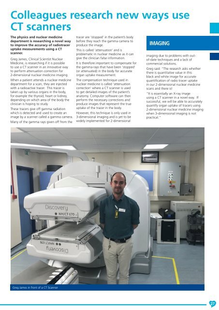
Greg James in front of a CT Scanner<br />
27