Available online at www.pharmresfoundation.com ISSN: 2229-3787 ...
Available online at www.pharmresfoundation.com ISSN: 2229-3787 ...
Available online at www.pharmresfoundation.com ISSN: 2229-3787 ...

You also want an ePaper? Increase the reach of your titles
YUMPU automatically turns print PDFs into web optimized ePapers that Google loves.
<strong>Available</strong> <strong>online</strong> <strong>at</strong> <strong>www</strong>.pharmresfound<strong>at</strong>ion.<strong>com</strong> <strong>ISSN</strong>: <strong>2229</strong>-<strong>3787</strong><br />
Journal of Advanced Pharmaceutical Research. 2011, 2(2), 64-75.<br />
Research paper<br />
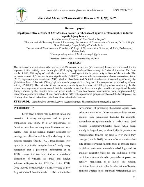
Hep<strong>at</strong>oprotective activity of Clerodendron inerme (Verbenaceae) against acetaminophen induced<br />
hep<strong>at</strong>ic injury in mice<br />
Ravindra kumar Chourasiya 1 , Siva Shankar Nayak 2,*<br />
1 Pharmaceutical Chemistry Research Labor<strong>at</strong>ory, Department of Pharmaceutical Sciences, Dr. Hari Singh<br />
Gour University, Sagar, Madhya Pradesh, India.<br />
2 Department of Pharmaceutical Chemistry, College of Pharmaceutical Sciences, Mohuda, Berhampur,<br />
Orissa, India<br />
*Corresponding author E.Mail: sivanayak@yahoo.<strong>com</strong><br />
Received: Feb 18, 2011; Accepted: May 22, 2011<br />
ABSTRACT<br />
The methanol and petroleum ether extracts of Clerodendron inerme (Verbenaceae) leaves were screened for its<br />
hep<strong>at</strong>oprotective activity in acetaminophen (250 mg/kg, i.p) induced liver damage in Swiss albino mice. The dose<br />
levels of 200, 300 mg/kg of both the extracts were used against the hep<strong>at</strong>otoxicity in liver of the animals. The<br />
methanol extract of C. inerme showed significantly (P
of phytoconstituents. Clerodendron inerme (L.)<br />
Gaertn (Family- Verbenaceae) is a scandent,<br />
straggling highly branched plant 0.9 - 2.1 m. long.<br />
The shrub is found in the wastelands, hedges, bank of<br />
rivers, sea and in various tropical parts of India<br />
(Kirtikar and Basu, 1998; Ch<strong>at</strong>terjee and Pakrashi,<br />
1995; Chopra et al., 1956) and is acknowledged under<br />
vernaculars as ‘Glory Bower genus, Garden Quinine’<br />
in English (Chourasiya et al., 2010); ‘Lanjai or<br />
Sangkupi’ in Hindi; ‘Cholora’ in Oriya; ‘Kundali,<br />
Samudrayuthika, Vanajai and Vanayuthika’ in<br />
Sanskrit. The poultice of the plant is used in buboes,<br />
vermifuge, anti periodic and as a substitute for quinine<br />
in remittent and intermittent fevers. Traditionally the<br />
leaves powder along with camphor, garlic or pepper<br />
was used for edema, muscular pains, rheum<strong>at</strong>ic pains<br />
and roots were also used for venereal diseases<br />
(Nadkarni, 1996, Kirtikar and Basu, 1998; Chopra et<br />
al., 1956). Additionally, the fresh juices obtained from<br />
powdered leaves are used as altern<strong>at</strong>ive in scrofula;<br />
venereal diseases and is used orally in the tre<strong>at</strong>ment of<br />
jaundice (Ch<strong>at</strong>terjee and Pakrashi, 1995). Alcoholic<br />
extract of leaves stimul<strong>at</strong>es pregnant uterus, raises<br />
blood pressure and increases intestinal movements in<br />
r<strong>at</strong>s. The main active chemical constituents include α-<br />
and β- amyrins, betulin, dehydroroyleanone,<br />
royleanone, neoclerodane (diterpenoids), Clerodermic<br />
acid, Cleroinermin, Acacetin, Apigenin (5-hydroxy-7,<br />
4’-dimethoxyflavone), Salvigenin (5-hydroxy-6, 7, 4’-<br />
trimethoxyflavone), Pectolinarigenin, Scutellarein and<br />
β-sitosterol (Ch<strong>at</strong>terjee and Pakrashi, 1995; Rastogi<br />
and Mehrotra, 1995). The liter<strong>at</strong>ure reveals th<strong>at</strong> C.<br />
inerme is used orally in the tre<strong>at</strong>ment of jaundice in<br />
traditional Indian medicine and, moreover, the<br />
hep<strong>at</strong>oprotective activity of the stem and leaf w<strong>at</strong>er<br />
extract of C. inerme against carbon tetrachloride-<br />
<strong>Available</strong> <strong>online</strong> <strong>at</strong> <strong>www</strong>.pharmresfound<strong>at</strong>ion.<strong>com</strong> <strong>ISSN</strong>: <strong>2229</strong>-<strong>3787</strong><br />
induced liver toxicity was reported by Gopal and<br />
Sengottuvelu (2008). To d<strong>at</strong>e, no system<strong>at</strong>ic study has<br />
been reported in an acute model regarding the<br />
Acetaminophen induced hep<strong>at</strong>otoxicity in mice. In the<br />
present study, the authors has been made to establish<br />
the hep<strong>at</strong>oprotective property of methanol and<br />
petroleum ether leaf extracts of C. inerme against<br />
Acetaminophen -induced hep<strong>at</strong>otoxicity in mice.<br />
MATERIALS AND METHODS<br />
Plant m<strong>at</strong>erial<br />
The plant m<strong>at</strong>erial (Leaves) used in the study.<br />
Clerodendron inerme (Verbenaceae) were collected in<br />
the month of August 2007 from the rural area of<br />
Mohuda in Ganjam District of Orissa, India. The plant<br />
was identified and authentic<strong>at</strong>ed by botanists Prof. S.<br />
K. Dash, HOD, PG Department of Bioscience,<br />
College of Pharmaceutical Sciences, Mohuda and<br />
<strong>com</strong>paring with the voucher specimen (CI-1) present<br />
in the herbarium, has been kept in the labor<strong>at</strong>ory for<br />
experimental purpose. The collected plant leaves were<br />
washed and air-dried under the shade, cut into small<br />
pieces, powdered by a mechanical grinder and passed<br />
through 40-mesh sieve and stored in a closed vessel<br />
for future use.<br />
Prepar<strong>at</strong>ion of C. inerme leave extracts<br />
The dried, powdered leaves of Clerodendron<br />
inerme (250 g) were extracted successively with<br />
petroleum ether 60–80°C (10 h) in soxhlet appar<strong>at</strong>us.<br />
A dark green colored petroleum ether extract was<br />
obtained. The same leave powder (marc), after proper<br />
air drying, again extracted with methanol (18 h) to<br />
produce a greenish brown semisolid mass. The<br />
extractions were carried out until the solvents be<strong>com</strong>e<br />
colorless. These extracts were further dried and<br />
concentr<strong>at</strong>ed by evapor<strong>at</strong>ing the solvent <strong>com</strong>pletely<br />
65
under vacuum <strong>at</strong> the range of boiling points of solvent<br />
(Methanol <strong>at</strong> 64°C and petroleum ether <strong>at</strong> 35°C) using<br />
rotary evapor<strong>at</strong>or (Jain Scientific glass works, DTC<br />
201, Ambala cantt, India) to get crude extracts. The<br />
methanol extract (CIME) yield 15.7% w/w and<br />
petroleum ether extract (CIPE) yield 3.0% w/w<br />
respectively. The dried mass of the extracts were<br />
stored <strong>at</strong> 4°C and prepared an emulsion by tritur<strong>at</strong>ing<br />
the accur<strong>at</strong>ely weighed quantity of the extract with 1%<br />
gum acacia and suspended in double distilled w<strong>at</strong>er <strong>at</strong><br />
definite concentr<strong>at</strong>ions separ<strong>at</strong>ely used for the<br />
pharmacological study.<br />
Chemical used<br />
Paracetamol (acetaminophen) was obtained<br />
from ALPA Labor<strong>at</strong>ories, Indore, India and silymarin<br />
was obtained from Micro labs, Hosur, India as free<br />
sample, The assay kits used for the estim<strong>at</strong>ions of<br />
alanine aminotransferase (ALT), aspart<strong>at</strong>e<br />
aminotransferase (AST), alkaline phosph<strong>at</strong>ase (ALP),<br />
total bilirubin were purchased from (AGAPPE<br />
Diagnostics Pvt. Ltd., Maharashtra, India). All<br />
extractive solvents are used as analytical grade<br />
reagents (AR) and purchased from S.D. Fine<br />
Chemicals, Mumbai, India.<br />
Preliminary phytochemical analysis<br />
The methanol (CIME) and petroleum ether<br />
(CIPE) extracts were subjected to preliminary<br />
phytochemical screening for detection of major<br />
chemical groups. In each test, 10% w/v solution of the<br />
extracts were used and unless otherwise mentioned in<br />
individual test (P<strong>at</strong>il, 2001).<br />
Experimental animals<br />
All animal procedures were in strict<br />
accordance with the CPCSEA Guidelines (Committee<br />
for the purpose of control and supervision of<br />
experiment on animal). Healthy Swiss albino mice of<br />
<strong>Available</strong> <strong>online</strong> <strong>at</strong> <strong>www</strong>.pharmresfound<strong>at</strong>ion.<strong>com</strong> <strong>ISSN</strong>: <strong>2229</strong>-<strong>3787</strong><br />
7-8 weeks old weighing 20-35g of either sex were<br />
used. They were obtained from Jawaharlal Nehru<br />
Cancer Hospital and Research Centre, Bhopal (M.P)<br />
India. The mice were housed <strong>at</strong> a temper<strong>at</strong>ure of 20–<br />
25 0 C under a 12-h light/dark cycle with 50% of<br />
rel<strong>at</strong>ive humidity. They were fed with standard animal<br />
feed e.g. Altromin pellets (Lipton’s India Ltd) and<br />
w<strong>at</strong>er ad libitum. This study <strong>com</strong>plied with current<br />
ethical regul<strong>at</strong>ions on animal research in Jawaharlal<br />
Nehru Cancer Hospital and Research Centre, Bhopal<br />
and all mice used in the experiments were tre<strong>at</strong>ed<br />
humanely. The use of animal was as per CPCSEA<br />
norms (CPCSEA Registr<strong>at</strong>ion No. -<br />
500/01/a/CPCSEA/2001). Approval for experimental<br />
work was as per ethical <strong>com</strong>mittee of Jawaharlal<br />
Nehru Cancer Hospital and Research Centre, Bhopal<br />
M.P., India (Ref. No– 670/225-IAEC/2008/Project<br />
No.-51) .Animals was acclim<strong>at</strong>ized to their<br />
environment for one week prior to experiment<strong>at</strong>ion.<br />
Acute Toxicity analysis<br />
Toxicity studies of the CIME and CIPE were<br />
performed to get the inform<strong>at</strong>ion, how safe is this<br />
extract for the therapeutic use. The LD50 value of<br />
CIME and CIPE were derived by the method of<br />
Litchfield and Wilcoxon 1949. The maximum non-<br />
lethal doses of both the extracts were found to be 3000<br />
mg/kg body weight, orally. The 1% gum acacia was<br />
used as a vehicle and showed no mortality. The<br />
determin<strong>at</strong>ion of acute toxicity by adopting fixed dose<br />
the guideline of CPCSEA and 1/10 th of LD50 cut off<br />
values (P<strong>at</strong>il, 2001; Shivakumar, 2007) of the extracts<br />
were taken as screening dose. i.e. 200, 300 mg/kg for<br />
subsequent studies (CIME 200, 300 mg/kg and CIPE -<br />
200, 300 mg/kg).<br />
Acetaminophen -induced hep<strong>at</strong>otoxicity in mice<br />
(Acute model)<br />
66
Animals were randomized and divided into<br />
seven groups (1-7) of six mice in each group. Animals<br />
of Group-1 (control) received only vehicle (1 % Gum<br />
Acacia in distilled w<strong>at</strong>er). Animals of Group- 2<br />
(Paracetamol-control) received acetaminophen 250<br />
mg/kg only. Animals of Group-3 fed with<br />
acetaminophen 250 mg/kg and the standard drug<br />
Silymarin 50 mg/kg. Animals of Group-4 and 5 were<br />
fed with a single dose of acetaminophen 250 mg/kg<br />
and CIME of 200, 300 mg/kg. Similarly, animals of<br />
Group-6 and 7 were fed with a single dose of<br />
acetaminophen 250 mg/kg and CIPE of 200, 300<br />
mg/kg. Plant extracts were administered three hours<br />
after the administr<strong>at</strong>ion of acetaminophen. All the<br />
tre<strong>at</strong>ments were done by means of a gavage or gastric<br />
tube. The tre<strong>at</strong>ments were continued for seven<br />
consecutive days and on the eighth day of the<br />
experiment, animals were anaesthetized with ether<br />
and blood sample was collected by puncturing the<br />
retro orbital plexus and serum was separ<strong>at</strong>ed by<br />
centrifug<strong>at</strong>ion <strong>at</strong> 1500 rpm for 10min. The serum was<br />
then estim<strong>at</strong>ed for alanine aminotransferase (ALT),<br />
aspart<strong>at</strong>e aminotransferase (AST), alkaline<br />
phosph<strong>at</strong>ase (ALP) and total bilirubin level using<br />
assay kits according to the supplier (AGAPPE<br />
Diagnostics Pvt. Ltd., Maharashtra, India)<br />
specific<strong>at</strong>ions (Sabir and Rocha 2008). The estim<strong>at</strong>ion<br />
was done by using a photoanalyser instrument (FT-2;<br />
AMS); each determin<strong>at</strong>ion was carried out with a<br />
suitable kit (AGAPPE Diagnostics Pvt. Ltd.,<br />
Maharashtra, India) containing the necessary reagents<br />
<strong>at</strong> a predetermined wavelength for each parameter.<br />
Histop<strong>at</strong>hological studies<br />
One animal belongs to the tre<strong>at</strong>ed groups<br />
showing maximal activity as indic<strong>at</strong>ed by exhibit<br />
biochemical parameters from each test, positive<br />
<strong>Available</strong> <strong>online</strong> <strong>at</strong> <strong>www</strong>.pharmresfound<strong>at</strong>ion.<strong>com</strong> <strong>ISSN</strong>: <strong>2229</strong>-<strong>3787</strong><br />
control, hep<strong>at</strong>otoxin and control groups were selected<br />
for this purpose. The animals were sacrificed by ether<br />
anaesthesia and the abdomen was cut open to remove<br />
the liver, (5mm thick pieces) of the liver were tre<strong>at</strong>ed<br />
in Bouin’s solution (mixture of 75 ml of s<strong>at</strong>ur<strong>at</strong>ed<br />
picric acid, 25 ml of 40% formaldehyde and 5ml of<br />
glacial acetic acid) for 12 hours, then embedded in<br />
paraffin wax and cut into 5 μm thick sections by using<br />
microtome and stained with haem<strong>at</strong>oxylin-eosin dye<br />
and mounted in diphenyl xylene. Then the liver<br />
sections were observed under microscope for<br />
histop<strong>at</strong>hological changes in liver architecture and<br />
photo microscopic were observed (100X) including<br />
necrosis, ste<strong>at</strong>osis and f<strong>at</strong>ty change of hep<strong>at</strong>ic cells<br />
(Shai 2001; Saeed 2002).<br />
St<strong>at</strong>istical Analysis<br />
The results were recorded as Mean ± S.E.M.<br />
and st<strong>at</strong>istical significance between tre<strong>at</strong>ed groups<br />
with a control group was evalu<strong>at</strong>ed by One-way<br />
analysis of variance (ANOVA) followed by Dunnett’s<br />
t-test (Woodson, 1986).<br />
RESULTS<br />
Preliminary phytochemical analysis<br />
Results of different chemical tests on the<br />
CIME showed the presence of phytoconstituents, i.e.,<br />
reducing sugars, flavonoids, phenolic <strong>com</strong>pounds,<br />
alkaloids and gums; whereas CIPE showed a positive<br />
test for the presence of steroids and terpenoids.<br />
Effect of tre<strong>at</strong>ment with acetaminophen on serum<br />
ALT activities<br />
The estim<strong>at</strong>ion of all the biochemical<br />
parameters were carried out by using a photoanalyser<br />
(FT-2; AMS); serum ALT determin<strong>at</strong>ion was done<br />
with a suitable kit (AGAPPE Diagnostics Reagent)<br />
containing the necessary reagents <strong>at</strong> a pre-determined<br />
67
wavelength 340 nm where as the r<strong>at</strong>e of oxid<strong>at</strong>ion of<br />
NADH was measured kinetically by monitoring the<br />
decrease in absorbance <strong>at</strong> 340 nm (Murray, 1994).<br />
Mice were tre<strong>at</strong>ed with paracetamol in group 2 alone<br />
developed significant liver cell damage as was evident<br />
for a significant (P < 0.001) increase in serum alanine<br />
aminotransferase enzyme activities. The oral<br />
administr<strong>at</strong>ions of CIME and CIPE <strong>at</strong> the different<br />
dose levels of 200 and 300 mg/kg (groups 4 to 7),<br />
were shown the lower significantly (P < 0.01)<br />
activities of marker enzymes and <strong>com</strong>parable to the<br />
group 3 of standard drug silymarin (50 mg/kg; P <<br />
0.01) as shown in Table 1.<br />
Effect of tre<strong>at</strong>ment with acetaminophen on the<br />
serum AST activities<br />
In the paracetamol tre<strong>at</strong>ed group 2; there was<br />
a significant (P < 0.001) increase in aspart<strong>at</strong>e<br />
aminotransferase activity <strong>com</strong>pared to the normal<br />
group 1. AST activity of CIME and CIPE groups of<br />
two different doses 200 and 300 mg/kg (groups 4 to 7)<br />
significantly (P < 0.01) recovered the level of enzyme<br />
activities which is <strong>com</strong>parable with standard drug<br />
silymarin group-3 (P < 0.01) as shown in Table 1.<br />
Effect of tre<strong>at</strong>ment with acetaminophen on the<br />
levels of ALP activities<br />
The level of ALP activities were carried out<br />
using the kit AGAPPE Diagnostics Reagent analyzed<br />
in a photoanalyser (FT-2; AMS). By using a specific<br />
reagent <strong>at</strong> pH 10.3, alkaline phosph<strong>at</strong>ase (ALP)<br />
c<strong>at</strong>alyses the hydrolysis of colorless p-nitro phenyl<br />
phosph<strong>at</strong>e (p-NPP) to yellow colored p-nitro phenol<br />
and phosph<strong>at</strong>e. Change in absorbance due to yellow<br />
color form<strong>at</strong>ion is measured kinetically <strong>at</strong> 405 nm and<br />
is proportional to ALP activity in the sample (Moss<br />
and Henderson, 1994). Mice were tre<strong>at</strong>ed with<br />
paracetamol (group 2) alone developed significant<br />
<strong>Available</strong> <strong>online</strong> <strong>at</strong> <strong>www</strong>.pharmresfound<strong>at</strong>ion.<strong>com</strong> <strong>ISSN</strong>: <strong>2229</strong>-<strong>3787</strong><br />
liver cell damage as was evident from a significant (P<br />
< 0.001) increase in serum enzyme levels of ALP<br />
<strong>com</strong>pared to the normal group 1. CIME and CIPE<br />
groups of two different doses, 200 and 300 mg/kg<br />
significantly (P < 0.01 group 4, 5, 7; P < 0.05 group<br />
6), recovered the ALP level as <strong>com</strong>pared to the<br />
standard drug silymarin group 3 (50 mg/kg; P < 0.01)<br />
as shown in Table 1.<br />
Effect of tre<strong>at</strong>ment with acetaminophen on the<br />
serum bilirubin levels<br />
Serum bilirubin levels in paracetamol-induced<br />
liver damage were estim<strong>at</strong>ed using an analytical kit<br />
from AGAPPE Diagnostics Reagent. The assay<br />
principle is based on the fact th<strong>at</strong> total bilirubin reacts<br />
in the presence of caffeine with diazotized sulfanilic<br />
acid to form azobilirubin. The determin<strong>at</strong>ion of direct<br />
bilirubin <strong>at</strong> 546 nm is proportional to the<br />
concentr<strong>at</strong>ion of bilirubin (Willard and Meites, 1982).<br />
A significant (P < 0.001) increase in serum bilirubin<br />
level was seen in paracetamol-tre<strong>at</strong>ed animals in<br />
group 2 <strong>com</strong>pared to th<strong>at</strong> of the normal group 1.<br />
Bilirubin levels for CIME and CIPE groups of two<br />
different doses 200 and 300 mg/kg significantly (P <<br />
0.01 group 4, 5, 7; P < 0.05 group 6) recovered<br />
against the paracetamol group 2 and both doses were<br />
<strong>com</strong>parable with the standard drug silymarin <strong>at</strong> 50<br />
mg/kg (P < 0.01) as shown in Table 1.<br />
Histop<strong>at</strong>hological studies<br />
Histop<strong>at</strong>hological examin<strong>at</strong>ion of liver<br />
sections of control group 1 and standard group 3<br />
showed normal cellular architecture with distinct<br />
hep<strong>at</strong>ic cells, sinusoidal spaces and central vein (Fig.<br />
1 and 3). Disarrangement of normal hep<strong>at</strong>ic cells with<br />
centrilobular necrosis, vacuoliz<strong>at</strong>ion of cytoplasm and<br />
f<strong>at</strong>ty degener<strong>at</strong>ion were observed in the paracetamol<br />
tre<strong>at</strong>ed mice of group 2 in Fig. 2.<br />
68
<strong>Available</strong> <strong>online</strong> <strong>at</strong> <strong>www</strong>.pharmresfound<strong>at</strong>ion.<strong>com</strong> <strong>ISSN</strong>: <strong>2229</strong>-<strong>3787</strong><br />
Table 1: Hep<strong>at</strong>oprotective Activity of C. inerme extracts on acetaminophen-induced Hep<strong>at</strong>otoxicity in mice.<br />
Serum biochemical parameter<br />
Groups Tre<strong>at</strong>ment<br />
AST<br />
ALT<br />
ALP Total bilirubin<br />
IU/L<br />
IU/L<br />
IU/L<br />
mg/dl<br />
Group 1 1 % Gum Acacia 49.50±1.36 65.33±1.40 83.42±1.75 2.23±0.08<br />
Group 2 Acetaminophen<br />
( 250 mg/kg) i.p.<br />
Group 3 Acetaminophen 250 mg/kg<br />
i.p. + Silymarin (50 mg/kg)<br />
a, b<br />
62.67±1.03<br />
a, b<br />
70.25±1.87<br />
a, b<br />
113.17±1.30<br />
a, b, c<br />
2.72±0.12<br />
Group 4 Acetaminophen 250 mg/kg<br />
i.p. CIME (200 mg/kg)<br />
a, b<br />
93.00±1.74<br />
a, b<br />
193.75±1.71<br />
a, b<br />
401.50±1.33<br />
a, b, c<br />
0.28±0.024<br />
Group 5 Acetaminophen 250 mg/kg<br />
i.p. + CIME (300 mg/kg)<br />
a, b<br />
78.25±1.40<br />
a, b<br />
151.50±1.60<br />
a, b<br />
367.25±2.04<br />
a, b, c<br />
0.25±0.019<br />
Group 6 Acetaminophen 250 mg/kg<br />
i.p. + CIPE (200 mg/kg)<br />
a, b<br />
110.75±1.75<br />
a, b<br />
196.25±1.66<br />
a, b<br />
486.75±1.02<br />
a, b, c<br />
0.29±0.017<br />
Group 7 Acetaminophen 250 mg/kg<br />
i.p. + CIPE (300 mg/kg)<br />
a, b<br />
76.75±1.17<br />
a, b<br />
179.50±1.90<br />
a, b<br />
397.50±2.02<br />
a, b, c<br />
0.26±0.019<br />
a b c<br />
P < 0.001, P < 0.01 and P < 0.05.<br />
All the values are expressed as Mean ± SEM.; n =6. The d<strong>at</strong>a was analyzed by evalu<strong>at</strong>ed by One-way analysis of variance<br />
(ANOVA) followed by Dunnett’s t-test, Group-2 <strong>com</strong>pared with normal group-1 with a P < 0.001. Experimental groups from-3 to<br />
7 <strong>com</strong>pared with group-2 with b P < 0.01 and c P < 0.05.<br />
158.50±1.33 a<br />
The liver sections of the mice tre<strong>at</strong>ed with<br />
CIME (200 and 300 mg/kg) of groups 4 and 5 showed<br />
signs of protection as was evident by the absence of<br />
necrosis and vacuoles (Fig. 4 and 5) while the liver<br />
sections of the mice tre<strong>at</strong>ed with CIPE (200 and 300<br />
mg/kg) of groups 6 and 7 showed reduction in f<strong>at</strong>ty<br />
degener<strong>at</strong>ion and absence of necrosis (Fig. 6 and 7).<br />
Fig. 1: Histop<strong>at</strong>hology of liver of Neg<strong>at</strong>ive control<br />
group. T.S. of liver revealed normochrom<strong>at</strong>ic<br />
hep<strong>at</strong>ocytes (H), occasionally apoptosis, cart wheel<br />
nucleus (CWN) and lipid vacuoles (CV). A good mitotic<br />
index has been observed. H and E, 100x.<br />
129.42±1.76 a<br />
257.83±1.27 a<br />
4.02±0.20 a<br />
Fig. 2: Histop<strong>at</strong>hology of liver of Positive control group.<br />
T.S. of liver revealed hyper chrom<strong>at</strong>ic and highly<br />
damaged hep<strong>at</strong>ocytes (H). Large numbers of lipid<br />
vacuoles (LV) have been observed which propelled<br />
nucleus to the periphery of the cell. Frequent apoptosis<br />
(Apop) and necrosis (CWN) with poor mitotic index<br />
have been observed. H and E, 100x.<br />
DISCUSSION<br />
In acute toxicity study, oral administr<strong>at</strong>ion of<br />
both CIME and CIPE did not produce any mortality in<br />
mice up to a dose level of 3 g/kg body weight. This<br />
may be due to broad non-toxic range of the plant,<br />
69
where the plant extract showed a high LD50 and<br />
rel<strong>at</strong>ively safety. Paracetamol (acetaminophen) is a<br />
widely used analgesic-antipyretic agent which is<br />
metabolized by the liver.<br />
Fig.3: Histop<strong>at</strong>hology of liver of Standard group. T.S.<br />
of liver revealed normochrom<strong>at</strong>ic hep<strong>at</strong>ocytes (H).<br />
Occasionally apoptosis, cart wheel nucleus (CWN) and<br />
lipid vacuoles (CV) have been observed with average<br />
mitotic index. H and E, 100x.<br />
Fig. 4: Histop<strong>at</strong>hology of liver of Test group-1<br />
(Methanol Extract 200mg/kg bw orally). T.S. of<br />
liver revealed normochrom<strong>at</strong>ic hep<strong>at</strong>ocytes(H)<br />
with good mitotic index. Occasionally lipid<br />
vacuoles (CV) have been observed without<br />
apoptosis (Apop), necrosis and cart wheel nucleus<br />
(CWN). H and E, 100x<br />
Over dosing of paracetamol to r<strong>at</strong>s is reported<br />
to decrease their sensitivity to its hep<strong>at</strong>otoxic effects,<br />
which are associ<strong>at</strong>ed with oxid<strong>at</strong>ive stress (Meredith,<br />
1996). So the studies on antioxidant enzymes,<br />
<strong>Available</strong> <strong>online</strong> <strong>at</strong> <strong>www</strong>.pharmresfound<strong>at</strong>ion.<strong>com</strong> <strong>ISSN</strong>: <strong>2229</strong>-<strong>3787</strong><br />
(Glut<strong>at</strong>hione peroxidase (GPx), glut<strong>at</strong>hione-S-<br />
transferase (GST), superoxide dismutase (SOD) and<br />
c<strong>at</strong>alase (CAT) have been found to be of gre<strong>at</strong><br />
importance in the assessment of liver damage. A small<br />
fraction of the drug is subject to oxid<strong>at</strong>ion reactions<br />
c<strong>at</strong>alyzed by cytochrome P-450 enzymes in the liver,<br />
resulting in the gener<strong>at</strong>ion of hep<strong>at</strong>otoxic N-acetyl-p-<br />
benzo-quinoneimine.<br />
Fig. 5: Histop<strong>at</strong>hology of liver of Test group-2<br />
(Methanol Extract 300mg/kg bw orally). T.S. of liver<br />
revealed normochrom<strong>at</strong>ic hep<strong>at</strong>ocytes with good mitotic<br />
index. Occasionally lipid vacuoles(CV), cart wheel<br />
nucleus(CWN) and apoptosis(Apop) have been<br />
observed without necrosis. H and E, 100x.<br />
Fig. 6: Histop<strong>at</strong>hology of liver of Test group-3 (Pet.<br />
Ether Extract 200mg/kg bw orally). T.S. of liver<br />
revealed hyper chrom<strong>at</strong>ic hep<strong>at</strong>ocytes(H) with average<br />
mitotic index. Frequently lipid vacuoles (CV) and cart<br />
wheel nucleus(CWN) have been observed. Few<br />
apoptotic hep<strong>at</strong>ocyts(Apop) have been observed. H and<br />
E, 100x.<br />
70
Fig. 7: Histop<strong>at</strong>hology of liver of Test group-4 (Pet.<br />
Ether Extract 300mg/kg bw orally). T.S. of liver<br />
revealed hyper chrom<strong>at</strong>ic hep<strong>at</strong>ocytes (H) with average<br />
mitotic index. Frequently lipid vacuoles (LV) and cart<br />
wheel nucleus (CWN) have been observed. Few<br />
apoptosis (Apop) with necrotic hep<strong>at</strong>ocytes have been<br />
observed. H and E, 100x<br />
Exposure to high doses of acetaminophen<br />
results in increased levels of N-acetyl-p-benzo-<br />
quinoneimine. Normally, the toxic oxid<strong>at</strong>ion<br />
metabolites gener<strong>at</strong>ed in the liver are converted into<br />
non-toxic metabolites excreted in urine via<br />
conjug<strong>at</strong>ion with GSH, which contains sulphydryl<br />
groups. However, the intake of high doses of<br />
acetaminophen limits the capacity of GSH to detoxify<br />
N-acetyl-p-benzo-quinoneimine and results in the<br />
consumption of liver GSH stores (Mitchell et al.,<br />
1973; Savides and Oehme, 1983). Oxid<strong>at</strong>ive stress is<br />
reported to constitute a major mechanism in the<br />
p<strong>at</strong>hogenesis of acetaminophen induced liver damage<br />
(Ozdemirler et al., 1994). The binding of NAPQI with<br />
cellular proteins leads to necrosis in liver (Dahlin et<br />
al., 1984), which subsequently alters the liver function<br />
tests (LFTs).<br />
<strong>Available</strong> <strong>online</strong> <strong>at</strong> <strong>www</strong>.pharmresfound<strong>at</strong>ion.<strong>com</strong> <strong>ISSN</strong>: <strong>2229</strong>-<strong>3787</strong><br />
Reactive oxygen species (ROS) and nitrogen<br />
intermedi<strong>at</strong>es, produced by hep<strong>at</strong>ic parenchyma and<br />
non-parenchyma cells are believed to be important<br />
factors contributing to Acetaminophen induced injury<br />
(Michael et al., 1999). In addition, the anti oxid<strong>at</strong>ive<br />
enzymes such as, super oxide dismutase (SOD) and<br />
c<strong>at</strong>alase (CAT) also play an important role in the<br />
modul<strong>at</strong>ion of acetaminophen induced oxid<strong>at</strong>ive<br />
damage and is a <strong>com</strong>monly and widely used<br />
analgesic/ antipyretic agent. Hep<strong>at</strong>otoxic dose of<br />
acetaminophen step up the normal levels of hep<strong>at</strong>ic<br />
enzymes (Morazzoni et al 1995).<br />
In living systems, liver is considered to be<br />
highly sensitive to toxic agents therefore it is easier to<br />
analyze hep<strong>at</strong>otoxicity/protection for short dur<strong>at</strong>ion<br />
research. The study of different enzyme activities such<br />
as alanine amino transferase (ALT), aspar<strong>at</strong>e amino<br />
transferase (AST), alkaline phosph<strong>at</strong>es (ALP) and<br />
total bilirubin have been found to be of gre<strong>at</strong> value in<br />
the assessment of clinical and experimental liver<br />
damage (Vermulen et al 1992). In the present<br />
investig<strong>at</strong>ion it was observed th<strong>at</strong> the animals tre<strong>at</strong>ed<br />
with acetaminophen resulted in significant hep<strong>at</strong>ic<br />
damage shown by the elev<strong>at</strong>ed levels of serum<br />
markers. These elev<strong>at</strong>ed serum hep<strong>at</strong>ic profile reflects<br />
hep<strong>at</strong>ic structural integrity or the architect of liver<br />
tissue. Assessment of liver function can be made by<br />
estim<strong>at</strong>ing the activities of serum ALT, AST, ALP<br />
and total bilirubin, which are enzymes originally<br />
present in higher concentr<strong>at</strong>ion in cytoplasm<br />
(Manokaran et al., 2008).<br />
When there is hep<strong>at</strong>op<strong>at</strong>hy, these enzymes<br />
leak into the blood stream which serves as an<br />
indic<strong>at</strong>or for the liver damage (Nkosi et al., 2005).The<br />
abnormally high level of serum ALT, AST, ALP and<br />
total bilirubin content observed in group 2 in our<br />
71
study are the consequence of paracetamol-induced<br />
liver dysfunction and denotes the damage to the<br />
hep<strong>at</strong>ic cells (Table 1). Tre<strong>at</strong>ment with CIME and<br />
CIPE <strong>at</strong> dose levels of 200 and 300 mg/kg reduced the<br />
enhanced level of serum ALT, AST, ALP and total<br />
bilirubin level which seemed to offer protection and<br />
maintain the functional integrity of hep<strong>at</strong>ic cells. The<br />
elev<strong>at</strong>ed levels of serum marker enzyme activities<br />
such as ALT, AST enzyme levels are a direct measure<br />
of hep<strong>at</strong>ic injury and they show the st<strong>at</strong>us of liver.<br />
The elev<strong>at</strong>ed levels of serum enzymes are<br />
indic<strong>at</strong>ive of cellular leakage and loss of functional<br />
integrity of cell membrane in liver. Thus the lowering<br />
of the enzyme content in serum is a definite indic<strong>at</strong>ion<br />
of the hep<strong>at</strong>oprotective action of a drug. CIME and<br />
CIPE <strong>at</strong> a dose of 200 mg/kg (P< 0.01) and 300 mg/kg<br />
decreased significantly (P < 0.01) the activity of both<br />
AST and ALT as <strong>com</strong>pared to the standard drug<br />
silymarin. Serum ALP and bilirubin levels are also<br />
rel<strong>at</strong>ed to the st<strong>at</strong>us and function of hep<strong>at</strong>ic cells.<br />
Increase in serum ALP level is due to increased<br />
synthesis, in the presence of increasing biliary<br />
pressure (Moss and Butterworth, 1974).<br />
In the present study, CIME and CIPE <strong>at</strong> a<br />
dose of 200 mg/ kg (P < 0.01 and P < 0.05) and CIME<br />
and CIPE <strong>at</strong> a dose 300 mg/kg, were found to reduce<br />
significantly (P < 0.01) both serum ALP and bilirubin<br />
levels in the tre<strong>at</strong>ed groups which are <strong>com</strong>parable to<br />
the standard drug silymarin (Table 1).<br />
Hep<strong>at</strong>oprotective activity of C. inerme was shown by<br />
its ability to inhibit paracetamol-induced liver damage<br />
in mice. The fall in serum marker level by CIME (300<br />
mg/kg) was significantly (P < 0.01) able to alter the<br />
toxic condition of the hep<strong>at</strong>ocytes as <strong>com</strong>pared to<br />
CIPE.<br />
<strong>Available</strong> <strong>online</strong> <strong>at</strong> <strong>www</strong>.pharmresfound<strong>at</strong>ion.<strong>com</strong> <strong>ISSN</strong>: <strong>2229</strong>-<strong>3787</strong><br />
The histop<strong>at</strong>hological study has been done in<br />
liver tissue. Neg<strong>at</strong>ive control group revealed normal<br />
hep<strong>at</strong>ocyte with normochrom<strong>at</strong>ic cellular <strong>com</strong>ponents<br />
(fig.1). The liver section of mice which was tre<strong>at</strong>ed<br />
with paracetamol (Acetaminophen) revealed hypo-<br />
chrom<strong>at</strong>ic cells, apoptotic bodies with more<br />
intracellular lipid vacuoles which was more prominent<br />
for shifting of nucleus to the periphery. Cart wheel<br />
nucleus was also seen (fig.2). When the animal was<br />
tre<strong>at</strong>ed with Silymarin there was a slight reduction of<br />
apoptotic cells and cart wheel nucleus (fig.3). On the<br />
other hand when the animal was administered with<br />
200 mg/kg bw orally CIME revealed good mitotic<br />
index, very few lipid vacuoles, cart wheel nucleus and<br />
very few apoptotic bodies. Section was suggestive of<br />
normochrom<strong>at</strong>ic cells (fig.4) when the animal was<br />
tre<strong>at</strong>ed with 300 mg/kg bw orally CIME. It has been<br />
also observed th<strong>at</strong> all the cells were normochrom<strong>at</strong>ic<br />
hep<strong>at</strong>ocytes with good mitotic index, negligible lipid<br />
vacuoles were present and there was no evidence of<br />
cellular toxicity (fig.5). On the other hand, CIPE @<br />
200 mg/kg b.w orally revealed hyper chrom<strong>at</strong>ic,<br />
dysplastic cells, distorted hep<strong>at</strong>ocyte and large no. of<br />
lipid vacuoles with low mitotic index (fig.6). When<br />
animal was tre<strong>at</strong>ed with CIPE @ 300 mg/kg b.w<br />
orally revealed hyper chrom<strong>at</strong>ic hep<strong>at</strong>ocytes with<br />
average mitotic index (fig.7). A <strong>com</strong>par<strong>at</strong>ive<br />
histop<strong>at</strong>hological study of livers from different<br />
experimental groups further corrobor<strong>at</strong>ed the<br />
hep<strong>at</strong>oprotective efficacy of CIME and CIPE.<br />
Hep<strong>at</strong>oprotective activity of C. inerme was<br />
shown by its ability to inhibit paracetamol-induced<br />
liver damage in mice. The fall in serum marker level<br />
by CIME (300 mg/kg) was significantly (P < 0.01)<br />
able to alter the toxic condition of the hep<strong>at</strong>ocytes as<br />
<strong>com</strong>pared to CIPE <strong>at</strong> a dose level of 200 and 300<br />
72
mg/kg, so as to protect the membrane integrity against<br />
acetaminophen-induced leakage of marker enzymes.<br />
This means there was a potential functional change in<br />
the hep<strong>at</strong>ocytes of the hep<strong>at</strong>ic cells. Hence, it has been<br />
concluded th<strong>at</strong> the methanol extract of C. inerme<br />
(CIME) <strong>at</strong> dose levels of 300 mg/kg revealed its<br />
potential significance as a hep<strong>at</strong>oprotection against<br />
acetaminophen-induced toxicity which was further<br />
supported by the presence of phytoconstituents such<br />
as flavonoids, polyphenols, and tannins. All the results<br />
can be <strong>com</strong>parable with the standard drug silymarin.<br />
Hence, we conclude th<strong>at</strong> CIME and CIPE <strong>at</strong> higher<br />
dose revealed its potential significant as a<br />
hep<strong>at</strong>oprotection against Acetaminophen induced<br />
toxicity. However, a mild hep<strong>at</strong>ic cell aberr<strong>at</strong>ion was<br />
found in lower doses. A <strong>com</strong>par<strong>at</strong>ive<br />
histop<strong>at</strong>hological study of liver from different<br />
experimental groups corrobor<strong>at</strong>ed the<br />
hep<strong>at</strong>oprotective efficacy of methanol extract which<br />
was more effective than the petroleum ether extract of<br />
Clerodendron inerme. Further detailed studies may,<br />
however, confirm the utility profile of this drug.<br />
ACKNOWLEDGEMENTS<br />
Authors are thankfully acknowledged to<br />
Jawaharlal Nehru Cancer Hospital and Research<br />
Centre, Bhopal (M.P) India and College of<br />
Pharmaceutical sciences, Mohuda, Berhampur, India<br />
for providing research facilities. The authors also wish<br />
to thank Dr. N.Ganesh, Head, Department of<br />
Research, Jawaharlal Nehru Cancer Hospital and<br />
Research Centre, Bhopal (M.P) India for his valuable<br />
help and suggestions during the course of work and<br />
also thankful to the Prof. S. K. Dash, HOD, PG<br />
Department of Biosciences, College of<br />
Pharmaceutical sciences, Mohuda, Berhampur, for the<br />
identific<strong>at</strong>ion of the plant.<br />
<strong>Available</strong> <strong>online</strong> <strong>at</strong> <strong>www</strong>.pharmresfound<strong>at</strong>ion.<strong>com</strong> <strong>ISSN</strong>: <strong>2229</strong>-<strong>3787</strong><br />
REFERENCES<br />
1. Ch<strong>at</strong>terjee A, Pakrashi SC. The Tre<strong>at</strong>ise on Indian<br />
Medicinal Plants, Vol. IV, 1st edition, CSIR,<br />
New Delhi, 1995, 222-223.<br />
2. Chopra IC, Nayar SL, Chopra RN. Glossary of<br />
Indian medicinal plants, 1st edition, 1956, 71.<br />
3. Chourasiya RK, Jain PK, Sharma S, Ganesh N,<br />
Nayak SS, Agrawal RK. Genomic stability and<br />
tissue protection of Clerodendron inerme (L.)<br />
Gaertn leaves. Med Chem Res DOI<br />
10.1007/s00044-010-9482-3. 2010.<br />
4. Dahlin DC, Miwa GT, Lu AY, Nelson SD. N-<br />
Acetyl-p-benzoquinoneimine: a cytochrome P-<br />
450-medi<strong>at</strong>ed oxid<strong>at</strong>ion product of<br />
acetaminophen. Proc N<strong>at</strong>l Acad Sci., 1984, 81,<br />
1327.<br />
5. Farrell GC. Drug-induced liver disease.<br />
Edinburgh, Scotland: Churchill Livingstone.<br />
1994, 615-618.<br />
6. Gopal N, Sengottuvelu S. Hep<strong>at</strong>oprotective<br />
activity of Clerodendron inerme against CCL4<br />
induced hep<strong>at</strong>ic injury in r<strong>at</strong>s. Fitoterapia, 2008,<br />
79, 24-26.<br />
7. Kaplowitz N. Drug metabolism and<br />
hep<strong>at</strong>otoxicity. In: Kaplowitz N, ed. Liver and<br />
biliary diseases. Baltimore: Williams and Wilkins.<br />
1991, 316-318.<br />
8. Kirtikar KR, Basu BD. Indian medicinal plants,<br />
Vol. I, 2nd edition, Bishen Singh Mahendrapal<br />
Singh, Dehradun, 1998, 1945-1947.<br />
9. Litchfield JT, Wilcoxon FA. A simplified method<br />
of evalu<strong>at</strong>ing dose-effect experiments. J<br />
Pharmacol Expt Ther., 1949, 96, 99-113.<br />
10. Lorelle IB, Jon<strong>at</strong>han FC, Daune LC, Frances NS,<br />
Herbert T. Hep<strong>at</strong>oprotection by L-Cysteine-<br />
glut<strong>at</strong>hione mixed disulfide: A sulfhydryl-<br />
73
modified prodrug of glut<strong>at</strong>hione. J Biochem Mol<br />
Toxic, 2003, 17(2), 95-97.<br />
11. Manokaran S, Jaswanth A, Sengottuvelu S,<br />
Nandhakumar J, Duraisamy R, Karthikeyan D,<br />
Mallegaswari R. Hep<strong>at</strong>oprotective activity of<br />
Aerva lan<strong>at</strong>a Linn. against paracetamol induced<br />
hep<strong>at</strong>otoxicity in r<strong>at</strong>s. Res J Pharm Technol, 2008,<br />
1, 398–400.<br />
12. Meredith TJ, Vale JA, Proudfoot AT. Poisoning<br />
caused by analgesics. In: Oxford textbook of<br />
medicine. We<strong>at</strong>herall DJ, Ledingham JGG,<br />
Warrell DA, 3rd ed Oxford, 1996, 219-229.<br />
13. Michael SL, Pumfod NR, Mayeux PR, Niesman<br />
MR, Hinson JA. Pretre<strong>at</strong>ment of mice with<br />
macrophage inactiv<strong>at</strong>ors decreases acetaminophen<br />
hep<strong>at</strong>otoxicity and the form<strong>at</strong>ion of reactive<br />
oxygen and nitrogen species. Hep<strong>at</strong>ology, 1999,<br />
30, 186.<br />
14. Mitchell JR, Jollow DJ, Potter WZ, Gillette JR,<br />
Brodie BB. Acetaminophen-induced hep<strong>at</strong>ic<br />
necrosis. IV. Protective role of glut<strong>at</strong>hione. J<br />
Pharmacol Exp Ther., 1973, 187, 211-217.<br />
15. Morazzoni P, Bombardelli E. Valeriana<br />
officinalis: traditional use and recent evalu<strong>at</strong>ion of<br />
activity. Fitoterapia, 1995, 66, 99–112.<br />
16. Moss DW, Butterworth PJ. Enzymology an<br />
Medicine. London: Pitman Medical, 1974, 139.<br />
17. Moss DW, Henderson AK. Clinical enzymology.<br />
In: Burtis CA and Ashwood ER, eds. Tietz<br />
Textbook of Clinical Chemistry, third edition.<br />
Philadephia: WB Saunders, 1994, 617–721.<br />
18. Murray RL. Enzymes. In: Kaplan LA & Pesce AJ,<br />
eds. Clinical Chemistry; Theory, Analysis and<br />
Correl<strong>at</strong>ion, Toronto: Mosby, 1994, 1079–1134.<br />
<strong>Available</strong> <strong>online</strong> <strong>at</strong> <strong>www</strong>.pharmresfound<strong>at</strong>ion.<strong>com</strong> <strong>ISSN</strong>: <strong>2229</strong>-<strong>3787</strong><br />
19. Nadkarni KM, Nadkarni AK, Chopra RN. Indian<br />
Meteria Medica. Vol. I, Popular Prakashan,<br />
Mumbai, 1996, 352.<br />
20. Nkosi CZ, Opoku AR, Terblanche SE. Effect of<br />
pumpkin seed (Cucurbita pepo) protein isol<strong>at</strong>e on<br />
the activity levels of certain plasma enzymes in<br />
CCl4-induced liver injury in low protein fed r<strong>at</strong>s.<br />
Phytother Res. 2005, 19, 341–345.<br />
21. Ozdemirler G, Aykac G, Uysal M, Oz H. Liver<br />
lipid peroxid<strong>at</strong>ion and glut<strong>at</strong>hione-rel<strong>at</strong>ed defence<br />
enzyme systems in mice tre<strong>at</strong>ed with paracetamol.<br />
J Appl Toxicol., 1994, 14, 297–299.<br />
22. P<strong>at</strong>il MB, Jalalpure SS, Ashraf A. Preliminary<br />
phytochemical investig<strong>at</strong>ion and wound healing<br />
activity of the leaves of Argemone mexicana Linn.<br />
Indian Drugs, 2001, 38, 288-293.<br />
23. Rastogi RP, Mehrotra BN. Compendium of Indian<br />
Medicinal Plants (1980-1984), Vol. III, 1st<br />
edition, CSIR: Lucknow; 1995, 184-185.<br />
24. Reddy PB, Reddy CK, Rambhau D,<br />
Venk<strong>at</strong>eshvaralu V, Murthy VN. Anti hep<strong>at</strong>otoxic<br />
activity of some Ayurvedic prepar<strong>at</strong>ions. Ind J<br />
Pharm Sci., 1993, 55(4), 137-140.<br />
25. Sabir SM, Rocha JBT. W<strong>at</strong>er-extractable<br />
Phytochemicals from Phyllanthus niruri exhibit<br />
distinct in vitro antioxidant and in vivo<br />
hep<strong>at</strong>oprotective activity against paracetamol-<br />
induced liver damage in mice. Food Chemistry,<br />
2008, 111, 845–851.<br />
26. Saeed MA, Sabir AW. Irritant potential of<br />
triterpenoids from Ficus carica leaves.<br />
Fitoterapia, 2002, 73, 417-420.<br />
27. Savides MC, Oehme FW. Acetaminophen and its<br />
toxicity. J Appl Toxicol., 1983, 3, 96–111.<br />
28. Shai R, Yoel K, Ruth R, Michael S, Raphael M.<br />
Suppressors of cancer cell prolifer<strong>at</strong>ion from fig<br />
74
(Ficus carica) resin: Isol<strong>at</strong>ion and structure<br />
elucid<strong>at</strong>ion. J N<strong>at</strong> Pro., 2001, 64, 993-996.<br />
29. Shivakumar H, Sankara SLVJ, Vaidya VP. Anti-<br />
inflamm<strong>at</strong>ory activity of the unripe fruits of Ficus<br />
glomer<strong>at</strong>a. Indian Drugs, 2007, 44, 48-50.<br />
30. Vermulen NPE, Bessems JGM, Van DE, Sta<strong>at</strong> R.<br />
Molecular aspects of paracetamol induced<br />
hep<strong>at</strong>otoxicity and its mechanism based<br />
prevention. Drug Metab Rev., 1992, 24, 367-407.<br />
31. Willard RF, Meites S. Selected methods for the<br />
small clinical chemistry labor<strong>at</strong>ory. 1982, 9, 113–<br />
118.<br />
<strong>Available</strong> <strong>online</strong> <strong>at</strong> <strong>www</strong>.pharmresfound<strong>at</strong>ion.<strong>com</strong> <strong>ISSN</strong>: <strong>2229</strong>-<strong>3787</strong><br />
32. Woodson RF. St<strong>at</strong>istical Methods for the analysis<br />
of biochemical d<strong>at</strong>a, series in Probability and<br />
M<strong>at</strong>hem<strong>at</strong>ical St<strong>at</strong>istics, Wiley, New York, 1986,<br />
315-316.<br />
33. Zimmerman HJ, Maddrey WC. Toxic and drug-<br />
induced hep<strong>at</strong>itis. In: Schiff L, Schiff ER, eds.<br />
Diseases of the liver. Philadelphia: J.B.<br />
Lippincott., 1993, 214-218.<br />
75