2009 Scientific Report

You also want an ePaper? Increase the reach of your titles
YUMPU automatically turns print PDFs into web optimized ePapers that Google loves.
Van Andel Research Institute | <strong>Scientific</strong> <strong>Report</strong><br />
We work with clinical collaborators at several institutions to address various clinical needs. One such need is to help doctors<br />
make a more accurate diagnosis of patients with suspected pancreatic cancer. Since pancreatic cancer can be difficult to<br />
distinguish from benign conditions of the gastrointestinal tract, highly accurate biomarkers are needed to match patients to the<br />
appropriate procedures at the earliest possible time. We also are developing biomarkers to screen for clinically undetectable<br />
pancreatic cancer. Among populations at an increased risk for developing pancreatic cancer—including those suffering from<br />
chronic pancreatitis or with a family history of pancreatic cancer—an accurate screening test could detect new cancers early<br />
enough to allow more effective treatment. Further, we are testing our novel biomarkers for use in drug trials. Biomarkers that<br />
give early indications of the effectiveness of a candidate drug could accelerate drug trials and better match patients with the<br />
drugs that benefit them most.<br />
Another novel class of biomarker we are developing is for the diagnosis of patients with pancreatic cysts. Cystic lesions of the<br />
pancreas are increasingly being recognized due to the widespread use of high-resolution abdominal imaging. Since certain<br />
cyst types are precursors of invasive cancer, this situation presents an opportunity to intervene prior to malignant progression.<br />
Effective implementation of that strategy has been hampered by difficulties in clearly distinguishing cystic lesions based on<br />
differences in their malignant potential. In collaboration with Dr. Diane Simeone at the University of Michigan, we have identified<br />
glycan variants of secreted mucins that distinguish benign from pre-cancerous cysts with an 87% accuracy—better than the best<br />
current markers. Ongoing work is aimed at validating and building upon these results. Ultimately, we hope to implement a test<br />
that could be used to determine which pancreatic cysts should be surgically removed in order to prevent progression to cancer.<br />
Origin and function of secreted glycan alterations in pancreatic cancer<br />
Our laboratory also studies the origins and functions of cancer-cell secretions bearing altered glycans. The carbohydrate<br />
alterations observed in pancreatic tumors are strongly associated with accelerated disease progression, but it is not known<br />
whether these alterations functionally contribute to that progression. We have shown that certain glycoprotein alterations<br />
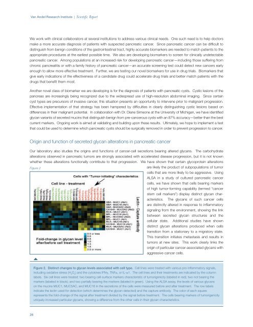
Figure 2<br />
are likely the product of subpopulations of tumor<br />
cells that are more likely to be aggressive. Using<br />
ALSA in a study of cultured pancreatic cancer<br />
cells, we have shown that cells bearing markers<br />
of high tumor-forming capability (termed “cancer<br />
stem cell markers”) display distinct glycan characteristics.<br />
The glycans of such cancer cells<br />
are distinctly altered in response to inflammatory<br />
signaling from the environment, showing the link<br />
between secreted glycan structures and the<br />
cellular state. Additional studies have shown<br />
distinct glycan alterations produced when cells<br />
transition from a stationary to a migratory state.<br />
This transition initiates metastasis and results in<br />
tumors at new sites. This work clearly links the<br />
origin of particular cancer-associated glycans with<br />
aggressive cancer cells.<br />
Figure 2. Distinct changes to glycan levels associated with cell type. Cell lines were treated with various pro-inflammatory signals,<br />
including oxidative stress (H 2<br />
O 2<br />
) and the cytokines IFNg, TNFa, or IL-a1. The cell lines and their treatments are indicated by the column<br />
labels. Six cell lines were treated: two bearing cell-surface markers characteristic of tumorigenicity (labeled in red); two not bearing the<br />
markers (labeled in black); and two partially bearing the markers (labeled in green). Using the ALSA assay, the levels of various glycans<br />
on the mucins MUC1, MUC5AC, and MUC16 in the secretions of the cells were measured before and after treatment. The row labels<br />
indicate the lectin used for detection (which determines the glycan detected) and the capture antibody. The color of each square<br />
represents the fold-change of the signal after treatment divided by the signal before treatment. The cells bearing markers of tumorigenicity<br />
uniquely increased particular glycans, showing a difference from the other cells in their glycan characteristics.<br />
28