Virtopsy: Postmortem minimally invasive angiography using ... - Library
Virtopsy: Postmortem minimally invasive angiography using ... - Library
Virtopsy: Postmortem minimally invasive angiography using ... - Library
You also want an ePaper? Increase the reach of your titles
YUMPU automatically turns print PDFs into web optimized ePapers that Google loves.
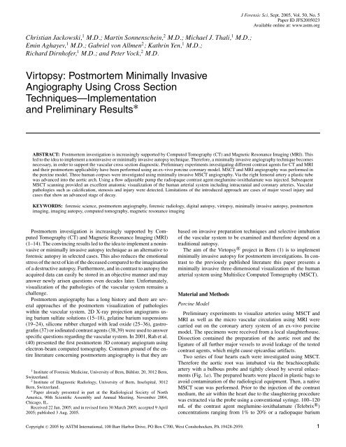
Christian Jackowski, 1 M.D.; Martin Sonnenschein, 2 M.D.; Michael J. Thali, 1 M.D.;<br />
Emin Aghayev, 1 M.D.; Gabriel von Allmen 2 ; Kathrin Yen, 1 M.D.;<br />
Richard Dirnhofer, 1 M.D.; and Peter Vock, 2 M.D.<br />
<strong>Virtopsy</strong>: <strong>Postmortem</strong> Minimally Invasive<br />
Angiography Using Cross Section<br />
Techniques—Implementation<br />
and Preliminary Results ∗<br />
JForensicSci,Sept. 2005, Vol. 50, No. 5<br />
Paper ID JFS2005023<br />
Available online at: www.astm.org<br />
ABSTRACT: <strong>Postmortem</strong> investigation is increasingly supported by Computed Tomography (CT) and Magnetic Resonance Imaging (MRI). This<br />
led to the idea to implement a non<strong>invasive</strong> or <strong>minimally</strong> <strong>invasive</strong> autopsy technique. Therefore, a <strong>minimally</strong> <strong>invasive</strong> <strong>angiography</strong> technique becomes<br />
necessary, in order to support the vascular cross section diagnostic. Preliminary experiments investigating different contrast agents for CT and MRI<br />
and their postmortem applicability have been performed <strong>using</strong> an ex-vivo porcine coronary model. MSCT and MRI <strong>angiography</strong> was performed in<br />
the porcine model. Three human corpses were investigated <strong>using</strong> <strong>minimally</strong> <strong>invasive</strong> MSCT <strong>angiography</strong>. Via the right femoral artery a plastic tube<br />
was advanced into the aortic arch. Using a flow adjustable pump the radiopaque contrast agent meglumine-ioxithalamate was injected. Subsequent<br />
MSCT scanning provided an excellent anatomic visualization of the human arterial system including intracranial and coronary arteries. Vascular<br />
pathologies such as calcification, stenosis and injury were detected. Limitations of the introduced approach are cases of major vessel injury and<br />
cases that show an advanced stage of decay.<br />
KEYWORDS: forensic science, postmortem <strong>angiography</strong>, forensic radiology, digital autopsy, virtopsy, <strong>minimally</strong> <strong>invasive</strong> autopsy, postmortem<br />
imaging, imaging autopsy, computed tomography, magnetic resonance imaging<br />
<strong>Postmortem</strong> investigation is increasingly supported by Computed<br />
Tomography (CT) and Magnetic Resonance Imaging (MRI)<br />
(1–14). The convincing results led to the idea to implement a non<strong>invasive</strong><br />
or <strong>minimally</strong> <strong>invasive</strong> autopsy technique as an alternative to<br />
forensic autopsy in selected cases. This also reduces the emotional<br />
stress of the next of kin of the deceased compared to the imagination<br />
of a destructive autopsy. Furthermore, and in contrast to autopsy the<br />
acquired data can easily be stored in an objective manner and may<br />
answer newly arisen questions even decades later. Unfortunately,<br />
visualization of the pathologies of the vascular system remains a<br />
challenge.<br />
<strong>Postmortem</strong> <strong>angiography</strong> has a long history and there are several<br />
approaches of the postmortem visualization of pathologies<br />
within the vascular system. 2D X-ray projection angiograms <strong>using</strong><br />
barium sulfate solutions (15–18), gelatine barium suspensions<br />
(19–24), silicone rubber charged with lead oxide (25–36), gastrografin<br />
(37) or iodinated contrast agents (38,39) were used to answer<br />
specific questions regarding the vascular system. In 2001, Rah et al.<br />
(40) presented the first postmortem 3D coronary angiogram <strong>using</strong><br />
electron-beam computed tomography. Common ground of the entire<br />
literature concerning postmortem <strong>angiography</strong> is that they are<br />
1 Institute of Forensic Medicine, University of Bern, Bühlstr. 20, 3012 Bern,<br />
Switzerland.<br />
2 Institute of Diagnostic Radiology, University of Bern, Inselspital, 3012<br />
Bern, Switzerland.<br />
∗ Paper already presented in part at the Radiological Society of North<br />
America, 90th Scientific Assembly and Annual Meeting, November 2004,<br />
Chicago, IL.<br />
Received 22 Jan. 2005; and in revised form 30 March 2005; accepted 9 April<br />
2005; published 3 Aug. 2005.<br />
based on <strong>invasive</strong> preparation techniques and selective intubation<br />
of the vascular system to be examined and therefore depend on a<br />
traditional autopsy.<br />
The aim of the <strong>Virtopsy</strong> R○ project in Bern (1) is to implement<br />
<strong>minimally</strong> <strong>invasive</strong> autopsy for postmortem investigations. In contrast<br />
to the previously published literature this paper presents a<br />
<strong>minimally</strong> <strong>invasive</strong> three-dimensional visualization of the human<br />
arterial system <strong>using</strong> Multislice Computed Tomography (MSCT).<br />
Material and Methods<br />
Porcine Model<br />
Preliminary experiments to visualize arteries <strong>using</strong> MSCT and<br />
MRI as well as the micro vascular circulation <strong>using</strong> MRI were<br />
carried out on the coronary artery system of an ex-vivo porcine<br />
model. The specimens were received from a local slaughterhouse.<br />
Dissection contained the preparation of the aortic root and the<br />
ligature of all further major vessels to avoid leakage of the tested<br />
contrast agents, which might cause epicardiac artifacts.<br />
Two series of four hearts each were investigated <strong>using</strong> MSCT.<br />
Therefore the aortic root was intubated via the brachiocephalic<br />
artery with a bulbous probe and tightly closed by several enlacements<br />
(Fig. 1a). The prepared hearts were placed in plastic bags to<br />
avoid contamination of the radiological equipment. Then, a native<br />
MSCT scan was performed. Prior to the injection of the contrast<br />
medium, the air within the heart due to the slaughtering procedure<br />
was extracted via the probe <strong>using</strong> a conventional syringe. 100–120<br />
mL of the contrast agent meglumine-ioxithalamate (Telebrix R○ )<br />
concentrations ranging from 1% to 20% or a radiopaque barium<br />
Copyright C○ 2005 by ASTM International, 100 Barr Harbor Drive, PO Box C700, West Conshohocken, PA 19428-2959. 1
2 JOURNAL OF FORENSIC SCIENCES<br />
FIG. 1—Vascular access in porcine and human corpse model: (a) Preparation of the porcine hearts with a plastic bulbous probe in the supravalvular<br />
ascending aorta and a three-way stopcock. Further vessels have been ligated or tightly sutured. (b) Angiography of the whole human corpse was performed<br />
via a small inguinal incision to get access to the right common femoral artery. The tube was advanced into the aortic arch in guide wire technique. (c) Using<br />
a T-piece within the left common femoral artery a pressure control during injection could be performed without cutting the left leg off the <strong>angiography</strong>.<br />
sulfate suspension (Micropaque R○ ) were injected. MSCT scanning<br />
was performed on a 16 row scanner (Sensation 16, Siemens)<br />
with a collimation of 16 × 0.75 mm, calculated slice thickness of<br />
0.8 mm and an increment of 0,3 mm. Postprocessing was performed<br />
on a workstation (Advantage Windows 4.1, General Electric Co.,<br />
Milwaukee, WI).<br />
For MRI the procedure remained the same except for a gadolinium<br />
based T1 shortening contrast agent (Dotarem R○ ) in an aqueous<br />
solution replacing the X-ray contrast agent. Two series of<br />
4 hearts each were investigated. Injected concentrations ranged<br />
from 0.1% to 1%. MR scanning was performed on a 1.5 Tesla system<br />
(Magnetom Sonata, Siemens) and sequences were as follows:<br />
axial T1-weighted (tse, TE-17 ms/TR-656 ms, flip angle 180 ◦ , slice<br />
thickness 3 mm) and flair 3D (TE-1.4 ms/TR-3.8 ms, flip angle 25 ◦ ,<br />
slice thickness 1 mm) for high resolution images.<br />
To visualize the intra myocardial micro vessels by MRI the<br />
preparation differed as follows: in a series of two hearts the aortic<br />
root was dissected and the coronary orifice of the left coronary<br />
artery was intubated with a smaller bulbous probe and tightly ligated.<br />
Injection of the gadolinium at a concentration of 0.5% was<br />
thereby limited to the myocardium fed by the left main coronary<br />
artery. MRI scanning was performed on a 1.5 Tesla Signa Echospeed<br />
Horizon unit (version 5.8, General Electric Medical Systems,<br />
Milwaukee, WI) and the sequence was used as follows; axial T1weighted<br />
fat saturated (TE-15 ms/TR-400 ms, slice thickness 4 mm,<br />
gap 1 mm).<br />
Human Corpse Model<br />
Three non-fixed corpses were investigated <strong>using</strong> MSCT in close<br />
collaboration with the Institute of Human Anatomy of the University<br />
of Bern. These came from persons who had dedicated their<br />
bodies for science and education. To visualize the human arterial<br />
system including the coronary arteries in a <strong>minimally</strong> <strong>invasive</strong> way,<br />
both common femoral arteries were dissected in supine position.<br />
Using the Seldinger technique, a rigid guide wire was placed into<br />
the aortic arch via the right femoral artery. Via the guide wire, a<br />
flexible tube of the maximal possible diameter ranging from 5 mm<br />
to 8 mm was placed into the aortic arch and closed with a clamp.<br />
The tube was fixed to the femoral artery by several ligating enlacements.<br />
Two of the three human cases showed severe atherosclerosis<br />
of the iliac arteries, complicating the placing of the tube. In these<br />
cases the problem was solved by simultaneous use of an intravascular<br />
catheter and a more flexible guide wire. After having placed the<br />
catheter correctly, the flexible guide wire was replaced by a backup<br />
guide wire of distinctively lower flexibility. The catheter was then<br />
removed and the backup guide wire was used to place the flexible<br />
tube into the aortic arch.<br />
A T-piece was inserted into the left femoral artery (Fig. 1c) and<br />
tightly fixed to observe the intravascular pressure while the injection<br />
of the contrast agent was performed. A conventional manometer<br />
was connected to the third access of the T-piece after the air<br />
within the system was removed. The preparation was performed<br />
at the autopsy room and afterwards the corpse was wrapped into<br />
two body bags that had been proven to cause no imaging artifacts.<br />
The tubes left the body bags via two small incisions. For the radiological<br />
investigation the corpse was transported to the Institute of<br />
Diagnostic Radiology, Inselspital, University of Bern.<br />
At the MSCT scanning room, the injection tube was connected to<br />
the tube from the flow adjustable injection pump (COBE perfusion<br />
system, Lakewood, CO) as used in heart-lung machines. The injection<br />
system was completely filled with contrast medium to ensure<br />
that no air was within the system or could enter it. <strong>Postmortem</strong><br />
whole body <strong>angiography</strong> was performed <strong>using</strong> an aqueous solution<br />
of meglumine-ioxithalamate at a concentration of 20%.<br />
The radiological examination started with a native scan. Thereby<br />
arterial calcifications could be detected. The contrast agent was injected<br />
on the scanner table (Fig. 2b). We started with 15–20 mL/kg<br />
body weight. The injection flow was adapted to vital conditions of<br />
a regular cardiac output and was limited by an intravascular pressure<br />
threshold of 50–60 mmHg. The flow was slowly increased to a<br />
maximum of 3–4 L/min resulting in an injection time of 40–60 sec.
JACKOWSKI ET AL. • VIRTOPSY 3<br />
FIG. 2—Scanning positioning in porcine and human model: (a) For MR-examination the porcine heart was placed in a conventional knee coil (usually<br />
used for MR examinations of the extremities). Injection of the gadolinium solution was performed via a to the three-way stopcock connected tube.<br />
(b) Setting of the whole corpse wrapped twice for <strong>angiography</strong> <strong>using</strong> MSCT. The flexible tubes were allowed to leave the wrapping bags. Injection prior to<br />
scanning was performed <strong>using</strong> a flow adjustable pump. The intravascular pressure was observed <strong>using</strong> a conventional manometer.<br />
Scanning was started immediately after the end of injection before<br />
a collapse of the vessels was possible. A second or even a<br />
third injection with subsequent scanning was performed when gas<br />
bubbles occurred within the vascular system to displace the gas<br />
and to differentiate these artifacts. The time needed for preparation<br />
of the corpses, logistics, injection and scanning ranged from 2 to<br />
3 h. Postprocessing of the MSCT data was performed on the same<br />
workstation as within the porcine model.<br />
After angiographic investigation, the corpses were partly dissected<br />
to validate postmortem angiographic findings.<br />
Results<br />
Porcine Model<br />
The ex-vivo porcine model, MSCT and meglumine-ioxithalamate<br />
provided for an excellent visualization of the coronary<br />
artery system, without selective intubation of the coronary orifices<br />
(Fig. 3). Concentrations above 10% were useful for MSCT. MRI<br />
results were inferior to MSCT because of the increased slice thickness,<br />
but nevertheless sufficient to display the three main coronary<br />
arteries in adequate quality <strong>using</strong> a 1% aqueous solution of the<br />
administered gadolinium (Fig. 4a). Selective intubation of the left<br />
main coronary artery visualized the perfused myocardium in good<br />
distinction to the remaining myocardium on axial T1-weighted images<br />
(Fig. 4b).<br />
Human Corpse<br />
<strong>Postmortem</strong> <strong>angiography</strong> of the human corpse was successful in<br />
visualizing the human arterial system including e.g., the intracranial<br />
arteries. A complete visualization of the arterial circle of Willis as<br />
well as its branches is exemplarily shown in Fig. 5 and is displayed<br />
in more detail than routine autopsies do. 3D reconstructed models<br />
allow for easy stenosis detection as seen in Fig. 6. Minimally<br />
<strong>invasive</strong> coronary <strong>angiography</strong> also became possible (Fig. 7). The<br />
main coronary branches became visualized and allowed for calcified<br />
plaque detection and for an assessment of the patency of the<br />
vessel. Soft tissue injury could also be assessed by extravasation<br />
of contrast medium as seen in a case with an agonal bruise on<br />
the forehead (Fig. 8). In addition to the detection of stenosis, its<br />
density and composition could be correlated to the histological appearance<br />
and showed hyperdense calcification parts and hypodense<br />
fatty areas (Fig. 6). For more detailed explanation see the figure<br />
legends.<br />
Discussion<br />
Method and Literature<br />
<strong>Postmortem</strong> <strong>angiography</strong> has a long history and goes back to<br />
the early decades of the twentieth century (41–46). The existing<br />
literature is based on traditional autopsies as it was used to investigate<br />
the vascular systems of isolated organs, such as the heart<br />
(16–24,31,37,38,41,44,46–100), the brain (29,30,32,34,36,101–<br />
112), the lung (113,114), the kidneys (45), the spleen (33), the<br />
intestine (27,115–117), the uterus (118), the spinal column (119)<br />
and the extremities (120,121). Exceptions to this are postmortem<br />
angiographic investigations of fetuses and newborn babies (39,43,<br />
122–128) as they predominantly utilized the umbilical vessels for<br />
injection of the radiopaque contrast agent without previous autopsy.<br />
Impressively, these studies imaged the entire arterial system of the<br />
fetus and this triggered the idea to transfer this <strong>minimally</strong> <strong>invasive</strong><br />
angiographic approach onto adult human corpses in order to assess<br />
the vascular pathology within the concept of a <strong>minimally</strong> <strong>invasive</strong><br />
autopsy (1).<br />
Contrary to fetuses and newborn babies, adult human corpses do<br />
not present with an existing vascular access. Therefore, an access to<br />
the arterial system must be created and we used two small inguinal<br />
incisions. We deemed this as acceptable, as similar incisions are<br />
performed in embalming or conservation procedures, which both<br />
aim at keeping the body intact (129–131). Future alternatives might<br />
be less <strong>invasive</strong> by <strong>using</strong> semiautomatic image-guided puncture<br />
systems; these have not been implemented yet to reach the aortic<br />
arch for the injection of the contrast agents (132–134). When these
4 JOURNAL OF FORENSIC SCIENCES<br />
FIG. 3—<strong>Postmortem</strong> coronary MSCT <strong>angiography</strong> in the ex-vivo porcine model: (a) The 4 images show an oblique anterior-posterior 3D volume<br />
rendering view with successive exclusion of the soft tissue within the reconstructed volume. Injection of the meglumine-ioxithalamate was performed into<br />
the aortic root. (b) Cranio-caudal view of the reconstructed coronaries: a-left anterior descending coronary artery (LAD), b-first diagonal branch, c-first<br />
septal perforator, d-circumflex coronary artery (CX), e-first posterolateral branch, f-second posterolateral branch and g-right coronary artery.<br />
FIG. 4—<strong>Postmortem</strong> coronary MRI <strong>angiography</strong> in the ex-vivo porcine model: (a) Cranio-caudal view of a 3D VR reconstruction of the porcine coronary<br />
artery system with the gadolinium solution injected into the aortic root: a-left anterior descending coronary artery, b-circumflex coronary artery, c-first<br />
posterolateral branch and d-right coronary artery. Specimen contained small amounts of air due to the slaughtering procedure that caused contrast agent<br />
defects within the vessel and thereby simulate occlusion of the right coronary artery (∗). (b) Short axis T1-weighted image (TE-15 ms/TR-400 ms) after<br />
selective injection of the left main coronary artery. It visualizes the myocardial distribution of the contrast agent depending on the anatomy of the left main<br />
coronary artery. Slight insufficiency of the aortic valve caused backflow of contrast agent into the left ventricle (arrow).
JACKOWSKI ET AL. • VIRTOPSY 5<br />
FIG. 5—Cranial 3D volume rendered MSCT <strong>angiography</strong> (same case as in Fig. 8): (a) Posterior-anterior view visualizes the intracranial arterial system<br />
on a slab of the whole data set; slab thickness and orientation is indicated in b (dashed frame). (b) Lateral view on a cranial slab of the same data set as in<br />
a; slab thickness and orientation is indicated in a (dashed frame). Note the vertebral arteries (c), the basilar artery (d), a small posterior cerebral artery<br />
(e), the arterial circle of Willis (f), anterior cerebral artery-pericallosal artery (h) and middle cerebral artery (g).<br />
FIG. 6—Human left internal carotid artery stenosis (same case as in Fig. 7): (a) Native axial MSCT image shows two calcifications within the wall of the<br />
left internal carotid artery (arrow). Right common carotid artery also shows calcifications (dashed arrow). (b) Postcontrast axial MSCT image visualizes<br />
the lumen of the left internal carotid artery distinctively narrowed between both calcifications (arrow) compared to the left external carotid artery (∗).<br />
Angiography of the right common carotid artery reveals a normal lumen (dashed arrow). (c) 3D volume rendered reconstruction of the left carotid artery<br />
illustrates the narrowing just above the bifurcation. (d) Magnification of the left internal carotid artery (dashed circle) in Fig. 6a. (e) Magnification of the<br />
left internal carotid artery (dashed circle) in Fig. 6b. (f) Histological cross section (H&E) of the presented stenosis. Calcifications (blue arrows) appear<br />
dense and therefore white within the vessel wall at MSCT (Fig. 6d,e). The lipid rich core of the lesions (yellow arrows) appears dark at MSCT (Fig. 6e) as<br />
the density of fat (∼−100 Hounsfield Units/HU) is distinctively lower than that of other soft tissue (0–100 HU). The narrowed lumen (black dashed arrow)<br />
is bright at MSCT due to the radiopaque contrast agent. (g) Autoptical appearance of the stenosis is shown.
6 JOURNAL OF FORENSIC SCIENCES<br />
FIG. 7—Human coronary MSCT <strong>angiography</strong> (same case as in Fig. 6): (a) Autoptical appearance of the coronary anatomy in this case with a branching<br />
of the left main coronary artery into a LAD (∗), a major intermediate branch (∗∗) and a small CX (arrow). Calcifications of the left orifice and within<br />
the proximal LAD cause narrowing of the vessel. (b) Reformatted MSCT data of the left orifice visualize the calcification and the moderate narrowing of<br />
the lumen. (c) 3D volume rendered aortic root and coronary arteries in a cranio-caudal view visualizes the lumen and the mural calcifications of the left<br />
coronary artery correlated to Fig. 7a. Due to the calcifications no assessment of the coronary lumen can be made. Note additional calcifications within<br />
the right coronary artery (dashed arrow) and the very small CX (arrow). (d) Curved reformatted MSCT data of the LAD show the vessel wall lesion as<br />
narrowing the vessel lumen. Nevertheless as nowhere totally occluding the LAD its distal segments are well visualized and show no further pathology.<br />
technical challenges are overcome, the open preparation procedure,<br />
as presented here, will become obsolet.<br />
Contrary to our own expectations it was in all of the 3 cases<br />
possible to get the flexible tube via the femoral and iliac artery<br />
through the abdominal aorta into the aortic arch. In two of the<br />
three corpses the procedure was slightly complicated by severe<br />
atherosclerosis of the iliac artery. But the technique as described in<br />
material and methods could easily overcome this problem.<br />
The access via the femoral artery has the disadvantage of necessitating<br />
a severing of the femoral artery before <strong>angiography</strong> is<br />
performed as a tight ligation is needed to fix the tube within the<br />
vessel. This current problem should also be overcome when the<br />
technical conditions for an automated transthoracic puncture are<br />
implemented. A possible method is the puncture of the ascending<br />
aorta under image guidance through upper intercostal spaces,<br />
thus simplifying the method. In the second vascular access for the<br />
pressure control, a T-piece with two ligations was inserted, thus<br />
allowing the contrast medium to reach the periphery of the second<br />
leg (Fig. 1c). This might also soon be obsolete, when a puncture<br />
for the placing of a pressure measurement probe will be possible.<br />
Discussing the minimal <strong>invasive</strong>ness of the approach, we questioned<br />
whether the visualization of the coronary arteries is sufficient<br />
for diagnostic purposes without separate intubation and injection of<br />
contrast medium in each coronary orifice. As long as an intra aortic<br />
pressure that reliably presses the contrast medium through the<br />
coronaries can be achieved, a selective orifice intubation may not to<br />
be necessary. In the literature, several studies have shown adequate<br />
results in coronary and bypass graft visualization by injecting the<br />
contrast medium into the aortic root (60,62,93). Selective intubation<br />
of the coronaries may result in an increased image quality but will<br />
no longer be practicable in a <strong>minimally</strong> <strong>invasive</strong> manner. This problem<br />
will be nearly intractable when aorto-coronary bypass grafts<br />
need to be investigated. Injected into the ascending aorta with a<br />
low physiological arterial pressure of 50–60 mmHg, the contrast<br />
medium will distribute within the entire arterial system including<br />
unknown bypass grafts.<br />
Injection Pressure<br />
In the literature different injection pressures are advised. Contrast<br />
media of increased viscosity require an elevated injection<br />
pressure as compared to vital conditions, ranging from 150 mmHg<br />
(21,24,49) to 200 mmHg (55,59,67,115) and up to 250 mmHg (45).<br />
Aqueous contrast agents of lower viscosity need lower injection
JACKOWSKI ET AL. • VIRTOPSY 7<br />
FIG. 8—Volume rendered cranial superficial MSCT angiogram (same case as in Fig. 5): (a) An agonal crush wound was present on the left forehead<br />
(arrow). (b) Anterior-posterior view of the MSCT <strong>angiography</strong> shows massive extravasation of the contrast medium within the tissue above the left orbita.<br />
(c) Left lateral view. (d) Cranio-caudal view.<br />
FIG. 9—Special postmortem MSCT angiographic characteristics: (a) Demonstrated is massive enhancement of the pancreas due to autolytic vulnerability<br />
of the pancreatic capillary bed (arrow) and enhancement of the bowel wall due to beginning putrefaction (dashed arrow). Note the enhancement of the<br />
renal cortex due to increased vascularization compared to the medulla. (b) Ruptures within the vulnerable capillary bed of the gastric and bowel wall<br />
cause the contrast agent to enter the gastric or bowel lumen (arrow). Note the enhancement of an intra muscular hematoma within the left latissimus dorsi<br />
muscle (∗).<br />
pressures such as 120 mmHg (17,106), 100 mmHg (74) or less<br />
(135). It is not advisable to reach these high pressures when <strong>angiography</strong><br />
of an entire corpse is performed. Especially the capillary<br />
system of the intestinal wall, being firstly exposed to the<br />
destructive putrefaction process, might not withstand “vital” pres-<br />
sures. Indeed, according to our experience, even at pressures of 60–<br />
70 mmHg the contrast medium will enter the bowel wall and penetrate<br />
into the intestinal lumen or cause a diffuse enhancement of<br />
the autolytic pancreas (Fig. 9). If the injection pressure is further<br />
increased, a relevant volume of contrast medium might get lost
8 JOURNAL OF FORENSIC SCIENCES<br />
within the intestine and cause different artifacts. Therefore, the<br />
maximal intravascular pressure during injection should be progressively<br />
reduced with increasing postmortem interval. In other words,<br />
angiographic investigations should probably be performed as early<br />
as possible after death.<br />
Injected Contrast Agent Volume<br />
The volume to be injected is certainly larger than for single organ<br />
or fetal <strong>angiography</strong>. To fill the entire arterial system unto the<br />
periphery, the injected volume needs to be more than the expected<br />
arterial volume of the corpse. The contrast agent might also enter<br />
the pulmonary veins due to regurgitation in insufficient aortic<br />
and mitral valves. Thereby, further volume may get lost. To inject<br />
volumes distinctively larger than the arterial volume results in a venous<br />
overlap that will complicate image interpretation and should<br />
therefore be avoided. In our experience it is advisable to start with<br />
volumes between 1–2 L, adapted to the sex and habitus of the corpse<br />
and to eventually add an increasing volume through an additional<br />
injection.<br />
Contrast Agents<br />
In the past different radiopaque agents have been utilized<br />
for postmortem <strong>angiography</strong>. Barium sulphate suspended in<br />
H2O has been very popular (16–18,38,72–77,81,83–85,102,104–<br />
107,113,118,121,123–125,127,135–138). The authors showed high<br />
quality angiographic x-ray images of the investigated vessels. As<br />
barium sulphate is available in different particle sizes, not all suspensions<br />
reach the capillary bed and this may lead to variable<br />
visualization of the capillary system. Meglumine-ioxithalamate, as<br />
used in our approach is a water soluble iodinated contrast agent<br />
(111,112), radiographic visualization of the capillary bed depends<br />
only on the sufficiency of the vascularization and the injection parameters.<br />
Nevertheless, we consider barium sulphate suspensions of<br />
adequate particle size as an applicable alternative, since our preliminary<br />
experiments on the porcine hearts included aqueous barium<br />
sulfate suspensions and were able to show comparable results to<br />
those of iodinated components—here not presented.<br />
Nearly as often, barium sulphate was used in suspension with<br />
gelatine (19–24,45,47,49–57,60–67,70,71,80,90,101,114,115,117,<br />
122,128,139–143). This combination is suited for radiological investigation<br />
of extracted organs or organ parts as it is a thin fluid<br />
when warmed up and can easily be injected into any kind of tubular<br />
system and hardens when cooling down. Thereby the barium<br />
sulphate remains within the vessel system and can be used for angiographic<br />
or micro angiographic X-ray investigations even when<br />
the organ is further dissected or cut into thin slices. This major advantage<br />
for autopsy accompanying radiological investigations will<br />
be futile when no cutting knives are needed, as the idea of virtual<br />
autopsy intends. The usefulness of gelatine suspensions for <strong>minimally</strong><br />
<strong>invasive</strong> investigations of an entire human corpse is further<br />
reduced by the limitation to one injection. When gelatine has hardened,<br />
the vessels are filled with a rigid medium and do not allow<br />
for an additional injection. But these additional injections are useful<br />
when small gas bubbles, whether by gas embolism or due to<br />
putrefaction, which can not be surely excluded within the corpse,<br />
occlude parts of the vascular system. Further injections move the<br />
bubbles within the vessel to the periphery and this can visualize the<br />
lumen of a vessel previously occluded by gas.<br />
Silicon as carrier substance demonstrates similar properties as<br />
gelatine (25–36,91,93,119). Once hardened, it can display the<br />
anatomy and the lumen of the investigated vessel after autopsy<br />
when extracted from the vessel as a cast. For postmortem <strong>minimally</strong><br />
<strong>invasive</strong> <strong>angiography</strong> this advantage is irrelevant, because a<br />
dissection of the vessel is not intended. Therefore, the limitation to<br />
one injection limits is use for the virtual approach.<br />
Besides hydrophilic contrast media and aqueous suspensions,<br />
lipophilic contrast media or oily suspensions have been used for<br />
different reasons to visualize the vascular system on postmortem<br />
X-rays (41,68,69,89,94–96,116,120). These media leave the vascular<br />
system within the capillary bed to a distinctively lesser degree,<br />
thus enabling a postmortem circulation <strong>using</strong> a peristaltic pump.<br />
Thereby angiographic investigations of the arterial, parenchymic<br />
and venous system might be possible (144,145). Comparison of<br />
both types of contrast agents showed that lipophilic contrast media<br />
systematically visualized larger diameters of investigated vessels<br />
compared to aqueous media (146). Additionally the investigated<br />
vessel diameter showed an increased standard deviation<br />
when lipophilic media were used (146). This may be a result of<br />
increased injection pressure caused by an increased viscosity, as<br />
compared to the aqueous solutions and higher pressure may inflate<br />
the postmortem vessel diameter more during and after injection. As<br />
a further reason for a systematic overestimation of the diameter, the<br />
interaction of vessel wall and lipophilic agents has to be discussed.<br />
Especially in vessel regions with atherosclerotic plaques and stenosis<br />
these media easily enter the vessel wall and solve the deposed<br />
lipids such as cholesterol (147). This leads to extravasations of the<br />
contrast medium into the plaque and causes diagnostic problems<br />
in assessment of the reduction of the vessel lumen particularly in<br />
those regions where <strong>angiography</strong> needs to be a definite diagnostic<br />
tool to gain routine application.<br />
Furthermore, lipophilic media show an increased temperature dependence<br />
of their viscosity ca<strong>using</strong> alternating injection pressures<br />
in contrast to the constantly low viscosity of the hydrophilic media<br />
within the range of usual corpse temperatures (0 ◦ C–40 ◦ C) (147).<br />
Aqueous media have the minor disadvantage of partly leaving the<br />
vascular capillary bed system and entering the interstitial tissue. To<br />
assess vascular morphology the CT-scan has to be performed immediately<br />
after injection has stopped as long as the applied pressure<br />
results in a “vital” shape of the arterial vessels avoiding collapsed<br />
vessels in dorsal regions of the corpse. Interstitial accumulation of<br />
the contrast media can result in histological signs of edema. As<br />
long as imaging is used in a combination of MRI and MSCT, the<br />
differentiation between real edema and angiographic artifacts can<br />
be made by assessing the native predominantly T2-weighted MRI<br />
scans for preexisting edema. Secondly, a comparison of the native<br />
MSCT scan with MSCT <strong>angiography</strong> will reveal tissue around<br />
vessels that accumulates contrast medium. In particular the ability<br />
of aqueous agents to pass the capillary bed offers the possibility<br />
to assess vascular territories with different tissue distribution depending<br />
on the sufficiency of the corresponding arterial vessel. As<br />
shown in the ex-vivo porcine model, contrast distribution within the<br />
myocardium reveals the coronary anatomy. We hypothesize, that<br />
local areas of missing myocardial contrast might reflect occluded<br />
coronaries in postmortem <strong>minimally</strong> <strong>invasive</strong> <strong>angiography</strong>.<br />
A final comment to contrast media may be made: the agents<br />
used in our approach do not leave the corpse nor do they interfere<br />
with postmortem procedures such as cremation, avoiding an<br />
environmental pollution on local cemeteries.<br />
Results and Perspectives<br />
The presented method is not restricted to the detection of stenosis<br />
or plaques but also can detect injuries, showing external or internal<br />
extravasation of the contrast agent. Elaborate preparations to find
the bleeding source might be replaced by a short radiological investigation.<br />
This application might gain special importance in the visualization<br />
of soft tissue injuries such as small liver or spleen ruptures.<br />
The detection of tiny hemorrhages, as often encountered in tumor<br />
lesions and often missed in a classical autopsy, may be facilitated.<br />
Unenhanced postmortem imaging is currently not able to display<br />
small tissue lesions with readapted wound margins. <strong>Postmortem</strong><br />
<strong>angiography</strong> may therefore detect previously missed lesions.<br />
As shown within the ex-vivo porcine model, postmortem <strong>angiography</strong><br />
based on magnetic resonance imaging provides nearly<br />
comparable results (Fig. 4). The major drawback of MRI in postmortem<br />
<strong>angiography</strong> is the distinctively longer acquisition time.<br />
Especially for whole body <strong>angiography</strong> MSCT is currently faster,<br />
although parallel imaging will allow for a fast whole body MR<br />
examination in the near future (148,149). Furthermore MSCT allows<br />
the acquisition of thinner slices down to 0.5 mm that result<br />
in an increased quality of the performed 3D reconstructions. It is<br />
therefore likely that in the near future MSCT will be more important<br />
for postmortem <strong>angiography</strong> than MRI, supported by the<br />
lower cost of contrast agents for MSCT. But MRI may serve as an<br />
alternative tool for detailed vascular questions, e.g. when calcified<br />
plaques and the vascular lumen can not be distinguished by MSCT.<br />
It may solve the problem by selectively enhancing the lumen and<br />
thereby distinguishing it from the calcified hypointense plaque.<br />
Studies have already shown that MRI is able to further discriminate<br />
the different micro structural components of an atherosclerotic<br />
plaque, such as lipids or fibro cellular tissue (150–154). Thereby,<br />
in MSCT detected lesions can be further investigated <strong>using</strong> the<br />
more elaborate but increased soft tissue resolution providing MRI<br />
technique. Especially postmortem <strong>minimally</strong> <strong>invasive</strong> cardiac diagnostics<br />
depends on vessel lumen assessment (11,155). Compared to<br />
the clinical MSCT assessment of the coronary artery disease, postmortem<br />
scans are not influenced by cardiac motion and ventilation<br />
and thereby acquire images of increased quality.<br />
We consider postmortem <strong>angiography</strong> primarily as a technique<br />
for the detection of macroscopic vascular pathologies, such as occlusions,<br />
stenosis or plaques. We assume that the definite diagnosis<br />
will often require histological investigation of the detected vascular<br />
alteration. Therefore image-guided biopsy has to be implemented<br />
in addition to postmortem <strong>angiography</strong> to maintain the <strong>minimally</strong><br />
<strong>invasive</strong> autopsy.<br />
Limitations of our approach are currently manifold: the low number<br />
of subjects in our initial experiments requires further studies<br />
with a larger spectrum of pathology to prove the reliability of the<br />
method. Forensic cases with major vessel injuries, such as seen in<br />
fatal hemorrhage, might prevent the required intravascular injection<br />
pressure. Cases presenting in an advanced stage of decay will limit<br />
the application of the postmortem <strong>angiography</strong> twofold; putrefaction<br />
gas will lead to intravascular artifacts and the injection pressure<br />
has to be decreased in view of the vulnerability of vessels predominantly<br />
in the mesenteric territory. Also, the introduced approach is<br />
clearly limited to systemic arterial diseases, whether a modification<br />
of the injection may have to be adapted for the detection of venous<br />
or pulmonary artery diseases.<br />
Summary<br />
Initial experiments <strong>using</strong> a porcine model were performed to display<br />
the coronary artery system. On three human corpses minimal<br />
<strong>invasive</strong> angiographic examinations were carried out. The anatomy<br />
of the arterial system was well displayed including the intracranial<br />
branches as well as the coronary branches. Stenosis and calcified<br />
plaques were detected. A soft tissue injury showed severe contrast<br />
JACKOWSKI ET AL. • VIRTOPSY 9<br />
agent extravasation. Limitations of the method are advanced stages<br />
of decay and causes of death with major vessel injury (e.g., rupture<br />
of aorta).<br />
Conclusion<br />
The first step to a minimal <strong>invasive</strong> postmortem assessment of<br />
the vascular pathology has been made. Especially the detection<br />
of minor bleeding sources will be simplified <strong>using</strong> the introduced<br />
approach.<br />
Acknowledgments<br />
We are particularly grateful to Urs Königsdorfer and Roland<br />
Dorn (both Institute of Forensic Medicine, University of Bern) for<br />
their experienced support in innumerable varying ways. Thanks go<br />
further to Therese Perinat (Institute of Forensic Medicine) for the<br />
preparation of the tissue specimen and to Susanne Boemke as well<br />
as Kati Haenssgen (both Institute of Human Anatomy, University<br />
of Bern) for the reliable collaboration. Furthermore we would like<br />
to express our gratitude to Verena Beutler and Karin Zwygart (Department<br />
of Clinical Research, Magnetic Resonance Spectroscopy<br />
and Methodology, University of Bern) for their assistance during<br />
the data acquisition in MRI. The authors also thank Andreas Hofer<br />
(Medical Technology Department, Inselspital, Bern) for the technical<br />
support and Dr. med. Stephan Bolliger for the support in<br />
manuscript preparation.<br />
References<br />
1. Thali MJ, Yen K, Schweitzer W, Vock P, Boesch C, Ozdoba C, Schroth<br />
G, Ith M, Sonnenschein M, Doernhoefer T, Scheurer E, Plattner T,<br />
Dirnhofer R. <strong>Virtopsy</strong>, a new imaging horizon in forensic pathology: virtual<br />
autopsy by postmortem multislice computed tomography (MSCT)<br />
and magnetic resonance imaging (MRI)-a feasibility study. J Forensic<br />
Sci 2003;48(2):386–403. [PubMed]<br />
2. Thali MJ, Yen K, Plattner T, Schweitzer W, Vock P, Ozdoba C, Dirnhofer<br />
R. Charred body: virtual autopsy with multi-slice computed tomography<br />
and magnetic resonance imaging. J Forensic Sci 2002;47(6):1326–<br />
31. [PubMed]<br />
3. Thali MJ, Schweitzer W, Yen K, Vock P, Ozdoba C, Spielvogel E,<br />
Dirnhofer R. New horizons in forensic radiology: the 60-second digital<br />
autopsy-full-body examination of a gunshot victim by multislice<br />
computed tomography. Am J Forensic Med Pathol 2003;24(1):22–7. [PubMed]<br />
4. Shiotani S, Kohno M, Ohashi N, Yamazaki K, Nakayama H, Watanabe<br />
K, Itai Y. Dilatation of the heart on postmortem computed tomography<br />
(PMCT): comparison with live CT. Radiat Med 2003;21(1):29–35. [PubMed]<br />
5. Shiotani S, Kohno M, Ohashi N, Yamazaki K, Nakayama H, Watanabe<br />
K, Oyake Y, Itai Y. Non-traumatic postmortem computed tomographic<br />
(PMCT) findings of the lung. Forensic Sci Int 2004;139(1):39–48. [PubMed]<br />
6. Bisset RA, Thomas NB, Turnbull IW, Lee S. <strong>Postmortem</strong> examinations<br />
<strong>using</strong> magnetic resonance imaging: four year review of a working<br />
service. BMJ 2002;324(7351):1423–4. [PubMed]<br />
7. Patriquin L, Kassarjian A, Barish M, Casserley L, O’Brien M, Andry<br />
C, Eustace S. <strong>Postmortem</strong> whole-body magnetic resonance imaging as<br />
an adjunct to autopsy: preliminary clinical experience. J Magn Reson<br />
Imaging 2001;13(2):277–87. [PubMed]<br />
8. Wallace SK, Cohen WA, Stern EJ, Reay DT. Judicial hanging: postmortem<br />
radiographic, CT, and MR imaging features with autopsy confirmation.<br />
Radiology 1994;193(1):263–7. [PubMed]<br />
9. Farkash U, Scope A, Lynn M, Kugel C, Maor R, Abargel A, Eldad<br />
A. Preliminary experience with postmortem computed tomography in<br />
military penetrating trauma. J Trauma 2000;48(2):303–8. [PubMed]<br />
10. Donchin Y, Rivkind AI, Bar-Ziv J, Hiss J, Almog J, Drescher M. Utility<br />
of postmortem computed tomography in trauma victims. J Trauma<br />
1994;37(4):552–5. [PubMed]
[PubMed]<br />
[PubMed]<br />
[PubMed]<br />
[PubMed]<br />
[PubMed]<br />
[PubMed]<br />
[PubMed]<br />
[PubMed]<br />
[PubMed]<br />
[PubMed]<br />
[PubMed]<br />
[PubMed]<br />
[PubMed]<br />
[PubMed]<br />
[PubMed]<br />
[PubMed]<br />
[PubMed]<br />
[PubMed]<br />
[PubMed]<br />
[PubMed]<br />
[PubMed]<br />
[PubMed]<br />
[PubMed]<br />
10 JOURNAL OF FORENSIC SCIENCES<br />
11. Jackowski C, Schweizer W, Thali M, Yen K, Aghayev E, Sonnenschein<br />
M, Vock P, Dirnhofer R. <strong>Virtopsy</strong>: <strong>Postmortem</strong> imaging of the human<br />
heart in situ <strong>using</strong> MSCT and MRI. Forensic Sci Int 2005;149(1):11–<br />
23.<br />
12. Aghayev E, Yen K, Sonnenschein M, Ozdoba C, Thali M, Jackowski C,<br />
Dirnhofer R. <strong>Virtopsy</strong> post-mortem multi-slice computed tomograhy<br />
(MSCT) and magnetic resonance imaging (MRI) demonstrating descending<br />
tonsillar herniation: comparison to clinical studies. Neuroradiology<br />
2004;46(7):559–64.<br />
13. Jackowski C, Thali M, Sonnenschein M, Aghayev E, Yen K, Dirnhofer<br />
R, Vock P. Visualization and quantification of air embolism structure by<br />
processing postmortem MSCT data. J Forensic Sci 2004;49(6):1339–<br />
42.<br />
14. Thali M, Vock P. Role of and techniques in forensic imaging. In: Payen-<br />
James J, Busuttil A, Smock W, editors. Forensic medicine: clinical<br />
and pathological aspects. London: Greenwich Medical Media, 2003;<br />
731–45.<br />
15. Nerantzis CE, Koutsaftis PN. Variant of the left coronary artery with<br />
an unusual origin and course: anatomic and postmortem angiographic<br />
findings. Clin Anat 1998;11(6):367–71.<br />
16. Schultz TC. Simple method for demonstrating coronary arteries at post-<br />
mortem examination. Am J Forensic Med Pathol 1987;8(4):313–6.<br />
17. Moberg A. Anastomoses between extracardiac vessels and coronary<br />
arteries. II. Via internal mammary arteries. Post-mortem angiographic<br />
study. Acta Radiol Diagn (Stockh) 1967;6(3):263–72.<br />
18. Nerantzis C, Avgoustakis D. An S-shaped atrial artery supplying the<br />
sinus node area. An anatomical study. Chest 1980;78(2):274–8.<br />
19. Thomas AC, Davies MJ. Post-mortem investigation and quantification<br />
of coronary artery disease. Histopathology 1985;9(9):959–76.<br />
20. Thomas AC, Pazios S. The postmortem detection of coronary artery<br />
lesions <strong>using</strong> coronary arteriography. Pathology 1992;24(1):5–11.<br />
21. Robbins SL, Solomon M, Bennett A. Demonstration of intercoronary<br />
anastomoses in human hearts with a low viscosity perfusion mass.<br />
Circulation 1966;33(5):733–43.<br />
22. Rodriguez FL, Robbins SL. <strong>Postmortem</strong> angiographic studies on the<br />
coronary arterial circulation; intercoronary arterial anastomoses in adult<br />
human hearts. Am Heart J 1965;70:348–64.<br />
23. Weiler G, Knieriem HJ. Contribution to the morphometry of coronary<br />
arteriosclerosis (author’s transl). Z Rechtsmed 1975;75(4):241–51.<br />
24. Levin DC, Fallon JT. Significance of the angiographic morphology of<br />
localized coronary stenoses: histopathologic correlations. Circulation<br />
1982;66(2):316–20.<br />
25. Karhunen PJ, Mannikko A, Penttila A, Liesto K. Diagnostic <strong>angiography</strong><br />
in postoperative autopsies. Am J Forensic Med Pathol 1989;<br />
10(4):303–9.<br />
26. Kauppila LI. Blood supply of the lower thoracic and lumbosacral regions.<br />
<strong>Postmortem</strong> aortography in 38 young adults. Acta Radiol 1994;<br />
35(6):541–4.<br />
27. Segerberg-Konttinen M. Demonstration of esophageal varices postmortem<br />
by gastroesophageal phlebography. J Forensic Sci 1987;<br />
32(3):703–10.<br />
28. Kauppila LI. Prevalence of stenotic changes in arteries supplying the<br />
lumbar spine. A postmortem angiographic study on 140 subjects. Ann<br />
Rheum Dis 1997;56(10):591–5.<br />
29. Karhunen PJ, Penttila A, Erkinjuntti T. Arteriovenous malformation<br />
of the brain: imaging by postmortem <strong>angiography</strong>. Forensic Sci Int<br />
1990;48(1):9–19.<br />
30. Karhunen PJ. Neurosurgical vascular complications associated with<br />
aneurysm clips evaluated by postmortem <strong>angiography</strong>. Forensic Sci Int<br />
1991;51(1):13–22.<br />
31. Weman SM, Salminen US, Penttila A, Mannikko A, Karhunen PJ. <strong>Postmortem</strong><br />
cast <strong>angiography</strong> in the diagnostics of graft complications in<br />
patients with fatal outcome following coronary artery bypass grafting<br />
(CABG). Int J Legal Med 1999;112(2):107–14.<br />
32. Karhunen PJ, Kauppila R, Penttila A, Erkinjuntti T. Vertebral artery<br />
rupture in traumatic subarachnoid haemorrhage detected by postmortem<br />
<strong>angiography</strong>. Forensic Sci Int 1990;44(2–3):107–15.<br />
33. Karhunen PJ, Penttila A. Diagnostic postmortem <strong>angiography</strong> of fatal<br />
splenic artery haemorrhage. Z Rechtsmed 1989;103(2):129–36.<br />
34. Karhunen PJ, Servo A. Sudden fatal or non-operable bleeding from ruptured<br />
intracranial aneurysm. Evaluation by post-mortem <strong>angiography</strong><br />
with vulcanising contrast medium. Int J Legal Med 1993;106(2):55–9.<br />
35. Kauppila LI. Ingrowth of blood vessels in disc degeneration. Angiographic<br />
and histological studies of cadaveric spines. J Bone Joint Surg<br />
Am 1995;77(1):26–31. [PubMed]<br />
36. Saimanen E, Jarvinen A, Pentitila A. Cerebral cast <strong>angiography</strong> as an<br />
aid to medicolegal autopsies in cases of death after adult cardiac surgery.<br />
Int J Legal Med 2001;114(3):163–8. [PubMed]<br />
37. Smith M, Trummel DE, Dolz M, Cina SJ. A simplified method for<br />
postmortem coronary <strong>angiography</strong> <strong>using</strong> gastrograffin. Arch Pathol Lab<br />
Med 1999;123(10):885–8. [PubMed]<br />
38. Prahlow JA, Scharling ES, Lantz PE. <strong>Postmortem</strong> coronary subtraction<br />
<strong>angiography</strong>. Am J Forensic Med Pathol 1996;17(3):225–30. [PubMed]<br />
39. Russell GA, Berry PJ. Post mortem radiology in children with congenital<br />
heart disease. J Clin Pathol 1988;41(8):830–6. [PubMed]<br />
40. Rah BR, Katz RJ, Wasserman AG, Reiner JS. Post-mortem threedimensional<br />
reconstruction of the entire coronary arterial circulation<br />
<strong>using</strong> electron-beam computed tomography. Circulation 2001;<br />
104(25):3168. [PubMed]<br />
41. Parade GW. Coronardarstellung. Verh dtsch Ges inn Med 1933;45:216–<br />
20.<br />
42. Miyata S. Aufbau und Gestalt der peripheren arteriellen Strombahn des<br />
kleinen Kreislaufs. Virchows Arch Path Anat 1939;304:608–24.<br />
43. Hintze A. Fehlbildungen im Blutgefässsystem und ihr Nachweis mittels<br />
der Röntgenuntersuchung. Virchows Arch Pathol Anat 1933;289:705–<br />
17.<br />
44. Schlesinger MJ. An injection plus dissection study of coronary artery<br />
occlusions and anastomoses. Am Heart J 1938;15:528–68.<br />
45. Hinman F, Morison DM. Comparative study of circulatory changes in<br />
hydronephrosis, caseo-cavernous tuberculosis, and polycystic kidney. a<br />
preliminary report. J Urol 1924;11:131–41.<br />
46. Crainicianu A. Anatomische Studien über die Coronararterien und<br />
experimentelle Untersuchungen über ihre Durchgängigkeit. Virchows<br />
Arch 1922;238:1–75.<br />
47. van Dantzig JM, Becker AE. Sudden cardiac death and acute pathology<br />
of coronary arteries. Eur Heart J 1986;7(11):987–91. [PubMed]<br />
48. Dock W. The capacity of the coronary bed in cardiac hypertrophy.<br />
J Exp Med 1941;74:177–86.<br />
49. Falk E. Unstable angina with fatal outcome: dynamic coronary thrombosis<br />
leading to infarction and/or sudden death. Autopsy evidence of<br />
recurrent mural thrombosis with peripheral embolization culminating<br />
in total vascular occlusion. Circulation 1985;71(4):699–708. [PubMed]<br />
50. Farb A, Tang AL, Burke AP, Sessums L, Liang Y, Virmani R. Sudden<br />
coronary death. Frequency of active coronary lesions, inactive coronary<br />
lesions, and myocardial infarction. Circulation 1995;92(7):1701–<br />
9. [PubMed]<br />
51. Freudenberg VH, Knieriem HJ, Moller C, Janzen C. Quantitative morphology<br />
of coronary arteriosclerosis and coronary insufficiency (author’s<br />
transl). Basic Res Cardiol 1974;69(2):161–203. [PubMed]<br />
52. Galbraith JE, Murphy ML, de Soyza N. Coronary angiogram interpretation.<br />
Interobserver variability. JAMA 1978;240(19):2053–<br />
56. [PubMed]<br />
53. Hochman JS, Phillips WJ, Ruggieri D, Ryan SF. The distribution of<br />
atherosclerotic lesions in the coronary arterial tree: relation to cardiac<br />
risk factors. Am Heart J 1988;116(5 Pt 1):1217–22. [PubMed]<br />
54. Hutchins GM, Bulkley BH, Ridolfi RL, Griffith LS, Lohr FT, Piasio<br />
MA. Correlation of coronary arteriograms and left ventriculograms<br />
with postmortem studies. Circulation 1977;56(1):32–7. [PubMed]<br />
55. Lagundoye SB, Edington GM, Ibeachum G, Cockshott WP. Post<br />
mortem coronary arteriography in Nigerians: a radiological review. Afr<br />
J Med Med Sci 1976;5(1):9–17. [PubMed]<br />
56. Laurie W, Woods JD. Interarterial coronary anastomoses in three race<br />
groups. Lancet 1962;1:13–7. [PubMed]<br />
57. Pepler WJ, Meyer BJ. Interarterial coronary anastomoses and coronary<br />
arterial pattern. A comparative study of South African Bantu and<br />
European hearts. Circulation 1960;22:14–24. [PubMed]<br />
58. Plachta A, Thompson SA, Speer FD. Pericardial and myocardial vascularization<br />
following cardiopericardiopexy; magnesium silicate technique.<br />
AMA Arch Pathol 1955;59(2):151–61. [PubMed]<br />
59. Prinzmetal M, Kayland S, Margoles C, Tragerman L J. A quantitative<br />
method for determining collateral coronary corculation. J Mt Sinai Hosp<br />
N Y 1942;8:933–45.<br />
60. Rissanen VT. Double contrast technique for postmortem coronary <strong>angiography</strong>.<br />
Lab Invest 1970;23(5):517–20. [PubMed]
[PubMed]<br />
[PubMed]<br />
[PubMed]<br />
[PubMed]<br />
[PubMed]<br />
[PubMed]<br />
[PubMed]<br />
[PubMed]<br />
[PubMed]<br />
[PubMed]<br />
[PubMed]<br />
[PubMed]<br />
[PubMed]<br />
[PubMed]<br />
[PubMed]<br />
[PubMed]<br />
[PubMed]<br />
[PubMed]<br />
[PubMed]<br />
[PubMed]<br />
[PubMed]<br />
[PubMed]<br />
[PubMed]<br />
61. Rozenberg VD, Nepomnyashchikh LM. Pathomorphology of myocardial<br />
bridges and their role in the pathogenesis of coronary disease. Bull<br />
Exp Biol Med 2002;134(6):593–6.<br />
62. Schoenmackers J. Die Angiomorphologie der Koronarangiogramme.<br />
Roentgenfortschritte 1965;102:349–68.<br />
63. Schoenmackers J, Bultmann FJ, Dechene G. Comparative planimetric<br />
and stereoscopic analysis of postmortal coronarogrammes. Z Kardiol<br />
1974;63(8):689–97.<br />
64. Trask N, Califf RM, Conley MJ, Kong Y, Peter R, Lee KL, Hackel<br />
DB, Wagner GS. Accuracy and interobserver variability of coronary<br />
cine<strong>angiography</strong>: a comparison with postmortem evaluation. J Am Coll<br />
Cardiol 1984;3(5):1145–54.<br />
65. Weiler G, Erkrath KD, Risse M. Contribution to anastomotic coronary<br />
circulation illustrated by a Bland-White-Garland-syndome in an adult<br />
(author’s transl). Z Kardiol 1979;68(10):717–9.<br />
66. Weitzman D. Post-mortem coronary arteriography and its correlation<br />
with electrocardiography. Br Heart J 1964;26:330–6.<br />
67. Allison RB, Rodriguez FL, Higgins EA, Jr., Leddy JP, Abelmann<br />
WH, Ellis LB, Robbins SL. Clinicopathologic correlations in coronary<br />
atherosclerosis. four hundred thirty patients studied with postmortem<br />
coronary <strong>angiography</strong>. Circulation 1963;27:170–84.<br />
68. Barmeyer J. [Post mortem coronary <strong>angiography</strong> and perfusion of normal<br />
and diseased hearts, perfusibility of intercoronary anastomoses].<br />
Beitr Pathol Anat 1968;137(4):373–90.<br />
69. Barmeyer J, Reindell H. Intercoronary anastomoses in postmortum <strong>angiography</strong>.<br />
Radiologe 1971;11(5):198–202.<br />
70. Bellman S, Frank HA. Intercoronary collaterals in normal hearts.<br />
J Thorac Surg 1958;36(4):584–603.<br />
71. Bulkely BH, Hutchins GM. Myocardial consequences of coronary<br />
artery bypass graft surgery. The paradox of necrosis in areas of revas-<br />
cularization. Circulation 1977;56(6):906–13.<br />
72. Coghill SB, Nicoll SM, McKimmie A, Houston I, Matthew BM. Revitalising<br />
postmortem coronary <strong>angiography</strong>. J Clin Pathol 1983;36(12):<br />
1406–9.<br />
73. Davis NA. A radioisotope dilution technique for the quantitative study<br />
of coronary artery disease postmortem. Lab Invest 1963;12:1198–<br />
203.<br />
74. Estes EH, Jr., Entman ML, Dixon HB, Hackel DB. The vascular supply<br />
of the left ventricular wall. Anatomic observations, plus a hypothesis<br />
regarding acute events in coronary artery disease. Am Heart<br />
J 1966;71(1):58–67.<br />
75. Giraldo AA, Higgins MJ, Humes JJ. Anatomical methods in the study<br />
of cardiovascular pathology: a refined technique. Ann Clin Lab Sci<br />
1986;16(1):13–25.<br />
76. Giraldo AA, Higgins MJ. Laboratory methods in the study of coronary<br />
atherosclerosis. Pathol Annu 1988;23 Pt 1:217–36.<br />
77. Gray CR, Hoffman HA, Hammond WS, Miller KL, Oseasohn RO.<br />
Correlation of arteriographic and pathologic findings in the coronary<br />
arteries in man. Circulation 1962;26:494–9.<br />
78. McNamara JJ, Molot MA, Stremple JF, Cutting RT. Coronary artery<br />
disease in combat casualties in Vietnam. JAMA 1971;216(7):1185–<br />
7.<br />
79. Muller-Mohnssen H. [Topography of septal arteries in the human heart<br />
and its significance in the origin of collateral circulation in coronary<br />
sclerosis.]. Beitr Pathol Anat 1957;118(1):121–42.<br />
80. Murphy ML, Galbraith JE, de Soyza N. The reliability of coronary<br />
angiogram interpretation: an angiographic-pathologic correlation with<br />
a comparison of radiographic views. Am Heart J 1979;97(5):578–<br />
84.<br />
81. Nerantzis CE, Koutsaftis PN. Variant of the left coronary artery with<br />
an unusual origin and course: anatomic and postmortem angiographic<br />
findings. Clin Anat 1998;11(6):367–71.<br />
82. Nerantzis CE, Lefkidis CA, Smirnoff TB, Agapitos EB, Davaris PS.<br />
Variations in the origin and course of the posterior interventricular<br />
artery in relation to the crux cordis and the posterior interventricular<br />
vein: an anatomical study. Anat Rec 1998;252(3):413–7.<br />
83. Nerantzis CE, Marianou SK. Ectopic “high” origin of both coronary<br />
arteries from the left aortic wall: anatomic and postmortem angiographic<br />
findings. Clin Anat 2000;13(5):383–6.<br />
84. Oosawa K. Clinicopathological studies on the coronary circulation with<br />
postmortem coronary <strong>angiography</strong>. Gunma J Med Sci 1967;16(3):146–<br />
76.<br />
JACKOWSKI ET AL. • VIRTOPSY 11<br />
85. Schwartz JN, Kong Y, Hackel DB, Bartel AG. Comparison of angiographic<br />
and postmortem findings in patients with coronary artery disease.<br />
Am J Cardiol 1975;36(2):174–8. [PubMed]<br />
86. Eusterman JH, Achor RWP, Kincaid OW, Brown AL. Atherosclerotic<br />
disease of the coronary arteries. A pathologic-radiolgic correlative<br />
study. Circulation 1962;26:1288–95.<br />
87. Farrer-Brown G, Rowles PM. Vascular supply of interventricular septum<br />
of human heart. Br Heart J 1969;31(6):727–34. [PubMed]<br />
88. Fulton WF. Arterial anastomoses in the coronary circulation. i. anatomical<br />
features in normal and diseased hearts demonstrated by stereoarteriography.<br />
Scott Med J 1963;143:420–34. [PubMed]<br />
89. Giese W. Anastomoses of coronary arteries in coronary arteriosclerosis.<br />
Dtsch Med Wochenschr 1957;82(17):602–4. [PubMed]<br />
90. Heard BE. Pathology of hearts after aortocoronary saphenous vein bypass<br />
grafting for coronary artery disease, studied by post-mortem coronary<br />
<strong>angiography</strong>. Br Heart J 1976;38(8):838–59. [PubMed]<br />
91. Jarvinen A, Manniko A, Ketonen P, Segerberg-Konttinen M, Luosto R.<br />
Surgical technique and operative mortality in coronary artery bypass. A<br />
postmortem analysis with cast<strong>angiography</strong>. Scand J Thorac Cardiovasc<br />
Surg 1989;23(2):103–9. [PubMed]<br />
92. Vesterby A. <strong>Postmortem</strong> coronary <strong>angiography</strong> and histological investigation<br />
of the conduction system of the heart in sudden unexpected<br />
death due to coronary heart disease. Acta Pathol Microbiol Scand [A]<br />
1981;89(2):157–63. [PubMed]<br />
93. Weman SM, Karhunen PJ, Penttila A, Jarvinen AA, Salminen US.<br />
Reperfusion injury associated with one-fourth of deaths after coronary<br />
artery bypass grafting. Ann Thorac Surg 2000;70(3):807–12. [PubMed]<br />
94. Melnick GS, Tuna N, Gilson MJ. <strong>Postmortem</strong> coronary arteriogram. A<br />
correlation with electrocardiographic and anatomic findings. Angiology<br />
1963;14:252–9. [PubMed]<br />
95. van der Straten PP. La Coronarographie post mortem de l’homme age.<br />
Acta Cardiol 1955;10(1):15–43. [PubMed]<br />
96. Brascho DJ. A technique for postmortem coronary arteriography. Am<br />
Heart J 1963;66:375–80. [PubMed]<br />
97. Huguet JF, Luccioni R, Navarro B, Colonna J. Coronary anastomoses.<br />
Post-mortem radiographic study. Ann Radiol (Paris) 1970;13(9):651–<br />
66. [PubMed]<br />
98. Kohlhardt M, Muller-Marienburg H, Vita G, Zeitler E. Determination of<br />
the coincidence between coronarography and pathologico-anatomical<br />
findings. Fortschr Geb Rontgenstr Nuklearmed 1963;98:399–408. [PubMed]<br />
99. Ravin A, Geever EF. Coronary arteriosclerosis, coronary anastomoses<br />
and myocardial infarction. Arch Intern Med 1946;78(2):125–38.<br />
100. Wexberg P, Gottsauner-Wolf M, Sulzbacher I, Birner P, Laggner A,<br />
Glogar D. Fatal late coronary thrombosis after implantation of a radioactive<br />
stent: postmortem angiographic and histologic findings–case<br />
report. Radiology 2001;220(1):142–4. [PubMed]<br />
101. Fujikura T, Kominato Y, Shimada I, Hata N, Takizawa H. Forensic application<br />
of <strong>angiography</strong> on injured brain. Med Sci Law 1990;30(2):127–<br />
32. [PubMed]<br />
102. Kormano M, Reijonen K. Microangiographic filling of the vascular<br />
system of the brain. Neuroradiology 1973;6(2):83–6. [PubMed]<br />
103. Saunders RL de CH. Micro<strong>angiography</strong> of the brain and spinal cord.<br />
X-raymicroscopy and microanalysis 1960;244–56.<br />
104. Maxeiner H. Demonstration and interpretation of bridging vein ruptures<br />
in cases of infantile subdural bleedings. J Forensic Sci 2001;46(1):85–<br />
93. [PubMed]<br />
105. Maxeiner H. Detection of ruptured cerebral bridging veins at autopsy.<br />
Forensic Sci Int 1997;89(1–2):103–10. [PubMed]<br />
106. Hassler O. Deep cerebral venous system in man. A microangiographic<br />
study on its areas of drainage and its anastomoses with the superficial<br />
cerebral veins. Neurology 1966;16(5):505–11. [PubMed]<br />
107. Ehrlich E, Maxheiner H, Lange J. <strong>Postmortem</strong> radiological investigation<br />
of bridging vein ruputres. Legal Med 2004;5:S225–7.<br />
108. Dowling G, Curry B. Traumatic basal subarachnoid hemorrhage. Report<br />
of six cases and review of the literature. Am J Forensic Med Pathol<br />
1988;9(1):23–31. [PubMed]<br />
109. Dor P, Salamon G. The arterioles and capillaries of the brain stem and<br />
cerebellum: a microangiographic study. Neuroradiology 1970;1:27–9.<br />
110. Mant AK. Traumatic subarachnoid haemorrhage following blows to the<br />
neck. J Forensic Sci Soc 1972;12(4):567–72. [PubMed]<br />
111. Stein BM, McCormick W, Rodriguez JN, Taveras JM. Incidence and<br />
significance of occlusive vascular disease of the extracranial arteries as
12 JOURNAL OF FORENSIC SCIENCES<br />
demonstrated by post-mortem <strong>angiography</strong>. Trans Am Neurol Assoc<br />
[PubMed] 1961;86:60–6.<br />
112. Stein BM, Svare GT. A technic of postmortem <strong>angiography</strong> for evaluating<br />
arteriosclerosis of the aortic arch and carotid and vertebral arteries.<br />
[PubMed] Radiology 1963;81:252–6.<br />
113. Charr R, Wooddrow Savacool J. Changes in the arteries in the walls of<br />
teberculous pulmonary cavities. Arch Pathol 1940;30(6):1159–71.<br />
114. Milne EN. Circulation of primary and metastatic pulmonary neoplasms.<br />
A postmortem microarteriographic study. Am J Roentgenol Radium<br />
[PubMed] Ther Nucl Med 1967;100(3):603–19.<br />
115. Gloor F. Vascularization of the esophagus. Thoraxchirurgie 1953;<br />
[PubMed] 1(2):146–67.<br />
116. Faller A. Behavior of arterial vessels in various parietal layers of the<br />
[PubMed] rectum in man. Acta Anat (Basel) 1957;30(1–4):275–86.<br />
117. Reiner L, Rodriguez FL, Platt R, Schlesinger MJ. Injection studies on<br />
the mesenteric arterial circulation. I. Technique and observations on<br />
[PubMed] collaterals. Surgery 1959;45(5):820–33.<br />
118. Aranyi Z, Patanyik M, Nemeth G, Scholz M, Stumpf J. Post mortem<br />
uterine arteriography and in vivo angiographic diagnosis. Acta Morphol<br />
[PubMed] Hung 1985;33(3–4):219–25.<br />
119. Kauppila LI, Karhunen PJ, Lahdenranta U. Intermittent medullary claudication:<br />
postmortem spinal angiographic findings in two cases and in<br />
[PubMed] six controls. J Spinal Disord 1994;7(3):242–7.<br />
120. Dejdar R, Roubkova H, Cachovan M, Kruml J, Linhart J. Comparison<br />
of postmortem angiograms with macro and microscopic findings on the<br />
A. femoralis and A. poplitea. Arch Kreislaufforsch 1967;54(3):309–<br />
35.<br />
121. Ross CF, Keele KD. Post mortem arteriography in “normal” lower<br />
[PubMed] limbs. Angiology 1951;2(5):374–85.<br />
122. Gronvall J, Graem N. Radiography in post-mortem examinations of<br />
fetuses and neonates. Findings on plain films and at arteriography.<br />
[PubMed] APMIS 1989;97(3):274–80.<br />
123. Richter E. <strong>Postmortem</strong> angiocardiography in newborn infants with congenital<br />
malformation of the heart and great vessels. Pediatr Radiol<br />
[PubMed] 1976;4(3):133–8.<br />
124. Foote GA, Wilson AJ, Stewart JH. Perinatal post-mortem radiography–<br />
[PubMed] experience with 2500 cases. Br J Radiol 1978;51(605):351–6.<br />
125. Stoeter P, Petersen D, Voigt K. Comparative roentgenological embryology<br />
of the supraaortic arteries of domestic mammals with a prenatal<br />
angiographic technique (author’s transl). Rofo 1977;126(2):150–<br />
[PubMed] 6.<br />
126. Jeanmart L. Study of the cerebral vascularization of the human fetus by<br />
[PubMed] transumbilical <strong>angiography</strong>. Angiology 1974;25(5):334–49.<br />
127. Beck BL. Two cases of congenital vascular malformations proved by<br />
post-mortem arteriography. Case reports. Acta Pathol Microbiol Im-<br />
[PubMed] munol Scand [A] 1987;95(1):17–21.<br />
128. Potts DG, Svare GT, Bergeron RT. The developing brain. Correlation between<br />
radiologic and anatomical findings. Acta Radiol Diagn (Stockh)<br />
[PubMed] 1969;9:430–9.<br />
129. Grabuschnigg P, Rous F. Preservation of human cadavers throughout<br />
history—a contribution to development and methodology. Beitr Gerichtl<br />
[PubMed] Med 1990;48:455–8.<br />
130. Hanzlick R. Embalming, body preparation, burial, and disinterment.<br />
An overview for forensic pathologists. Am J Forensic Med Pathol<br />
[PubMed] 1994;15(2):122–31.<br />
131. Macdonald GJ, MacGregor DB. Procedures for embalming cadavers for<br />
[PubMed] the dissecting laboratory. Proc Soc Exp Biol Med 1997;215(4):363–5.<br />
132. Brown KT, Getrajdman GI, Botet JF. Clinical trial of the Bard CT guide<br />
[PubMed] system. J Vasc Interv Radiol 1995;6(3):405–10.<br />
133. Fichtinger G, DeWeese TL, Patriciu A, Tanacs A, Mazilu D, Anderson<br />
JH, Masamune K, Taylor RH, Stoianovici D. System for robotically<br />
assisted prostate biopsy and therapy with intraoperative CT guidance.<br />
[PubMed] Acad Radiol 2002;9(1):60–74.<br />
134. Masamune K, Fichtinger G, Patriciu A, Susil RC, Taylor RH, Kavoussi<br />
LR, Anderson JH, Sakuma I, Dohi T, Stoianovici D. System for robotically<br />
assisted percutaneous procedures with computed tomography<br />
[PubMed] guidance. Comput Aided Surg 2001;6(6):370–83.<br />
135. Bohm E. Results of postmortem organ and tissue perfusions. Beitr<br />
[PubMed] Gerichtl Med 1983;41:449–58.<br />
136. Stoeter P, Buchhocker M, Bruzek W, Drews U, Schulze K. Angiographic<br />
examinations of the circulatory development of living chick embryos<br />
(author’s transl). Rofo 1980;133(1):83–91. [PubMed]<br />
137. Bohm E. A preparative technique for morphological analysis of the<br />
vessels in head and neck for medico-legal examinations. Scan Electron<br />
Microsc 1983;(Pt 4):1973–81. [PubMed]<br />
138. Bohm E, Hubner F. Microradiographic findings in death by hanging.<br />
Beitr Gerichtl Med 1983;41:465–73. [PubMed]<br />
139. Hübner F, Böhm E. Zur forensischen Bedeutung postmortaler<br />
Injektions- und Perfusionstechniken. Zacchia 1985;48:95–120.<br />
140. Rozenberg VD. Pathomorphological data of differential diagnosis if<br />
dilatational cardiomyopathy. Vrach Delo 1989;12:18–20. [PubMed]<br />
141. Rozenberg VD. <strong>Postmortem</strong> contrast cardioventriculography. Arkh Patol<br />
1987;49(6):82–4. [PubMed]<br />
142. Rubin P, Casarett GW, Kurohara SS, Fujii M. Micro<strong>angiography</strong> as a<br />
technique; radiation effect versus artifact. Am J Roentgenol Radium<br />
Ther Nucl Med 1964;92:378–87. [PubMed]<br />
143. Bergquist E, Rammer L. <strong>Postmortem</strong> vertebral <strong>angiography</strong> in cases of<br />
traumatic subarachnoid hemorrhage. Radiology 1974;110(3):709–10. [PubMed]<br />
144. Barmeyer J. [Measurement of the blood flow capacity of intercoronary<br />
anastomoses in normal pathologic heart by means of postmortal<br />
perfusion of coronary arteries]. Verh Dtsch Ges Kreislaufforsch<br />
1968;34:381–5. [PubMed]<br />
145. Grabher S. Postmortaler Kreislauf mit angiographischer Darstellung der<br />
arteriellen, kapillären und venösen Strombahn. Innsbruck 2004;Thesis.<br />
146. Frik W, Persch WF. The effect of contrast media type on the vascular caliber<br />
in experimental <strong>angiography</strong>. Fortschr Geb Rontgenstr Nuklearmed<br />
1969;111(5):620–9. [PubMed]<br />
147. Schoenmackers J. Technik der postmortalen Angiographie<br />
mit Berücksichtigung verwandter Methoden postmortaler<br />
Gefässdarstellung. Ergebn allg Pathol Anat 1960;39:53–151.<br />
148. Quick HH, Vogt FM, Maderwald S, Herborn CU, Bosk S, Gohde S, Debatin<br />
JF, Ladd ME. High spatial resolution whole—body MR <strong>angiography</strong><br />
featuring parallel imaging: initial experience. Rofo 2004;176(2):<br />
163–9. [PubMed]<br />
149. Kramer H, Schoenberg SO, Nikolaou K, Huber A, Struwe A, Winnik<br />
E, Wintersperger B, Dietrich O, Kiefer B, Reiser MF. Cardiovascular<br />
whole body MRI with parallel imaging. Radiologe 2004;44(9):835–43. [PubMed]<br />
150. Toussaint JF, LaMuraglia GM, Southern JF, Fuster V, Kantor HL.<br />
Magnetic resonance images lipid, fibrous, calcified, hemorrhagic, and<br />
thrombotic components of human atherosclerosis in vivo. Circulation<br />
1996;94(5):932–8. [PubMed]<br />
151. Wasserman BA, Smith WI, Trout HH, III, Cannon RO, III, Balaban RS,<br />
Arai AE. Carotid artery atherosclerosis: in vivo morphologic characterization<br />
with gadolinium-enhanced double-oblique MR imaging initial<br />
results. Radiology 2002;223(2):566–73. [PubMed]<br />
152. Ruehm SG. Magnetic resonance imaging of atherosclerotic plaque.<br />
Herz 2003;28(6):513–20. [PubMed]<br />
153. Worthley SG, Helft G, Fuster V, Fayad ZA, Rodriguez OJ, Zaman AG,<br />
Fallon JT, Badimon JJ. Non<strong>invasive</strong> in vivo magnetic resonance imaging<br />
of experimental coronary artery lesions in a porcine model. Circulation<br />
2000;101(25):2956–61. [PubMed]<br />
154. Worthley SG, Helft G, Fuster V, Fayad ZA, Fallon JT, Osende JI, Roque<br />
M, Shinnar M, Zaman AG, Rodriguez OJ, Verhallen P, Badimon JJ.<br />
High resolution ex vivo magnetic resonance imaging of in situ coronary<br />
and aortic atherosclerotic plaque in a porcine model. Atherosclerosis<br />
2000;150(2):321–9. [PubMed]<br />
155. Jachau K, Heinrichs T, Kuchheuser W, Krause D, Wittig H, Schoening<br />
R, Beck N, Beuing O, Doehring W, Jackowski C. Computed tomography<br />
and magnetic resonance imaging compared to pathoanatomic findings<br />
in isolated human autopsy hearts. Rechtsmedizin 2004;14(2):109–16.<br />
Additional information and reprint requests:<br />
Christian Jackowski, M.D.<br />
University of Bern<br />
Institute of Forensic Medicine<br />
IRM, Buehlstrasse 20<br />
CH 3012 Bern<br />
Switzerland<br />
E-mail: christian.jackowski@irm.unibe.ch


