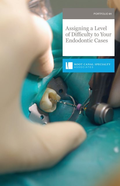
Portfolio 01: Assigning a Level of Difficulty to Your Endodontic Cases

You also want an ePaper? Increase the reach of your titles
YUMPU automatically turns print PDFs into web optimized ePapers that Google loves.
PORTFOLIO <strong>01</strong><br />
<strong>Assigning</strong> a <strong>Level</strong><br />
<strong>of</strong> <strong>Difficulty</strong> <strong>to</strong> <strong>Your</strong><br />
<strong>Endodontic</strong> <strong>Cases</strong>
Root Canal Specialty Associates<br />
provides care in all phases <strong>of</strong> surgical<br />
and nonsurgical endodontics. With<br />
decades <strong>of</strong> combined experience, four<br />
locations, and nine endodontists,<br />
we have you covered.<br />
LOCATIONS<br />
Ann Arbor<br />
Brigh<strong>to</strong>n<br />
Livonia<br />
West Bloomfield<br />
DOCTORS<br />
Dr. Young Bin Bok<br />
Dr. Steven Edlund*<br />
Dr. Chris<strong>to</strong>pher McWatters<br />
Dr. Andrew Racek•<br />
Dr. Michael Shapiro<br />
Dr. Jae M. Shin<br />
Dr. Dmitry Vodopyanov<br />
Dr. Martha Zinderman<br />
* Active Diplomate <strong>of</strong> the American<br />
Board <strong>of</strong> Endodontists
PATIENTS COME FIRST<br />
You care about your patients. We care<br />
about your patients <strong>to</strong>o – and promise <strong>to</strong><br />
return them <strong>to</strong> your <strong>of</strong>fice feeling better.<br />
Our priority is <strong>to</strong> put them at ease and<br />
make them feel comfortable from the<br />
moment they connect with our team.<br />
We pride ourselves in consistently<br />
delivering the highest standard<br />
<strong>of</strong> endodontic care no matter which<br />
location or doc<strong>to</strong>r your patient<br />
visits. We <strong>of</strong>fer extensive scheduling<br />
availability for both routine and<br />
urgent care. <strong>Your</strong> patients will receive<br />
the prompt and specialized attention<br />
they deserve, when they need it.<br />
“I was nervous before my<br />
appointment since I had<br />
never had any sort <strong>of</strong> dental<br />
work done aside from<br />
six-month cleanings – not<br />
even a cavity filling. I didn’t<br />
know what <strong>to</strong> expect. Root<br />
Canal Specialty Associates<br />
was very thorough and<br />
went through everything<br />
step-by- step before my<br />
appointment. It was a<br />
great experience–very<br />
quick and painless. Their<br />
staff put me at ease…”<br />
Angelina T.,<br />
Patient, Root Canal<br />
See more s<strong>to</strong>ries from satisfied patients and<br />
referring doc<strong>to</strong>rs at rootcanaldocs.com. 1
TRUE PARTNERS<br />
We take our referral partnerships<br />
very seriously. Consider us an extension<br />
<strong>of</strong> your team.<br />
In many cases, a compromised <strong>to</strong>oth<br />
can be retained with endodontic<br />
treatment. If you have concerns about<br />
complicated conditions or treatment<br />
options for your patient, our doc<strong>to</strong>rs<br />
are available for endodontic case<br />
consultation whenever you need it.<br />
Together we can relieve pain, save<br />
teeth and provide your patients with<br />
optimal, quality care. And because <strong>of</strong><br />
our focus, procedures can be performed<br />
in an efficient and predictable manner –<br />
creating positive experiences and<br />
favorable outcomes.<br />
“My experience with Root<br />
Canal Specialty Associates<br />
(RCSA) has been<br />
wonderful as a patient and<br />
referring doc<strong>to</strong>r. In the<br />
30+ years <strong>of</strong> referring <strong>to</strong><br />
RCSA, feedback from<br />
my patients has been 99%<br />
positive. The doc<strong>to</strong>rs are<br />
always available <strong>to</strong> discuss<br />
cases, and the staff is<br />
always helpful and pleasant.<br />
As a patient myself, I was<br />
surprised how efficiently<br />
the doc<strong>to</strong>rs worked.”<br />
Dr. Michael Pardonnet<br />
Great Lakes Family<br />
Dental Group<br />
2
ONLY THE BEST FOR YOU<br />
Our doc<strong>to</strong>rs have an extensive<br />
and diverse mix <strong>of</strong> educational and<br />
clinical experience.<br />
Trained at <strong>to</strong>p endodontic programs<br />
across the country – and with over<br />
170 years <strong>of</strong> combined experience –<br />
our doc<strong>to</strong>rs deliver superb results.<br />
We are dedicated <strong>to</strong> advancing the<br />
specialty <strong>of</strong> endodontics through<br />
teaching and staying at the forefront<br />
<strong>of</strong> technology and innovation.<br />
DOCTORS<br />
Dr. Robert Coleman*<br />
Dr. Steven Edlund*<br />
Dr. Martin Goode<br />
Dr. Wesley Ichesco<br />
Dr. Alexandra Martella<br />
Dr. Chris<strong>to</strong>pher McWatters<br />
Dr. Andrew Racek<br />
Dr. Michael Shapiro<br />
Dr. Martha Zinderman<br />
“Root Canal Specialty<br />
Associates (RCSA) is a<br />
team <strong>of</strong> excellent clinicians<br />
with strong diagnostic<br />
and treatment skills. RCSA<br />
employs up-<strong>to</strong>-date<br />
technology <strong>to</strong> help in all<br />
phases <strong>of</strong> treatment. They<br />
also continually strive <strong>to</strong><br />
incorporate changes within<br />
the practice <strong>to</strong> consistently<br />
deliver clinical excellence<br />
<strong>to</strong> their patients.”<br />
Dr. Neville J. McDonald<br />
Pr<strong>of</strong>essor, Division Head<br />
and ASE Program Direc<strong>to</strong>r,<br />
<strong>Endodontic</strong>s CRSE,<br />
University <strong>of</strong> Michigan,<br />
School <strong>of</strong> Dentistry<br />
* Active Diplomate <strong>of</strong> the American<br />
Board <strong>of</strong> Endodontists<br />
We <strong>of</strong>fer Cone Beam Computed Tomography<br />
(CBCT) at all four locations – a 3-dimensional imaging<br />
technology that helps us make better diagnoses and<br />
treatment decisions for our patients.<br />
Want <strong>to</strong> hear more about the indications for and<br />
benefits <strong>of</strong> CBCT imaging? Call (734) 261-7800<br />
<strong>to</strong> schedule a time <strong>to</strong> talk.
GUIDELINES FOR ASSESSMENT<br />
The cases shared here have been treated by Root Canal Specialty Associates and have<br />
been deemed moderate <strong>to</strong> difficult based on guidelines from the American Association<br />
<strong>of</strong> <strong>Endodontic</strong>s (AAE) shown below. These guidelines also include an assessment form,<br />
making it easier <strong>to</strong> decide whether a referral is the best choice – enabling you <strong>to</strong> assign<br />
a level <strong>of</strong> difficulty <strong>to</strong> your case.<br />
Reference the AAE assessment form we’ve included for you, or visit rootcanaldocs.com/<br />
patient-referral <strong>to</strong> download a PDF and see a complete list <strong>of</strong> considerations <strong>to</strong> properly<br />
evaluate whether a case meets minimal, moderate, or high levels <strong>of</strong> difficulty.<br />
If you have concerns about complicated conditions or treatment options for your patient,<br />
our doc<strong>to</strong>rs are available for endodontic case consultation whenever you need it.<br />
AAE LEVELS OF DIFFICULTY<br />
MINIMAL DIFFICULTY<br />
Preoperative condition<br />
indicates routine complexity<br />
(uncomplicated). These<br />
types <strong>of</strong> cases would<br />
exhibit only those fac<strong>to</strong>rs<br />
listed in the minimal<br />
difficulty category.<br />
Achieving a predictable<br />
treatment outcome<br />
should be attainable by<br />
a competent practitioner<br />
with limited experience.<br />
MODERATE DIFFICULTY<br />
Preoperative condition<br />
is complicated, exhibiting<br />
one or more patient or<br />
treatment fac<strong>to</strong>rs listed in<br />
the moderate difficulty<br />
category. Achieving<br />
a predictable treatment<br />
outcome will be challenging<br />
for a competent, experienced<br />
practitioner.<br />
HIGH DIFFICULTY<br />
Preoperative condition<br />
is exceptionally<br />
complicated, exhibiting<br />
several fac<strong>to</strong>rs listed in<br />
the moderate difficulty<br />
category or at least one<br />
in the high difficulty<br />
category. Achieving a<br />
predictable treatment<br />
outcome will be challenging<br />
for even the most experienced<br />
practitioner with<br />
an extensive his<strong>to</strong>ry <strong>of</strong><br />
favorable outcomes.<br />
4
CASE <strong>01</strong><br />
Conventional endodontic therapy <strong>of</strong> a mandibular second<br />
molar with atypical ana<strong>to</strong>my (s-shaped mesial root).<br />
TOOTH #18<br />
Pre-Op<br />
Post-Op<br />
DIAGNOSTIC & TREATMENT<br />
CONSIDERATIONS<br />
POSITION IN THE ARCH<br />
Mandibular second molar<br />
TOOTH ISOLATION<br />
Simple pretreatment modification<br />
required for rubber dam isolation<br />
CANAL & ROOT MORPHOLOGY<br />
Extreme curvature (>30°)<br />
or S-shaped curve<br />
Preoperative evaluation revealed extensive caries and<br />
a coronal fracture. Clinical testing supported a diagnosis<br />
<strong>of</strong> symp<strong>to</strong>matic irreversible pulpitis with symp<strong>to</strong>matic<br />
apical periodontitis.<br />
While there were no patient conditions that exceeded<br />
a minimal level <strong>of</strong> difficulty, there were several diagnostic<br />
and treatment considerations that could potentially<br />
complicate and adversely affect the outcome <strong>of</strong> this case.<br />
The presence <strong>of</strong> caries and a fractured mesiolingual cusp<br />
made rubber dam isolation moderately difficult. In addition,<br />
being a second molar with an s-shaped mesial root created<br />
a very high level <strong>of</strong> treatment difficulty.<br />
5
CASE 02<br />
<strong>Endodontic</strong> retreatment <strong>of</strong> a maxillary second bicuspid<br />
with overextended filling material.<br />
TOOTH #4<br />
Pre-Op Gutta Percha Removed Post-Op<br />
PATIENT CONSIDERATIONS<br />
EMERGENCY CONDITION<br />
Severe pain<br />
ADDITIONAL CONSIDERATIONS<br />
ENDODONTIC<br />
TREATMENT HISTORY<br />
Previous nonsurgical endodontic<br />
treatment with complications<br />
Patient presented with severe pain in the maxillary right<br />
quadrant. Radiographic examination revealed previous<br />
nonsurgical endodontic treatment with the canal filling<br />
material extended beyond the apex and perforating<br />
the Schneiderian membrane.<br />
Patient considerations such as severe pain requiring<br />
emergency care can create a high level <strong>of</strong> treatment<br />
difficulty. In addition, the his<strong>to</strong>ry <strong>of</strong> previous endodontic<br />
therapy complicated by the overextension <strong>of</strong> canal filling<br />
material make the management <strong>of</strong> this case extremely<br />
challenging.<br />
6
CASE 03<br />
Conventional endodontic therapy with the surgical repair <strong>of</strong> an<br />
external resorptive defect involving a mandibular first molar.<br />
TOOTH #19<br />
Pre-Op<br />
CBCT Axial View<br />
Conventional RCT Post-Op<br />
Resorptive Defect Exposed<br />
Defect Res<strong>to</strong>red<br />
One Year Post-Op<br />
DIAGNOSTIC & TREATMENT<br />
CONSIDERATIONS<br />
RESORPTION<br />
External resorption<br />
Patient presented with an asymp<strong>to</strong>matic, unres<strong>to</strong>red and<br />
vital mandibular first molar. Radiographic examination<br />
revealed invasive cervical external resorption on the mesial<br />
surface <strong>of</strong> the root extending below the alveolar crest.<br />
Cone Beam Computerized Tomography (CBCT) showed<br />
that this defect was extensive and communicated with<br />
the pulp. Depending on the size and location <strong>of</strong> the defect,<br />
teeth exhibiting external resorption can be very complex<br />
and difficult <strong>to</strong> treat predictably.<br />
A multi-stage treatment approach, including both conventional<br />
endodontic therapy and surgical intervention was planned<br />
<strong>to</strong> try and obtain a successful outcome. After isolating the<br />
defect, endodontic treatment and a core build-up was placed.<br />
A surgical approach allowed complete removal <strong>of</strong> the resorptive<br />
tissue and res<strong>to</strong>ration <strong>of</strong> the defect. One year follow-up shows<br />
excellent healing and the patient remains asymp<strong>to</strong>matic.<br />
7
CASE 04<br />
Cone beam computerized <strong>to</strong>mography (CBCT) used <strong>to</strong><br />
enhance diagnosis and treatment planning.<br />
TOOTH #29<br />
Pre-Op CBCT Sagittal View CBCT Coronal View<br />
DIAGNOSTIC & TREATMENT<br />
CONSIDERATIONS<br />
RADIOGRAPHIC DIFFICULTIES<br />
Moderate difficulty interpreting<br />
radiographs<br />
CANAL & ROOT MORPHOLOGY<br />
Mandibular premolar with two roots<br />
RADIOGRAPHIC APPEARANCE<br />
OF CANALS<br />
Indistinct canal path<br />
Preoperative evaluation and clinical testing supported<br />
a diagnosis <strong>of</strong> pulpal necrosis with symp<strong>to</strong>matic<br />
apical periodontitis. Conventional radiography displayed<br />
a mandibular premolar with atypical root morphology. Further<br />
study using Cone Beam Computerized Tomography (CBCT)<br />
disclosed the presence <strong>of</strong> a horizontal root fracture.<br />
All other considerations aside, the presence <strong>of</strong> a mandibular<br />
premolar with two roots would have made endodontic<br />
treatment very difficult. However, the additional diagnostic<br />
information obtained by the CBCT helped <strong>to</strong> determine<br />
a more appropriate treatment plan (extraction) avoiding<br />
endodontic failure and further complications.<br />
8
CASE 05<br />
<strong>Endodontic</strong> retreatment <strong>of</strong> a maxillary first premolar with<br />
atypical ana<strong>to</strong>my and previously separated instruments.<br />
TOOTH #5<br />
Pre-Op<br />
Post-Op<br />
PATIENT CONSIDERATIONS<br />
MEDICAL HISTORY<br />
One or more medical problems<br />
GAG REFLEX<br />
Gags occasionally with<br />
radiographs/treatment<br />
DIAGNOSTIC & TREATMENT<br />
CONSIDERATIONS<br />
CANAL & ROOT MORPHOLOGY<br />
Maxillary premolar with three roots<br />
Patient presented with a medical his<strong>to</strong>ry that included<br />
controlled diabetes and a previous myocardial infarction<br />
followed by an angioplasty procedure with stent placement.<br />
In addition, he reported that he frequently gags during the<br />
exposure <strong>of</strong> dental radiographs. The radiographic examination<br />
reveled incomplete endodontic therapy complicated<br />
by separated instruments and atypical root morphology.<br />
The patient’s multiple medical problems and elevated<br />
gag reflex make this case moderately difficult. In addition,<br />
maxillary premolars with three roots and previous<br />
treatment complications (such as separated instruments)<br />
make achieving a predictable treatment outcome highly<br />
challenging for even the most experienced practitioner.<br />
ADDITIONAL CONSIDERATIONS<br />
ENDODONTIC<br />
TREATMENT HISTORY<br />
Previous access with complications<br />
(separated instruments)<br />
9
CASE 06<br />
Medically compromised patient with complex endodonticres<strong>to</strong>rative<br />
needs.<br />
TOOTH #31<br />
Pre-Op<br />
Caries Removed<br />
Distal Res<strong>to</strong>red<br />
Post-Op<br />
One Year Post-Op<br />
PATIENT CONSIDERATIONS<br />
MEDICAL HISTORY<br />
Complex medical his<strong>to</strong>ry/serious<br />
illness/disability<br />
DIAGNOSTIC & TREATMENT<br />
CONSIDERATIONS<br />
POSITION IN THE ARCH<br />
2nd or third molar; extreme<br />
inclination (>30degrees)<br />
TOOTH ISOLATION<br />
Extensive pretreatment<br />
modification (res<strong>to</strong>ration<br />
<strong>of</strong> distal caries)<br />
An elderly patient presented with a symp<strong>to</strong>matic<br />
mandibular second molar. Their medical his<strong>to</strong>ry indicated<br />
that they were receiving a regimen <strong>of</strong> IV bisphosphonate<br />
therapy administered every three months. Preoperative<br />
evaluation revealed that <strong>to</strong>oth #31 was extremely inclined,<br />
had subgingival distal root caries and approximated an<br />
impacted bicuspid. Additionally, the <strong>to</strong>oth was res<strong>to</strong>red with<br />
a crown that served as the key abutment for a mandibular<br />
removable denture. Clinical testing supported a diagnosis<br />
<strong>of</strong> pulpal necrosis with symp<strong>to</strong>matic apical periodontitis.<br />
This case exhibits numerous complicating fac<strong>to</strong>rs creating<br />
an extremely high level <strong>of</strong> difficulty. Due <strong>to</strong> the risk <strong>of</strong><br />
osteonecrosis (MRONJ), extraction was not a favorable<br />
treatment option. Prior <strong>to</strong> endodontic therapy, the distal<br />
caries was removed and res<strong>to</strong>red without eliminating<br />
access <strong>to</strong> the canal space. Due <strong>to</strong> the severe inclination,<br />
endodontic treatment was completed through the mesial<br />
rest seat. A one year follow-up exhibited excellent healing.<br />
10
CASE 07<br />
Maxillary lateral incisor with dens invaginatus.<br />
TOOTH #7<br />
Pre-Op CBCT Axial View Post-Op<br />
DIAGNOSTIC & TREATMENT<br />
CONSIDERATIONS<br />
RADIOGRAPHIC DIFFICULTIES<br />
Moderate difficulty interpreting<br />
radiographs<br />
CROWN MORPHOLOGY<br />
Significant deviation from normal<br />
<strong>to</strong>oth/root form (dens invaginatus)<br />
Patient presented with pain, tenderness <strong>to</strong> percussion,<br />
and intraoral swelling associated with <strong>to</strong>oth #7. Clinical<br />
testing supported a diagnosis <strong>of</strong> pulpal necrosis<br />
with symp<strong>to</strong>matic apical periodontitis. The radiographic<br />
examination revealed the presence <strong>of</strong> dens invaginatus<br />
(dens in dente) with two deep infoldings <strong>of</strong> enamel and<br />
dentin mesial and distal <strong>to</strong> a main canal. This finding<br />
was confirmed and better visualized for treatment planning<br />
with Cone Beam Computerized Tomography (CBCT).<br />
While there were no patient considerations that exceeded<br />
a moderate level <strong>of</strong> difficulty, the atypical crown and root<br />
morphology associated with dens invaginatus makes these<br />
cases highly difficult <strong>to</strong> treat. Accurate pretreatment<br />
visualization and an understanding <strong>of</strong> the aberrant morphology<br />
are key fac<strong>to</strong>rs in obtaining clinical success in cases such<br />
as this.<br />
11
CASE 08<br />
Conventional endodontic therapy <strong>of</strong> a maxillary second molar<br />
with atypical ana<strong>to</strong>my.<br />
TOOTH #2<br />
Pre-Op Post-Op One Year Post-Op<br />
DIAGNOSTIC & TREATMENT<br />
CONSIDERATIONS<br />
POSITION IN THE ARCH<br />
2nd or 3rd molars<br />
CROWN & ROOT MORPHOLOGY<br />
Maxillary molar with atypical<br />
ana<strong>to</strong>my<br />
The patient presented with a his<strong>to</strong>ry <strong>of</strong> pain in the<br />
maxillary right quadrant. Clinical examination revealed<br />
that <strong>to</strong>oth #2 had a deep palatal groove and caries was<br />
present. Clinical testing supported a diagnosis <strong>of</strong> pulpal<br />
necrosis with symp<strong>to</strong>matic apical periodontitis. During<br />
endodontic treatment, a second palatal was located with<br />
the aid <strong>of</strong> a surgical operating microscope.<br />
Although the patient was young and healthy, maxillary<br />
molars may <strong>of</strong>fer significant ana<strong>to</strong>mical variability.<br />
Due <strong>to</strong> their position in the arch, locating and negotiating<br />
all canals without supplementary magnification and<br />
illumination can be very challenging, especially if atypical<br />
ana<strong>to</strong>my is present.<br />
12
WHEN TO REFER<br />
Sometimes it’s difficult <strong>to</strong> know when a referral is best for<br />
your patient. Guidelines from The American Association<br />
<strong>of</strong> Endodontists (AAE) enable you <strong>to</strong> assign a level <strong>of</strong> difficulty<br />
<strong>to</strong> your case, making it easier <strong>to</strong> decide whether a referral is<br />
the best choice.<br />
Reference the AAE assessment form we’ve included for you,<br />
or visit rootcanaldocs.com/patient-referral <strong>to</strong> download<br />
a PDF and see a complete list <strong>of</strong> considerations <strong>to</strong> properly<br />
evaluate whether a case meets minimal, moderate, or high<br />
levels <strong>of</strong> difficulty.<br />
READY TO REFER?<br />
Visit us at rootcanaldocs.com <strong>to</strong> fill<br />
out our online referral form.
FOUR LOCATIONS, ONE GREAT EXPERIENCE.<br />
ANN ARBOR<br />
(734) 973-2727<br />
BRIGHTON<br />
(810) 229-7800<br />
LIVONIA<br />
(734) 261-7800<br />
WEST BLOOMFIELD<br />
(248) 626-0600<br />
As one <strong>of</strong> the largest endodontic specialty practices in the state, we have<br />
four <strong>of</strong>fices in SE Michigan <strong>to</strong> better accommodate your patients. All <strong>of</strong> our<br />
<strong>of</strong>fices have hours Monday through Friday with early morning (7am) openings<br />
and evening appointments (until 7pm), and availability on Saturdays in Livonia.<br />
Patients can make an appointment at the location that’s most convenient<br />
for them.<br />
We also participate with most major dental benefit plans so your patient’s<br />
experience will not only be pleasant, but hassle-free.<br />
Visit rootcanaldocs.com for more information about each <strong>of</strong> our locations.<br />
ROOTCANALDOCS.COM