Portfolio 02: The Challenge Surrounding Tooth Resorption

Create successful ePaper yourself
Turn your PDF publications into a flip-book with our unique Google optimized e-Paper software.
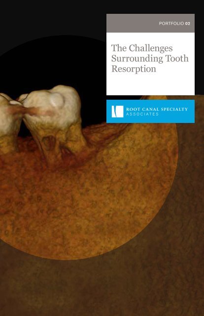
PORTFOLIO <strong>02</strong><br />
<strong>The</strong> <strong>Challenge</strong>s<br />
<strong>Surrounding</strong> <strong>Tooth</strong><br />
<strong>Resorption</strong>
Root Canal Specialty Associates<br />
provides care in all phases of surgical<br />
and nonsurgical endodontics. With<br />
decades of combined experience, four<br />
locations, and eleven endodontists,<br />
we have you covered.<br />
LOCATIONS<br />
Ann Arbor<br />
Brighton<br />
Livonia<br />
West Bloomfield<br />
DOCTORS<br />
Dr. Young Bin Bok<br />
Dr. Steven Edlund*<br />
Dr. Christopher McWatters<br />
Dr. Andrew Racek*<br />
Dr. Michael Shapiro*<br />
Dr. Jae M. Shin<br />
Dr. Dmitry Vodopyanov Dr.<br />
Martha Zinderman<br />
* Active Diplomate of the American<br />
Board of Endodontists
TRUE PARTNERS<br />
We take our referral partnerships<br />
very seriously. Consider us an extension<br />
of your team.<br />
We work collaboratively with our referrers to alleviate<br />
pain, save teeth and provide patients with optimal, quality<br />
care. In many cases, even a severely compromised tooth<br />
can be retained with endodontic treatment.<br />
Root Canal Specialty Associates doctors have extensive<br />
experience treating difficult cases (such as internal<br />
and external resorption). Our goal is to preserve the natural<br />
dentition and we employ both surgical and non-surgical<br />
approaches for treating resorptive defects. If you have<br />
questions or concerns about treatment options for your<br />
patient, our doctors are available for endodontic case<br />
consultation whenever you need it.<br />
“I have frequently consulted with Root Canal Specialty<br />
Associates doctors over the years. In addition to<br />
providing routine endodontic treatment, they have<br />
diagnosed and successfully partnered with me to provide<br />
optimal care for several patients with internal and<br />
external resorptive defects.”<br />
Dr. Robert Borowiec<br />
D.D.S., South Lyon Dental Group<br />
See more stories from satisfied patients and referring doctors at rootcanaldocs.com/stories<br />
1
THE CHALLENGES OF RESORPTION<br />
Etiology. It is thought that tooth resorption may occur following injuries to or irritation to<br />
the periodontal ligament and/or dental pulp and it is a frequent sequelae following traumatic<br />
injuries, orthodontic tooth movement, or chronic infections of the periodontal structures.<br />
Diagnosis. <strong>The</strong>re may be no external signs, and the resorptive condition is often detected<br />
by routine radiographic examination. Where the lesion is visible, the clinical features vary<br />
from a small defect at the gingival margin to a pink coronal discoloration of the tooth crown<br />
resulting in ultimate cavitation of the overlying enamel. <strong>The</strong> condition is usually painless<br />
unless pulpal or periodontal infection develops. Radiographic features of resorptive lesions<br />
vary from well-delineated to irregularly bordered mottled radiolucencies, and these can be<br />
confused with dental caries.<br />
Treatment. <strong>Resorption</strong> can occur as a single entity or a combination of internal and external<br />
defects can occur simultaneously on the same tooth. Effective management and appropriate<br />
treatment can only be carried out if the true nature, history and exact location of the resorptive<br />
defect is known. Cone Beam Computed Tomography (CBCT) has been shown to be an<br />
important adjunct for the diagnosis and treatment of resorptive defects.<br />
Classification. <strong>The</strong> various types of root resorptive defects have been organized and<br />
classified in a variety of ways. In an attempt to eliminate confusion, a clinical related classification<br />
system based on stimulation factors has been developed and is outlined below:<br />
RESORPTION CLASSIFICATIONS<br />
1. PULPAL INFECTION ROOT RESORPTION (internal and/or external)<br />
2. PERIODONTAL INFECTION ROOT RESORPTION<br />
3. ORTHODONTIC PRESSURE ROOT RESORPTION<br />
4. IMPACTED TOOTH OR TUMOR PRESSURE ROOT RESORPTION<br />
5. ANKYLOTIC ROOT RESORPTION<br />
To learn more about these classifications, reference the article “Root resorption –<br />
diagnosis, classification and treatment choices based on stimulation factors.”<br />
Copyright © Blackwell Munksgaard 2003<br />
If you have questions about this complicated condition or treatment options for your<br />
patient, our doctors are always available for endodontic case consultation.<br />
2
DIFFERENTIATING INTERNAL FROM EXTERNAL RESORPTION<br />
Internal<br />
External<br />
Original site<br />
of resorption<br />
starts in the<br />
pulp<br />
Original<br />
site is the<br />
periodontal<br />
ligament<br />
<strong>The</strong> following diagnostic features can often help distinguish internal<br />
from external resorption in clinical practice:<br />
INTERNAL<br />
EXTERNAL<br />
An internal resorptive lesion appears to be<br />
close or continuous with the pulp whatever<br />
the angle of the x-ray.<br />
In internal resorption, the outline of the<br />
canal is often distorted and the canal and<br />
resorptive defect are contiguous.<br />
Internal resorption is rarely accompanied<br />
by bone loss around the tooth.<br />
An external resorptive lesion typically<br />
moves away from the canal as the angle<br />
of the radiograph changes. In addition,<br />
the buccal object rule can often help<br />
distinguish if the defect is on the buccal<br />
or lingual.<br />
When a resorptive defect is external,<br />
the root canal outline appears to be normal<br />
and can often be seen running through<br />
the defect.<br />
External resorption is frequently<br />
accompanied by bone loss around<br />
the tooth.<br />
3
TECHNOLOGY AND BETTER DIAGNOSES<br />
Cone Beam Computed Technology (CBCT) –<br />
a 3-dimensional imaging technology that helps us make<br />
better diagnoses and treatment decisions – is a great<br />
adjunct to making an accurate diagnosis of internal versus<br />
external resorption. We offer Cone Beam Computed<br />
Technology (CBCT) at all four locations:<br />
Ann Arbor<br />
Brighton<br />
Livonia<br />
West Bloomfield<br />
If you’d like to hear more about the indications for<br />
and benefits of CBCT imaging, call (734) 261-7800<br />
to schedule a time to talk with us.<br />
4
CASE 01<br />
Pulpal Infection Root <strong>Resorption</strong> (External)<br />
Conventional endodontic treatment with surgical repair<br />
of external resorptive defect.<br />
TOOTH #30 | AGE 67<br />
DIAGNOSIS<br />
Patient in good health.<br />
Chief complaint of lingering<br />
pain to cold and tenderness<br />
to palpation over the buccal<br />
of #30. Examination revealed<br />
#30 to have a symptomatic<br />
irreversible pulpitis with<br />
external resorption on the<br />
mesiobuccal root.<br />
CHALLENGE<br />
Pre-Op<br />
Conventional RCT Post-Op<br />
A 6mm pocket was present<br />
over the mesiobuccal root<br />
in the area of the resorption.<br />
<strong>The</strong> subgingival extent of the<br />
resorptive defect will require<br />
periapical surgery to repair.<br />
Defect Restored<br />
Defect Repaired<br />
TREATMENT<br />
Due to the irreversible<br />
pulpitis, root canal therapy<br />
was completed and the<br />
tooth was restored with a<br />
bonded core. Flap surgery<br />
was completed at a follow-up<br />
visit, during which the area<br />
was exposed and the defect<br />
was cleaned of all resorptive<br />
tissue. <strong>The</strong> defect was<br />
repaired with Geristore<br />
to allow for gingival<br />
reattachment and the area<br />
healed uneventfully. <strong>The</strong><br />
periodontal defect resolved<br />
leaving a 3mm probing<br />
depth.<br />
5
CASE <strong>02</strong><br />
Conventional endodontic treatment followed by surgical<br />
repair of resorptive defect.<br />
TOOTH #18 | AGE 52<br />
CBCT Axial Slice<br />
CBCT Coronal Slice<br />
6
Pulpal Infection Root <strong>Resorption</strong> (External)<br />
Pre-Op<br />
Post-Op<br />
DIAGNOSIS<br />
Patient with intermittent<br />
pain on the lower left side.<br />
Examination revealed what<br />
appeared to be extensive<br />
external resorption on #18<br />
near the pulp, with lingering<br />
pain to cold and pain on<br />
percussion consistent with<br />
a symptomatic irreversible<br />
pulpitis. CBCT showed<br />
the defect to be on the<br />
mesiolingual aspect, just<br />
above the crestal bone and<br />
involving the pulp. As the<br />
patient was already missing<br />
#19, he desired to save<br />
the tooth. Prognosis:<br />
questionable.<br />
CHALLENGE<br />
Due to the pulpal involvement<br />
of the resorption, hemostasis<br />
and field isolation can be<br />
difficult. <strong>The</strong> location of the<br />
defect on the lingual aspect<br />
also make surgical<br />
correction and restoration<br />
difficult.<br />
TREATMENT<br />
Root canal treatment was<br />
completed and a permanent<br />
core was placed to seal<br />
the defect internally. This<br />
was followed by flap surgery,<br />
during which the defect<br />
was fully repaired with<br />
Geristore. A full coverage<br />
restoration, with margin<br />
replacement on the Geristore,<br />
was recommended.<br />
7
CASE 03<br />
Conventional endodontic treatment with surgical repair<br />
of external resorptive defect.<br />
TOOTH #30 | AGE 47<br />
CBCT Axial Slice<br />
CBCT Coronal Slice<br />
8
Pulpal Infection Root <strong>Resorption</strong> (External)<br />
Pre-Op<br />
Post-Op<br />
DIAGNOSIS<br />
Patient presented with<br />
a history of pain on the<br />
lower right. Radiographic<br />
examination exhibited<br />
external resorption of<br />
the mesiobuccal root.<br />
Clinical testing showed<br />
the tooth to have lingering<br />
pain to cold, consistent<br />
with a diagnosis of<br />
irreversible pulpitis.<br />
CHALLENGE<br />
<strong>The</strong> resorption of the<br />
mesial root communicates<br />
with the mesiobuccal canal.<br />
This makes the endodontic<br />
treatment of the mesial root<br />
very complicated.<br />
TREATMENT<br />
Non-surgical root canal<br />
therapy was completed<br />
to alleviate the patient’s<br />
symptoms, and the<br />
mesiobuccal root was<br />
initially sealed with MTA.<br />
Post operative CBCT<br />
revealed a large defect on<br />
the mesiobuccal root. After<br />
placing a permanent core<br />
buildup, the area was<br />
exposed surgically and was<br />
restored with Bioceramic<br />
Root Repair Material<br />
and Geristore, to allow<br />
gingival reattachment.<br />
9
CASE 04<br />
Conventional endodontic treatment with internal repair<br />
of resorptive defect.<br />
TOOTH #9 | AGE 47<br />
CBCT Axial Slice<br />
CBCT Sagittal Slice<br />
10
Periodontal Infection Root <strong>Resorption</strong><br />
DIAGNOSIS<br />
Patient presented with<br />
no symptoms. Radiographs<br />
and CBCT scan show<br />
invasive cervical external<br />
resorption.<br />
CHALLENGE<br />
<strong>The</strong> location of the resorptive<br />
defect (on the palatal aspect<br />
of the root) would make a<br />
surgical repair very difficult.<br />
TREATMENT<br />
Prophylactic endo completed<br />
along with orthograde<br />
Geristore root repair. No<br />
surgical intervention was<br />
needed. Repair was made<br />
through the endo access<br />
on the palatal side of tooth.<br />
Pre-Op<br />
Post-Op<br />
11
CASE 05<br />
Conventional endodontic treatment including<br />
calcium hydroxide therapy.<br />
TOOTH #10 | AGE 14<br />
CBCT Axial Slice<br />
CBCT Sagittal Slice<br />
12
Orthodontic Pressure and/or Impacted <strong>Tooth</strong> Pressure <strong>Resorption</strong><br />
Pre-Op Calcium Hydroxide Post-Op<br />
DIAGNOSIS<br />
Patient has had several<br />
phases or orthodontic<br />
treatment that commenced<br />
when he was seven years<br />
old. He also had a history<br />
of a labially positioned and<br />
impacted maxillary cuspid<br />
that was surgically exposed<br />
and orthodontically moved<br />
into position. He had a<br />
traumatic injury to his face<br />
that resulted in a broken<br />
nose. As a part of this<br />
evaluation, periapical<br />
radiographs and a CBCT<br />
scan was taken by an oral<br />
and maxillofacial surgeon.<br />
<strong>The</strong>se radiographs disclosed<br />
extensive external root<br />
resorption of tooth #10.<br />
<strong>Tooth</strong> #10 was asymptomatic,<br />
vital and exhibited moderate<br />
clinical mobility.<br />
CHALLENGE<br />
<strong>The</strong> distal aspect of tooth<br />
#10 had extensive root<br />
resorption and the tooth<br />
structure overlying the pulp<br />
is very thin. Extreme care<br />
must be exercised to avoid<br />
root perforation.<br />
TREATMENT<br />
<strong>The</strong> treatment plan for<br />
tooth #10 consisted of<br />
removing the orthodontic<br />
forces on tooth #10,<br />
initiating endodontic<br />
treatment and placing<br />
calcium hydroxide for several<br />
months (to attempt to arrest<br />
resorptive process) and<br />
completing endodontic<br />
therapy. Due to the extensive<br />
amount of root resorption,<br />
the prognosis will be<br />
guarded.<br />
13
CASE 06<br />
Conventional endodontic treatment including<br />
calcium hydroxide therapy.<br />
TOOTH #24 | AGE 51<br />
CBCT Axial Slice<br />
Three-Dimensional Reconstruction<br />
14
Pulpal Infection Root <strong>Resorption</strong> (External)<br />
DIAGNOSIS<br />
Patient presented the<br />
chief complaint of a loose<br />
lower front tooth. Clinical<br />
examination revealed a nonrestored<br />
tooth #24 with<br />
class 3 mobility and<br />
tenderness to percussion<br />
and palpation. Radiographs<br />
revealed a mid-root radiolucency<br />
communicating with<br />
the apex and bone loss<br />
extending up the mesial root<br />
surface to the crestal bone.<br />
CBCT displayed external<br />
inflammatory resorption<br />
on the lingual of tooth that<br />
communicated with the<br />
pulp. <strong>The</strong>se findings were<br />
consistent with a diagnosis<br />
of pulpal necrosis with<br />
symptomatic apical periodontitis<br />
and external resorption.<br />
Pre-Op X-Ray<br />
After Endodontic Treatment<br />
CHALLENGE<br />
<strong>The</strong> extensive bone loss<br />
and excessive clinical<br />
mobility made the prognosis<br />
for treatment very poor. In<br />
addition, the location of the<br />
external resorptive defect<br />
(on the lingual of tooth #24)<br />
magnified the difficulty of<br />
this case.<br />
Post-Surgery<br />
TREATMENT<br />
Root canal treatment was<br />
completed over two visits,<br />
medicating with calcium<br />
hydroxide. A lingual splint<br />
was placed by the patient’s<br />
general dentist to stabilize<br />
the tooth. Surgical repair of<br />
the lingual resorptive defect<br />
6-Month Recall<br />
TREATMENT<br />
with Geristore, apical<br />
surgery with a bioceramic<br />
putty retrofilling, scaling<br />
and root planning were also<br />
performed. 6-month recall<br />
shows resolution of the<br />
radiolucency with bone fill<br />
up to the crestal bone.<br />
15
CASE 07<br />
Pulpal Infection Root <strong>Resorption</strong> (External)<br />
Arrested external resorption.<br />
TOOTH #31 | AGE 69<br />
Radiograph: 10 Years Ago<br />
CBCT Axial Slice<br />
DIAGNOSIS<br />
Patient is asymptomatic;<br />
but, aware that tooth #31<br />
has had a resorptive defect<br />
for over ten years. <strong>The</strong>re is<br />
no evidence of the defect<br />
upon visual inspection and<br />
clinical findings are within<br />
normal limits.<br />
CBCT images display a<br />
resorptive defect on the<br />
distal aspect involving the<br />
dentin and communicating<br />
with the periodontal ligament.<br />
This defect extends into<br />
the middle third of the root;<br />
but, does not communicate<br />
with the pulp chamber or<br />
distal canal. <strong>The</strong>se findings<br />
support a diagnosis of normal<br />
pulpal and periapical tissues,<br />
with an external resorptive<br />
defect.<br />
16<br />
CBCT Sagittal Slice<br />
CHALLENGE<br />
It is often difficult to<br />
ascertain if the resorptive<br />
process is active or arrested.<br />
Comparing the current<br />
radiograph with those taken<br />
several years in the past<br />
can assist in this evaluation<br />
and help determine the most<br />
optimal treatment plan.<br />
TREATMENT<br />
<strong>The</strong> lesion has been<br />
present and unchanged on<br />
radiographs (10+ years).<br />
Patient opted to re-evaluate<br />
in 6 months with a repeat<br />
CBCT. If the lesion increases<br />
in size, treatment options<br />
would be considered at<br />
that time.
WHEN TO REFER<br />
Sometimes it’s difficult to know when a referral is best for<br />
your patient. Guidelines from <strong>The</strong> American Association<br />
of Endodontists (AAE) enable you to assign a level of difficulty<br />
to your case, making it easier to decide whether a referral is<br />
the best choice.<br />
Visit rootcanaldocs.com/patient-referral to download<br />
a PDF and see a complete list of considerations to properly<br />
evaluate whether a case meets minimal, moderate, or high<br />
levels of difficulty.<br />
READY TO REFER?<br />
Visit us at rootcanaldocs.com to fill<br />
out our online referral form.
FOUR LOCATIONS, ONE GREAT EXPERIENCE.<br />
ANN ARBOR<br />
(734) 973-2727<br />
BRIGHTON<br />
(810) 229-7800<br />
LIVONIA<br />
(734) 261-7800<br />
WEST BLOOMFIELD<br />
(248) 626-0600<br />
As one of the largest endodontic specialty practices in the state, we have<br />
four offices in SE Michigan to better accommodate your patients. All of our<br />
offices have hours Monday through Friday with early morning (7am) openings<br />
and evening appointments (until 7pm), and availability on Saturdays in Livonia.<br />
Patients can make an appointment at the location that’s most convenient<br />
for them.<br />
We also participate with most major dental benefit plans so your patient’s<br />
experience will not only be pleasant, but hassle-free.<br />
Visit rootcanaldocs.com for more information about each of our locations.<br />
ROOTCANALDOCS.COM