Portfolio 02: The Challenge Surrounding Tooth Resorption

You also want an ePaper? Increase the reach of your titles
YUMPU automatically turns print PDFs into web optimized ePapers that Google loves.
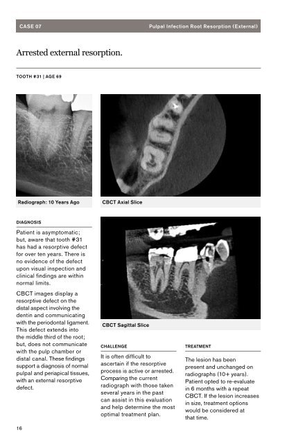
CASE 07<br />
Pulpal Infection Root <strong>Resorption</strong> (External)<br />
Arrested external resorption.<br />
TOOTH #31 | AGE 69<br />
Radiograph: 10 Years Ago<br />
CBCT Axial Slice<br />
DIAGNOSIS<br />
Patient is asymptomatic;<br />
but, aware that tooth #31<br />
has had a resorptive defect<br />
for over ten years. <strong>The</strong>re is<br />
no evidence of the defect<br />
upon visual inspection and<br />
clinical findings are within<br />
normal limits.<br />
CBCT images display a<br />
resorptive defect on the<br />
distal aspect involving the<br />
dentin and communicating<br />
with the periodontal ligament.<br />
This defect extends into<br />
the middle third of the root;<br />
but, does not communicate<br />
with the pulp chamber or<br />
distal canal. <strong>The</strong>se findings<br />
support a diagnosis of normal<br />
pulpal and periapical tissues,<br />
with an external resorptive<br />
defect.<br />
16<br />
CBCT Sagittal Slice<br />
CHALLENGE<br />
It is often difficult to<br />
ascertain if the resorptive<br />
process is active or arrested.<br />
Comparing the current<br />
radiograph with those taken<br />
several years in the past<br />
can assist in this evaluation<br />
and help determine the most<br />
optimal treatment plan.<br />
TREATMENT<br />
<strong>The</strong> lesion has been<br />
present and unchanged on<br />
radiographs (10+ years).<br />
Patient opted to re-evaluate<br />
in 6 months with a repeat<br />
CBCT. If the lesion increases<br />
in size, treatment options<br />
would be considered at<br />
that time.