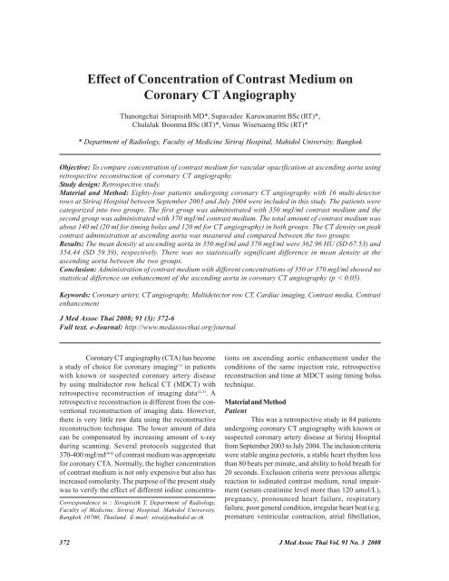
Effect of Concentration of Contrast Medium on Coronary CT ...
Effect of Concentration of Contrast Medium on Coronary CT ...
Effect of Concentration of Contrast Medium on Coronary CT ...
You also want an ePaper? Increase the reach of your titles
YUMPU automatically turns print PDFs into web optimized ePapers that Google loves.
<str<strong>on</strong>g>Effect</str<strong>on</strong>g> <str<strong>on</strong>g>of</str<strong>on</strong>g> <str<strong>on</strong>g>C<strong>on</strong>centrati<strong>on</strong></str<strong>on</strong>g> <str<strong>on</strong>g>of</str<strong>on</strong>g> <str<strong>on</strong>g>C<strong>on</strong>trast</str<strong>on</strong>g> <str<strong>on</strong>g>Medium</str<strong>on</strong>g> <strong>on</strong><br />
Cor<strong>on</strong>ary <strong>CT</strong> Angiography<br />
Than<strong>on</strong>gchai Siriapisith MD*, Supavadee Karuwanarint BSc (RT)*,<br />
Chulaluk Bo<strong>on</strong>ma BSc (RT)*, Venus Wisetsaeng BSc (RT)*<br />
* Department <str<strong>on</strong>g>of</str<strong>on</strong>g> Radiology, Faculty <str<strong>on</strong>g>of</str<strong>on</strong>g> Medicine Siriraj Hospital, Mahidol University, Bangkok<br />
Objective: To compare c<strong>on</strong>centrati<strong>on</strong> <str<strong>on</strong>g>of</str<strong>on</strong>g> c<strong>on</strong>trast medium for vascular opacificati<strong>on</strong> at ascending aorta using<br />
retrospective rec<strong>on</strong>structi<strong>on</strong> <str<strong>on</strong>g>of</str<strong>on</strong>g> cor<strong>on</strong>ary <strong>CT</strong> angiography.<br />
Study design: Retrospective study.<br />
Material and Method: Eighty-four patients undergoing cor<strong>on</strong>ary <strong>CT</strong> angiography with 16 multi-detector<br />
rows at Siriraj Hospital between September 2003 and July 2004 were included in this study. The patients were<br />
categorized into two groups. The first group was administrated with 350 mgI/ml c<strong>on</strong>trast medium and the<br />
sec<strong>on</strong>d group was administrated with 370 mgI/ml c<strong>on</strong>trast medium. The total amount <str<strong>on</strong>g>of</str<strong>on</strong>g> c<strong>on</strong>trast medium was<br />
about 140 ml (20 ml for timing bolus and 120 ml for <strong>CT</strong> angiography) in both groups. The <strong>CT</strong> density <strong>on</strong> peak<br />
c<strong>on</strong>trast administrati<strong>on</strong> at ascending aorta was measured and compared between the two groups.<br />
Results: The mean density at ascending aorta in 350 mgI/ml and 370 mgI/ml were 362.96 HU (SD 67.53) and<br />
354.44 (SD 59.39), respectively. There was no statistically significant difference in mean density at the<br />
ascending aorta between the two groups.<br />
C<strong>on</strong>clusi<strong>on</strong>: Administrati<strong>on</strong> <str<strong>on</strong>g>of</str<strong>on</strong>g> c<strong>on</strong>trast medium with different c<strong>on</strong>centrati<strong>on</strong>s <str<strong>on</strong>g>of</str<strong>on</strong>g> 350 or 370 mgI/ml showed no<br />
statistical difference <strong>on</strong> enhancement <str<strong>on</strong>g>of</str<strong>on</strong>g> the ascending aorta in cor<strong>on</strong>ary <strong>CT</strong> angiography (p < 0.05).<br />
Keywords: Cor<strong>on</strong>ary artery, <strong>CT</strong> angiography, Multidetector row <strong>CT</strong>, Cardiac imaging, <str<strong>on</strong>g>C<strong>on</strong>trast</str<strong>on</strong>g> media, <str<strong>on</strong>g>C<strong>on</strong>trast</str<strong>on</strong>g><br />
enhancement<br />
J Med Assoc Thai 2008; 91 (3): 372-6<br />
Full text. e-Journal: http://www.medassocthai.org/journal<br />
Cor<strong>on</strong>ary <strong>CT</strong> angiography (<strong>CT</strong>A) has become<br />
a study <str<strong>on</strong>g>of</str<strong>on</strong>g> choice for cor<strong>on</strong>ary imaging (1) in patients<br />
with known or suspected cor<strong>on</strong>ary artery disease<br />
by using multidector row helical <strong>CT</strong> (MD<strong>CT</strong>) with<br />
retrospective rec<strong>on</strong>structi<strong>on</strong> <str<strong>on</strong>g>of</str<strong>on</strong>g> imaging data (2,3) . A<br />
retrospective rec<strong>on</strong>structi<strong>on</strong> is different from the c<strong>on</strong>venti<strong>on</strong>al<br />
rec<strong>on</strong>structi<strong>on</strong> <str<strong>on</strong>g>of</str<strong>on</strong>g> imaging data. However,<br />
there is very little raw data using the rec<strong>on</strong>structive<br />
rec<strong>on</strong>structi<strong>on</strong> technique. The lower amount <str<strong>on</strong>g>of</str<strong>on</strong>g> data<br />
can be compensated by increasing amount <str<strong>on</strong>g>of</str<strong>on</strong>g> x-ray<br />
during scanning. Several protocols suggested that<br />
370-400 mgI/ml (4-6) <str<strong>on</strong>g>of</str<strong>on</strong>g> c<strong>on</strong>trast medium was appropriate<br />
for cor<strong>on</strong>ary <strong>CT</strong>A. Normally, the higher c<strong>on</strong>centrati<strong>on</strong><br />
<str<strong>on</strong>g>of</str<strong>on</strong>g> c<strong>on</strong>trast medium is not <strong>on</strong>ly expensive but also has<br />
increased osmolarity. The purpose <str<strong>on</strong>g>of</str<strong>on</strong>g> the present study<br />
was to verify the effect <str<strong>on</strong>g>of</str<strong>on</strong>g> different iodine c<strong>on</strong>centra-<br />
Corresp<strong>on</strong>dence to : Siriapisith T, Department <str<strong>on</strong>g>of</str<strong>on</strong>g> Radiology,<br />
Faculty <str<strong>on</strong>g>of</str<strong>on</strong>g> Medicine, Siriraj Hospital, Mahidol University,<br />
Bangkok 10700, Thailand. E-mail: sitsa@mahidol.ac.th<br />
ti<strong>on</strong>s <strong>on</strong> ascending aortic enhancement under the<br />
c<strong>on</strong>diti<strong>on</strong>s <str<strong>on</strong>g>of</str<strong>on</strong>g> the same injecti<strong>on</strong> rate, retrospective<br />
rec<strong>on</strong>structi<strong>on</strong> and time at MD<strong>CT</strong> using timing bolus<br />
technique.<br />
Material and Method<br />
Patient<br />
This was a retrospective study in 84 patients<br />
undergoing cor<strong>on</strong>ary <strong>CT</strong> angiography with known or<br />
suspected cor<strong>on</strong>ary artery disease at Siriraj Hospital<br />
from September 2003 to July 2004. The inclusi<strong>on</strong> criteria<br />
were stable angina pectoris, a stable heart rhythm less<br />
than 80 beats per minute, and ability to hold breath for<br />
20 sec<strong>on</strong>ds. Exclusi<strong>on</strong> criteria were previous allergic<br />
reacti<strong>on</strong> to iodinated c<strong>on</strong>trast medium, renal impairment<br />
(serum creatinine level more than 120 umol/L),<br />
pregnancy, pr<strong>on</strong>ounced heart failure, respiratory<br />
failure, poor general c<strong>on</strong>diti<strong>on</strong>, irregular heart beat (e.g.<br />
premature ventricular c<strong>on</strong>tracti<strong>on</strong>, atrial fibrillati<strong>on</strong>,<br />
372 J Med Assoc Thai Vol. 91 No. 3 2008
supraventricular trachycardia) and technical failure.<br />
All patients had written informed c<strong>on</strong>sent. Up<strong>on</strong> review<br />
<str<strong>on</strong>g>of</str<strong>on</strong>g> the <strong>CT</strong> images, 17 patients were excluded because<br />
technical failure scanning became apparent. Then the<br />
84 enrolled patients were intervened a c<strong>on</strong>trast medium<br />
<str<strong>on</strong>g>of</str<strong>on</strong>g> 350 mgI/ml (group A) or 370 mgI/ml (group B) <str<strong>on</strong>g>of</str<strong>on</strong>g><br />
iodine c<strong>on</strong>centrati<strong>on</strong>s.<br />
<strong>CT</strong> scanning<br />
The routine <strong>CT</strong> angiography was performed<br />
in all patients with 16 slices multidetector <strong>CT</strong> (MD<strong>CT</strong>)<br />
using 0.625 mm interval from 1 cm just below the carina<br />
to the base <str<strong>on</strong>g>of</str<strong>on</strong>g> the heart or 1.25 mm interval from the<br />
supraclavicular regi<strong>on</strong> to the base <str<strong>on</strong>g>of</str<strong>on</strong>g> the heart for<br />
patients who had a history <str<strong>on</strong>g>of</str<strong>on</strong>g> cor<strong>on</strong>ary bypass graft.<br />
The radiati<strong>on</strong> exposure was about 120 kV, 440 mA. The<br />
scan time ranged from 18-25 sec<strong>on</strong>ds. The rec<strong>on</strong>structi<strong>on</strong><br />
<str<strong>on</strong>g>of</str<strong>on</strong>g> images was retrospective rec<strong>on</strong>structi<strong>on</strong><br />
depending <strong>on</strong> heart rate.<br />
<str<strong>on</strong>g>C<strong>on</strong>trast</str<strong>on</strong>g> medium<br />
The c<strong>on</strong>trast medium <str<strong>on</strong>g>of</str<strong>on</strong>g> 350 mgI/ml and 370<br />
mgI/ml was used in group A and group B, respectively.<br />
The total amount <str<strong>on</strong>g>of</str<strong>on</strong>g> c<strong>on</strong>trast medium was the same in<br />
all groups (120 ml for scanning plus 20 ml for timing<br />
bolus). The c<strong>on</strong>trast medium was intravenously administrated<br />
with single syringe power injector via anticubital<br />
or wrist veins using a 20 or 22 gauge catheter.<br />
The scanning delay was determined by testing c<strong>on</strong>trast<br />
medium about 20 ml. The peak c<strong>on</strong>centrati<strong>on</strong> was measured<br />
at the ascending aorta just below the carina 1 cm<br />
using a circular regi<strong>on</strong>-<str<strong>on</strong>g>of</str<strong>on</strong>g>-interest (ROI) (Fig. 1, 2). Then<br />
cor<strong>on</strong>ary scanning was performed after delay times and<br />
a bolus <str<strong>on</strong>g>of</str<strong>on</strong>g> 120 ml <str<strong>on</strong>g>of</str<strong>on</strong>g> c<strong>on</strong>trast medium injecti<strong>on</strong>.<br />
Statistical analysis<br />
Descriptive statistics were to describe the<br />
c<strong>on</strong>tinuous variables. The peak c<strong>on</strong>centrati<strong>on</strong> <str<strong>on</strong>g>of</str<strong>on</strong>g><br />
c<strong>on</strong>trast medium was measured in mean Hounsfield<br />
unit (HU) at the ascending aorta by placing ROI at first<br />
rec<strong>on</strong>structi<strong>on</strong> image. The differences between the<br />
two groups were compared by unpaired t-test. The<br />
differences were c<strong>on</strong>sidered significant when p-value<br />
was less than 0.05.<br />
Results<br />
The characteristic <str<strong>on</strong>g>of</str<strong>on</strong>g> 84 patients were described<br />
in Table 1. The mean density at the ascending<br />
aorta <str<strong>on</strong>g>of</str<strong>on</strong>g> males and females in group A was 354.64 + 55.7<br />
(range 260.95-508.22) and 374.80 + 77.1 (range 283.70-<br />
584.33), respectively. The mean density at the ascend-<br />
Fig. 1 The first image <str<strong>on</strong>g>of</str<strong>on</strong>g> retrospective rec<strong>on</strong>structi<strong>on</strong> <str<strong>on</strong>g>of</str<strong>on</strong>g><br />
cor<strong>on</strong>ary <strong>CT</strong> angiography<br />
The ROI is placed at ascending aorta<br />
Fig. 2 Curved reformati<strong>on</strong> and 3D volume rendering <str<strong>on</strong>g>of</str<strong>on</strong>g><br />
cor<strong>on</strong>ary <strong>CT</strong> angiography using 350-370 mgI/ml<br />
c<strong>on</strong>trast medium with retrospective rec<strong>on</strong>tructi<strong>on</strong><br />
The size <str<strong>on</strong>g>of</str<strong>on</strong>g> cor<strong>on</strong>ary is small about 2-3 mm<br />
There is well opacificati<strong>on</strong> <str<strong>on</strong>g>of</str<strong>on</strong>g> cor<strong>on</strong>ary arteries form<br />
proximal to distalparts<br />
ing aorta <str<strong>on</strong>g>of</str<strong>on</strong>g> males and females in group B was 336.05 +<br />
37.7 (range 291.16-401.46) and 391.23 + 77.5 (range<br />
279.12-496.80) respectively (Table 2). There were no<br />
significant differences in gender.<br />
J Med Assoc Thai Vol. 91 No. 3 2008 373
Table 1. Characteristic <str<strong>on</strong>g>of</str<strong>on</strong>g> 84 patients<br />
Group A (n = 66) Group B (n = 18) Total (n = 84)<br />
Sex<br />
Male 37 12 49<br />
Female 29 6 35<br />
Age (years)<br />
Mean + SD 64.3 + 12.8 60.6 + 10.4 63.5 + 12.4<br />
Median 67 63 65<br />
Range 15-89 39-84 15-89<br />
Table 2. <str<strong>on</strong>g>C<strong>on</strong>trast</str<strong>on</strong>g> density at ascending aorta: The group A is c<strong>on</strong>centrati<strong>on</strong> <str<strong>on</strong>g>of</str<strong>on</strong>g> c<strong>on</strong>trast medium 350 mgI/ml and group B is<br />
c<strong>on</strong>centrati<strong>on</strong> <str<strong>on</strong>g>of</str<strong>on</strong>g> c<strong>on</strong>trast medium 370 mgI/ml. The patients were deviated by sex and c<strong>on</strong>centrati<strong>on</strong> <str<strong>on</strong>g>of</str<strong>on</strong>g> c<strong>on</strong>trast<br />
medium<br />
Group A (n = 66) Group B (n = 18) Total (n = 84)<br />
Sex<br />
Male 354.64 + 55.7 (n = 37) 336.05 + 37.7 (n = 12) 350.09 + 52.5 (n = 49)<br />
Female 374.80 + 77.1 (n = 29) 391.23 + 75.5 (n = 6) 377.61 + 77.09 (n = 35)<br />
Both 362.96 + 67.53 (n = 66) 354.44 + 59.39 (n = 18) 361.13 + 65.96 (n = 84)<br />
The mean enhancement <str<strong>on</strong>g>of</str<strong>on</strong>g> ascending aorta<br />
in the HU was 362.96 + 67.53 in group A and 354.44 +<br />
59.39 in group B (Fig. 3) with no statistical significance<br />
(p > 0.05).<br />
Discussi<strong>on</strong><br />
Cor<strong>on</strong>ary <strong>CT</strong> angiography has become the<br />
<strong>CT</strong> technique for excluding cor<strong>on</strong>ary artery disease.<br />
The retrospective rec<strong>on</strong>structi<strong>on</strong> is different from the<br />
Fig. 3 The distributi<strong>on</strong> graph (y-axis: <strong>CT</strong> density at<br />
ascending aorta, x-axis:number <str<strong>on</strong>g>of</str<strong>on</strong>g> patients) shows<br />
similar distributi<strong>on</strong> <str<strong>on</strong>g>of</str<strong>on</strong>g> mean density (HU) at<br />
ascending aortain group A (350 mgI/ml) and group B<br />
(370 mgI/ml)<br />
c<strong>on</strong>venti<strong>on</strong>al method. The c<strong>on</strong>venti<strong>on</strong>al <strong>CT</strong> and spiral<br />
<strong>CT</strong> have completely 360 degrees <str<strong>on</strong>g>of</str<strong>on</strong>g> data acquisiti<strong>on</strong><br />
for c<strong>on</strong>venti<strong>on</strong>al rec<strong>on</strong>structi<strong>on</strong> but retrospective<br />
technique for cor<strong>on</strong>ary <strong>CT</strong> angiography has <strong>on</strong>ly 270<br />
degrees, so the data acquisiti<strong>on</strong>s are limited for retrospective<br />
rec<strong>on</strong>structi<strong>on</strong>. The c<strong>on</strong>centrati<strong>on</strong> <str<strong>on</strong>g>of</str<strong>on</strong>g> c<strong>on</strong>trast<br />
should be c<strong>on</strong>cerned for cor<strong>on</strong>ary <strong>CT</strong> angiography<br />
because <str<strong>on</strong>g>of</str<strong>on</strong>g> two reas<strong>on</strong>s; the first <strong>on</strong>e is the very small<br />
size <str<strong>on</strong>g>of</str<strong>on</strong>g> the cor<strong>on</strong>ary artery about 2-3 millimeters that<br />
needs more c<strong>on</strong>trast for visualizati<strong>on</strong> (Fig. 2) and the<br />
sec<strong>on</strong>d <strong>on</strong>e is fewer amounts <str<strong>on</strong>g>of</str<strong>on</strong>g> data acquisiti<strong>on</strong> for<br />
rec<strong>on</strong>structi<strong>on</strong> that need more c<strong>on</strong>trast for data compensati<strong>on</strong>.<br />
In additi<strong>on</strong>, the retrospective rec<strong>on</strong>structi<strong>on</strong>,<br />
the c<strong>on</strong>centrati<strong>on</strong> <str<strong>on</strong>g>of</str<strong>on</strong>g> c<strong>on</strong>trast may interfere with<br />
<strong>CT</strong> density in vascular structures. The authors believe<br />
that the lower c<strong>on</strong>centrati<strong>on</strong> <str<strong>on</strong>g>of</str<strong>on</strong>g> c<strong>on</strong>trast should yield<br />
the same cor<strong>on</strong>ary <strong>CT</strong> images the same as higher c<strong>on</strong>centrati<strong>on</strong>.<br />
The ascending aorta is the easiest part <str<strong>on</strong>g>of</str<strong>on</strong>g><br />
vascular structures for comparis<strong>on</strong> <str<strong>on</strong>g>of</str<strong>on</strong>g> the effect <str<strong>on</strong>g>of</str<strong>on</strong>g> different<br />
c<strong>on</strong>trast medium c<strong>on</strong>centrati<strong>on</strong>s <strong>on</strong> this retrospective<br />
rec<strong>on</strong>structi<strong>on</strong> <str<strong>on</strong>g>of</str<strong>on</strong>g> cor<strong>on</strong>ary <strong>CT</strong> angiography.<br />
The first image <str<strong>on</strong>g>of</str<strong>on</strong>g> scanning is at the peak <str<strong>on</strong>g>of</str<strong>on</strong>g> c<strong>on</strong>trast<br />
following calculating timing bolus curve, so the first<br />
image is proved to be the maximum c<strong>on</strong>centrati<strong>on</strong> <str<strong>on</strong>g>of</str<strong>on</strong>g><br />
c<strong>on</strong>trast in the present study. There is no statistical<br />
difference between two iodine c<strong>on</strong>centrati<strong>on</strong>s at the<br />
374 J Med Assoc Thai Vol. 91 No. 3 2008
ascending aorta (p < 0.05). Thus, the higher c<strong>on</strong>centrati<strong>on</strong><br />
<str<strong>on</strong>g>of</str<strong>on</strong>g> c<strong>on</strong>trast medium (370 mgI/ml) does not produce<br />
more enhancements at the ascending aorta.<br />
The previous comparis<strong>on</strong> study for two iodine<br />
c<strong>on</strong>centrati<strong>on</strong>s in abdominal aorta, portal vein and liver<br />
dem<strong>on</strong>strated no significant difference between iodine<br />
c<strong>on</strong>centrati<strong>on</strong>s <str<strong>on</strong>g>of</str<strong>on</strong>g> 300 and 370 mgI/ml (8,9) . In patients<br />
with cirrhosis, an increased c<strong>on</strong>centrati<strong>on</strong> <str<strong>on</strong>g>of</str<strong>on</strong>g> iodine<br />
improves liver-to-lesi<strong>on</strong> c<strong>on</strong>trast and might improve<br />
the detecti<strong>on</strong> <str<strong>on</strong>g>of</str<strong>on</strong>g> hepatocellular carcinoma (10) . There<br />
was a recent report (11) that the rapid administrati<strong>on</strong> <str<strong>on</strong>g>of</str<strong>on</strong>g><br />
moderate c<strong>on</strong>centrati<strong>on</strong> <str<strong>on</strong>g>of</str<strong>on</strong>g> c<strong>on</strong>trast medium was more<br />
effective than that <str<strong>on</strong>g>of</str<strong>on</strong>g> high c<strong>on</strong>centrati<strong>on</strong> <str<strong>on</strong>g>of</str<strong>on</strong>g> c<strong>on</strong>trast<br />
medium.<br />
However, Flippo et al (12) showed that the increasing<br />
iodine c<strong>on</strong>centrati<strong>on</strong> yielded proporti<strong>on</strong>ally<br />
higher vascular attenuati<strong>on</strong>. Awai et al (13) recently<br />
reported that significantly higher aortic enhancement<br />
was obtained with a 370 mgI/ml material than with 300<br />
mgI/ml.<br />
Becker et al (14) c<strong>on</strong>sidered attenuati<strong>on</strong> <str<strong>on</strong>g>of</str<strong>on</strong>g><br />
250-300 HU to be optimal for cor<strong>on</strong>ary <strong>CT</strong> angiography,<br />
so both c<strong>on</strong>centrati<strong>on</strong>s <str<strong>on</strong>g>of</str<strong>on</strong>g> c<strong>on</strong>trast medium in the<br />
present study were enough for vascular enhancement<br />
(mean 362.96 for 350mgI/ml and 354.44 mgI/ml for 370<br />
mgI/ml). The lower c<strong>on</strong>centrati<strong>on</strong> <str<strong>on</strong>g>of</str<strong>on</strong>g> c<strong>on</strong>trast medium<br />
has several advantages such as being less expensive<br />
and has less osmolarity.<br />
Ideally, the best way to assess the efficacy <str<strong>on</strong>g>of</str<strong>on</strong>g><br />
c<strong>on</strong>trast medium <strong>on</strong> cor<strong>on</strong>ary artery should be to measure<br />
attenuati<strong>on</strong> <strong>on</strong> each branch <str<strong>on</strong>g>of</str<strong>on</strong>g> cor<strong>on</strong>ary from the<br />
proximal part to the distal part. The presence <str<strong>on</strong>g>of</str<strong>on</strong>g> stenosis<br />
or occluded vessels can affect enhancement <str<strong>on</strong>g>of</str<strong>on</strong>g> the<br />
cor<strong>on</strong>ary artery, particularly <strong>on</strong> the distal part.<br />
C<strong>on</strong>clusi<strong>on</strong><br />
Administrati<strong>on</strong> <str<strong>on</strong>g>of</str<strong>on</strong>g> c<strong>on</strong>trast medium with an<br />
iodine c<strong>on</strong>centrati<strong>on</strong> <str<strong>on</strong>g>of</str<strong>on</strong>g> 350 or 370 mgI/ml had the<br />
same effect <strong>on</strong> enhancement <str<strong>on</strong>g>of</str<strong>on</strong>g> ascending aorta in<br />
retrospective rec<strong>on</strong>structi<strong>on</strong> images <str<strong>on</strong>g>of</str<strong>on</strong>g> cor<strong>on</strong>ary <strong>CT</strong><br />
angiography. The lower c<strong>on</strong>centrati<strong>on</strong> <str<strong>on</strong>g>of</str<strong>on</strong>g> c<strong>on</strong>trast gains<br />
popularity <str<strong>on</strong>g>of</str<strong>on</strong>g> lower cost, less osmolarity and less toxicity.<br />
References<br />
1. Schoepf UJ, Becker CR, Ohnesorge BM, Yucel EK.<br />
<strong>CT</strong> <str<strong>on</strong>g>of</str<strong>on</strong>g> cor<strong>on</strong>ary artery disease. Radiology 2004;<br />
232: 18-37.<br />
2. Ohnesorge B, Flohr T, Becker C, Kopp AF, Schoepf<br />
UJ, Baum U, et al. Cardiac imaging by means <str<strong>on</strong>g>of</str<strong>on</strong>g><br />
electrocardiographically gated multisecti<strong>on</strong> spiral<br />
<strong>CT</strong>: initial experience. Radiology 2000; 217: 564-71.<br />
3. Becker CR, Ohnesorge BM, Schoepf UJ, Reiser<br />
MF. Current development <str<strong>on</strong>g>of</str<strong>on</strong>g> cardiac imaging with<br />
multidetector-row <strong>CT</strong>. Eur J Radiol 2000; 36: 97-103.<br />
4. van Hoe L, Marchal G, Baert AL, Gryspeerdt S,<br />
Mertens L. Determinati<strong>on</strong> <str<strong>on</strong>g>of</str<strong>on</strong>g> scan delay time in<br />
spiral <strong>CT</strong>-angiography: utility <str<strong>on</strong>g>of</str<strong>on</strong>g> a test bolus injecti<strong>on</strong>.<br />
J Comput Assist Tomogr 1995; 19: 216-20.<br />
5. Haage P, Schmitz-Rode T, Hubner D, Piroth W,<br />
Gunther RW. Reducti<strong>on</strong> <str<strong>on</strong>g>of</str<strong>on</strong>g> c<strong>on</strong>trast material dose<br />
and artifacts by a saline flush using a double power<br />
injector in helical <strong>CT</strong> <str<strong>on</strong>g>of</str<strong>on</strong>g> the thorax. AJR Am J<br />
Roentgenol 2000; 174: 1049-53.<br />
6. Cademartiri F, van der LA, Luccichenti G, Pav<strong>on</strong>e<br />
P, Krestin GP. Parameters affecting bolus geometry<br />
in <strong>CT</strong>A: a review. J Comput Assist Tomogr 2002;<br />
26: 598-607.<br />
7. Platt JF, Reige KA, Ellis JH. Aortic enhancement<br />
during abdominal <strong>CT</strong> angiography: correlati<strong>on</strong><br />
with test injecti<strong>on</strong>s, flow rates, and patient demographics.<br />
AJR Am J Roentgenol 1999; 172: 53-6.<br />
8. Suzuki H, Oshima H, Shiraki N, Ikeya C, Shibamoto<br />
Y. Comparis<strong>on</strong> <str<strong>on</strong>g>of</str<strong>on</strong>g> two c<strong>on</strong>trast materials with<br />
different iodine c<strong>on</strong>centrati<strong>on</strong>s in enhancing the<br />
density <str<strong>on</strong>g>of</str<strong>on</strong>g> the the aorta, portal vein and liver at<br />
multi-detector row <strong>CT</strong>: a randomized study. Eur<br />
Radiol 2004; 14: 2099-104.<br />
9. Fleischmann D, Rubin GD, Bankier AA, Hittmair<br />
K. Improved uniformity <str<strong>on</strong>g>of</str<strong>on</strong>g> aortic enhancement with<br />
customized c<strong>on</strong>trast medium injecti<strong>on</strong> protocols<br />
at <strong>CT</strong> angiography. Radiology 2000; 214: 363-71.<br />
10. Marchiano A, Spreafico C, Lanocita R, Frigerio L,<br />
Di Tolla G, Patelli G, et al. Does iodine c<strong>on</strong>centrati<strong>on</strong><br />
affect the diagnostic efficacy <str<strong>on</strong>g>of</str<strong>on</strong>g> biphasic spiral <strong>CT</strong><br />
in patients with hepatocellular carcinoma? Abdom<br />
Imaging 2005; 30: 274-80.<br />
11. Awai K, Inoue M, Yagyu Y, Watanabe M, Sano T,<br />
Nin S, et al. Moderate versus high c<strong>on</strong>centrati<strong>on</strong><br />
<str<strong>on</strong>g>of</str<strong>on</strong>g> c<strong>on</strong>trast material for aortic and hepatic enhancement<br />
and tumor-to-liver c<strong>on</strong>trast at multi-detector<br />
row <strong>CT</strong>. Radiology 2004; 233: 682-8.<br />
12. Cademartiri F, Mollet NR, van der LA, McFadden<br />
EP, Stijnen T, de Feyter PJ, et al. Intravenous c<strong>on</strong>trast<br />
material administrati<strong>on</strong> at helical 16-detector<br />
row <strong>CT</strong> cor<strong>on</strong>ary angiography: effect <str<strong>on</strong>g>of</str<strong>on</strong>g> iodine<br />
c<strong>on</strong>centrati<strong>on</strong> <strong>on</strong> vascular attenuati<strong>on</strong>. Radiology<br />
2005; 236: 661-5.<br />
13. Awai K, Takada K, Onishi H, Hori S. Aortic and<br />
hepatic enhancement and tumor-to-liver c<strong>on</strong>trast:<br />
analysis <str<strong>on</strong>g>of</str<strong>on</strong>g> the effect <str<strong>on</strong>g>of</str<strong>on</strong>g> different c<strong>on</strong>centrati<strong>on</strong>s<br />
<str<strong>on</strong>g>of</str<strong>on</strong>g> c<strong>on</strong>trast material at multi-detector row helical<br />
<strong>CT</strong>. Radiology 2002; 224: 757-63.<br />
J Med Assoc Thai Vol. 91 No. 3 2008 375
14. Becker CR, H<strong>on</strong>g C, Knez A, Leber A, Bruening R,<br />
Schoepf UJ, et al. Optimal c<strong>on</strong>trast applicati<strong>on</strong> for<br />
การศึกษาผลของการใช้สารทึบรังสีในการตรวจหลอดเลือดหัวใจด้วยเครื ่องเอกซเรย์คอมพิวเตอร์<br />
ชนิด 16 หัววัด<br />
ทนงชัย สิริอภิสิทธิ ์, สุภาวดี คุรุวนารินทร์, จุฬาลักษณ์ บุญมา, วีนัส วิเศษแสง<br />
cardiac 4-detector-row computed tomography.<br />
Invest Radiol 2003; 38: 690-4.<br />
วัตถุประสงค์: เพื่อศึกษาเปรียบเทียบความเข้มข้นของ c<strong>on</strong>trast medium ในหลอดเลือดแดงใหญ่ส่วน ascending<br />
aorta ในการตรวจด้วยเอกซเรย์คอมพิวเตอร์หลอดเลือดหัวใจ<br />
รูปแบบการศึกษา: การศึกษาแบบย้อนหลัง<br />
วัสดุและวิธีการ: ผู้ป่วยจำนวน 84 รายที่มีอาการสงสัยว่าจะมีโรคหลอดเลือดหัวใจ ซึ่งได้รับการตรวจด้วยเอกซเรย์<br />
คอมพิวเตอร์หลอดเลือดหัวใจที่โรงพยาบาลศิริราช ตั้งแต่กันยายน พ.ศ. 2546 จนถึง กรกฏาคม พ.ศ.2547 ผู้ป่วย<br />
เหล่านี ้ได้รับ c<strong>on</strong>trast medium ที ่แตกต่างกัน 2 ความเข้มข้น กลุ ่มแรกได้รับ c<strong>on</strong>trast medium ที ่มีความเข้มข้น 350<br />
mgI/mL ส่วนกลุ ่มที ่สองได้รับ c<strong>on</strong>trast medium ที ่มีความเข้มข้น 370 mgI/mL โดยทั ้งสองกลุ ่มได้รับปริมาณ c<strong>on</strong>trast<br />
medium เท่ากันประมาณ 140 มิลลิลิตร (20 mL สำหรับวัดค่าเวลาที่ c<strong>on</strong>trast medium เข้าสู่ตำแหน่งที่ต้องการ<br />
และ 120 ml สำหรับ ใช้ตรวจจริง) จากนั ้นจึงทำการวัดค่าความเข้มข้นที ่หลอดเลือดแดงใหญ่ส่วน ascending aorta<br />
เปรียบเทียบกันระหว่างสองกลุ่ม<br />
ผลการศึกษา: จากการศึกษาพบว่าความเข้มข้นที่หลอดเลือดแดงใหญ่ส่วน ascending aorta ที่ความเข้มข้นของ<br />
c<strong>on</strong>trast medium 350 และ 370 mgI/mL มีค่าเท่ากับ 362.96HU (SD 67.53) และ 354.44 (SD 59.39) ตามลำดับ<br />
ซึ่งไม่พบมีความแตกต่างกันอย่างมีนัยสำคัญทางสถิติสำหรับการใช้ c<strong>on</strong>trast medium ทั้งสองกลุ่ม<br />
สรุป: ความเข้มข้นของหลอดเลือดแดงใหญ่ส่วน ascending aorta ด้วยการใช้ c<strong>on</strong>trast medium ทั้งสองชนิด<br />
ในการตรวจเอกซเรย์คอมพิวเตอร์หลอดเลือดหัวใจ ไม่พบมีความแตกต่างกันอย่างมีนัยสำคัญทางสถิติ<br />
376 J Med Assoc Thai Vol. 91 No. 3 2008


