Spine and Spinal cord injury - Mahidol University
Spine and Spinal cord injury - Mahidol University
Spine and Spinal cord injury - Mahidol University
Create successful ePaper yourself
Turn your PDF publications into a flip-book with our unique Google optimized e-Paper software.
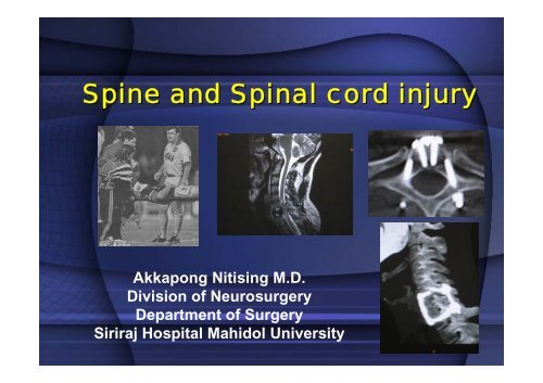
<strong>Spine</strong> <strong>and</strong> <strong>Spinal</strong> <strong>cord</strong> <strong>injury</strong><br />
Akkapong Nitising M.D.<br />
Division of Neurosurgery<br />
Department of Surgery<br />
Siriraj Hospital <strong>Mahidol</strong> <strong>University</strong>
Epidemiology & cost of<br />
care<br />
• In the US<br />
- About 10,000 new cases/year, 10%<br />
are pediatric cases; 8,000 survive the<br />
acute period; 5,000 involve the cervical<br />
spines; while 82% of patients are male.<br />
- a prevalence of 191,000 cases.<br />
- 5.6 billion US$ per year.
• Level of spinal <strong>injury</strong><br />
55 % cervical<br />
15 % thoracic<br />
15 % thoracolumbar<br />
15 % lumbosacral area<br />
• Approximately 10% of pts w/ C spine fracture have<br />
a second noncontiguous vertebral column fracture<br />
• Five percent of brain-injured pts have associated<br />
spinal <strong>injury</strong>, while 25% of spine <strong>injury</strong> patients<br />
have at least a mild brain <strong>injury</strong>.
• Etiology<br />
- Motor vehicle accidents & Road<br />
traffic accidents.<br />
- Falls (greater than 10 feet)<br />
- Sports-related injuries<br />
- Recreational injuries<br />
- Violent crimes
Primary <strong>and</strong> secondary<br />
tissue damage<br />
• Primary <strong>injury</strong>: mechanical disruption of<br />
axons as a result of stretch or<br />
laceration.<br />
• Secondary <strong>injury</strong>: free radical<br />
formation, uncontrolled calcium influx,<br />
ischemia & lipid peroxidation, <strong>and</strong><br />
apoptosis.
The Primary goal is to<br />
Save Life<br />
• Excessive manipulation <strong>and</strong> inadequate<br />
immoblization of a patient with a spinal <strong>injury</strong><br />
can cause additional neurologic damage.<br />
• As long as the patient’s spine is protected,<br />
evaluation of the spine <strong>and</strong> exclusion of spine<br />
<strong>injury</strong> may be safely deferred, especially in<br />
the presence of systemic instability, eg,<br />
respiratory inadequacy <strong>and</strong> hypotension.
Prehospital management<br />
Immobilization <strong>and</strong> transportation
• A hard cervical<br />
collar should be<br />
applied on the<br />
scene.
• Log-rolling<br />
techniques during<br />
extrication.
• Further stabilization<br />
with s<strong>and</strong>bags <strong>and</strong><br />
taping.<br />
• Beware of vomiting<br />
<strong>and</strong> aspiration,<br />
- NG tube
• Immobilization <strong>and</strong> transportation by<br />
the backboard.<br />
• The patient should be evaluated <strong>and</strong><br />
removed from the backboard as quickly<br />
as possible. If not within 2 hrs, the<br />
patient should be logrolled every 2 hrs,<br />
to reduce the risk of decubitus ulcer<br />
formation.
Pediatric trauma<br />
Plane of face is paralelled to<br />
<strong>Spine</strong> board<br />
• The spine board<br />
need padding.<br />
• A young child’s<br />
chest is raised,<br />
allowing for safe<br />
cervical spine<br />
positioning <strong>and</strong><br />
maintenance of the<br />
airway.
Emergency room <strong>and</strong><br />
acute management.<br />
• Multidisciplinary team<br />
• Continue to identify <strong>and</strong><br />
manage hypoxia,<br />
hypotension, systemic<br />
<strong>and</strong> spinal injuries.<br />
• Thorough <strong>and</strong> complete<br />
spinal <strong>and</strong> neurological<br />
examination.
ATLS<br />
• Primary survey & Resuscitation<br />
A Airway maintenance with<br />
cervical spine protection<br />
B Breathing <strong>and</strong> ventilation<br />
C Circulation with hemorrhage control<br />
D Disability: neurologic status<br />
E Exposure/Environment:<br />
completely undress the patient,<br />
but prevent hypothermia
• Nasal vs. Oral - controversy<br />
dependent on the skill <strong>and</strong> expertise<br />
(Rhee KJ 1990, Talucci 1988 <strong>and</strong> Wright 1992)<br />
Keep the neck neutral.
Hypovolemic vs.<br />
neurogenic shock
Neurogenic shock<br />
• Impairment of the descending sympathetic<br />
pathways in the spinal <strong>cord</strong> causes<br />
vasodilatation of visceral <strong>and</strong> lower extremity<br />
blood vessels; pooling of blood, <strong>and</strong>,<br />
consequently, hypotension.<br />
• Loss of sympathetic innervation to the heart,<br />
results in the patient become bradycardic, or<br />
at least fail to become tachycardic in<br />
response to hypovolemia.
Neurogenic shock<br />
• Massive fluid resuscitation may result in fluid<br />
overload <strong>and</strong> pulmonary edema.<br />
• The blood pressure can often be restored by<br />
the judicious use of vasopressors (dopamine<br />
is agent of choice) after moderate volume<br />
replacement.<br />
• Atropine for bradycardia associated with<br />
hypotension
Findings associated with<br />
spinal injuries<br />
- laceration on face or forehead; vertex<br />
- abrasion of the upper neck<br />
- fractures of m<strong>and</strong>ible<br />
- bruises about the abdomen, from seat belt.<br />
- multiple long bone <strong>and</strong> pelvic fractures<br />
- tenderness, focal hematoma; widening<br />
spinous processes
Completeness of <strong>injury</strong> <strong>and</strong><br />
Neurologic level<br />
• Complete vs. Incomplete<br />
• Level of the <strong>injury</strong>
• Key muscle groups<br />
C5 = Elbow flexors<br />
C6 = Wrist extensors<br />
C7 = Elbow extensors<br />
C8 = Finger flexors<br />
T1 = Finger abductors<br />
L2 = Hip flexors<br />
L3 = Knee extensors<br />
L4 = Ankle dorsiflexors<br />
L5 = Long toe extensors<br />
S1 = Ankle plantar flexors
ASIA (American <strong>Spinal</strong> Injury Association)<br />
score
Completeness<br />
• Complete (ASIA grade A)<br />
Complete loss of motor <strong>and</strong> sensory<br />
function below level of lesion<br />
• Incomplete (ASIA B – D)<br />
Any neurological function below the level<br />
of lesion, including preservation of<br />
perineal sensation (sacral sparing)
Level<br />
• Motor level<br />
The most caudal key muscle with at least gr.<br />
3 power. The remaining cephalad key<br />
muscle groups must have grade 5 strength.<br />
• Sensory level<br />
the most caudal segment of the spinal <strong>cord</strong><br />
with normal sensory function.
<strong>Spinal</strong> shock<br />
• “ .. All phenomena surrounding physiologic or<br />
anatomic transection of the spinal <strong>cord</strong> that<br />
results in temporary loss or depression of all<br />
or most spinal reflex activity below the level of<br />
<strong>injury</strong>.”<br />
(Atkinson PP Atkinson JLD Mayo Clin Pro 1996;71:384-9)<br />
• Loss of somatic motor, sensory, <strong>and</strong><br />
sympathetic autonomic function due to spinal<br />
<strong>cord</strong> <strong>injury</strong>.<br />
(Kiss Z, Tator CH 1993)
<strong>Spinal</strong> shock<br />
• The exact mechanism is unknown but may<br />
be related to temporary electrolyte or<br />
neurotransmitter effects on impulse<br />
conduction<br />
• Etiologies: primary axonal <strong>and</strong> cellular<br />
dysfunction, ionic conduction block caused<br />
by sodium/potassium shift, maintenance of<br />
spinal inhibitory pathways, hyperpolarization<br />
of caudal neurons, <strong>and</strong> loss of fusimotor<br />
drive in caudal spinal segments.
<strong>Spinal</strong> shock<br />
• Return of the Bulbocavernosus reflex<br />
heralds time course out of spinal shock.<br />
• However, in practical motor <strong>and</strong><br />
sensory deficits detected one hour<br />
or later after SCI are due to<br />
physical <strong>cord</strong> <strong>injury</strong> rather than to<br />
spinal shock.
Incomplete spinal <strong>cord</strong><br />
syndromes
Central <strong>cord</strong> syndrome<br />
• First described by<br />
Schneider in 1954.<br />
• Typically occurs in<br />
hyperextension <strong>injury</strong><br />
with inbucking of the<br />
ligamentum flavum<br />
compressing the spinal<br />
<strong>cord</strong> within an area of<br />
preexisting cervical<br />
spine stenosis
Central <strong>cord</strong> syndrome<br />
• Greater motor weakness in the upper<br />
extremities than in the lower extremities with<br />
varying degree of sensory loss.<br />
• Somatotopically organized corticospinal tract,<br />
with the leg fibers lateral <strong>and</strong> the arm fibers<br />
medial in the <strong>cord</strong><br />
• Involvement of the anterior horn cells in the<br />
necrotic center of the <strong>cord</strong>.
Anterior <strong>cord</strong> syndrome<br />
• Patients present with motor<br />
paralysis, loss of pain <strong>and</strong><br />
temperature, but are spared<br />
proprioception <strong>and</strong> vibration<br />
sense.<br />
• It occurs with hyperflexion<br />
<strong>and</strong> axial-loading injuries;<br />
central disk herniation,<br />
teardrop fracture, <strong>and</strong> burst<br />
fracture.<br />
• Blockage of the Anterior<br />
spinal a.?<br />
• Less favorable prognosis
Brown-Séquard syndrome<br />
• First described in 1850<br />
• Hemisection of the spinal <strong>cord</strong><br />
• Ipsilateral motor weakness,<br />
ipsilateral loss of<br />
proprioception, <strong>and</strong><br />
contralateral pain <strong>and</strong><br />
temperature loss beginning 1<br />
to 2 levels below the level of<br />
<strong>injury</strong>.<br />
• Good prognosis for recovery
Conus medullaris syndrome<br />
• Typically found between spinal levels of T11 <strong>and</strong> L2 –<br />
transition zone<br />
• Both upper <strong>and</strong> lower motor finding – mixtures of <strong>cord</strong><br />
<strong>and</strong> root syndrome.<br />
• The most common - complete sacral <strong>cord</strong> damage with<br />
variable <strong>injury</strong> <strong>and</strong> sparing of lumbar roots.<br />
• The prognosis for recovery of bladder <strong>and</strong> bowel<br />
function is relatively poor.
Cauda equina syndrome<br />
• An <strong>injury</strong> to the nerve roots<br />
• Can be seen with a fracture<br />
or acute disk herniation<br />
from L2 <strong>and</strong> below.<br />
• Asymmetrical paralysis,<br />
sensory loss, <strong>and</strong> areflexia;<br />
including loss of bowel <strong>and</strong><br />
bladder control.<br />
• Has a significant capacity<br />
for recovery.
Bell’s cruciate paralysis<br />
• First described by Bell in<br />
1970<br />
• Weakness or paralysis of<br />
the h<strong>and</strong>s <strong>and</strong> arms with<br />
relative preservation of<br />
lower extremity strength.<br />
• cervicomedullary <strong>injury</strong> –<br />
the most common is C2<br />
fracture.<br />
• Midline damage to the<br />
rostral portion of the<br />
pyramidal decussation
Guidelines for screening pts<br />
with suspect C spine (ATLS)<br />
1. para/quadriplegia –> unstable spine<br />
2. Awake/alert/intact/no neck pain/no tender ness –<br />
remove c collar <strong>and</strong> pts move their neck <strong>and</strong> no<br />
pain<br />
- > no x ray<br />
3. awake/alert/intact with neck pain/tenderness<br />
- > x rays with or without CT<br />
if normal<br />
- > flex/ext<br />
4. Altered LOC or pediatric<br />
- > x rays with or without CT <strong>and</strong> evaluated by<br />
specialist
Radiographic evaluation<br />
cervical spine<br />
• indicated in patients with<br />
- midline neck pain<br />
- palpation tenderness<br />
- neurologic deficits referable to the cervical<br />
spine<br />
- altered level of consciousness<br />
• Lateral, AP, <strong>and</strong> open-mouth odontoid<br />
view.
Plain x-ray – Cervical spine<br />
C1
CT scan<br />
• Delineate bony<br />
anatomy <strong>and</strong><br />
evaluation of canal<br />
compromise.<br />
• Subluxation may be<br />
missed – overcome<br />
by sagittal<br />
reconstruction.
• CT scan at 3-mm interval should be obtained<br />
through suspicious areas identified on the<br />
plain films, or through the lower cervical<br />
spine, if it is not adequately visualized on the<br />
plain films.<br />
• CT images through occiput – C2 may also be<br />
more sensitive than plain film for detection of<br />
fractures of these vertebrae
Lateral flexion/extension<br />
• If the screening<br />
radiographs (x-ray +<br />
CT) are normal, C<br />
spine flex/ext may be<br />
done in<br />
- pts w/o altered LOC<br />
who c/o neck pain to<br />
detect occult stability<br />
• All movement should<br />
be voluntary<br />
• Pts w/ pure<br />
ligamentous <strong>injury</strong>
MRI<br />
• Ability to image soft<br />
tissue injuries;<br />
muscle, ligaments<br />
<strong>and</strong> discs.<br />
• To detect the<br />
presence of<br />
hematoma in the<br />
spinal <strong>cord</strong> or<br />
canal.
Radiographic evaluation<br />
thoracic <strong>and</strong> lumbar spine<br />
• Indications for screening radiographs –<br />
same as cervical spine.<br />
• AP <strong>and</strong> lateral plain x-ray with axial CT<br />
at 3-mm intervals through suspicious<br />
areas.
Treatment<br />
• Surgery - Indications<br />
- Neural decompression<br />
- Stabilization of the spine<br />
- Deformity correction<br />
• Timing of surgery: Early vs. Late ?<br />
- Traction of cervical spine dislocation<br />
(Hadley MN et al. Neurosurgery 1992)<br />
- Others - controversy
Prognostic factors for<br />
recovery<br />
• Complete or incomplete:<br />
complete C injuries 24 hrs: 1-3%<br />
regain ambulatory function<br />
• Level: C > TL<br />
• Age: young > old<br />
• MRI finding: Intramedullary hemorrhage<br />
– worse outcome
Emergency room<br />
management<br />
• Cervical Traction<br />
- realign <strong>and</strong> stabilize the spine<br />
- one of the fastest way to increase the spinal<br />
canal<br />
- Two absolute contraindications<br />
1) Occiput-C1 dislocation<br />
2) Comminuted skull (temporal bone<br />
fracture)
Occipito-Cervical Dislocation HIGH<br />
ENERGY<br />
RESPIRATORY ARREST<br />
NEUROGENIC SHOCK<br />
25 year old, high speed MVA<br />
Apneic, hypotensive, <strong>and</strong><br />
unconscious at scene - intubated<br />
<strong>and</strong> rapidly transported to<br />
emergency room<br />
ASSOCIATED<br />
HEAD INJURY<br />
Facial fractures, closed head <strong>injury</strong>,<br />
R femur fracture<br />
No upper or lower extremity motor /<br />
sensory function (spinal shock)<br />
SIGNIFICANT<br />
NEUROLOGIC<br />
INJURY
• Five pounds per level for reduction; i.e.<br />
a C5-6 dislocation would require 30 lb<br />
traction. Serial imaging <strong>and</strong><br />
neurological examination are<br />
m<strong>and</strong>atory.<br />
• Use of muscle relaxants, analgesics,<br />
<strong>and</strong> sedatives may facilitate the<br />
process.<br />
• To maintain stabilization use 5-10 lbs.
• 30 yrs old female<br />
• Motor vehicle<br />
accident<br />
• C4 complete <strong>cord</strong><br />
lesion<br />
• Respiratory<br />
insuffiency
C3 C3-C4 facet<br />
C5<br />
C6<br />
C7<br />
C5-C6<br />
C4
Closed reduction
Odontoid fracture
Atlantoaxial dislocation
os odontoideum w/ C1-2<br />
dislocation
C1 lateral mass & C2 pedicle<br />
screws
Hangman fracture
Subaxial (C3-C7) Cervical<br />
spine <strong>injury</strong>
Thoracic spine <strong>injury</strong>
Research<br />
• Two categories<br />
1. Agents that can be given during the<br />
acute phase of the <strong>injury</strong>.<br />
2. those that limit secondary <strong>injury</strong><br />
mechanisms or promote regeneration.
• Drugs of the future<br />
Neurotrophins – promote the survival <strong>and</strong><br />
regeneration of injured nerve cells<br />
Drugs that prevent apoptotic cell death<br />
• Transplantation – cellular therapy<br />
Schwann cells<br />
OSG (Olfactory ensheathing glia)<br />
Embryonic spinal <strong>cord</strong> cells<br />
Neural progenitor (stem) cells<br />
Antibodies that neutralize the inhibitory proteins<br />
within myelin


