In situ identification and analysis of automotive paint ... - Library
In situ identification and analysis of automotive paint ... - Library
In situ identification and analysis of automotive paint ... - Library

Create successful ePaper yourself
Turn your PDF publications into a flip-book with our unique Google optimized e-Paper software.
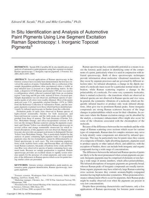
Edward M. Suzuki, 1 Ph.D. <strong>and</strong> Mike Carrabba, 2 Ph.D.<br />
<strong>In</strong> Situ Identification <strong>and</strong> Analysis <strong>of</strong> Automotive<br />
Paint Pigments Using Line Segment Excitation<br />
Raman Spectroscopy: I. <strong>In</strong>organic Topcoat<br />
Pigments 3<br />
REFERENCE: Suzuki EM, Carrabba M. <strong>In</strong> <strong>situ</strong> <strong>identification</strong> <strong>and</strong><br />
<strong>analysis</strong> <strong>of</strong> <strong>automotive</strong> <strong>paint</strong> pigments using line segment excitation<br />
Raman spectroscopy: I. <strong>In</strong>organic topcoat pigments. J Forensic Sci<br />
2001;46(5):1053–1069.<br />
ABSTRACT: Several applications <strong>of</strong> Raman spectroscopy in the<br />
forensic sciences have recently been demonstrated, but few have involved<br />
the <strong>analysis</strong> <strong>of</strong> <strong>paint</strong>s. Undoubtedly, this is a reflection <strong>of</strong> the<br />
sample degradation problems <strong>of</strong>ten encountered when a visible or<br />
near-infrared laser is focused on a light-absorbing matrix. <strong>In</strong> this<br />
study, a dispersive CCD Raman spectrometer (785 nm) was used in<br />
a configuration which collected scattered light from an excitation<br />
region 3 mm long <strong>and</strong> 80 �m wide, instead <strong>of</strong> from a focused spot.<br />
Sample degradation was not observed, <strong>and</strong> Raman spectra <strong>of</strong> <strong>automotive</strong><br />
<strong>paint</strong>s <strong>of</strong> all colors were readily obtained. Most <strong>of</strong> the <strong>paint</strong>s<br />
analyzed were U.S. automobile original finishes (1974 to 1989)<br />
from the Reference Collection <strong>of</strong> Automotive Paints, <strong>and</strong> the inorganic<br />
pigments examined were those which had been identified previously<br />
by infrared spectroscopy in finishes from this collection.<br />
Prominent peaks <strong>of</strong> rutile were observed in Raman spectra <strong>of</strong><br />
light-colored nonmetallic finishes for both monocoats <strong>and</strong><br />
basecoat/clearcoat systems, <strong>and</strong> the rutile peaks are readily distinguished<br />
from those <strong>of</strong> anatase. The lead chromates (Chrome Yellow,<br />
Molybdate Orange, <strong>and</strong> silica-encapsulated versions <strong>of</strong> the<br />
two) are the strongest Raman scatterers among the pigments examined,<br />
<strong>and</strong> Chrome Yellow was identified by Raman spectroscopy in<br />
several yellow <strong>and</strong> orange nonmetallic monocoats for which infrared<br />
absorptions <strong>of</strong> this pigment were not observed. Raman spectroscopy<br />
also provides an unequivocal means to distinguish Chrome<br />
Yellow from Molybdate Orange. This is particularly helpful for the<br />
<strong>analysis</strong> <strong>of</strong> <strong>paint</strong>s containing light pigment loads or encapsulated<br />
pigments since the two formulations cannot be differentiated by infrared<br />
spectroscopy in such cases. The iron-containing pigments,<br />
ferric oxide, hydrous ferric oxide, <strong>and</strong> Prussian Blue, are relatively<br />
weak Raman scatterers, but peaks <strong>of</strong> hydrous ferric oxide <strong>and</strong> Prussian<br />
Blue were observed in spectra <strong>of</strong> <strong>paint</strong>s containing heavy pigment<br />
loads. Because no sample preparation is required, Raman<br />
spectroscopy provides an excellent means to rapidly screen reference<br />
panels for the presence <strong>of</strong> certain pigments, <strong>and</strong> some examples<br />
<strong>of</strong> the differences in Raman spectra which occur for <strong>paint</strong>s having<br />
similar colors are presented.<br />
KEYWORDS: forensic science, criminalistics, <strong>paint</strong> <strong>analysis</strong>, <strong>automotive</strong><br />
<strong>paint</strong>, pigment <strong>identification</strong>, Raman spectroscopy, inorganic<br />
pigments<br />
1<br />
Forensic scientist, Washington State Crime Laboratory, Public Safety<br />
Building, 2nd Floor, Seattle, WA.<br />
2<br />
Marketing manager, Chromex, <strong>In</strong>c., 2705 B Pan American Freeway NE,<br />
Albuquerque, NM.<br />
3 Presented at the 52nd Annual Meeting <strong>of</strong> the American Academy <strong>of</strong> Forensic<br />
Sciences, February 2000, Reno, NV.<br />
Received 20 June 2000; accepted 8 Jan. 2001.<br />
Copyright © 2001 by ASTM <strong>In</strong>ternational<br />
1053<br />
Raman spectroscopy has considerable potential as a means to assist<br />
the forensic <strong>paint</strong> analyst in identifying some <strong>of</strong> the components<br />
<strong>of</strong> a <strong>paint</strong>, particularly when it is used in conjunction with infrared<br />
spectroscopy. Both <strong>of</strong> these spectroscopic techniques<br />
provide information about molecular vibrational transitions, but<br />
they occur by separate processes <strong>and</strong> are governed by different selection<br />
rules: for infrared absorption, a change in the dipole moment<br />
<strong>of</strong> a molecule must occur for a particular normal mode <strong>of</strong> vibration,<br />
while Raman scattering requires a change in the<br />
polarizability <strong>of</strong> a molecule. For some very symmetric molecules,<br />
there is mutual exclusivity—the transitions which are observed in<br />
infrared spectra are not observed in Raman spectra <strong>and</strong> vice versa.<br />
<strong>In</strong> general, the symmetric vibrations <strong>of</strong> a molecule, which are frequently<br />
infrared inactive or produce only weak infrared absorptions,<br />
<strong>of</strong>ten give rise to prominent Raman peaks. Some inorganic<br />
compounds are strong Raman scatterers because <strong>of</strong> the large<br />
changes in polarizabilities which occur for their vibrations. <strong>In</strong> certain<br />
cases where the Raman excitation energy can be absorbed by<br />
the analyte, a resonance enhancement effect might also occur for<br />
some <strong>of</strong> the vibrations associated with the chromophore <strong>of</strong> the<br />
molecule.<br />
Because <strong>of</strong> the differences between the two methods <strong>and</strong> the wide<br />
range <strong>of</strong> Raman scattering cross sections which occur for various<br />
types <strong>of</strong> compounds, Raman data for complex mixtures may serve<br />
to help identify some components not detected by infrared spectroscopy.<br />
This may be particularly true for <strong>paint</strong>s since they consist<br />
<strong>of</strong> a composite <strong>of</strong> binders, light-absorbing pigments, pigments used<br />
to produce opacity or other optical effects, <strong>and</strong> additives; with the<br />
exception <strong>of</strong> binders, these can include both inorganic <strong>and</strong> organic<br />
compounds having a wide range <strong>of</strong> concentrations.<br />
The differences between the infrared <strong>and</strong> Raman spectra <strong>of</strong> <strong>paint</strong>s<br />
are likely to be the most pronounced when certain pigments are present.<br />
<strong>In</strong>organic pigments may be comprised <strong>of</strong> elements or ions having<br />
a wide range <strong>of</strong> atomic numbers, which affect polarizabilities.<br />
The infrared absorptions <strong>of</strong> inorganic pigments are generally broad,<br />
whereas Raman peaks <strong>of</strong> inorganic compounds are usually narrow.<br />
Some organic pigments may have relatively large Raman scattering<br />
cross sections because they contain aromatic/highly-conjugated<br />
moieties having high molecular symmetries. When pronounced resonance<br />
enhancement effects occur for pigments, their Raman peaks<br />
might be readily observed even when the pigments are present in<br />
very low concentrations.<br />
Despite these promising characteristics <strong>and</strong> features, only a few<br />
applications <strong>of</strong> Raman spectroscopy for forensic <strong>paint</strong> examina-
1054 JOURNAL OF FORENSIC SCIENCES<br />
tions have been reported (1–3), although this method has been used<br />
for the <strong>analysis</strong> <strong>of</strong> several other types <strong>of</strong> evidence (4–25). This<br />
paucity undoubtedly reflects the difficulties encountered when a<br />
visible or near-infrared laser beam is focused on a light-absorbing<br />
matrix. Thermal destruction <strong>of</strong> the sample <strong>of</strong>ten occurs, or strong<br />
fluorescence, produced either by pigments or other <strong>paint</strong> components,<br />
may overwhelm the weaker Raman scattering peaks.<br />
To minimize fluorescence, most <strong>of</strong> the recent Raman studies <strong>of</strong><br />
<strong>paint</strong> have used Fourier transform (FT) Raman instruments, which<br />
use near-infrared excitation. Kuptsov (2) presented FT Raman data<br />
for some inorganic pigments <strong>and</strong> alkyds resins, but indicated that<br />
for actual <strong>paint</strong>s, the method is usually limited to lighter colors<br />
(whites <strong>and</strong> yellows). Massonnet <strong>and</strong> Stoecklein (3) were able to<br />
obtain FT Raman data for some yellow, orange, <strong>and</strong> red organic<br />
pigments used in <strong>automotive</strong> <strong>paint</strong>s, along with spectra <strong>of</strong> some red<br />
finishes which contain these pigments. Data for <strong>automotive</strong> <strong>paint</strong>s<br />
<strong>of</strong> other colors were not presented, however, <strong>and</strong> these authors reported<br />
thermal degradation problems with blue <strong>and</strong> green phthalocyanine<br />
pigments. Massonnet <strong>and</strong> Stoecklein noted that the Raman<br />
spectra <strong>of</strong> the finishes were dominated by pigment peaks, as was<br />
observed in some other recent Raman studies involving non-<strong>automotive</strong><br />
<strong>paint</strong>s (26–31).<br />
The intensity <strong>of</strong> Raman scattering is proportional to the fourth<br />
power <strong>of</strong> the frequency <strong>of</strong> scattered light. Consequently, FT Raman<br />
instruments, with their near-infrared lasers, require higher power<br />
levels than instruments employing visible lasers. <strong>In</strong> addition, the<br />
near-infrared detectors used with FT Raman spectrometers are less<br />
sensitive than the charge-coupled device (CCD) array detectors<br />
which are used with visible excitation instruments. The use <strong>of</strong> minimal<br />
laser power levels was particularly desirable in this study because<br />
<strong>automotive</strong> <strong>paint</strong>s typically contain heavier loads <strong>of</strong> colorimparting<br />
pigments than most other types <strong>of</strong> coatings. A dispersive<br />
CCD Raman spectrometer equipped with a visible laser (785 nm)<br />
was therefore used, <strong>and</strong> this instrument was additionally operated<br />
in a collection mode which produced an excitation area consisting<br />
<strong>of</strong> a line segment rather than a focused spot. This resulted in much<br />
lower power density levels directed onto the sample surface, <strong>and</strong><br />
Raman spectra <strong>of</strong> <strong>automotive</strong> finishes <strong>of</strong> all colors were readily obtained.<br />
The <strong>paint</strong>s examined in this study included U.S. automobile<br />
original (OEM) finishes (1974 to 1989) from the Reference Collection<br />
<strong>of</strong> Automotive Paints, more recent finishes obtained from<br />
some <strong>paint</strong> manufacturers, <strong>and</strong> <strong>paint</strong>s removed from vehicles. The<br />
inorganic pigments examined were those previously identified by<br />
infrared spectroscopy in finishes from the Reference Collection <strong>of</strong><br />
Automotive Paints (32,33). Because Raman data are complementary<br />
to infrared data, infrared spectra (4000 to 220 cm �1 ) are also<br />
presented for the pigments <strong>and</strong> <strong>paint</strong>s discussed in this work, <strong>and</strong><br />
the features <strong>of</strong> both spectra are compared <strong>and</strong> contrasted.<br />
Experimental<br />
Raman Analyses<br />
Raman spectra were acquired using a Chromex Raman 2000<br />
spectrometer equipped with a 785 nm solid state diode laser.<br />
Rayleigh scattered radiation was removed with a thin-film dielectric<br />
edge filter, <strong>and</strong> the Stokes scattered radiation having Raman<br />
shifts between 3317 <strong>and</strong> 160 cm �1 was collected at a spectral b<strong>and</strong>width<br />
<strong>of</strong> 10 cm �1 . The instrument uses a single grating <strong>and</strong> the detector<br />
is a thermoelectrically cooled CCD system consisting <strong>of</strong> a<br />
1024 by 256 array <strong>of</strong> pixels. The spectrometer is configured with<br />
both a microscope attachment <strong>and</strong> a macro sampling chamber, <strong>and</strong><br />
the latter was used for all analyses. Two sample excitation modes,<br />
consisting <strong>of</strong> a spot focus <strong>and</strong> a line segment focus, are available.<br />
Using the macro chamber, the spot focus illuminates an area 80 �m<br />
in diameter, while the line segment focus illuminates a segment 3<br />
mm long <strong>and</strong> 80 �m wide. The energy density (energy/area) produced<br />
by the line segment excitation is approximately 2% <strong>of</strong> the<br />
spot focus, <strong>and</strong> the line segment excitation mode was used for all<br />
analyses.<br />
Automotive <strong>paint</strong> panels <strong>and</strong> <strong>paint</strong> chips were analyzed directly<br />
with no sample preparation by placing them horizontally in the<br />
macro chamber. Pigments powders were analyzed neat by placing<br />
them on a glass slide wrapped with aluminum foil; the powders<br />
were compressed with a second glass slide to form a flat surface.<br />
Laser power levels between 30 <strong>and</strong> 60 mW (at the sample) were<br />
used for all <strong>of</strong> the <strong>paint</strong>s <strong>and</strong> pigments. Total collection times for<br />
most samples were between 10 <strong>and</strong> 60 s.<br />
Grams32 ® s<strong>of</strong>tware was used for data collection <strong>and</strong> preliminary<br />
data processing. Spectral data were then converted to JCAMP files<br />
<strong>and</strong> imported to Spectra Calc ® , which was used for further mathematical<br />
processing <strong>and</strong> for preparation <strong>of</strong> data presentation formats.<br />
Currently, there is no convention regarding the direction <strong>of</strong><br />
the abscissa scale for presentation <strong>of</strong> Raman data. All <strong>of</strong> the Raman<br />
spectra presented in this work are depicted together with infrared<br />
data, so both are shown with Raman shifts/frequencies decreasing<br />
from left to right, as is normally used for infrared data.<br />
<strong>In</strong>frared Analyses<br />
<strong>In</strong>frared spectra were acquired at a resolution <strong>of</strong> 4 cm �1 using an<br />
extended range (4000 to 220 cm �1 ) Digilab FTS-7 Fourier transform<br />
infrared (FT-IR) spectrometer. Paint samples were analyzed<br />
as thin slices placed over a 1 mm diameter circular aperture in a<br />
metal disk, which was mounted in a Digilab 5X beam condenser,<br />
<strong>and</strong> 1000 scans were collected. Spectra <strong>of</strong> pigments were acquired<br />
using a low pressure diamond anvil cell (DAC) mounted in the 5X<br />
beam condenser. Pigment powders were ground with excess CsI,<br />
<strong>and</strong> this mixture was then either pressed onto a single anvil <strong>of</strong> the<br />
DAC (34,35) or pressed between both anvils. Reference spectra<br />
were obtained <strong>of</strong> CsI pressed onto the same single anvil, or pressed<br />
between both anvils, <strong>and</strong> 1000 scans were collected for both references<br />
<strong>and</strong> samples. Spectral processing functions were performed<br />
using Spectra Calc s<strong>of</strong>tware. More details regarding the FT-IR<br />
spectrometer, the instrument parameters used for data collection,<br />
the <strong>paint</strong> <strong>and</strong> pigment sample preparation techniques, <strong>and</strong> spectral<br />
processing are presented elsewhere (33).<br />
Automotive Paints <strong>and</strong> Pigments<br />
Most <strong>of</strong> the <strong>paint</strong>s analyzed in this study consisted <strong>of</strong> panels<br />
from the Reference Collection <strong>of</strong> Automotive Paints (Collaborative<br />
Testing Services, <strong>In</strong>c., Herndon, Virginia); the cited colors <strong>of</strong> these<br />
<strong>paint</strong>s are based on the classification system used in this collection.<br />
Both monocoats <strong>and</strong> basecoat/clearcoat finishes were examined<br />
<strong>and</strong> they will be referred to by their seven or nine character <strong>identification</strong><br />
codes, which are described elsewhere (32,36). Panels <strong>of</strong><br />
original finishes used on more recent vehicles were obtained from<br />
BASF, DuPont, PPG, <strong>and</strong> Mazda, while other samples were removed<br />
from salvage vehicles.<br />
The pigments which were examined in this study included rutile,<br />
anatase, Chrome Yellow, Molybdate Orange, silica-encapsulated<br />
Molybdate Orange, diatomaceous silica, synthetic silica, ferric oxide,<br />
hydrous ferric oxide, <strong>and</strong> Prussian Blue. They are all described<br />
elsewhere (32,33).
Results <strong>and</strong> Discussion<br />
<strong>In</strong>frared <strong>and</strong> Raman spectra <strong>of</strong> <strong>paint</strong>s containing various pigments<br />
are presented in this work, but the details <strong>of</strong> the <strong>identification</strong><br />
<strong>of</strong> these pigments based on their infrared absorptions are presented<br />
elsewhere (32,33,37–40). Although organic pigments are<br />
not the focus <strong>of</strong> this paper, absorptions <strong>of</strong> a few <strong>of</strong> these are observed<br />
in some <strong>of</strong> the infrared spectra <strong>and</strong> their presence is noted to<br />
facilitate comparisons to Raman spectra.<br />
<strong>In</strong>strument Response Function <strong>and</strong> Spectral Corrections<br />
The instrument response function <strong>of</strong> the spectrometer used<br />
in this study is depicted in Fig. 1f. Ideally, this function is produced<br />
by measuring the spectrum <strong>of</strong> a white light source having<br />
a constant intensity output for the spectral region <strong>of</strong> interest (for<br />
an excitation wavelength <strong>of</strong> 785 nm, the Stokes Raman shifts<br />
from 160 to 3317 cm �1 correspond to near-infrared radiation between<br />
795 <strong>and</strong> 1061 nm). Such a source was not available, so<br />
the light <strong>of</strong> an inc<strong>and</strong>escent lamp was used to produce Fig. 1f.<br />
The decrease in this function for high Raman shifts reflects the insensitivity<br />
<strong>of</strong> the CCD detector to near-infrared radiation, while<br />
the drop-<strong>of</strong>f for low Raman shifts is caused by the filter system<br />
used to remove the Rayleigh line. This filter system, which is<br />
based on interference effects, also produces the sinusoidal fringes<br />
<strong>of</strong> Fig. 1f.<br />
To obtain Raman spectra having relative peak intensities that<br />
are not dependent on the detection efficiency <strong>of</strong> the instrument,<br />
acquired data can be divided by the instrument response function<br />
to give normalized spectra. <strong>In</strong> the absence <strong>of</strong> a fluorescence background,<br />
this simply serves to alter the relative peak intensities.<br />
When a strong fluorescence background occurs, as observed for<br />
the <strong>paint</strong> spectrum depicted in Fig. 1c, fringes caused by the filter<br />
are present, which can make interpretation <strong>of</strong> data more difficult.<br />
For such cases, an instrument correction is necessary to remove<br />
the fringes <strong>and</strong> the resulting spectral contour represents the<br />
shape <strong>of</strong> the fluorescence b<strong>and</strong> (which may have sharper Raman<br />
peaks superimposed on it). The normalized spectrum <strong>of</strong> Fig. 1d,<br />
for example, was obtained by dividing the uncorrected spectrum<br />
(Fig. 1c) by the instrument response function (Fig. 1f); the apparent<br />
rise in the baseline <strong>of</strong> this normalized spectrum above 2800<br />
cm �1 is an artifact resulting from the nonlinear output <strong>of</strong> the lamp<br />
used to produce Fig. 1f. For the various spectra presented in this<br />
study, instrument corrections were only used when a pronounced<br />
fluorescence background was observed, <strong>and</strong> these spectra are denoted<br />
as “normalized.”<br />
Rutile<br />
Rutile, the polymorph <strong>of</strong> titanium dioxide most <strong>of</strong>ten used in<br />
<strong>paint</strong>s, has an infrared spectrum consisting <strong>of</strong> a very broad low frequency<br />
absorption having two shoulder features (Fig. 2g). <strong>In</strong> contrast,<br />
the Raman spectrum <strong>of</strong> rutile (Fig. 2d) has two relatively narrow<br />
peaks at 613 <strong>and</strong> 451 cm �1 <strong>and</strong> is easily distinguished from<br />
that <strong>of</strong> anatase (Fig. 2e), a second polymorph <strong>of</strong> titanium dioxide<br />
that may also be used in <strong>paint</strong>s (the infrared spectrum <strong>of</strong> anatase is<br />
shown in Fig. 2f). Strong absorptions <strong>of</strong> rutile are observed in infrared<br />
spectra <strong>of</strong> all <strong>of</strong> the white nonmetallic monocoats in the Reference<br />
Collection <strong>of</strong> Automotive Paints (33), as illustrated by the<br />
spectrum <strong>of</strong> DC82A0466 (Fig. 2a), an acrylic melamine enamel.<br />
The Raman spectrum <strong>of</strong> DC82A0466 (Fig. 2b) is comprised primarily<br />
<strong>of</strong> rutile peaks <strong>and</strong> only very weak binder features, including<br />
three peaks at 1449, 1002, <strong>and</strong> 980 cm �1 , are observed. The<br />
SUZUKI AND CARRABBA • AUTOMOTIVE PAINT PIGMENTS 1055<br />
1002 <strong>and</strong> 980 cm �1 peaks have previously been assigned to ring<br />
expansion modes <strong>of</strong> the phenyl group <strong>of</strong> styrene <strong>and</strong> the triazine<br />
ring <strong>of</strong> melamine, respectively (2).<br />
The Raman spectrum <strong>of</strong> CC77A0364, a white nonmetallic<br />
basecoat/clearcoat finish which has acrylic melamine enamel<br />
binders for both the basecoat <strong>and</strong> the clearcoat layers, is depicted in<br />
Fig. 1c. Other than relative increases in the weak binder peak intensities<br />
<strong>and</strong> fluorescence, results similar to those <strong>of</strong> the monocoat<br />
are obtained. It is thus evident that pigments in basecoats can be<br />
readily analyzed through clearcoats since the clearcoat layer is a<br />
weakly-scattering medium.<br />
Lead Chromate Pigments<br />
The two main lead chromate pigments used in <strong>automotive</strong> finishes<br />
are Chrome Yellow (PbCrO4 · xPbSO4) <strong>and</strong> Molybdate Orange<br />
(PbCrO4 · xPbMoO4 · yPbSO4). Both consist <strong>of</strong> solid solutions<br />
containing mostly lead chromate together with some other lead<br />
salts. Lead chromate pigments are no longer used in U.S. automobile<br />
OEM finishes, having last been used (33) in the early 1990s<br />
(<strong>and</strong> mostly phased-out in the 1980s), but they continued to be used<br />
in <strong>automotive</strong> <strong>paint</strong>s in Europe (3).<br />
Chrome Yellow—<strong>In</strong>frared <strong>and</strong> Raman spectra <strong>of</strong> Chrome Yellow<br />
are shown in Figs. 3f <strong>and</strong> 3d, respectively. Two relatively narrow<br />
peaks at 843 <strong>and</strong> 365 cm �1 are observed in the Raman spectrum,<br />
together with a very weak peak at 406 cm �1 . <strong>In</strong>frared <strong>and</strong><br />
Raman spectra <strong>of</strong> a yellow nonmetallic monocoat (NN78H0476),<br />
which contains a heavy pigment load <strong>of</strong> Chrome Yellow, are depicted<br />
in Figs. 3b <strong>and</strong> 3c. As observed for white <strong>paint</strong>s, the Raman<br />
spectrum <strong>of</strong> this monocoat is primarily that <strong>of</strong> the pigment;<br />
NN78H0476 has an acrylic melamine enamel binder, but the<br />
binder peaks are very weak in Fig. 3c.<br />
The infrared spectrum <strong>of</strong> a second yellow nonmetallic acrylic<br />
melamine enamel monocoat, DC76H0043, is shown in Fig. 3a.<br />
This monocoat contains a large amount <strong>of</strong> rutile <strong>and</strong> what is likely<br />
a lead chromate pigment based on the weak broad feature at 868<br />
cm �1 . The Raman spectrum <strong>of</strong> DC76H0043 (Fig. 3e) clearly indicates<br />
the presence <strong>of</strong> both Chrome Yellow <strong>and</strong> rutile. Moreover,<br />
the main Chrome Yellow peak at 841 cm �1 is nearly as intense as<br />
that <strong>of</strong> the rutile peaks, even though the infrared spectrum suggests<br />
that much more rutile than Chrome Yellow is present.<br />
Chrome Yellow is thus a relatively strong Raman scatterer, <strong>and</strong><br />
this pigment was identified by Raman spectroscopy in several Reference<br />
Collection <strong>of</strong> Automotive Paints yellow <strong>and</strong> orange nonmetallic<br />
monocoats for which infrared absorptions <strong>of</strong> Chrome Yellow<br />
were not observed. Raman spectroscopy is therefore<br />
particularly helpful for the <strong>analysis</strong> <strong>of</strong> <strong>paint</strong>s having infrared spectra<br />
which do not provide a clear indication <strong>of</strong> whether a lead chromate<br />
pigment is present or not. The infrared spectrum <strong>of</strong> one <strong>of</strong><br />
these <strong>paint</strong>s, a yellow nonmetallic acrylic melamine enamel monocoat<br />
(74H0117), is shown in Fig. 4c. The weak 851 cm �1 peak <strong>of</strong><br />
74H0117 does not appear to be that <strong>of</strong> a lead chromate pigment because<br />
<strong>of</strong> its low frequency <strong>and</strong> relative sharpness, but the Raman<br />
spectrum <strong>of</strong> this finish (Fig. 4d) shows clearly that Chrome Yellow<br />
is present.<br />
Molybdate Orange—<strong>In</strong>frared <strong>and</strong> Raman spectra <strong>of</strong> Molybdate<br />
Orange are depicted in Figs. 5a <strong>and</strong> 5d, respectively. The Raman<br />
spectrum <strong>of</strong> Molybdate Orange is similar to that <strong>of</strong> Chrome Yellow<br />
(Fig. 5h), but the two can be distinguished based on the Raman shift<br />
<strong>of</strong> the strongest peak (826 versus 843 cm �1 ), the 360/346 cm �1
1056 JOURNAL OF FORENSIC SCIENCES<br />
FIG. 1—(a) <strong>In</strong>frared spectrum <strong>of</strong> ferric oxide; (b) infrared spectrum <strong>of</strong> a red nonmetallic acrylic melamine enamel monocoat, FN86 1055, which contains<br />
a relatively large amount <strong>of</strong> ferric oxide; (c) Raman spectrum <strong>of</strong> FN86 1055, uncorrected; (d) normalized Raman spectrum <strong>of</strong> FN86 1055 (the spectrum<br />
<strong>of</strong> (c) was divided by the spectrum <strong>of</strong> (f)); (e) Raman spectrum <strong>of</strong> ferric oxide; <strong>and</strong> (f) Raman spectrum <strong>of</strong> a lamp used to approximate a source having<br />
a linear emission output for the range covered by the instrument.
SUZUKI AND CARRABBA • AUTOMOTIVE PAINT PIGMENTS 1057<br />
FIG. 2—(a) <strong>In</strong>frared spectrum <strong>of</strong> a white nonmetallic acrylic melamine enamel monocoat, DC82A0466, which contains a large amount <strong>of</strong> rutile; (b) Raman<br />
spectrum <strong>of</strong> DC82A0466; (c) Raman spectrum <strong>of</strong> CC77A0364, a white nonmetallic basecoat/clearcoat finish; both the basecoat <strong>and</strong> the clearcoat layers<br />
have acrylic melamine enamel binders <strong>and</strong> the basecoat contains a large amount <strong>of</strong> rutile; (d) Raman spectrum <strong>of</strong> rutile; (e) Raman spectrum <strong>of</strong><br />
anatase; (f) infrared spectrum <strong>of</strong> anatase; <strong>and</strong> (g) infrared spectrum <strong>of</strong> rutile.
1058 JOURNAL OF FORENSIC SCIENCES<br />
FIG. 3—(a) <strong>In</strong>frared spectrum <strong>of</strong> a yellow nonmetallic acrylic melamine enamel monocoat, DC76H0043, which contains a large amount <strong>of</strong> rutile <strong>and</strong> a<br />
small amount <strong>of</strong> Chrome Yellow (the Chrome Yellow absorption is marked with its frequency); (b) infrared spectrum <strong>of</strong> a yellow nonmetallic acrylic<br />
melamine enamel monocoat, NN78H0476, which contains a large amount <strong>of</strong> Chrome Yellow; (c) Raman spectrum <strong>of</strong> NN78H0476; (d) Raman spectrum <strong>of</strong><br />
Chrome Yellow; (e) Raman spectrum <strong>of</strong> DC76H0043; (f) infrared spectrum <strong>of</strong> Chrome Yellow; <strong>and</strong> (g) Raman spectrum <strong>of</strong> rutile.
SUZUKI AND CARRABBA • AUTOMOTIVE PAINT PIGMENTS 1059<br />
FIG. 4—(a) <strong>In</strong>frared spectrum <strong>of</strong> hydrous ferric oxide; (b) infrared spectrum <strong>of</strong> a yellow nonmetallic acrylic melamine enamel monocoat, CC76H0036,<br />
which contains large amounts <strong>of</strong> rutile <strong>and</strong> hydrous ferric oxide; (c) infrared spectrum <strong>of</strong> a yellow nonmetallic acrylic melamine enamel monocoat,<br />
74H0117, which contains a large amount <strong>of</strong> hydrous ferric oxide, some rutile, <strong>and</strong> a small amount <strong>of</strong> Chrome Yellow; (d) Raman spectrum <strong>of</strong> 74H0117;<br />
(e) Raman spectrum CC76H0036; (f) Raman spectrum <strong>of</strong> hydrous ferric oxide; (g) Raman spectrum <strong>of</strong> rutile; <strong>and</strong> (h) Raman spectrum <strong>of</strong> Chrome Yellow.
1060 JOURNAL OF FORENSIC SCIENCES<br />
FIG. 5—(a) <strong>In</strong>frared spectrum <strong>of</strong> Molybdate Orange; (b) infrared spectrum <strong>of</strong> a red nonmetallic acrylic melamine enamel monocoat, KN79E0533, which<br />
contains a relatively large amount <strong>of</strong> Molybdate Orange; (c) Raman spectrum <strong>of</strong> KN79E0533; (d) Raman spectrum <strong>of</strong> Molybdate Orange; (e) infrared<br />
spectrum <strong>of</strong> a red nonmetallic acrylic lacquer monocoat, NA76E0076, which contains small amounts Molybdate Orange, Quinacridone Red Y, <strong>and</strong> ferric<br />
oxide; (f) normalized Raman spectrum <strong>of</strong> NA76E0076; (g) uncorrected Raman spectrum <strong>of</strong> NA760076; <strong>and</strong> (h) Raman spectrum <strong>of</strong> Chrome Yellow.
doublet <strong>of</strong> Molybdate Orange, <strong>and</strong> the weak 406 cm �1 peak <strong>of</strong><br />
Chrome Yellow.<br />
<strong>In</strong>frared <strong>and</strong> Raman spectra <strong>of</strong> KN79E0533, a red nonmetallic<br />
acrylic melamine enamel monocoat that contains a large amount <strong>of</strong><br />
Molybdate Orange, are shown in Figs. 5b <strong>and</strong> 5c. As observed for<br />
the example involving a heavy pigment load <strong>of</strong> Chrome Yellow<br />
(Fig. 3c), the Raman spectrum <strong>of</strong> KN79E0533 is essentially that <strong>of</strong><br />
the pigment.<br />
The infrared spectrum <strong>of</strong> a red nonmetallic acrylic lacquer<br />
monocoat (NA76E0076), which contains small amounts <strong>of</strong><br />
Quinacridone Red Y, ferric oxide, <strong>and</strong> a lead chromate pigment, is<br />
depicted in Fig. 5e. The Raman spectrum <strong>of</strong> this monocoat (Fig.<br />
5g) has a strong fluorescence background, <strong>and</strong> this spectrum was<br />
normalized to give the result shown in Fig. 5f. Peaks <strong>of</strong> Molybdate<br />
Orange can now be seen superimposed on the fluorescence contour,<br />
<strong>and</strong> although they are weak, they can be distinguished from<br />
those <strong>of</strong> Chrome Yellow (Fig. 5h).<br />
Raman spectroscopy thus provides an excellent means to differentiate<br />
between Chrome Yellow <strong>and</strong> Molybdate Orange in <strong>paint</strong>s<br />
containing light pigment loads, or for <strong>paint</strong>s with infrared spectra<br />
having interfering absorptions <strong>of</strong> binders or other pigments present.<br />
With light pigment loads, the infrared absorptions <strong>of</strong> Chrome Yellow<br />
<strong>and</strong> Molybdate Orange both consist <strong>of</strong> a single fairly broad indistinct<br />
feature between 870 <strong>and</strong> 855 cm �1 (see Figs. 3a <strong>and</strong> 5e),<br />
<strong>and</strong> the two are not easily differentiated. <strong>In</strong> contrast, the Raman<br />
spectra <strong>of</strong> such <strong>paint</strong>s allow a distinction even when the observed<br />
peaks have low intensities.<br />
<strong>In</strong>frared spectra <strong>of</strong> some acrylic melamine enamel monocoats<br />
also have weak absorptions near 870 cm �1 , as observed for<br />
DC82A0466 (Fig. 2a), DC82E0617 (Fig. 6b), <strong>and</strong> NC84 0830 (Fig.<br />
7b), but these are not from pigments. Lead chromate absorptions are<br />
usually broader, as observed for the spectrum <strong>of</strong> KC80E0617 (Fig.<br />
6a), another monocoat which has the same color as DC82E0617.<br />
This is a subtle distinction, however, <strong>and</strong> the two absorptions can be<br />
easily confused. <strong>In</strong> contrast, the Raman spectra <strong>of</strong> KC80E0617 (Fig.<br />
6d) <strong>and</strong> DC82E0617 (Fig. 6e) indicate clearly that the former contains<br />
Molybdate Orange <strong>and</strong> that the latter does not contain a lead<br />
chromate pigment. Note that Molybdate Orange can be identified<br />
from Fig. 6d even though the low frequency doublet <strong>of</strong> this pigment<br />
is partially obscured by a peak <strong>of</strong> an organic pigment.<br />
Massonnet <strong>and</strong> Stoecklein (3) obtained a Molybdate Orange<br />
spectrum similar to Fig. 5d using an FT Raman instrument. These<br />
authors identified Molybdate Orange peaks in Raman spectra <strong>of</strong> 5<br />
<strong>of</strong> 27 light red nonmetallic finishes, <strong>and</strong> 10 <strong>of</strong> 27 dark red nonmetallic<br />
finishes used on European vehicles spanning the model<br />
years 1988 to 1995. As was observed for red nonmetallic monocoats<br />
(33) used on U.S. automobiles (1974 to 1989), Molybdate Orange<br />
was also a common pigment for European vehicles. <strong>In</strong> addition,<br />
it was used for some European models manufactured after<br />
formulations involving lead-containing pigments were discontinued<br />
in the United States.<br />
Silica-Encapsulated Lead Chromate Pigments—Some <strong>of</strong> the<br />
lead chromate pigments used in <strong>automotive</strong> <strong>paint</strong>s were encapsulated<br />
with a layer <strong>of</strong> silica to minimize the possibility <strong>of</strong> a reaction<br />
between the chromates (which are oxidants) <strong>and</strong> <strong>paint</strong> binders. The<br />
infrared spectra <strong>of</strong> these pigments, such as that <strong>of</strong> silica-encapsulated<br />
Molybdate Orange (Fig. 8a), have strong absorptions <strong>of</strong> silica<br />
(compare Figs. 8a <strong>and</strong> 9g). Silica is a weak Raman scatterer, however,<br />
<strong>and</strong> a comparison <strong>of</strong> the Raman spectra <strong>of</strong> Molybdate Orange<br />
(Fig. 8g) <strong>and</strong> silica-encapsulated Molybdate Orange (Fig. 8f) indicates<br />
that they are virtually identical, with no apparent peaks <strong>of</strong> sil-<br />
SUZUKI AND CARRABBA • AUTOMOTIVE PAINT PIGMENTS 1061<br />
ica present. Some formulation information about the lead chromate<br />
pigments is thus not obtained with Raman spectroscopy, but this information<br />
can usually be deduced from infrared spectra when<br />
heavy pigment loads <strong>of</strong> encapsulated pigments are present. The absorptions<br />
<strong>of</strong> silica are quite evident, for example, in the infrared<br />
spectra <strong>of</strong> the two orange nonmetallic acrylic lacquer monocoats<br />
74G0070 (Fig. 8b) <strong>and</strong> DA75G0189 (Fig. 8c).<br />
Because the silica coating has little influence on the Raman<br />
spectra <strong>of</strong> the encapsulated lead chromates, this can be <strong>of</strong> benefit<br />
when interpreting data. The infrared absorptions <strong>of</strong> Chrome Yellow<br />
<strong>and</strong> Molybdate Orange in <strong>paint</strong> spectra can usually be distinguished<br />
when large quantities <strong>of</strong> the pigments are present (compare<br />
Figs. 3f, 3b, 5a, <strong>and</strong> 5b), but this is not the case for the encapsulated<br />
versions. The details <strong>of</strong> the main chromate absorption are mostly<br />
lost with encapsulation, <strong>and</strong> it is not clear, for example, which lead<br />
chromate pigment is present in DA75G0189 or 74G0070 based on<br />
their infrared spectra. The Raman spectra <strong>of</strong> DA75G0189 (Fig. 8d)<br />
<strong>and</strong> 74G0070 (Fig. 8e uncorrected, Fig. 8h normalized), however,<br />
clearly indicate that DA75G0189 contains (silica-encapsulated)<br />
Molybdate Orange <strong>and</strong> 74G0070 contains (silica-encapsulated)<br />
Chrome Yellow. The low Raman shift shoulders <strong>of</strong> the 840 <strong>and</strong> 363<br />
cm �1 peaks <strong>of</strong> Fig. 8e (compare to Fig. 5h) further suggest that a<br />
lesser amount <strong>of</strong> Molybdate Orange may also present in 74G0070<br />
(which is not unexpected for this orange <strong>paint</strong> since Molybdate Orange<br />
has a very red shade). This can be confirmed using elemental<br />
<strong>analysis</strong>, but such detailed pigment formulation information cannot<br />
be easily deduced from the broad chromate infrared absorption.<br />
High Resolution Data—Chrome Yellow, Molybdate Orange,<br />
<strong>and</strong> some monocoats which contain heavy pigment loads <strong>of</strong> the two<br />
pigments were also analyzed using a higher spectral resolution (this<br />
was not used for routine <strong>analysis</strong> since this limits the spectral range<br />
<strong>of</strong> the instrument). The range <strong>of</strong> the instrument was also adjusted to<br />
allow the collection <strong>of</strong> data having lower Raman shifts. For resolutions<br />
<strong>of</strong> approximately 4 cm �1 or higher, the Chrome Yellow 365<br />
cm �1 peak (Fig. 5h) was resolved into a main peak at 359 cm �1<br />
with satellite peaks at 328, 339, <strong>and</strong> 377 cm �1 , <strong>and</strong> a lower Raman<br />
shift peak at 135 cm �1 was observed. For Molybdate Orange (Fig.<br />
5d), a shoulder peak at 377 cm �1 was resolved <strong>and</strong> a lower Raman<br />
shift peak at 151 cm �1 was seen. The resolved pigment features<br />
were also observed in spectra <strong>of</strong> the monocoats. When high laser<br />
power levels were used, the Molybdate Orange 360/346 cm �1 doublet<br />
(377/360/346 cm �1 for higher resolutions) coalesced into a<br />
single unresolved b<strong>and</strong>, <strong>and</strong> this was observed for spectra <strong>of</strong> both<br />
the neat pigment <strong>and</strong> monocoats containing various levels <strong>of</strong> the<br />
pigment. This likely arises from laser heating, as this effect generally<br />
causes b<strong>and</strong> broadening in Raman spectra (41).<br />
Diatomaceous Silica <strong>and</strong> Synthetic Silica<br />
The use <strong>of</strong> large amounts <strong>of</strong> silica in <strong>automotive</strong> <strong>paint</strong>s is much<br />
more common for undercoats (42) than for finish layers, but diatomaceous<br />
silica <strong>and</strong> synthetic silica were identified in a few U.S.<br />
automobile (1974 to 1989) OEM black nonmetallic monocoats<br />
(33). The two were used as flatting agents in these particular <strong>paint</strong>s,<br />
which have semigloss or eggshell finishes. <strong>In</strong>frared spectra <strong>of</strong> two<br />
<strong>of</strong> these monocoats, DC83 1031 <strong>and</strong> KC83 1031, are presented in<br />
Figs. 9b <strong>and</strong> 9h, respectively. DC83 1031 contains diatomaceous<br />
silica (Fig. 9a) <strong>and</strong> KC83 1031 contains synthetic silica (Fig. 9g).<br />
The Raman spectra <strong>of</strong> DC83 1031 <strong>and</strong> KC83 1031 are shown in<br />
Figs. 9c <strong>and</strong> 9e, respectively, together with Raman spectra <strong>of</strong> diatomaceous<br />
silica (Fig. 9d) <strong>and</strong> synthetic silica (Fig. 9f). As noted,
1062 JOURNAL OF FORENSIC SCIENCES<br />
FIG. 6—<strong>In</strong>frared spectra <strong>of</strong> three red nonmetallic acrylic melamine enamel monocoats <strong>of</strong> Reference Collection <strong>of</strong> Automotive Paints color 0617: (a)<br />
KC80E0617; (b) DC82E0617; <strong>and</strong> (c) AC81E0617; all three monocoats contain ferric oxide, <strong>and</strong> KC80E0617 <strong>and</strong> AC81E0617 also contain small amounts<br />
<strong>of</strong> Molybdate Orange <strong>and</strong> Quinacridone Violet, respectively. Normalized Raman spectra <strong>of</strong> the three monocoats: (d) KC80E0617; (e) DC82E0617; <strong>and</strong><br />
(f) AC81E0617.
SUZUKI AND CARRABBA • AUTOMOTIVE PAINT PIGMENTS 1063<br />
FIG. 7—(a) <strong>In</strong>frared spectrum <strong>of</strong> Prussian Blue; (b) infrared spectrum <strong>of</strong> a dark blue nonmetallic acrylic melamine enamel monocoat, NC84 0830, which<br />
contains a large amount <strong>of</strong> Prussian Blue; (c) infrared spectrum <strong>of</strong> a dark blue nonmetallic acrylic melamine enamel monocoat, KN82L0830, which contains<br />
a small amount <strong>of</strong> Prussian Blue; this monocoat has the same Reference Collection <strong>of</strong> Automotive Paints color as NC84 0830; (d) Raman spectrum<br />
<strong>of</strong> KN82L0830; (e) normalized Raman spectrum <strong>of</strong> NC84 0830; <strong>and</strong> (f) normalized Raman spectrum <strong>of</strong> Prussian Blue.
1064 JOURNAL OF FORENSIC SCIENCES<br />
FIG. 8—(a) <strong>In</strong>frared spectrum <strong>of</strong> silica-encapsulated Molybdate Orange; (b) infrared spectrum <strong>of</strong> an orange nonmetallic acrylic lacquer monocoat,<br />
74G0070, which contains a large amount <strong>of</strong> silica-encapsulated Chrome Yellow; silica-encapsulated Molybdate Orange is likely also present; (c) infrared<br />
spectrum <strong>of</strong> a second orange nonmetallic acrylic lacquer monocoat, DA75G0189, which contains a large amount <strong>of</strong> silica-encapsulated Molybdate Orange;<br />
(d) normalized Raman spectrum <strong>of</strong> DA75G0189; (e) uncorrected Raman spectrum <strong>of</strong> 74G0070; (f) Raman spectrum <strong>of</strong> silica-encapsulated Molybdate<br />
Orange; (g) Raman spectrum <strong>of</strong> Molybdate Orange; <strong>and</strong> (h) normalized Raman spectrum <strong>of</strong> 74G0070.
SUZUKI AND CARRABBA • AUTOMOTIVE PAINT PIGMENTS 1065<br />
FIG. 9—(a) <strong>In</strong>frared spectrum <strong>of</strong> diatomaceous silica; (b) infrared spectrum <strong>of</strong> a black nonmetallic acrylic melamine enamel monocoat, DC83 1031,<br />
which contains diatomaceous silica; (c) Raman spectrum <strong>of</strong> DC83 1031; (d) Raman spectrum <strong>of</strong> diatomaceous silica; (e) Raman spectrum <strong>of</strong> KC83 1031,<br />
a black nonmetallic acrylic melamine enamel monocoat which contains synthetic silica; (f) Raman spectrum <strong>of</strong> synthetic silica; (g) infrared spectrum <strong>of</strong><br />
synthetic silica; <strong>and</strong> (h) infrared spectrum <strong>of</strong> KC83 1031.
1066 JOURNAL OF FORENSIC SCIENCES<br />
silica is a weak Raman scatterer (which is one <strong>of</strong> the reasons objects<br />
in glass containers can <strong>of</strong>ten be analyzed intact using Raman<br />
spectroscopy), <strong>and</strong> all four spectra consist primarily <strong>of</strong> the broad<br />
fluorescence b<strong>and</strong> <strong>of</strong> silica. The very weak sharp peaks at 2320<br />
cm �1 in the spectra <strong>of</strong> the <strong>paint</strong>s (Figs. 9c <strong>and</strong> 9e) are from atmospheric<br />
nitrogen; they appear in these particular spectra because<br />
longer collection times (1000 sec) were used. Because the silica<br />
pigments are such weak Raman scatterers, infrared spectroscopy is<br />
clearly the method <strong>of</strong> choice when considering vibrational spectroscopic<br />
techniques to identify these extender pigments.<br />
Iron-Containing Pigments<br />
Three iron-containing pigments, ferric oxide (Fe2O3), hydrous ferric<br />
oxide (FeO�OH), <strong>and</strong> Prussian Blue (Fe4[Fe(CN)6]3), may be<br />
found in <strong>automotive</strong> <strong>paint</strong>s. For U.S. automobile (1974 to 1989)<br />
OEM monocoats, ferric oxide (rust colored), <strong>and</strong> hydrous ferric oxide<br />
(yellow) were very common pigments used in nonmetallic <strong>and</strong><br />
metallic finishes having a wide range <strong>of</strong> colors (33), while Prussian<br />
Blue was used primarily to create a dark blue shade in some nonmetallic<br />
finishes (32). Ferric oxide <strong>and</strong> hydrous ferric oxide continue<br />
to be widely used in <strong>automotive</strong> <strong>paint</strong>s. Formulations involving Prussian<br />
Blue have been mostly discontinued for U.S. automobile OEM<br />
finishes, although they are still used for refinishes (personal communication,<br />
DuPont Automotive Products, July 1999). The iron-containing<br />
pigments are relatively weak Raman scatterers, although they<br />
are stronger scatterers than the silica pigments.<br />
Ferric Oxide—<strong>In</strong>frared spectra <strong>of</strong> ferric oxide <strong>and</strong> FN86 1055, a<br />
red nonmetallic acrylic melamine enamel monocoat that contains a<br />
relatively large amount <strong>of</strong> this pigment, are depicted in Figs. 1a <strong>and</strong><br />
1b, respectively. Raman spectra <strong>of</strong> ferric oxide <strong>and</strong> FN86 1055<br />
(normalized data) are shown in Figs. 1e <strong>and</strong> 1d. The Raman spectrum<br />
<strong>of</strong> this pigment has more peaks than the infrared spectrum, but<br />
these are not likely to be observed for most <strong>paint</strong>s. Two ferric oxide<br />
Raman peaks are seen as weak or very weak features superimposed<br />
on the fluorescence contour <strong>of</strong> Fig. 1d, <strong>and</strong> two more can be<br />
detected only with very close scrutiny. The monocoat (FN86 1055)<br />
chosen for this example has stronger infrared absorptions <strong>of</strong> ferric<br />
oxide than most <strong>of</strong> the other finishes in which this pigment was<br />
identified, so Raman spectroscopy (employing the excitation conditions<br />
used in this study, at least) is not likely to be useful for the<br />
<strong>identification</strong> <strong>of</strong> ferric oxide in most <strong>automotive</strong> <strong>paint</strong>s.<br />
Hydrous Ferric Oxide—<strong>In</strong>frared <strong>and</strong> Raman spectra <strong>of</strong> hydrous<br />
ferric oxide are shown in Figs. 4a <strong>and</strong> 4f, respectively, while similar<br />
data for 74H0117, a yellow nonmetallic acrylic melamine<br />
enamel monocoat that contains a heavy load <strong>of</strong> this pigment, are<br />
shown in Figs. 4c <strong>and</strong> 4d. The two strongest Raman peaks <strong>of</strong> hydrous<br />
ferric oxide are observed at 390 <strong>and</strong> 302 cm �1 in Fig. 4d, <strong>and</strong><br />
the weaker 554 cm �1 feature occurs as a shoulder to a rutile 611<br />
cm �1 peak. The second rutile peak is observed at 445 cm �1 , <strong>and</strong><br />
two peaks <strong>of</strong> Chrome Yellow occur at 842 <strong>and</strong> 364 cm �1 .<br />
A comparison <strong>of</strong> the infrared <strong>and</strong> Raman spectra <strong>of</strong> 74H0117 illustrates<br />
the effects <strong>of</strong> differences in the relative infrared absorption<br />
coefficients <strong>of</strong> hydrous ferric oxide, rutile, <strong>and</strong> Chrome Yellow<br />
versus differences in their relative Raman scattering cross<br />
sections. The low frequency region <strong>of</strong> the infrared spectrum <strong>of</strong> this<br />
monocoat is dominated by very strong absorptions <strong>of</strong> hydrous ferric<br />
oxide (Fig. 4a), <strong>and</strong> the broad dipping background on which<br />
these features appear strongly suggest that rutile absorptions are<br />
also present. As noted, it is questionable whether the 851 cm �1<br />
peak is due to Chrome Yellow, but in any case, it is a weak feature.<br />
The Raman spectrum <strong>of</strong> 74H0117 (Fig. 4d) indicates the unmistakable<br />
presence <strong>of</strong> both rutile <strong>and</strong> Chrome Yellow along with hydrous<br />
ferric oxide, <strong>and</strong> the intensities <strong>of</strong> the main Raman peaks <strong>of</strong><br />
all three pigments are comparable. Clearly, a more revealing <strong>and</strong><br />
definitive picture <strong>of</strong> the pigment composition <strong>of</strong> this particular<br />
<strong>paint</strong> is obtained by considering both infrared <strong>and</strong> Raman data.<br />
The infrared spectrum <strong>of</strong> another yellow nonmetallic acrylic<br />
melamine enamel monocoat (CC76H0036) that has strong absorptions<br />
<strong>of</strong> both rutile <strong>and</strong> hydrous ferric oxide is shown in Fig. 4b.<br />
The Raman spectrum <strong>of</strong> CC76H0036 (Fig. 4e) consists primarily<br />
<strong>of</strong> rutile peaks, with the main hydrous ferric oxide peak manifested<br />
as a weaker shoulder peak at 395 cm �1 . Note that binder peaks are<br />
relatively strong in this spectrum.<br />
Prussian Blue—The cyano C�N stretching fundamental near<br />
2100 cm �1 is the most conspicuous feature <strong>of</strong> the infrared spectrum<br />
<strong>of</strong> Prussian Blue (Fig. 7a), <strong>and</strong> <strong>of</strong> spectra <strong>of</strong> many <strong>paint</strong>s which<br />
contain this pigment (Figs. 7b <strong>and</strong> 7c). The Raman spectrum <strong>of</strong><br />
Prussian Blue (Fig. 7f) includes cyano peaks at 2149 <strong>and</strong> 2090<br />
cm �1 together with a low Raman shift peak at 273 cm �1 , but Prussian<br />
Blue is a relatively weak Raman scatterer (evidenced, for example,<br />
by the presence <strong>of</strong> the 2320 cm �1 atmospheric nitrogen<br />
peak <strong>of</strong> Fig. 7f). At higher laser power levels, a very broad fluorescence<br />
b<strong>and</strong> is also observed (centered near 1400 cm �1 ) for both the<br />
pigment <strong>and</strong> monocoats containing heavy pigment loads <strong>of</strong> Prussian<br />
Blue, suggesting that this feature may arise from a photo- or<br />
thermal-decomposition product <strong>of</strong> the pigment.<br />
<strong>In</strong>frared <strong>and</strong> Raman spectra <strong>of</strong> NC84 0830, a dark blue nonmetallic<br />
acrylic melamine enamel monocoat that contains a large<br />
amount <strong>of</strong> Prussian Blue, are shown in Figs. 7b <strong>and</strong> 7e, respectively.<br />
The three Prussian Blue pigment peaks can be seen in the Raman<br />
spectrum. <strong>In</strong>frared <strong>and</strong> Raman spectra <strong>of</strong> a second dark blue nonmetallic<br />
acrylic melamine enamel monocoat (KN82L0830) that<br />
contains a lesser amount <strong>of</strong> Prussian Blue are shown in Figs. 7c <strong>and</strong><br />
7d. The cyano stretching absorption is readily observed in the infrared<br />
spectrum <strong>of</strong> this monocoat, but the cyano Raman peak at 2154<br />
cm �1 is quite weak. Raman spectroscopy can thus serve to corroborate<br />
the presence <strong>of</strong> Prussian Blue in some <strong>paint</strong>s, but low levels <strong>of</strong><br />
this pigment are more easily detected using infrared spectroscopy.<br />
Relative Raman Scattering Cross Sections <strong>of</strong> <strong>In</strong>organic Pigments<br />
The relative Raman scattering cross sections <strong>of</strong> the pigments examined<br />
in this study were gaged using several criteria, including: the<br />
peak intensities <strong>of</strong> spectra <strong>of</strong> neat pigment powders collected for<br />
similar time periods; the pigment peak intensities observed for spectra<br />
<strong>of</strong> <strong>paint</strong>s containing various concentrations <strong>of</strong> a particular pigment;<br />
<strong>and</strong> the relative pigment peak intensities observed for spectra<br />
<strong>of</strong> <strong>paint</strong>s containing various combinations <strong>of</strong> two or more pigments<br />
<strong>of</strong> interest; infrared spectra <strong>of</strong> these same <strong>paint</strong>s were used to provide<br />
some measure <strong>of</strong> the relative amounts <strong>of</strong> pigments present.<br />
From these comparisons, it was evident that the lead chromate<br />
pigments—<strong>and</strong> Chrome Yellow in particular—are the strongest<br />
Raman scatterers among the inorganic pigments examined, followed<br />
by (in order <strong>of</strong> decreasing scattering strengths) rutile, the<br />
iron-containing pigments, <strong>and</strong> silica. The scattering efficiencies <strong>of</strong><br />
the pigments, however, seem to be quite dependent on the color <strong>of</strong><br />
the finish in which they occur. This most likely reflects differences<br />
in the penetration depth <strong>of</strong> the exciting laser beam for <strong>paint</strong>s <strong>of</strong> various<br />
colors. Chrome Yellow <strong>and</strong> Molybdate Orange, for example,<br />
have similar chemical compositions <strong>and</strong> one might reasonably ex-
SUZUKI AND CARRABBA • AUTOMOTIVE PAINT PIGMENTS 1067<br />
FIG. 10—(a) <strong>In</strong>frared spectrum <strong>of</strong> a red nonmetallic acrylic melamine enamel monocoat, DC77E0054, which contains Quinacridone Red Y, Benzimidazolone<br />
Orange, ferric oxide, <strong>and</strong> rutile; (b) normalized Raman spectrum <strong>of</strong> DC77E0054; (c) uncorrected Raman spectrum <strong>of</strong> DC77E0054; (d) normalized<br />
Raman spectrum <strong>of</strong> a red nonmetallic acrylic lacquer monocoat, NA77E0054, which contains Quinacridone Red Y <strong>and</strong> Molybdate Orange; this monocoat<br />
has the same Reference Collection <strong>of</strong> Automotive Paints color as DC77E0054; (e) uncorrected Raman spectrum <strong>of</strong> NA77E0054; (f) infrared spectrum<br />
<strong>of</strong> NA77E0054; <strong>and</strong> (g) Raman spectrum <strong>of</strong> Molybdate Orange.
1068 JOURNAL OF FORENSIC SCIENCES<br />
pect them to have comparable Raman scattering cross-sections, but<br />
Chrome Yellow is clearly a stronger scatterer. The 785 nm laser<br />
light would be expected to penetrate deeper into yellow <strong>paint</strong>s<br />
compared to red ones, since the absorption maxima <strong>of</strong> the red samples<br />
are closer to the laser wavelength. Consequently, the effective<br />
sampling volume would be greater for yellow samples compared to<br />
those having red colors, <strong>and</strong> more scattered light can be collected<br />
(Molybdate Orange, despite its name, is actually much more common<br />
in red finishes (33) than orange ones).<br />
Another example <strong>of</strong> this effect involves rutile, which unlike the<br />
other pigments, is not colored. <strong>In</strong>frared <strong>and</strong> Raman spectra <strong>of</strong><br />
DC77E0054, a red nonmetallic monocoat which contains a moderate<br />
amount <strong>of</strong> rutile, are depicted in Figs. 10a <strong>and</strong> 10b, respectively.<br />
The rutile peaks are barely noticeable in the Raman spectrum<br />
<strong>of</strong> this <strong>paint</strong>, even though such peaks are usually readily<br />
observed in spectra <strong>of</strong> yellow <strong>paint</strong>s which contain comparable<br />
amounts <strong>of</strong> this pigment (all white <strong>automotive</strong> finishes contain<br />
much more rutile so they are not as useful for comparison).<br />
Screening <strong>of</strong> Reference Panels<br />
Because intact <strong>paint</strong> samples can be analyzed directly with no<br />
sample preparation using Raman spectroscopy, this method provides<br />
an excellent means to screen reference <strong>paint</strong> panels to determine<br />
if certain pigments are present. This can be helpful when attempting<br />
to identify an unknown finish that is compared to various<br />
reference panels, or when generating a database <strong>of</strong> pigment compositions<br />
involving a large number <strong>of</strong> samples. The presence <strong>of</strong> pigments<br />
in both monocoats <strong>and</strong> basecoat layers <strong>of</strong> basecoat/clearcoat<br />
finishes can be determined by this method.<br />
The color <strong>and</strong> type <strong>of</strong> finish (nonmetallic or metallic) <strong>of</strong> an <strong>automotive</strong><br />
<strong>paint</strong> are among its most distinctive <strong>and</strong> easily-determined<br />
characteristics (43,44). A screening process intended to help<br />
identify an unknown <strong>paint</strong> would thus normally be used to differentiate<br />
between reference panels having similar colors. Examples<br />
<strong>of</strong> some <strong>of</strong> the differences that occur in Raman spectra <strong>of</strong> monocoats<br />
<strong>of</strong> three Reference Collection <strong>of</strong> Automotive Paints colors are<br />
therefore presented.<br />
Raman spectra (uncorrected <strong>and</strong> normalized) <strong>of</strong> two red nonmetallic<br />
monocoats <strong>of</strong> color 0054 are shown in Figs. 10b to 10e.<br />
NA77E0054 (Fig. 10d) contains Molybdate Orange (Fig. 10g),<br />
<strong>and</strong> the peaks <strong>of</strong> this pigment are easily seen in the uncorrected spectrum<br />
as well (Fig. 10e); DC77E0054 (Fig. 10b) does not contain a<br />
lead chromate pigment. <strong>In</strong>frared spectra <strong>of</strong> DC77E0054 <strong>and</strong><br />
NA77E0054 are shown in Figs. 10a <strong>and</strong> 10f, respectively.<br />
DC77E0054 contains Benzimidazolone Orange, Quinacridone Red<br />
Y, ferric oxide, <strong>and</strong> rutile, while NA77E0054 contains<br />
Quinacridone Red Y <strong>and</strong> a lead chromate pigment (which, as discussed,<br />
cannot be easily identified by infrared spectroscopy when<br />
present in small quantities). As mentioned, only very weak rutile<br />
peaks are observed in the Raman spectrum <strong>of</strong> DC77E0054 (Fig.<br />
10b), together with very weak peaks <strong>of</strong> an organic pigment.<br />
Raman spectra <strong>of</strong> two dark blue nonmetallic monocoats <strong>of</strong> color<br />
0830, KN82L0830 <strong>and</strong> NC84 0830, are depicted in Figs. 7d <strong>and</strong> 7e,<br />
respectively. As noted previously, both contain Prussian Blue, but<br />
at different levels, <strong>and</strong> the Raman spectra <strong>of</strong> the two <strong>paint</strong>s suggest<br />
that this is probably also the case. More significantly for screening<br />
purposes, however, the spectrum <strong>of</strong> KN82L0830 (Fig. 7d) contains<br />
strong peaks <strong>of</strong> an organic pigment that clearly distinguishes the<br />
two <strong>paint</strong>s.<br />
Raman spectra <strong>of</strong> three red nonmetallic monocoats <strong>of</strong> color 0617<br />
are shown in Figs. 6d to 6f. As noted, KC80E0617 (Fig. 6d) contains<br />
Molybdate Orange but DC82E0617 (Fig. 6e) <strong>and</strong> AC81E0617 (Fig.<br />
6f) do not. The infrared spectra <strong>of</strong> the monocoats (Figs. 6a to 6c) indicate<br />
that all three contain ferric oxide, <strong>and</strong> the Raman spectrum <strong>of</strong><br />
AC81E0617 (Fig. 6f) appears to contain two very weak peaks <strong>of</strong> this<br />
pigment near 300 <strong>and</strong> 230 cm �1 (see Fig. 1e). The infrared spectrum<br />
<strong>of</strong> AC81E0617 (Fig. 6c) also has weak peaks <strong>of</strong> Quinacridone Violet,<br />
but its Raman spectrum (Fig. 6f) has little indication <strong>of</strong> the presence<br />
<strong>of</strong> organic pigment peaks. The Raman spectra <strong>of</strong> KC80E0617<br />
(Fig. 6d) <strong>and</strong> DC82E0617 (Fig. 6e), in contrast, have prominent<br />
peaks <strong>of</strong> an (unidentified) organic pigment.<br />
Even without a knowledge <strong>of</strong> which specific organic pigments<br />
are responsible for producing the Raman peaks <strong>of</strong> Figs. 6d, 6e, <strong>and</strong><br />
7d, these spectral features can still be used to help differentiate<br />
<strong>paint</strong>s having similar colors. These particular Raman spectra are<br />
especially noteworthy, however, since the corresponding infrared<br />
spectra (Figs. 6a, 6b, <strong>and</strong> 7c, respectively) lack significant absorptions<br />
<strong>of</strong> organic pigments. These Raman peaks can thus be used to<br />
identify some organic pigments that are present in concentrations<br />
too low to be determined by infrared spectroscopy, <strong>and</strong> this topic<br />
will be explored further in subsequent papers in this series.<br />
A Note <strong>of</strong> Caution<br />
During one prolonged <strong>analysis</strong> <strong>of</strong> Prussian Blue using relatively<br />
high laser power levels (more than 100 mW at the sample), the neat<br />
powder, which has a dark blue hue, ignited. This was not realized until<br />
after the sample chamber was opened, at which time considerable<br />
smoke was observed <strong>and</strong> most <strong>of</strong> the pigment had developed a crusty<br />
rust-colored surface. The reaction continued after the sample was removed<br />
from the instrument <strong>and</strong> the heat generated from it caused the<br />
glass slide, on which the sample was <strong>situ</strong>ated, to crack. Fortunately,<br />
the reaction <strong>of</strong> this pigment (Fe4[Fe(CN)6]3) did not appear to involve<br />
the generation <strong>of</strong> hydrogen cyanide, but clearly this incident<br />
indicates the need for caution when certain samples are subjected to<br />
<strong>analysis</strong> using a laser (which in this case had a very diffuse focus).<br />
<strong>In</strong> their study <strong>of</strong> explosives <strong>and</strong> propellants using an FT Raman<br />
instrument, McNesby et al. (15) noted that M30, a dark gray product<br />
which contains 40% nitroguanidine, 28% nitrocellulose, <strong>and</strong><br />
22% nitroglycerin, also combusted during <strong>analysis</strong>. A 1.06 �m Nd:<br />
YAG laser was used with 400 mW <strong>of</strong> power at the sample, <strong>and</strong> the<br />
M30 ignited after a few seconds <strong>of</strong> exposure to the laser light. Lewis<br />
et al., in their study <strong>of</strong> explosives using various Raman instruments<br />
(18), reported that HNS-II, a product which contains 2,2�, 4,4�,6,6�hexanitrostilbene,<br />
readily burned when exposed to 50 mW <strong>of</strong> laser<br />
power (these authors did not indicate, however, whether this product<br />
was colored or not). None <strong>of</strong> the other materials examined by either<br />
McNesby et al. or Lewis et al. was cited as producing such problems.<br />
Acknowledgments<br />
The authors would like to thank: Cookson Pigments <strong>In</strong>c., Degussa<br />
Corp., DuPont White Pigment <strong>and</strong> Mineral Products, Farwest<br />
Paint Manufacturing Co., Heubach <strong>In</strong>c., <strong>and</strong> Wayne Pigment<br />
Corp. for providing samples <strong>and</strong> information; DuPont Automotive<br />
Products for providing information; <strong>and</strong> Ms. Chesterene Cwiklik<br />
(Cwiklik & Associates), Ms. <strong>In</strong>grid Dearmore (WSCL), Dr. Bill<br />
Gresham (WSCL), Ms. Cynthia Graff (WSCL), Ms. Helen Griffin<br />
(WSCL), Ms. Becky Huff (FBI Laboratory), <strong>and</strong> Ms. Marianne<br />
Stam (California Department <strong>of</strong> Justice Forensic Science Laboratory,<br />
Riverside) for taking the time to review this manuscript. One<br />
<strong>of</strong> the authors would also like to thank a descendant <strong>of</strong> the “Head<br />
<strong>of</strong> the Cararra Marble Mines” family <strong>of</strong> Italy, for allowing this<br />
FTIRUser to become a RamanUser once again.
References<br />
1. Guineau B. Micro<strong>analysis</strong> <strong>of</strong> <strong>paint</strong>ed manuscripts <strong>and</strong> colored archeological<br />
materials by Raman laser microprobe. J Forensic Sci 1984;29:<br />
471–85.<br />
2. Kuptsov AH. Applications <strong>of</strong> Fourier transform Raman spectroscopy in<br />
forensic science. J Forensic Sci 1994;39:305–18.<br />
3. Massonnet G, Stoecklein W. Identification <strong>of</strong> organic pigments in coatings:<br />
applications to red <strong>automotive</strong> topcoats. Part III: Raman spectroscopy<br />
(NIR FT-Raman). Sci <strong>and</strong> Justice 1999;39:181–7.<br />
4. Carver FWS, Sinclair TJ. Spectroscopic studies <strong>of</strong> explosives. 1. Detection<br />
<strong>of</strong> nitro compounds on silica gel <strong>and</strong> carbon by non-resonant Raman<br />
spectroscopy. J Raman Spectrosc 1983;14:410–4.<br />
5. Lang PL, Katon JE, O’Keefe JF, Schiering DW. The <strong>identification</strong> <strong>of</strong><br />
fibers by infrared <strong>and</strong> Raman microspectroscopy. Microchem J 1986;<br />
34:319–31.<br />
6. Hodges CM, Hendra PJ, Willis HA, Farley T. Fourier transform Raman<br />
spectroscopy <strong>of</strong> illicit drugs. J Raman Spectrosc 1989;20:745–9.<br />
7. Bourgeois D, Church SP. Studies <strong>of</strong> dyestuffs by Fourier transform Raman<br />
spectroscopy. Spectrochim Acta 1990;46A:295–301.<br />
8. Hodges CM, Akhavan J. The use <strong>of</strong> Fourier transform Raman spectroscopy<br />
in the forensic <strong>identification</strong> <strong>of</strong> illicit drugs <strong>and</strong> explosives.<br />
Spectrochim Acta 1990;46A:303–7.<br />
9. Hendra PJ, Maddams WF, Royaud IAM, Willis HA, Zichy V. The application<br />
<strong>of</strong> Fourier transform Raman spectroscopy to the <strong>identification</strong><br />
<strong>and</strong> characterization <strong>of</strong> polyamides—I. Single number nylons. Spectrochim<br />
Acta 1990;46A:747–56.<br />
10. Maddams WF, Royaud IAM. The application <strong>of</strong> Fourier transform Raman<br />
spectroscopy to the <strong>identification</strong> <strong>and</strong> characterization <strong>of</strong><br />
polyamides—II. Double number nylons. Spectrochim Acta 1990;46A:<br />
1327–33.<br />
11. Neville GA, Shurvell HF. Fourier transform Raman <strong>and</strong> infrared vibrational<br />
study <strong>of</strong> diazepam <strong>and</strong> four closely related 1,4-benzodiazepines. J<br />
Raman Spectrosc 1990;21:9–19.<br />
12. Akhavan J. Analysis <strong>of</strong> high-explosive samples by Fourier transform Raman<br />
spectroscopy. Spectrochim Acta 1991;47A:1247–50.<br />
13. By A, Neville GA, Shurvell HF. Fourier transform infrared/Raman differentiation<br />
<strong>and</strong> characterization <strong>of</strong> cis- <strong>and</strong> trans- 2,5-dimethoxy-4, �dimethyl-�-nitrostyrenes:<br />
precursors to the street drug STP. J Forensic<br />
Sci 1992;37:503–12.<br />
14. Bouffard SP, Sommer AJ, Katon JE, Godber S. Use <strong>of</strong> molecular microspectroscopy<br />
to characterize pigment-loaded polypropylene single<br />
fibers. Appl Spectrosc 1994;48:1387–93.<br />
15. McNesby KL, Wolfe JE, Morris JB, Pesce-Rodriguez RA. Fourier transform<br />
Raman spectroscopy <strong>of</strong> some energetic materials <strong>and</strong> propellant<br />
formulations. J Raman Spectrosc 1994;25:75–87.<br />
16. Cheng C, Kirkbride TE, Batchelder DN, Lacey RJ, Sheldon TG. <strong>In</strong> <strong>situ</strong><br />
detection <strong>and</strong> <strong>identification</strong> <strong>of</strong> trace explosives by Raman microscopy. J<br />
Forensic Sci 1995;40:31–7.<br />
17. Hayward IP, Kirkbride TE, Batchelder DN, Lacey RJ. Use <strong>of</strong> a fiber optic<br />
probe for the detection <strong>and</strong> <strong>identification</strong> <strong>of</strong> explosive materials by<br />
Raman spectroscopy. J Forensic Sci 1995;40:883–4.<br />
18. Lewis IR, Daniel NW, Chaffin NC, Griffiths PR, Tungol MW. Raman<br />
spectroscopic studies <strong>of</strong> explosive materials towards a fieldable explosive<br />
detector. Spectrochim Acta 1995;51A:1985–2000.<br />
19. Pestaner JP, Mullick FG, Centeno JA. Characterization <strong>of</strong> acetaminophen:<br />
molecular micro<strong>analysis</strong> with Raman microprobe spectroscopy.<br />
J Forensic Sci 1996;41:1060–3.<br />
20. Coupry C, Brissaud D. Raman microscopy: applications in art, jewelry<br />
<strong>and</strong> forensic science. <strong>In</strong>: Turrell G, Corset J, editors. Raman microscopy:<br />
developments <strong>and</strong> applications. New York: Academic Press, 1996;<br />
421–50.<br />
21. Tsuchihashi H, Katagi M, Nishikawa M, Tatsuno M, Nishioka H, Nara<br />
A, Nishio E, Petty C. Determination <strong>of</strong> methamphetamine <strong>and</strong> its related<br />
compounds using Fourier transform Raman spectroscopy. Appl Spectrosc<br />
1997;51:1796–9.<br />
22. Keen IP, White GW, Fredericks PM. Characterization <strong>of</strong> fibers by Raman<br />
microprobe spectroscopy. J Forensic Sci 1998;43:82–9.<br />
23. S<strong>and</strong>s HS, Hayward IP, Kirkbride TE, Bennett R, Lacey RJ, Batchelder<br />
DN. UV-excited resonance Raman spectroscopy <strong>of</strong> narcotics <strong>and</strong> explosives.<br />
J Forensic Sci 1998;43:509–13.<br />
24. Ryder AG, O’Connor GM, Glynn TJ. Identifications <strong>and</strong> quantitative<br />
measurements <strong>of</strong> narcotics in solid mixtures using near-IR Raman<br />
spectroscopy <strong>and</strong> multivariate <strong>analysis</strong>. J Forensic Sci 1999;44:<br />
1013–9.<br />
SUZUKI AND CARRABBA • AUTOMOTIVE PAINT PIGMENTS 1069<br />
25. Wilson AS, Edwards HGM, Farwell DW, Janaway RC. Fourier transform<br />
Raman spectroscopy: evaluation as a non-destructive technique for<br />
studying the degradation <strong>of</strong> human hair from archaeological <strong>and</strong> forensic<br />
environments. J Raman Spectrosc 1999;30:367–73.<br />
26. Ellis G, Claybourn M, Richards SE. The applications <strong>of</strong> Fourier transform<br />
Raman spectroscopy to the study <strong>of</strong> <strong>paint</strong> systems. Spectrochim<br />
Acta 1990;46A:227–41.<br />
27. Singer BW, Gardiner DJ, Derow JP. Analysis <strong>of</strong> white <strong>and</strong> blue pigments<br />
from watercolors by Raman microscopy. The Paper Conservator<br />
1993;17:13–9.<br />
28. Burgio L, Clark RJH, Gibbs PJ. Pigment <strong>identification</strong> studies in <strong>situ</strong> <strong>of</strong><br />
Javanese, Thai, Korean, Chinese <strong>and</strong> Uighur manuscripts by Raman microscopy.<br />
J Raman Spectrosc 1999;30:181–4.<br />
29. Edwards HGM, Farwell DW, Rozenberg S. Raman spectroscopic study<br />
<strong>of</strong> red pigment <strong>and</strong> fresco fragments from King Herod’s palace at Jericho.<br />
J Raman Spectrosc 1999;30:361–6.<br />
30. Zuo J, Xu C, Wang C, Yushi Z. Identification <strong>of</strong> the pigment in <strong>paint</strong>ed<br />
pottery from the Xishan site by Raman microscopy. J Raman Spectrosc<br />
1999;30:1053–5.<br />
31. Burgio L, Clark RJH, Stratoudaki T, Doulgeridis M, Anglos D. Pigment<br />
<strong>identification</strong> in <strong>paint</strong>ed artworks: A dual analytical approach employing<br />
laser-induced breakdown spectroscopy <strong>and</strong> Raman microscopy. Appl<br />
Spectrosc 2000;54:463–9.<br />
32. Suzuki EM. <strong>In</strong>frared spectra <strong>of</strong> U.S. automobile original topcoats<br />
(1974–1989): I. Differentiation <strong>and</strong> <strong>identification</strong> based on acrylonitrile<br />
<strong>and</strong> ferrocyanide C�N stretching absorptions. J Forensic Sci 1996;41:<br />
376–92.<br />
33. Suzuki EM. <strong>In</strong>frared spectra <strong>of</strong> U.S. automobile original topcoats<br />
(1974–1989): II. Identification <strong>of</strong> some topcoat inorganic pigments using<br />
an extended range (4000-220 cm �1 ) Fourier transform spectrometer.<br />
J Forensic Sci 1996;41:393–406.<br />
34. Lin-Vien D, Bl<strong>and</strong> BJ, Spence VJ. An improved method <strong>of</strong> using the diamond<br />
anvil cell for infrared microprobe <strong>analysis</strong>. Appl Spectrosc<br />
1990;44:1227–8.<br />
35. Suzuki EM. Fourier transform infrared analyses <strong>of</strong> some particulate drug<br />
mixtures using a diamond anvil cell with a beam condenser <strong>and</strong> an infrared<br />
microscope. J Forensic Sci 1992;37:467–87.<br />
36. Collaborative Testing Services, <strong>In</strong>c. Reference collection <strong>of</strong> <strong>automotive</strong><br />
<strong>paint</strong>s technical data. Herndon, VA: Collaborative Testing Services,<br />
Sept. 1989.<br />
37. Suzuki EM, Marshall WP. <strong>In</strong>frared spectra <strong>of</strong> U.S. automobile original<br />
topcoats (1974–1989): III. <strong>In</strong> <strong>situ</strong> <strong>identification</strong> <strong>of</strong> some organic pigments<br />
used in yellow, orange, red, <strong>and</strong> brown nonmetallic <strong>and</strong> brown<br />
metallic finishes—benzimidazolones. J Forensic Sci 1997;42:619–48.<br />
38. Suzuki EM, Marshall WP. <strong>In</strong>frared spectra <strong>of</strong> U.S. automobile original<br />
topcoats (1974–1989): IV. Identification <strong>of</strong> some organic pigments used<br />
in red <strong>and</strong> brown nonmetallic <strong>and</strong> metallic monocoats—quinacridones. J<br />
Forensic Sci 1998;43:514–42.<br />
39. Suzuki EM. <strong>In</strong>frared spectra <strong>of</strong> U.S. automobile original topcoats<br />
(1974–1989): V. Identification <strong>of</strong> organic pigments used in red nonmetallic<br />
<strong>and</strong> brown nonmetallic <strong>and</strong> metallic monocoats—DPP Red BO<br />
<strong>and</strong> Thioindigo Bordeaux. J Forensic Sci 1999;44:297–313.<br />
40. Suzuki EM. <strong>In</strong>frared spectra <strong>of</strong> U.S. automobile original topcoats<br />
(1974–1989): VI. Identification <strong>and</strong> <strong>analysis</strong> <strong>of</strong> yellow organic <strong>automotive</strong><br />
<strong>paint</strong> pigments—Isoindolinone Yellow 3R, Isoindoline Yellow, Anthrapyrimidine<br />
Yellow, <strong>and</strong> miscellaneous yellows. J Forensic Sci<br />
1999;44:1151–75.<br />
41. Bowie BT, Chase B, Griffiths PR. Factors affecting the performance <strong>of</strong><br />
bench-top Raman spectrometers. Part I: instrumental effects. Appl Spectrosc<br />
2000;54:164A–73A.<br />
42. Rodgers PG, Cameron R, Cartwright NS, Clark WH, Deak JS, Norman<br />
EWW. The classification <strong>of</strong> <strong>automotive</strong> <strong>paint</strong> by diamond window infrared<br />
spectrophotometry. Part I: binders <strong>and</strong> pigments. Can Soc Forensic<br />
Sci J 1976;9:1–14.<br />
43. Gothard JA. Evaluation <strong>of</strong> <strong>automotive</strong> <strong>paint</strong> flakes as evidence. J Forensic<br />
Sci 1976;21:636–41.<br />
44. Ryl<strong>and</strong> SG, Kopec RJ. The evidential value <strong>of</strong> automobile <strong>paint</strong> chips. J<br />
Forensic Sci 1979;24:140–7.<br />
Additional information <strong>and</strong> reprint requests:<br />
Ed Suzuki<br />
Washington State Crime Laboratory<br />
Public Safety Building, 2nd Floor<br />
Seattle, WA 98104<br />
E-Mail address: FTIRUser@aol.com